Eye surgery
Cataracts, the cornea, glaucoma, the retina, eye muscles, eyelids and tear ducts: tiny instruments under the microscope, drugs that look alike, and an awake patient who hears every word.
1What eye surgery covers
Eye surgery aims to bring back sight lost to disease, injury or a defect present from birth. It works on the outside of the eye, such as the lids, the tear ducts and the eye muscles, and on the inside, such as the lens, the cornea and the retina. It is done in hospitals and in outpatient centers, and cataract surgery is the operation done most often in ambulatory surgery in the United States.
The surgeon is an ophthalmologist. Some focus on one area: children's eye problems, the cornea, the retina, or oculoplastics, the blend of plastic surgery and eye surgery that treats the lids, tear ducts and orbit. Many eye surgeons hire their own surgical technologists, and ophthalmic surgical technologists may work in the eye clinic as well as the OR.
Eye operations are small, but they are not minor. Even a slight jar of an instrument touching the eye can injure it, the patient is often awake, and the number of cases is high. You learn to work in tiny detail, keep a steady hand and speak clearly to the surgeon.
The Core Curriculum groups eye operations by the part of the eye they treat: the conjunctiva, cornea and iris; the eyelid; the globe; the lens; the muscles; the retina; and the tear duct. Cases that share instruments, supplies, positions and steps are co-related procedures, so learn the pattern once and each case becomes a new middle. Each case below links to its setup card, which holds the full instrument list and every step; here you get the reasons, and the moments when the scrub matters most.
The Core Curriculum lists every eye operation under the open approach, and for cataract surgery it adds a laser-assisted form. A dacryocystorhinostomy can also be done endoscopically, through the nose, usually by an ear, nose and throat surgeon.
2The eye in brief
Chapter 4 teaches the eye's anatomy in full. This section reviews only the parts each operation touches.
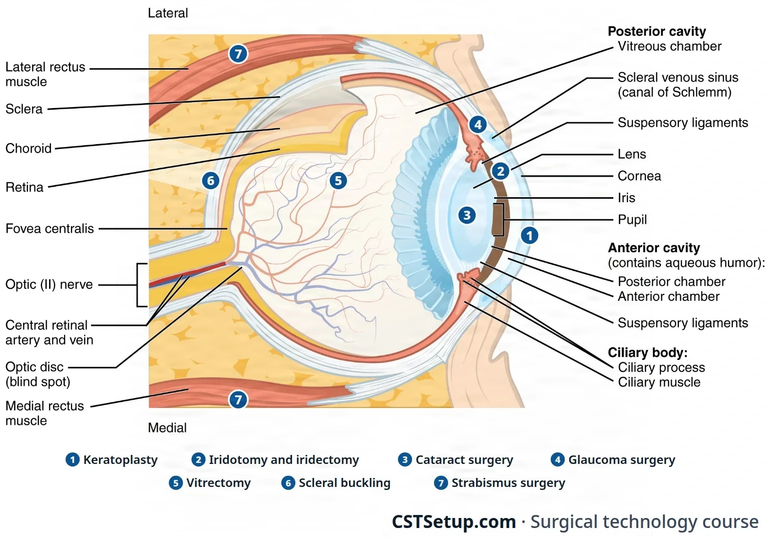
The orbit, lids and conjunctiva
- Orbit: the eyeball, or globe, sits in a bony socket built from seven bones (frontal, lacrimal, sphenoid, ethmoid, maxillary, zygomatic and palatine), lined with connective tissue that cushions it. The rim is thick and protective, and the optic nerve enters at the back through the optic foramen.
- Eyelids: thin skin over a scaffold of dense fibrous tissue, the tarsal plate. The plates hold the meibomian glands, tiny oil glands along the lid margin, about 30 in the upper lid and 20 in the lower, whose oil slows the evaporation of tears. The gap between the open lids is the palpebral fissure, and each corner where the lids meet is a canthus, inner (medial) and outer (lateral).
- Eyelid muscles: the orbicularis oculi closes the lids, and the levator palpebrae superioris raises the upper lid.
- Conjunctiva: a thin, clear mucous membrane. Its palpebral part lines the inside of the lids and folds back at the fornix as the bulbar part over the white of the eye, up to the limbus, where the cornea meets the sclera. Under it lies Tenon capsule, the fibrous layer surgeons dissect to reach the muscles and sclera.
The wall of the globe
- Cornea: the clear window at the front, with no blood vessels. It does most of the bending of light, about 65 percent of the eye's focusing power. It has five layers: the epithelium, the Bowman membrane, the stroma, the Descemet membrane and the endothelium, a single layer of cells that pumps fluid out to keep the cornea clear. Endothelial cells do not grow back, so damage to them leaves a swollen, cloudy cornea. Some references count only the three main layers: epithelium, stroma and endothelium.
- Sclera: the tough white coat over most of the globe. It gives the eye its shape and takes the tendons of the eye muscles.
- Choroid and ciliary body: the choroid is a dark layer full of blood vessels under the sclera. At the front it becomes the ciliary body, a ring of muscle that holds the lens on fine fibers and makes aqueous humor. Its rear band, the pars plana, lies just in front of where the retina begins, which is why surgeons enter the back of the eye there.
- Iris and pupil: the iris is the colored ring of muscle between the cornea and the lens; the pupil is the opening in its center. The iris narrows the pupil in bright light and widens it in dim light.
The lens and the fluids
- Lens: a clear disc, curved on both sides, inside a thin, clear capsule. It sits behind the iris, held to the ciliary body by fine fibers called zonules (the suspensory ligament). Its center is the nucleus and its outer part the cortex, like the pit and flesh of a peach, with the capsule as the skin. Cataract surgery takes out the nucleus and cortex and leaves the capsule to hold the new lens.
- Chambers and aqueous humor: the space in front of the lens holds two chambers: the anterior chamber between the cornea and the iris, and the posterior chamber between the iris and the lens. Both hold aqueous humor, a clear watery fluid made by the ciliary body. It flows from the posterior chamber through the pupil into the anterior chamber, then drains through the trabecular meshwork, in the angle where the iris meets the cornea, into the canal of Schlemm and the veins. The balance between what is made and what drains sets the intraocular pressure (IOP).
- Vitreous humor: a clear jelly, about 99 percent water, that fills the back 80 percent of the eye and holds it in shape. It sticks to the retina at its base. Unlike aqueous humor, the body does not replace it.
The retina and optic nerve
- Retina: the inner, light-sensing layer. Its center, the macula, gives sharp central vision, and the fovea at the center of the macula is where vision is sharpest. The optic nerve (cranial nerve II) leaves at the optic disc, which has no light-sensing cells and so makes a blind spot. The central retinal artery and vein run inside the optic nerve.
The muscles and the tear system
- Eye muscles: six muscles move each eye: four recti (superior, inferior, medial and lateral) and two obliques (superior and inferior). The oculomotor nerve (III) supplies the superior, inferior and medial recti, the trochlear nerve (IV) the superior oblique, and the abducens nerve (VI) the lateral rectus. The muscles let both eyes aim at one point, so the brain fuses two images into one (binocular vision).
- Lacrimal apparatus: the lacrimal gland, in the upper outer orbit, makes tears. Tears run across the eye to the puncta, the small openings at the inner ends of the lids, then through the canaliculi into the lacrimal sac and down the nasolacrimal duct into the nose. Tears keep the cornea moist, bring it nutrients and antibodies, and hold enzymes that break down bacteria.
How the eye focuses
Light bends (refraction) as it passes through the cornea, the aqueous humor, the lens and the vitreous. The rays meet at the focal point, which should fall on the retina. To keep near and far objects sharp, the lens changes shape, a process called accommodation.
When the focus misses the retina, the eye has a refractive error:
- Myopia (nearsightedness): the rays focus in front of the retina, so faraway things look blurry.
- Hyperopia (farsightedness): the rays focus behind the retina.
- Astigmatism: an unevenly curved cornea scatters the focus.
- Presbyopia: after about age 40 the lens and zonules stiffen, so the lens can no longer change shape enough for near vision.
Glasses and contact lenses correct most refractive errors; corneal laser surgery and lens implants are the surgical options.
3Eye disease
The Core Curriculum lists eight conditions of the eye that lead to surgery. Each is taught again with the operation that treats it.
Cataract
A cataract is a clouding of the lens or its capsule, so light no longer focuses sharply on the retina. With age, the proteins of the lens clump together and the lens turns from clear to yellow or brown. Vision blurs and slowly worsens, colors fade, headlights glare, night vision drops, and some patients see double.
Age-related cataracts are the most common. Others are present at birth (congenital), follow an injury (traumatic), or come with diabetes, long use of corticosteroids, smoking, ultraviolet light or earlier eye surgery (secondary). Surgery is the only treatment, and it is advised when the cataract gets in the way of daily life, such as driving and reading, or blocks the surgeon's view of the retina.
Conjunctivitis
Conjunctivitis is inflammation of the conjunctiva, often called pinkeye. It may be bacterial, with thick, sticky discharge, or viral, with a more watery discharge, and it spreads easily to the other eye and to other people, so hand washing matters. Bacterial conjunctivitis is treated with antibiotic drops. It usually does not harm vision for good.
Corneal damage requiring transplant
The cornea must stay clear to let light through. It can be clouded or scarred by chemical and heat burns, infection, injury, inherited diseases (corneal dystrophies), swelling after eye surgery, or keratoconus, a cornea that thins and bulges into a cone. When the cornea cannot be cleared any other way, a corneal transplant (keratoplasty) replaces it, as long as the retina and optic nerve still work.
Eye muscle disorders
In strabismus, the extraocular muscles do not work together, so the eyes do not line up and binocular vision is lost. An eye may turn in (esotropia), out (exotropia), up (hypertropia) or down (hypotropia). By one estimate, about 2 to 4 percent of people have it, more often with conditions that affect the brain, such as cerebral palsy, hydrocephalus and Down syndrome. In a child, the brain begins to ignore the image from the turned eye to avoid double vision (diplopia). Left untreated, this becomes amblyopia, a lasting loss of vision in that eye, which is why strabismus is treated early.
Glaucoma
Glaucoma is a group of diseases that damage the optic nerve and narrow the field of vision, usually because the intraocular pressure is too high. In nearly all cases the problem is poor drainage of aqueous humor, not making too much of it. Normal pressure is about 10 to 22 mm Hg. Vision is lost from the edges inward, and what is lost cannot be restored, so glaucoma surgery protects the sight that is left.
- Open-angle glaucoma, the most common type, develops slowly in both eyes as the trabecular meshwork drains poorly.
- Angle-closure glaucoma happens when the iris lies against the drainage angle and blocks it. A sudden attack, with halos or rainbow rings around lights, is an emergency, because the eye can go blind without quick treatment.
- Normal-tension glaucoma damages the nerve even though the pressure reads normal.
- Congenital glaucoma: the drainage system is abnormal at birth. The baby's eye stretches, the cornea turns hazy, and light hurts (photophobia), with heavy tearing. Surgery prevents blindness.
Macular degeneration
Age-related macular degeneration damages the macula, so central vision blurs while side vision stays. It is the leading cause of vision loss in people 50 and older. The dry form has no treatment; the wet (exudative) form, with leaking vessels, can be treated with laser.
Retinal detachment and diabetic retinopathy
In a retinal detachment, the light-sensing layer of the retina separates from the pigment layer beneath it. The warning signs are a sudden shower of floaters, flashes of light, and a curtain or shadow falling across part of the vision. It needs prompt repair, because vision is lost wherever the retina lifts, and the detachment can spread until all useful vision in that eye is gone.
- Rhegmatogenous, the most common type: a tear or hole lets fluid from the vitreous seep under the retina and lift it. Aging is the usual cause, as the shrinking vitreous pulls on the retina; nearsightedness, injury and earlier cataract surgery raise the risk.
- Tractional: scar tissue on the retina pulls it away, most often from diabetic retinopathy.
- Exudative (serous): fluid collects behind the retina without a tear, as with inflammation, tumors or macular degeneration.
In diabetic retinopathy, high blood sugar damages the retina's vessels, which leak and swell the retina, and in the advanced (proliferative) stage fragile new vessels bleed into the vitreous and form scar tissue that can pull the retina loose. Laser treatment, drug injections into the eye and vitrectomy treat it.
Trauma
Eye injuries include blunt and sharp injuries to the lids, chemical and heat burns, foreign bodies, and a ruptured globe, which may tear where it was struck or, more often, at its weaker points such as the limbus. By one report, a third of serious eye injuries lead to blindness. Before repair, imaging rules out hidden damage: CT for foreign bodies and the bony orbit, ultrasound for a detached retina and for blood in the front of the eye (hyphema), and MRI for wood and other plant material. Chapter 37 teaches orbital floor (blowout) fractures.
4Diagnostic tests
Most of these tests are done before the day of surgery. Know what each one does, because its results shape the plan and the supplies, such as the power of the lens implant.
- Visual acuity and refraction: the patient reads an eye chart; a phoropter, a device holding many corrective lenses, finds the lens that gives the sharpest sight.
- Slit lamp: a microscope with a narrow beam of light for the front of the eye: the lid margins, conjunctiva, tear film, cornea, iris and, with the pupil dilated, the lens and front of the vitreous.
- Fluorescein stain: a dye that turns scratched or bare areas of the cornea bright green under a cobalt blue light.
- Tonometry: measures the intraocular pressure, a key test in finding and following glaucoma.
- Ophthalmoscopy: a handheld ophthalmoscope magnifies the fundus, the back of the eye; an indirect binocular ophthalmoscope shows a wider view of the retina and is used in the OR during retinal surgery. A dilated exam is the only way to find the exact cause of a retinal detachment.
- Fluorescein angiography: dye injected into a vein lights up the vessels of the retina and choroid in a series of photos, to find leaks and plan laser treatment.
- Ultrasound: the A-scan shows tissue density as a wave and measures the eye's length and the depth of its front chamber; the B-scan draws a picture of the tissues in spots whose brightness shows their density.
- Biometry for a lens implant: before cataract surgery, the surgeon measures the curve of the cornea (keratometry) and the eye's length from cornea to retina (axial length) and calculates the power of the intraocular lens (IOL). In a child this is done with an A-scan in an exam under anesthesia.
- Optical coherence tomography (OCT): high-resolution cross-section images of the retina, optic nerve and front of the eye, widely used to follow glaucoma.
- CT and MRI: show the orbit and the structures behind it. CT can miss very small structures and ones of similar density.
- History and examination: for problems of the lids and surface, such as a chalazion, an entropion or a strabismus, the history and the physical exam usually make the diagnosis.
5Before the case: the patient, the room and the team
An awake patient who hears everything
Most eye operations are done with the eye numbed and the patient sedated but awake. The patient hears the room and can see shapes and instruments coming toward the eye. Many look forward to seeing better, yet fear quietly that something will go wrong and leave them blind.
A calm room helps the eye as well as the patient: anxiety raises the pressure inside the eye and can increase bleeding. Keep talking and movement to a minimum, because the patient is awake, and give simple warnings of what they will feel, such as the sting of an injection or pressure on the eye. The patient is asked to lie still throughout, because movement while instruments touch the eye can injure it.
Because so many of these patients are sedated, an extra nurse or an anesthesia provider is assigned to watch the patient and give support.
The right eye, the right lens
Eye surgery carries a high risk of errors, such as the wrong eye or the wrong lens, partly because so many cases are done. The patient is identified with at least two identifiers, never the room number, and states which eye is having surgery. The consent, the orders, the schedule and the surgeon's mark must all agree, and any difference is settled before the case. The mark near the eye can disappear under the drapes, so the team is especially careful at the time-out. A time-out is also done before a regional block is given to the eye.
An eye checklist adds items to the usual time-out. The surgeon and the nurse confirm that the implant style and power, any antineoplastic drug, devices, donor tissue, gas or silicone oil and dyes are available, and the counts and specimens are confirmed before the patient leaves.
The wrong intraocular lens (IOL) is one of the most serious errors in eye surgery. Treat the lens like a drug passed to the field. A typical protocol, based on the American Academy of Ophthalmology's:
- Before surgery, the surgeon chooses the IOL from the patient's records in the room, and its details are written where the whole team can see them, such as on a whiteboard.
- The circulator shows the surgeon the box and reads the model number and lens power aloud, and the surgeon confirms.
- The circulator repeats this with you.
- As you pass the lens, you state its model number and power aloud, and the team checks it once more just before it goes in.
The lens, like every implant, is recorded in the patient's chart. A secondary IOL is kept available, with an anterior vitrectomy handpiece, because a torn capsule can leave nothing to hold the planned lens.
Anesthesia, as the scrub sees it
Most eye surgery is done under regional anesthesia with monitored sedation (monitored anesthesia care, MAC). Chapter 13 teaches topical anesthesia, the retrobulbar block and MAC; here is what you will see, with the other eye blocks.
- Drops: dilating drops and a topical anesthetic, such as tetracaine or proparacaine, are put in the eye in the holding area or the OR, often more than once. Lidocaine gel and preservative-free lidocaine injected into the anterior chamber can add to them. Topical anesthesia does not stop the eye from moving or the lids from closing.
- Blocks: a retrobulbar block (behind the globe, into the cone of muscles) or a peribulbar block (around the globe) numbs the eye and stops it moving; a sub-Tenon block puts the drug between the sclera and Tenon capsule. The ophthalmologist or the anesthesia provider gives them. A retrobulbar block, now used only in selected patients, can bleed behind the eye, pierce the globe, or set off the oculocardiac reflex, a vagal response that slows the heart.
- Block setup: a separate setup holds the anesthetic, syringes, 25- to 27-gauge needles, transfer needles and sponges, as the surgeon prefers, with any adjunct the surgeon adds (see the eye drugs below). The circulator stays at the patient's side.
- General anesthesia: used for children, usually for eye removal, and for long cases such as some retinal surgery.
an awake patient is listening. Keep the room quiet and still, and warn the patient before anything they will feel.
Position
- Supine, with the head on a gel donut or a headrest made for eye surgery, sometimes with a strip of tape across the forehead to the bed. A wrist rest at the head of the table steadies the surgeon's hands.
- Eye stretchers serve as both stretcher and operating table, so the patient does not have to be moved after surgery, when shifting can raise the eye pressure. With a regular stretcher, move the patient slowly and warn them about the narrow table.
- Arms are tucked at the sides away from the table frame, or the arm on the operative side is tucked and the other rests on an armboard. The safety strap goes on early.
- Comfort: support for the lower back and under the knees, padding as needed and warm blankets. An uncomfortable patient grows restless, and a patient who moves can be hurt. Older patients may have arthritis and not say they hurt, so watch their body language.
Prep
Dilute povidone-iodine, 5 percent or as the surgeon orders, is the prep approved for use around the eye. Ask about iodine allergy first; for a patient with an iodine allergy, a mix of baby shampoo and balanced salt solution (BSS) may be used. Chlorhexidine (CHG) is never used on the face or in or around the eyes or ears, and alcohol preps never go near the eye, because they can badly injure the cornea.
- Have the supplies ready before the block, so the prep is not delayed: small basins of prep solution and saline, towels, an adhesive towel drape, lint-free gauze, cotton balls, cotton-tipped applicators, eye sponges, a bulb syringe and BSS.
- Turn the head slightly toward the operative side, with a cotton ball at the ear opening and a small adhesive drape so solution cannot run into the ear.
- The surgeon may order one or two drops of 5 percent povidone-iodine into the eye first, after a drop of topical anesthetic to ease the sting, and left in place for at least 2 minutes, to cut down germs on the conjunctiva and the risk of infection inside the eye.
- Clean the lid margins and lashes with cotton-tipped applicators, then prep from the eyelid outward: the lids, the inner and outer corners, the brow and the face to the chin line, to about an inch from the hairline. The canthus, the corner of the eye, is treated as contaminated, so a sponge that touches it is thrown away. Repeat at least three times with fresh sponges, and squeeze extra solution from each sponge so it cannot run off or pool in the eye. Prep both sides of the face for both eyes, and for strabismus, so the eyes can be compared.
- Rinse with warm saline, flush the eye with BSS, and pat the skin dry so the adhesive drape will stick.
Eyebrows are never shaved, and long lashes are trimmed carefully with small scissors only when needed.
AST's 2008 skin prep standard rules out both CHG and iodine (iodophor) preps for the eye and face, because either can injure the cornea, and suggests a nontoxic agent such as PCMX with a warm sterile water rinse, kept out of the eyes.
Drape
Eye drapes repel water, shed no lint and leave the awake patient enough air to breathe comfortably.
- Globe and intraocular cases: a head drape, then an eye drape with a plastic, self-adhering section over the operative eye, large enough to cover the body. One scrubbed person spreads the lids with cotton-tipped applicators while the other presses the adhesive down, so the lashes stay under it and out of the field. Some eye drapes have a pouch that catches fluid. Press the edges down with sterile swabs rather than fingers, to protect the soft skin around the eye.
- Lid and conjunctiva cases: a head drape, such as an adhesive U drape around the site and a smaller sheet across the forehead, placed after the towels.
- The other eye, when both are exposed, is covered with a moist eye pad or taped shut.
6The microscope and the eye equipment
Chapter 23 teaches the operating microscope as equipment, how to move, set up, test and care for it, and introduces the phacoemulsifier and the cryotherapy unit; chapter 28 teaches draping the microscope. This section covers what is special about each machine in eye surgery.
The operating microscope
Most eye operations are done under the operating microscope. It magnifies the field, but the area the surgeon can see through it is very small. Set it up and care for it as chapter 23 teaches, and drape it as chapter 28 teaches; these points are particular to the eye.
- Use a microscope meant for the eye. Some microscopes built for other work, such as plastic surgery, have lights bright enough to damage the retina.
- Set it for the right or left eye, and test it, before the patient comes in.
- Position it before the prep: for some cases the microscope is brought over the patient, adjusted by the surgeon, locked and swung out of the way until it is needed. A portable microscope comes in from the side opposite the operative eye.
- Never bump it. The patient and the field must be completely still, and the slightest jar of the microscope or of an instrument touching the eye can injure it.
- The room may be darkened to improve the view, and the scrub learns to work while looking through the microscope, to irrigate the eye or cut a suture.
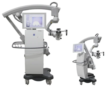
The phacoemulsification system
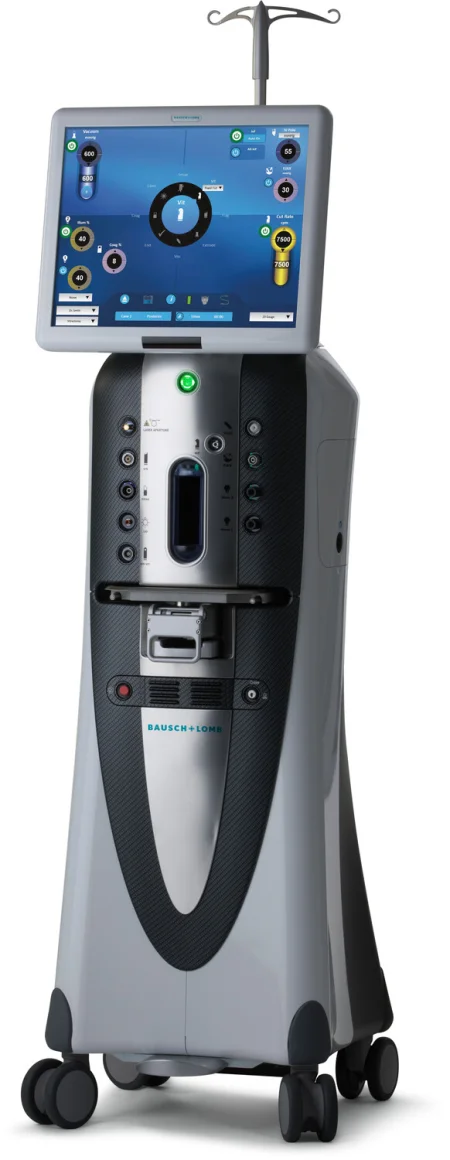
Phacoemulsification breaks up the lens with ultrasonic vibration and suctions it out through a handpiece, while balanced salt solution flows in to keep the front chamber filled. The handpieces and tips come in several sizes, straight or curved. Chapter 20 teaches ultrasound as an energy source.
- The settings for power, suction (vacuum), the height of the irrigation bottle and the foot pedal are set for each machine, each surgeon and each patient, and they cannot be copied from one brand to another.
- Everyone who runs the machine needs the knowledge and skill to use it correctly, by its maker's instructions.
- A machine that is not working properly can burn the cornea, and some chopping tips run so fast that one wrong move can shatter the cataract.
- Most phaco machines switch to a vitrectomy mode when vitreous comes forward during cataract surgery.
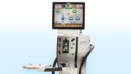
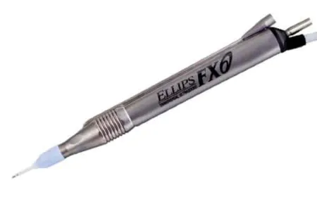
The vitrectomy machine
The vitrectomy machine draws the vitreous into a tiny port, cuts it and suctions it away, while an infusion of BSS flows in; raising or lowering the bottle changes the eye's pressure. It also supplies light inside the eye, and a unit can add an air-fluid exchange, diathermy and a laser. The vitreous is then replaced with saline, a gas bubble or silicone oil to hold the retina in place. Some units combine phaco and vitrectomy.
Cryotherapy, diathermy and lasers
- Cryotherapy unit: a probe chilled by a gas under pressure, such as nitrous oxide or carbon dioxide, freezes tissue. In retinal surgery the frozen spot heals as a scar that seals a tear. The surgeon runs it with a foot switch.
- Diathermy: low-power heat applied to the sclera over a retinal tear or detachment, which also leaves a sealing scar.
- Lasers: the argon and Nd:YAG lasers seal leaking vessels and retinal tears (photocoagulation), and the YAG laser also opens a capsule that clouds after cataract surgery. The excimer laser reshapes the cornea, and the femtosecond laser makes the cuts for cataract surgery. A laser on an ophthalmoscope, or one passed into the eye during vitrectomy, treats the retina.
Chapter 20 teaches laser safety: eyewear marked for the laser's wavelength; windows always covered for the argon, Nd:YAG and other lasers whose beams pass through glass, and for other lasers as facility policy sets; and, for argon or Nd:YAG under the microscope, a shutter that drops a filter into place to protect the surgeon's eyes.
Electrosurgery
Two kinds are common in eye surgery. A single-use, battery-powered cautery pen has a fine wire tip that heats up and seals tiny vessels; it is a true cautery and cannot cut. Bipolar electrosurgery, with fine bipolar forceps, gives precise cutting and coagulation. Monopolar units may also be used. A smoke evacuator is used whenever energy devices make smoke.
7Microinstruments, sponges, sutures and supplies
Handling microinstruments
Eye instruments are small, delicate and expensive, and many have finishes that cut glare under the microscope light. A bent tip or a dull edge can damage the eye.
- Inspect before the case: tips aligned, edges smooth, scissor blades meeting properly, needle holder catches and springs working, suction tips open. Look for burrs (rough spots on sharp edges) under magnification, or run a lint-free microsurgical wipe gently along the edge to feel for one.
- Keep a fixed order: lay instruments on the Mayo in a set order and always put each one back in its place, so you can hand the next one without looking away.
- Pass in position of use, placed firmly in the surgeon's hand, because the surgeon must not look up from the microscope to take it. Looking away breaks the surgeon's concentration and the rhythm of the case.
- Keep them clean during the case: wipe off blood, tissue and viscoelastic with a lint-free instrument wipe moistened with sterile water, and flush instruments with channels with sterile water. Chapter 27 teaches how sterile processing cleans and protects microinstruments, and how leftover residue causes toxic anterior segment syndrome (TASS).
- Keep lint and powder out: powder-free gloves, lint-free drapes and sponges, and suture kept off cloth towels. A clear plastic cover over the draped Mayo keeps dust off and protects the instruments. Lint or powder left in the eye can cause a granuloma, a small inflamed lump, after surgery.
- Steady hands: a tremor too small to notice is large under magnification, so keep your hand steady and make as little movement as possible near the field.
jar the microscope or an instrument while it touches the eye. Even a slight movement can injure the eye, so pass and take instruments with small, steady movements.
The instrument families
Eye sets vary by surgeon, but the same families appear in most of them.
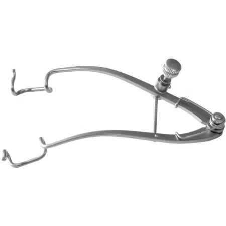
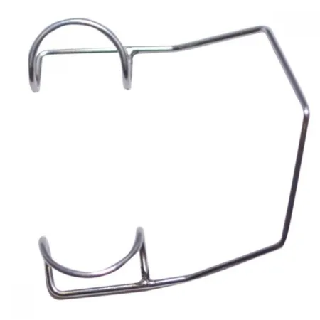
- Specula hold the lids open. A wire speculum, such as the Barraquer, is common for cataract surgery; adjustable ones, such as the Castroviejo, Williams and Lieberman, spread the lids wider for strabismus and eye removal. A Desmarres retractor holds a lid by hand.
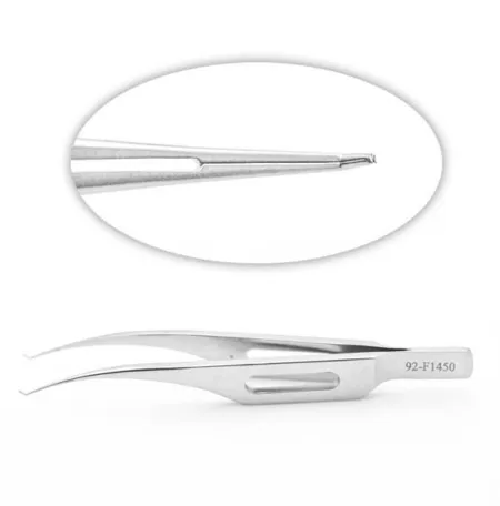
- Tissue forceps: Castroviejo suturing forceps have tiny teeth at the tip, 0.12, 0.3 or 0.5 mm, and a tying platform; Colibri forceps hold the cut edges of the cornea and sclera while the surgeon sews, and tie the suture on the platform behind the teeth; Bishop-Harmon forceps grasp delicate tissue.
- Tying forceps, such as the McPherson, have smooth platforms for tying fine sutures and are never used to grasp tissue, which they would crush.
- Scissors: Westcott spring scissors to cut the conjunctiva, cornea, sclera and iris and to divide the muscles; Vannas spring micro scissors to cut into the capsule; corneal scissors to finish a corneal cut; Stevens tenotomy scissors for the muscles and tendons; and curved enucleation scissors to cut the optic nerve. Eye suture scissors cut suture only.
- Needle holders: the Castroviejo, locking or nonlocking; the McPherson, nonlocking; and the Barraquer, locking with curved jaws.
- Knives: diamond knives, disposable keratomes and slit knives for corneal incisions, and fine blades such as Beaver blades and microblades.
- Hooks, spatulas and cannulas: muscle hooks lift the eye muscles; a Sinskey hook moves the lens implant; an iris spatula sweeps the iris back into place; blunt cannulas on syringes irrigate the eye and inject fluid or viscoelastic.
- Calipers measure the eye in millimeters, such as the Castroviejo caliper, 0 to 20 mm.
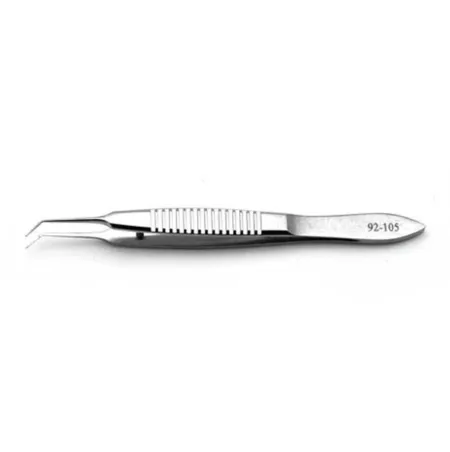
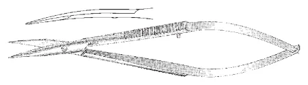
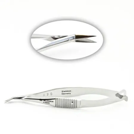
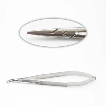
Eye sponges
Eye sponges are small spears of lint-free cellulose on a short plastic rod. Keep them apart from anything that sheds lint. To dry the field, touch the tip to the fluid and let it wick the fluid up; never wipe. A sponge is never used on the cornea.
Keeping the cornea wet
The open eye cannot keep itself moist during surgery. Irrigate the cornea with BSS as the surgeon wants, from a small squeeze bottle, an irrigating needle or a bulb syringe. Hold the tip over the eye without touching it, and let the fluid run from the inner corner to the outer corner so it does not pool; with the head turned toward the operative side, the runoff stays away from the other eye.
Sutures and needles
- Sizes: eye sutures are very fine, from 4-0 down to 10-0, and some references list sizes as small as 12-0. Fine suture causes less reaction and discomfort. Handle it as little as possible, keep it off cloth towels, and protect the needle points. Double-armed sutures, with a needle at each end, are common.
- Needles: the spatula needle, fine with a thin, flat profile, is the common needle for the cornea and sclera; other shapes, such as taper-cut and reverse cutting needles, are chosen for other tissues. Some eye needles are only about 4 mm at their widest. Inspect needles for burrs before use.
- Materials: nylon is the usual choice for the cornea, polypropylene for fixing a lens, polyester for the sclera in retinal surgery, silk for traction (bridle) sutures, and absorbable polyglactin or gut for the conjunctiva and lids.
- Loading: press a locking needle holder gently; too much pressure, or a needle too large for the holder, stops it locking.
- Cutting under the microscope: first bring the scissors into the microscope's field of view, then move the tips to the suture, rest your forefinger on the shank to steady the scissors, and lower the tips gently to cut.
- Passing sharps: the surgeon cannot look away from the microscope, so a hands-free pass into a neutral zone is not possible on the way in. Use a modified neutral zone, as chapter 25 teaches: tell the surgeon a sharp is coming, place the loaded needle holder in the surgeon's hand, say when your hand is out of the way, and the surgeon returns it to the neutral zone.
one reference has the scrub take used sharps back from the surgeon's hand in eye cases.
Counts
All sharps, including every suture needle however small, are counted and accounted for in every operation that uses them, and so are radiopaque sponges and other soft goods. Instruments must be counted whenever a body cavity is open, which eye surgery does not do, so instrument counts follow facility policy. The eye checklist confirms the counts before the patient leaves the room. Chapter 28 teaches the count itself.
an older AST standard lists eye surgery among the cases where sponge counts may not be required.
Specialty supplies
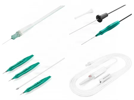
The Core Curriculum's list of eye supplies:
- Conformer: a plastic shell placed over the implant, behind the lids, after an eye is removed; it supports the lids while the socket heals.
- Corneal shield: a collagen shield shaped like a contact lens that carries antibiotic or anti-inflammatory drug to the eye and protects it after surgery; it often falls out on its own within 4 to 12 hours, which is expected.
- Eye pad and eye shield: a soft, lint-free gauze pad absorbs fluid and keeps debris out; a rigid perforated metal or plastic shield, such as the Fox shield, is taped over it to protect the eye from bumps and pressure.
- Eye-port: the Core Curriculum names it in its list; check your facility's card for the product it uses.
- Intraocular lens: the artificial lens implant.
- Microblade: fine knife blades for eye incisions.
- Phacoemulsification kit: the phaco and irrigation-aspiration tips, BSS and the other supplies the phaco machine uses, as its card lists.
- Scleral buckle: silicone bands, sponges and tires sewn to the sclera in retinal detachment repair.
- Silastic tube or lacrimal intubation kit: soft silicone tubes on metal probes that stent the tear ducts.
- Sphere: the round implant that fills the socket after the eye is removed.
- Vitrectomy kit: the single-use cutter, infusion line, light pipe and tubing for the vitrectomy machine.
Dressings
Some eye operations need no dressing at all. For many, the surgeon or scrub cleans the area with saline-wet sponges, pats it dry and tapes on a sterile eye pad. After surgery inside the eye, where pressure on the eye could do harm, a perforated shield goes over the pad. A pressure dressing is used when the surgeon wants pressure, as after an eye removal. After lid or tear duct surgery, a thin layer of antibiotic ointment on the skin and lashes keeps the dressing from sticking.
8Eye drugs on the field
Chapter 12 teaches the drug classes and how every drug reaches the field: verified aloud by two people, labeled at once and tracked to the end. Eye drugs need all of that, and more. A single mistake can blind the patient, many eye drugs come in small single-dose bottles that look alike, and several go inside the eye, where any impurity does harm.
- Label every drug the moment it reaches the field, and know what each one is for, so you can pass it at the right moment without the surgeon looking up. State its name as you pass it; the surgeon acknowledges every drug.
- Keep intraocular drugs apart. Drugs and solutions that go inside the eye are set apart from those that do not, such as ointments and the prep.
- Preservative-free inside the eye. Drugs injected into the eye must be free of preservatives, often labeled MPF (methylparaben free). Preservatives, the wrong pH, too high a concentration of dissolved particles and contaminated drugs or dyes are among the causes of TASS.
- Know the cap colors, then read the label. The caps of eye drops are color coded by class, following the American Academy of Ophthalmology: red for drugs that dilate the pupil, dark green for miotics, pink for steroids, gray for NSAIDs and tan for anti-infectives.
- Mix some drugs at the last minute. Acetylcholine (Miochol-E) is reconstituted immediately before use, and BSS Plus is made by adding its Part II to Part I with the transfer device and is used within 6 hours.
- Record every drug and amount used, and report the totals at the end of the case, for the intraoperative record.
- Watch the patient. Many drugs are used together, so report at once any change you see in the patient's appearance or behavior, or anything the patient says they feel.
The Core Curriculum lists these classes of eye drugs.
- Irrigating solutions: balanced salt solution (BSS) is a sterile irrigating fluid balanced to suit the eye's tissues. It keeps the cornea moist and irrigates inside the eye. Small 15 and 30 mL bottles are used on the field, and 250 and 500 mL bottles run through tubing into the eye during phaco and vitrectomy. BSS Plus, enriched with bicarbonate, dextrose and glutathione, supports the eye's tissues in long cases. Neither is ever given into a vein, and a bottle is used for one patient only. The surgeon may order preservative-free epinephrine added to the bottle, to keep the pupil wide or to limit bleeding.
- Lubricants: ointments that keep the cornea moist and protected. They are used under general anesthesia for any surgery, because the lids relax and the corneal reflex, which makes the eye blink, is gone; the lids are then taped shut. On waking, the patient's vision may be blurred by the ointment, and the patient must not rub the eyes.
- Mydriatics dilate the pupil (mydriasis) but still let the eye focus. Phenylephrine (2.5% or 10%) acts like the sympathetic nervous system, and it can raise the blood pressure.
- Cycloplegics dilate the pupil and also paralyze the focusing muscle (accommodation): tropicamide, cyclopentolate, atropine (which lasts 7 to 14 days), homatropine and scopolamine. They block acetylcholine. Drugs that dilate the pupil may be contraindicated in angle-closure (narrow-angle) glaucoma, and pressing gently over the lacrimal sac after drops cuts the amount absorbed into the body.
- Miotics constrict the pupil (miosis). Acetylcholine (Miochol-E) and carbachol (Miostat) are injected into the front chamber during surgery, for example right after the lens implant goes in, to constrict the pupil, which pulls the iris back into place and helps keep the lens in position; pilocarpine and carbachol drops lower the pressure in glaucoma.
- Viscoelastics, also called ophthalmic viscosurgical devices (OVDs): thick gels such as sodium hyaluronate, or a mix of chondroitin and hyaluronate, that fill the front chamber, keep it deep, hold tissues apart and protect the cornea's inner lining; they can also coat the lens implant. They come in prefilled syringes with blunt cannulas. Many brands are kept refrigerated and need about 20 to 40 minutes to warm to room temperature, so plan ahead. They are washed out at the end of the case, because gel left behind raises the eye pressure.
- Local anesthesia adjuncts: hyaluronidase, an enzyme mixed with the block's anesthetic so it spreads through the tissue, and not used in infected, inflamed or cancerous tissue; and epinephrine, which narrows vessels, makes the block last longer and reduces bleeding. The topical anesthetics and block drugs themselves are chapter 13, and the block drugs count toward the maximum doses that chapter 13 teaches.
- Enzymes and zonulysis: zonulysis (zonulolysis) is the use of an enzyme, alpha-chymotrypsin, to dissolve the zonules, so the whole lens can be lifted out in its capsule in an intracapsular cataract extraction, an older operation rarely done today.
- Dyes and stains: trypan blue is injected into the front chamber to stain the lens capsule so the surgeon can see it to open it, and the extra is washed out at once; fluorescein strips stain the cornea; a marking pen with methylene blue can mark a retinal tear on the sclera. Fluorescein and indocyanine green are also given into a vein for angiography, a diagnostic test.
- Antibiotics: drops and ointments (such as polymyxin B with bacitracin, neomycin combinations, gentamicin, tobramycin and fluoroquinolones such as moxifloxacin), injections under the conjunctiva at the end of the case (such as cefazolin or gentamicin), and antibiotics injected into the front chamber (intracameral), which a review of the research found well supported by evidence for preventing endophthalmitis, an infection inside the eye.
Other drugs you will see in eye cases:
- Steroids and NSAIDs: dexamethasone, betamethasone and methylprednisolone injected under the conjunctiva, and steroid drops, calm inflammation after surgery; some come combined with an antibiotic. NSAID drops such as ketorolac reduce inflammation, and flurbiprofen helps keep the pupil from narrowing during surgery.
- Antimetabolites: mitomycin and 5-fluorouracil (5-FU) are applied on small sponges to slow scarring in glaucoma filtering surgery and pterygium excision. They are hazardous drugs: handle and discard them by OSHA rules and your facility's policy for antineoplastics, take the exposed instruments off the field in a basin, and change gloves.
- Pressure-lowering drugs: acetazolamide (Diamox) and mannitol, given into a vein by the anesthesia provider, and timolol drops. A patient who receives mannitol often needs a urinary catheter.
- Gases and liquids that hold the retina in place: sulfur hexafluoride (SF6) and perfluoropropane (C3F8) gases, silicone oil and liquid perfluorocarbons, taught with retinal surgery below.
9Cataract surgery: the lens
Phacoemulsification with an intraocular lens
What and why: cataract extraction removes the cloudy lens and replaces it with an intraocular lens (IOL), restoring the focusing power the lens provided. More than three million lens extractions are done each year in the United States. Today it is almost always an extracapsular cataract extraction (ECCE): the nucleus and cortex come out and the capsule stays, as a bag to hold the new lens. Phacoemulsification, breaking up the lens with ultrasound and suctioning it out, is the usual way. Bilateral cataracts are done at separate times, usually 4 to 8 weeks apart, so the patient may see unevenly in between.
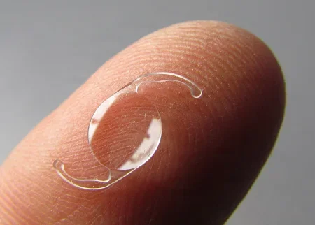
The intraocular lens: IOLs are made of silicone or acrylic and come in powers measured in diopters. The surgeon works out the power from the cornea's curve and the eye's length before the day of surgery.
- Types: a monofocal lens focuses at one distance, usually far, so the patient still wears glasses to read; multifocal and accommodating lenses give near and far vision; a toric lens also corrects astigmatism.
- Where it goes: a posterior chamber lens sits in the capsular bag behind the iris, its most natural place. An anterior chamber lens, in front of the iris, is used when there is no capsule to hold a lens, as after a torn capsule with vitreous loss.
- Folding it: foldable lenses go through an incision of 2.6 mm or less. The scrub folds the lens into the injector exactly as its maker directs, because a lens folded wrong can open upside down or backward, spoiling the result and inflaming the eye when the surgeon has to fix it. Some lenses come already loaded in their injector.
Before surgery: in the holding area, drops dilate the pupil (phenylephrine, with a cycloplegic such as tropicamide), an NSAID drop such as flurbiprofen helps keep it wide, and a topical anesthetic such as tetracaine numbs the eye. The patient keeps the eye closed so the cornea does not dry. In the OR, the patient is asked to look at the microscope light; if the patient cannot keep the eye open and still, the surgeon may give a facial nerve or retrobulbar block. A patient on long-term glaucoma treatment or an alpha-blocker such as tamsulosin, or with diabetes, is more prone to a floppy iris that prolapses and a pupil that narrows despite the drops; the setup includes a pupil expander.
Position, prep and drape: supine, the head steady on its headrest; povidone-iodine drops in the eye and a routine eye prep; a head drape and an adhesive eye drape.
Setup: a cataract set with the phaco handpiece and tips, the irrigation and aspiration (I/A) handpiece and tips, BSS, capsulorhexis forceps or a cystotome, a hydrodissection cannula, a chopper or second instrument, lens manipulators and a positioning hook such as the Sinskey, a pupil expander such as iris hooks or a ring, IOL forceps, and the IOL with its injector. On hold: an anterior vitrectomy handpiece, 10-0 nylon or polyglactin suture and a second IOL.
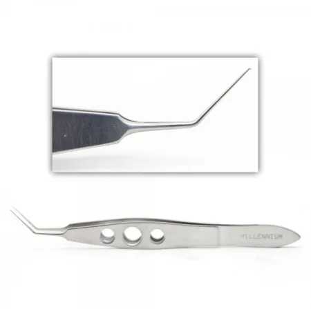
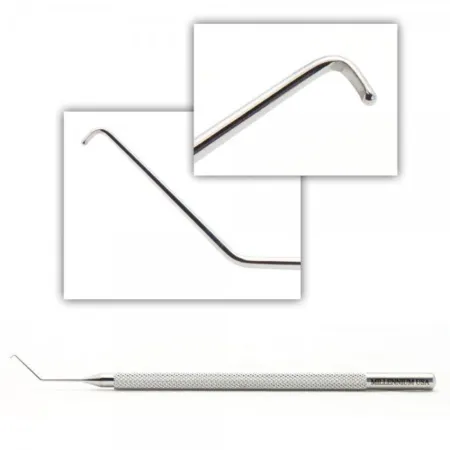
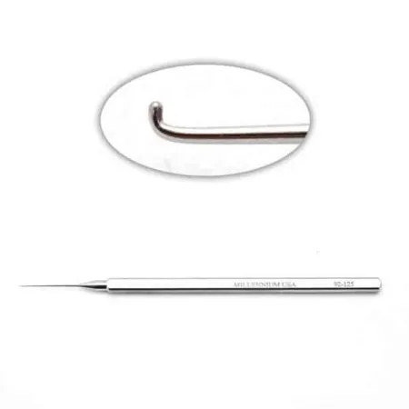
Medications: BSS for irrigation, sometimes with epinephrine added, preservative-free lidocaine for the front chamber, viscoelastic, trypan blue to make the capsule easier to see, a miotic, an antibiotic for injection into the front chamber at the end, and antibiotic and steroid drops.
Key steps:
- The speculum goes in, and the surgeon makes a 1 mm side-port stab incision (a paracentesis) into the front chamber. Preservative-free lidocaine may be injected through it.
- The instant the chamber is opened, aqueous leaks out and its pressure falls, so the surgeon fills it with viscoelastic to keep it deep and protect the cornea's lining.
- The main incision is made with a keratome or diamond knife, in clear cornea or near the limbus. An incision in clear cornea can be placed to reduce the patient's astigmatism.
- Capsulorhexis: a continuous round opening is torn in the front of the capsule with capsulorhexis forceps or a cystotome. This step must give a round, stable opening into the bag.
- Hydrodissection: BSS squirted under the capsule through a fine blunt cannula frees the nucleus so it turns in the bag.
- Phaco: the probe sculpts or grooves the nucleus, and a chopper or second instrument passed through the side port helps crack it into quarters, which the probe emulsifies and suctions away. If the nucleus is too hard to emulsify, the surgeon can press it out by hand instead (a manual extracapsular extraction).
- The I/A tip strips the remaining cortex from the capsule, and the capsule may be polished. Working from the edges, not the center, avoids drawing vitreous forward.
- Viscoelastic fills the bag. You fold the lens into the injector, check it, and state its model and power as you pass it; the surgeon injects it into the bag, where it unfolds, and centers it with a hook or the I/A tip.
- The I/A tip washes out the viscoelastic. Many incisions seal themselves; others get a 10-0 nylon stitch. An antibiotic may be injected into the front chamber, a miotic may be used, and antibiotic or combination drops go on; protocols against TASS avoid ointment after cataract surgery, because it can slip into the front chamber.
Throughout the case, keep the cornea moist with BSS.
at the lens implant, you say the model and power out loud as you pass it. The lens is checked against the patient's record just before it goes in, every time.
Specimen, counts and dressing: the lens is suctioned away in fragments, and facility policy decides whether anything goes to pathology. Sharps are counted, and any sponges used. An eye pad and a shield, or a shield alone.
Wound class: I, clean.
After surgery: most patients go home within a few hours. Teach them to wear the shield as the surgeon directs, not to sleep on the operative side or the stomach for the first week, not to rub or squeeze the eye, and to avoid strenuous exercise for 2 to 3 weeks. Sudden pain, sudden loss of vision, light sensitivity or a severe headache must be reported at once. Complications are rare: infection (endophthalmitis), bleeding, inflammation, high or low eye pressure, double vision and vision loss. Months or years later, cells can regrow on the capsule and cloud it; a YAG laser then opens a hole in the center of the capsule (a capsulotomy). Cataract surgery also raises the risk of retinal detachment.
When the capsule tears: anterior vitrectomy
If the back of the capsule tears, vitreous can push forward into the front chamber. The surgeon stops phaco and switches to vitrectomy, either with the phaco machine's vitrectomy mode or a separate cutter. With irrigation running, the cutter removes the vitreous from the front of the eye until none is left there and the iris falls back; vitreous can also be cut with Westcott scissors after drawing it up with a cellulose sponge. The pupil is constricted with acetylcholine, the chamber is refilled, and a lens is placed where support allows. Have the vitrectomy setup ready on every cataract case.
Femtosecond laser-assisted cataract surgery (FLACS)
A femtosecond laser fires pulses one quadrillionth of a second long, which cut precisely without heat. Before phaco, the laser is docked on the eye through a cone, with a speculum in place, and it makes accurate incisions and an even, predictable capsulorhexis and can precut the nucleus. Less ultrasound energy is then needed, and one study found less damage to the cornea's lining and a faster return of sight. The laser helps most with hard, dense cataracts. It also helps with eyes that have had corneal surgery to correct vision, such as LASIK, and it can correct a small amount of astigmatism. Complication rates match those of standard surgery, but docking can leave the eye bloodshot for a few days. The phaco steps then follow as above, and the wound class (I, clean) and the aftercare are those of phacoemulsification.
Two more lens cases
- Intracapsular extraction (ICCE): the whole lens, capsule and all, is lifted out, once with a cryoprobe after an enzyme had loosened the zonules. It leaves no capsule to hold a posterior chamber lens and is now rarely done, except for a dislocated lens.
- Children's cataracts: a cataract that affects a child's sight is removed, because a child's vision develops only with clear images; otherwise amblyopia follows. After an exam under anesthesia with an A-scan, the soft lens is removed with a vitrector and an IOL is placed or the child is left without a lens; a child with an IOL wears bifocals, because the IOL cannot accommodate.
10The cornea, conjunctiva and iris
Penetrating keratoplasty (corneal transplant)
What and why: a penetrating keratoplasty replaces the full thickness of a scarred or cloudy cornea with a donor cornea, to restore sight when the retina and optic nerve still work. The common reasons are burns, infection, scarring after injury, inherited corneal disease, swelling after earlier eye surgery and keratoconus. The cornea has no blood vessels, so donor tissue needs no antigen matching, and corneal transplants are among the most successful of all transplants.
Partial-thickness grafts: when only some layers are diseased, the surgeon replaces just those. A deep anterior lamellar keratoplasty (DALK) replaces the front layers and keeps the patient's own endothelium, lowering the risk of rejection; it suits keratoconus and scars that do not go all the way through. An endothelial keratoplasty, such as Descemet stripping endothelial keratoplasty (DSEK), replaces only the inner layer, for diseases such as Fuchs dystrophy, in which the endothelial cells die and the cornea swells. It heals faster with less astigmatism, and an air bubble injected under the graft presses it into place.
The donor cornea: it comes from an eye bank, cut as a corneoscleral button or as a whole eye, refrigerated in a storage medium such as Optisol-GS, which keeps it up to 14 days. The donor is screened for diseases such as HIV and hepatitis B and C. Handle it as the gift it is. Eye donation and recovery are taught with the eye removals below.
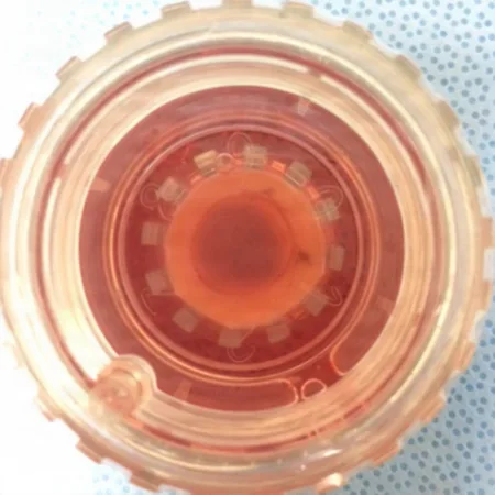
Before surgery: local anesthesia with a block, often with a facial nerve block, or general anesthesia. The patient must not be able to squeeze the lids once the cornea is out, because pressure from behind can push the eye's contents out or cause bleeding inside the eye, so many surgeons give mannitol beforehand to lower the pressure. If a block is planned, the skin behind the ear is prepped too. The microscope is positioned and locked before the prep, then swung aside.
Setup: a corneal transplant set with trephines, a punch block, corneal scissors, a fixation ring (such as a Flieringa ring), fine forceps, 10-0 nylon, viscoelastic and BSS. The donor tissue is prepared on its own separate table and Mayo, and instruments are not shared between the two, so nothing passes from one eye to the other.
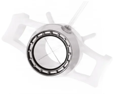
Key steps:
- A speculum, then a fixation ring or bridle sutures to support the eye.
- The donor button is placed on a block with its outer surface down, and a trephine punches a disc a little larger than the opening to be made in the patient's eye, for a proper fit. A drop of storage medium keeps it moist until it is used, and some of the medium may go to the lab for culture.
- The surgeon trephines the patient's cornea, by hand or with a suction trephine, and finishes the cut with corneal scissors. Receive the patient's cornea as a specimen. The eye is now "open sky", wide open to contamination, so have the graft, the next instruments and the sutures ready at once.
- Any extra work is done now, such as a peripheral iridectomy or a cataract extraction with a lens implant.
- The graft is laid in the opening and anchored with four stitches at the cardinal points, then sewn with interrupted or running 10-0 nylon.
- Air, BSS or viscoelastic re-forms the front chamber and keeps the iris off the suture line; the wound is checked for a leak.
- Antibiotic and steroid injections or drops, ointment, an eye pad and a shield.
Specimen: the patient's cornea, and a culture of the storage medium if the surgeon orders it.
Wound class: I, clean.
After surgery: full recovery of sight can take up to a year. The risks are bleeding, infection, glaucoma, swelling and rejection, which can happen weeks or years later. Pain, light sensitivity, redness and dimming vision are warning signs that need care at once, because steroids started early can stop a rejection.
Laceration repair
- Cornea: a cut through the cornea is closed with 10-0 sutures placed edge to edge, or with a tissue adhesive approved for the eye, which sets on contact and seals the wound. Antibiotics may be injected under the conjunctiva before the dressing.
- Conjunctiva: tears, and gaps left after removing a growth, are usually closed by freeing the edges and sewing them.
- A ruptured globe: the eye is imaged first to look for foreign bodies and hidden damage. An eye too damaged to see again, or a blind, painful eye, may have to be removed.
- The repair: done under the microscope with 9-0 or 10-0 sutures. When a cut crosses both the cornea and the sclera, a first stitch at the limbus lines up the edges, and the cornea is closed before the sclera. Antibiotic drops or ointment follow.
Wound class: a fresh accidental wound is Class III; an old one with dead (devitalized) tissue, or an infected one, is Class IV.
Pterygium excision
What and why: a pterygium is a fleshy, triangular growth of conjunctiva that spreads from the inner corner onto the cornea, more common in people who work outdoors. It is not cancer, but it tends to come back, so surgery usually waits until it affects vision.
Steps: under topical, peribulbar or retrobulbar anesthesia, the surgeon cuts the growth off the cornea and sclera, seals bleeding at the limbus with a battery cautery or bipolar, and may smooth the surface with a diamond burr. The bare sclera is covered with a graft of the patient's own conjunctiva or with amniotic membrane, held with fine sutures or fibrin glue. Mitomycin may be applied to lower the chance of regrowth, handled as a hazardous drug.
Wound class: I, clean; no tract is entered.
Iridotomy and iridectomy
What and why: both make an opening in the iris, so aqueous can pass from the posterior to the anterior chamber again. This relieves the block in angle-closure glaucoma: the iris falls back from the drainage angle and fluid can reach the trabecular meshwork. An iridotomy is a small hole, usually made with a laser; an iridectomy removes a piece of iris. A miotic may be used to narrow the pupil for a laser iridotomy, and the pressure may rise or the eye may inflame afterward.
Before a surgical iridectomy: a retrobulbar block or general anesthesia, supine with the head on a headrest, and the routine eye prep and drape; if the pressure is very high, drugs to lower it are given before the incision.
Surgical peripheral iridectomy: done when a laser cannot be used, as with a cloudy cornea or an inflamed eye, and often as part of a trabeculectomy. The surgeon makes a small incision at the upper limbus or in clear cornea, draws a fold of iris out with forceps and snips it off, then settles the iris back in place by stroking the cornea gently with a blunt instrument, such as a spatula or muscle hook, or with a stream of BSS. The incision is closed with 10-0 nonabsorbable suture, or absorbable suture at the limbus, and an antibiotic may be injected into the front chamber before the eye pad.
Wound class: I, clean.
Refractive surgery
Laser vision correction, such as LASIK and PRK, reshapes the cornea with an excimer laser to correct nearsightedness, farsightedness and astigmatism. It is mostly done in physicians' offices or physician-owned centers rather than in hospitals or ambulatory surgery centers.
11Glaucoma surgery
Glaucoma operations lower the pressure in the eye, either by opening a new drain for aqueous humor or by making less of it. They save the sight that is left; they do not bring back what is lost. Glaucoma operations are clean cases, Class I.
Trabeculectomy
What and why: a trabeculectomy is a filtering operation. The surgeon removes a small block of tissue containing part of the trabecular meshwork, under a flap of sclera, to make a new channel from the front chamber. Fluid seeps out under the flap into a small blister under the conjunctiva, the bleb, where the body absorbs it. It is done when other treatment does not control the pressure, and it is often combined with cataract extraction and a lens implant.
Before surgery: local anesthesia with a retrobulbar block, or general anesthesia; if the pressure is very high, drugs to lower it are given before the incision.
Setup: trabeculectomy instruments with a diathermy or cautery unit, fine blades such as Beaver blades, Vannas scissors or a punch, the antimetabolite drug, eye sponges for applying it, and 10-0 nylon and absorbable suture; a drainage device on hold if the surgeon may need one.
Key steps:
- A speculum, and often a traction suture of 4-0 silk under the superior rectus muscle, to steady the eye.
- The conjunctiva and Tenon capsule are opened at the limbus with toothed forceps and a blade, then freed from the sclera with Westcott scissors to make a conjunctival flap; bleeding is sealed with bipolar or cautery.
- The antimetabolite: small sponges soaked in mitomycin or 5-FU are placed between the flap and the sclera for the time the surgeon directs (the references give about 1 to 5 minutes), then removed and the area is washed with plenty of BSS. Hand off the instruments they touched, in a basin, and change gloves.
- A partial-thickness flap of sclera, square or triangular, is outlined with cautery and cut with a fine blade, then dissected forward from its tip.
- A small stab through the cornea (a paracentesis) lets aqueous out and is used later to refill the chamber.
- Under the flap, a block of deep tissue at the limbus, including part of the meshwork, is cut out with Vannas scissors or a punch, opening the new channel into the front chamber.
- A peripheral iridectomy is made, and one is always made if the iris prolapses into the wound.
- Interrupted 10-0 nylon sutures close the scleral flap; when you follow the suture, do not put too much tension on it. Tenon capsule and the conjunctiva are closed with fine absorbable suture. BSS is injected into the chamber, and an eye sponge held on the wound checks for a leak.
- Antibiotic and steroid injections under the conjunctiva, ointment, a pad and a shield.
After surgery: the operation works only if a bleb forms in the first days. A leaking bleb may need a bandage contact lens or repair, and a cataract may form later.
Laser trabeculoplasty
A trabeculoplasty is a laser treatment of the trabecular meshwork that helps aqueous drain out. It can be the first treatment for open-angle glaucoma or be used when drops fail or are not tolerated. Argon laser trabeculoplasty (ALT) is one type; selective laser trabeculoplasty (SLT), with a frequency-doubled Nd:YAG laser, is preferred because it uses less energy and causes less inflammation, and it can be repeated. One review reports a good pressure drop at first in more than three quarters of eyes treated with the argon laser, but 30 percent to more than half need more surgery within 5 years.
Glaucoma drainage device (tube shunt)
What and why: when a filtering operation fails, the surgeon implants an aqueous shunt, or glaucoma drainage device: a fine tube whose tip sits in the front chamber, attached to a plate sewn to the sclera far back under the conjunctiva. Aqueous drains out through the tube to the plate, which acts as a reservoir. Some devices have a valve and some do not.
Before surgery: usually local anesthesia with a retrobulbar or peribulbar block. The device and its patch graft (donor sclera or pericardium) may be soaked in an antibiotic solution, and both are recorded as implants.
Priming: one maker's valved device must be primed before it is implanted, by injecting about 1 mL of BSS or sterile water through the tube with a blunt 26-gauge cannula; a sharp needle can puncture the tube. A device that will not prime is not used. Follow the maker's instructions for the device in your room.
Key steps: the rectus muscles are looped with traction sutures, the conjunctiva is opened, and the plate is placed by measurement and sewn to the sclera. The surgeon checks that the tube is open, then may close it for now with an occluding suture. A needle makes a tunnel into the front chamber, the tube is trimmed, passed into the chamber and sewn down, and a patch graft covers it. The conjunctiva is closed, and an antibiotic may be injected under it before the pad.
After surgery: inflammation and a rise in pressure may follow, usually settling within 4 to 6 weeks. Pressure that drops too low, clogging, scarring around the plate, trouble moving the eye and injury to the cornea are the other risks.
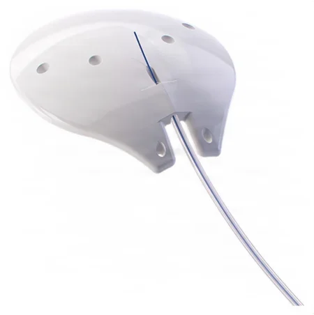
Other glaucoma operations
- A stent at cataract surgery: for open-angle glaucoma, a tiny titanium tube can be placed in the trabecular meshwork through the cataract incision at the end of the case, viewed through a gonio prism with the head turned.
- Canaloplasty: a microcatheter is threaded into the canal of Schlemm and viscoelastic is injected to widen it.
- Children: in congenital glaucoma, a goniotomy (cutting into the meshwork) or a trabeculotomy (opening the canal of Schlemm into the chamber from outside, through a scleral flap) comes first; trabeculectomy, drainage devices and cyclodestruction come next.
- Cyclodestruction: freezing (cryotherapy) or a diode laser destroys part of the ciliary body, so it makes less aqueous.
12The retina and vitreous
Retinal surgery is often urgent, because a detached retina threatens sight, and some of these cases are long.
Pars plana vitrectomy
What and why: a vitrectomy removes all or part of the vitreous. Through the pars plana, where there is no retina, the surgeon reaches the back of the eye with little risk of tearing the retina. It is done to clear blood or clouding from the vitreous, for advanced diabetic eye disease, severe injuries and foreign bodies, scar tissue on the retina, detachments from large tears, macular holes, infection inside the eye (endophthalmitis), and to take a sample of the vitreous. The surgeon may also peel membranes off the retina and seal vessels.
Before surgery: local anesthesia with a retrobulbar block and sedation. A scleral buckle may be needed in the same operation, so be ready for one.
Setup: the vitrectomy machine and pack, the microscope with a contact lens or a wide-angle viewing system mounted on it, small trocars and cannulas (such as 23 or 25 gauge), a light pipe and sometimes a chandelier light, an infusion of buffered BSS, picks, microforceps and microscissors, an endolaser and cryoprobe, soft-tipped cannulas for draining fluid, scleral plugs, and the tamponade the surgeon plans: air, gas or silicone oil. Culture tubes are kept ready.
Your jobs: attach the handpieces to their sterile tubing and hand the ends off to be connected; prime the infusion line and clear every air bubble; test the cutter and the suction before it reaches the eye; know that the surgeon runs the cutter with a foot switch; and remember that the infusion is stopped before the instruments come out of the eye.
Key steps:
- A speculum. Three openings are made through the pars plana, about 3 to 4 mm behind the limbus, often with small trocar-cannulas: one for the infusion, one for the light and one for the cutter or other instruments. The infusion cannula's position inside the eye is checked.
- The microscope and viewing system are brought into place. If a dense cataract blocks the view, the lens is removed first (a lensectomy).
- The surgeon cuts and removes the vitreous under direct view, then treats the problem: peeling membranes, sealing tears with the endolaser, draining fluid from under the retina and flattening it with a heavy liquid, then exchanging the fluid for air, gas or silicone oil.
- The cannulas come out; small sites often seal without sutures, and larger ones are closed with fine suture. Vitreous washings may go for culture.
- Steroid or antibiotic injections under the conjunctiva, a pad and a shield.
Holding the retina in place (tamponade):
- Gas: sulfur hexafluoride (SF6) or perfluoropropane (C3F8), drawn through a 0.22-micron filter and mixed with filtered air; the stronger the mix, the longer the bubble lasts. Both gases expand after injection, SF6 to about 2.5 times its volume and C3F8 to about 4 times, over about 2 days, and the eye's own fluid slowly replaces them over weeks to a few months.
- Silicone oil: for complicated cases or patients who cannot keep their head down. It can stay in the eye, but it should be taken out within about a year once the retina is attached and stable, which needs another operation.
- Liquid perfluorocarbon: heavier than BSS, it presses the retina flat during surgery and is removed after the repair.
let nitrous oxide run when a gas bubble is in the eye. Nitrous oxide floods into the bubble, which can swell to three times its size and drive the eye pressure up. The anesthesia provider stops it at least 20 minutes before the gas is injected.
After surgery: a patient with a gas bubble must keep the head in a set position, often facedown, for days to weeks, so the bubble presses on the tear. The patient wears a wristband naming the gas and when it was placed, and must tell any anesthesia provider about it, and must not fly or travel to high places until the gas is gone, because lower air pressure makes the bubble swell. Complications include new retinal tears, cataract, high pressure, bleeding and infection.
Wound class: I, clean.
Scleral buckling
What and why: a scleral buckle repairs a retinal detachment from outside the eye. A piece of silicone, a sponge or a band, is sewn onto the sclera over the tear, so it pushes the wall of the eye in against the detached retina, and cryotherapy, diathermy or laser seals the tear with a scar. An encircling band around the whole eye also eases the vitreous's pull on the retina.
Before surgery: general anesthesia, or MAC with a block; a long case may call for general anesthesia.
Setup: a scleral buckle set; the silicone bands, sponges, tires and sleeves the surgeon chooses, often soaked in an antibiotic; the cryotherapy unit; an indirect ophthalmoscope with handheld lenses and a headlight; nonabsorbable mattress sutures; gases; and a vitrectomy setup on hold.
Key steps:
- The conjunctiva is opened at the limbus, and the four rectus muscles are lifted on a muscle hook and looped with traction sutures, tagged with small clamps. You may steady the eye by the traction sutures.
- The surgeon finds the tear with the indirect ophthalmoscope while pressing on the sclera, and marks the spot on the sclera with diathermy or a marking pen.
- The cryoprobe freezes the area under direct view until an ice ball forms over the whole lesion; diathermy or laser are alternatives.
- The buckle is sewn to the sclera with double-armed nonabsorbable mattress sutures. For an encircling band, the band passes under all four muscles and is held with a sleeve at the size the surgeon wants.
- If needed, fluid under the retina is drained through a tiny opening in the sclera, and air or gas may be injected through the pars plana.
- The traction sutures come out, the conjunctiva is closed with fine absorbable suture, and an antibiotic or steroid may be injected under it before the pad.
After surgery: some discomfort is common. Vigorous activity is avoided for about a month, and no flying while gas is in the eye. Infection, inflammation, too little blood flow to the front of the eye and a detached choroid are possible complications.
Wound class: I, clean; no tract is entered.
Pneumatic retinopexy
In a pneumatic retinopexy, the surgeon injects a bubble of air or gas into the vitreous through the pars plana. The bubble rises against the retinal break and holds it shut, and after about 7 to 10 days of healing, laser or cryotherapy seals it. The patient's head position afterward depends on where the tear is, so the bubble floats onto it, and the same gas precautions apply. It may be combined with a buckle or a vitrectomy. It enters no tract, so it is Class I, clean.
Laser treatment of the retina
For proliferative diabetic retinopathy, laser burns scattered over the retina (panretinal photocoagulation) are the main treatment; the laser is aimed through an ophthalmoscope or a slit lamp, or with a probe inside the eye during vitrectomy. In premature babies with retinopathy of prematurity, a laser mounted on an indirect ophthalmoscope or drug injections into the eye are used, and a buckle or vitrectomy for the most advanced stage.
13Strabismus correction: recession and resection
What and why: strabismus surgery lines up the eyes by changing where an eye muscle pulls. A recession weakens a muscle by detaching it and sewing it back farther behind its original insertion. A resection strengthens a muscle by cutting out a piece and sewing the shortened muscle back at its original insertion; a plication strengthens it by folding it instead. Glasses and eye exercises are the treatments without surgery, and any other eye problem, such as a cataract, is treated before the strabismus. In children the goal is binocular vision before amblyopia sets in.
Before surgery: general anesthesia for children, sometimes local anesthesia for adults, and a retrobulbar block may be added. The patient lies supine with the shoulders slightly raised. Both sides of the face are prepped with 5 percent povidone-iodine and both eyes are left exposed, so the surgeon can compare them; the eye not being operated on is covered with a moist pad or taped shut.
Setup: a strabismus set: a wide speculum, locking fixation forceps, Westcott and Stevens tenotomy scissors, muscle hooks of several sizes, a Jameson muscle clamp, a caliper and a sterile marking pen, cautery, 4-0 silk traction sutures and double-armed 6-0 muscle sutures, absorbable or nonabsorbable, on the needle the surgeon prefers.
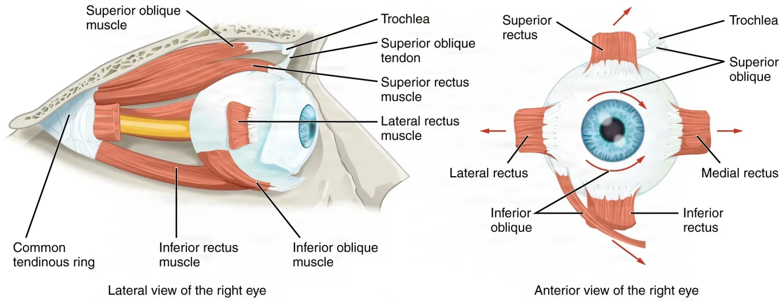
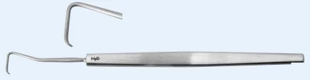
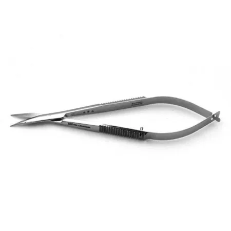
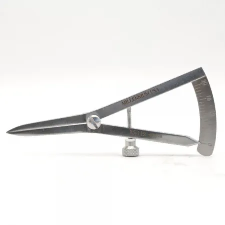
Key steps (recession):
- The speculum goes in, and the surgeon may move the eye with forceps in each direction to test whether it is held back (a forced duction test).
- Locking forceps at the limbus turn the eye, and the conjunctiva is opened over the muscle, at the limbus or in the fold behind it, with Westcott scissors.
- Tenon capsule is dissected down to bare sclera, and a muscle hook is passed under the muscle and swept along it to free it from the sclera.
- A double-armed absorbable suture is woven through the muscle close to its insertion and locked at each edge. A small clamp may be left on the muscle for up to 3 minutes to stop bleeding.
- The muscle is cut from the globe with scissors.
- The caliper measures the new spot behind the old insertion, which is marked. The surgeon passes the sutures through the surface of the sclera there, pulls the muscle up to it and ties it, then measures again. Pass an empty needle holder and smooth forceps for tying.
- The conjunctiva is closed with fine absorbable suture, often on a spatula needle, and an antibiotic or combination ointment goes on. Many surgeons use no dressing; others add a pad and shield.
Resection: the first steps are the same. The surgeon measures the length to remove, places the sutures at that point behind a resection clamp, cuts the muscle in front of the clamp, removes the stump from the globe, and sews the shortened muscle back at its original insertion. The piece of muscle removed is a specimen.
Adjustable sutures: some surgeons tie the muscle with a slipknot left where it can be reached. Within about a day, with the patient awake and the eye numbed with drops, the surgeon adjusts the suture while the patient reports when the eyes line up, then ties it for good.
pulling on an eye muscle can set off the oculocardiac reflex, a vagal response that slows the heart. If the anesthesia provider calls a slow heart rate, the surgeon lets go of the muscle until the rate recovers.
Wound class: I, clean.
After surgery: nausea and vomiting are common, especially in children, from the vagal reflex during surgery; they are treated with antiemetics and pass on their own.
14The eyelid
Lid operations are often done under local anesthesia, sometimes in the office. A head drape is used, and a corneal shield or protector may be placed on the eye to guard it while the surgeon works on the lid. Chapter 38 teaches blepharoplasty and the other oculoplastic operations that plastic surgeons do.
Chalazion excision
What and why: a chalazion is a firm lump in the eyelid from a blocked meibomian gland. The trapped oil causes inflammation, and a granuloma (a lump of inflamed tissue) forms; it is not an infection, though it can become infected. Surgery is for one that does not settle with simpler treatment.
Before surgery: local anesthesia, with MAC for children and anxious adults.
Setup: a chalazion set: a chalazion clamp, chalazion curettes, fine-toothed forceps, a Beaver #67 or a #11 blade, Westcott scissors, a meibomian expression paddle, and bipolar or a cautery pen.
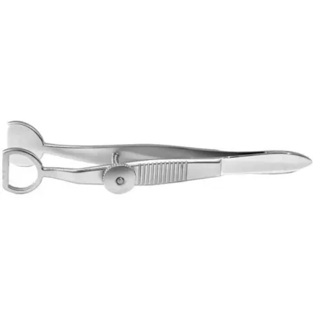
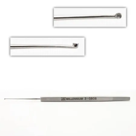
Key steps:
- After local anesthetic, the clamp grips the lid around the lump and everts it, which also controls bleeding and gives a firm surface to cut against.
- A vertical cut is made through the tarsal plate on the inner side of the lid; a chalazion near the skin may be opened from the outside instead.
- The contents are scraped out with a curette. Wipe the curette clean with a damp sponge between passes.
- Bleeding is sealed with bipolar or cautery; drops of BSS show the surgeon where it is bleeding. The wound is left open to drain or closed, and an eye pad goes on. The patient may put ice on it the first day.
Wound class: Class I by the CDC's definitions, because a chalazion is a chronic granuloma, not an acute inflammation or an infection, and no tract is entered; your facility's policy decides. One reference lists it as Class III, contaminated.
Entropion and ectropion repair
What and why: in an entropion the lid margin, usually the lower one, turns in, so the lashes rub the cornea (trichiasis), causing irritation, pain and tearing. The most common type comes with age, as the lid's supports loosen (involutional); scarring of the inner lid can also pull it in (cicatricial). In an ectropion the lower lid sags and turns out, so tears spill over and the exposed conjunctiva and cornea dry and grow irritated, and can ulcerate. It comes mostly with age, as the orbicularis muscle and the canthal tendons relax, but also from scarring, a facial nerve paralysis such as Bell palsy, or birth.
Before surgery: local anesthetic injected into the lid; general anesthesia when needed. Both eyes may be prepped. A corneal shield protects the eye.
Setup: an oculoplastic set, a sterile marking pen, smooth and tying forceps, iris and tenotomy scissors, a #15 blade, needle-tip cautery or bipolar, and fine absorbable and nonabsorbable sutures.
Key steps: the repair fits the cause, and there are many methods.
- Tightening the lid at the outer corner (a lateral tarsal strip or canthal sling): the surgeon opens the outer corner, frees a strip of tarsal plate and tendon, pulls the lid out to the side to tighten it, and sews the strip to the periosteum on the inside of the outer orbital rim. This corrects both involutional entropion and ectropion.
- Plicating the lid's retractor muscles for entropion: through an incision just below the tarsal plate, the retractors are exposed and folded with sutures, and the extra skin is trimmed.
- Everting sutures (a Wies procedure) for scarring entropion: a full-thickness cut across the lid and mattress sutures that turn the margin back out.
- A wedge resection for ectropion: a full-thickness wedge of lid is removed and the edges of the tarsal plate are sewn together to shorten and tighten the lid.
During the case, irrigate the cornea and blot blood and fluid as the surgeon works. Antibiotic ointment goes on the wound, and an ice-cold compress helps the swelling.
Wound class: I, clean.
Eyelid laceration repair
What and why: a cut lid must be repaired exactly, because a lid margin that heals out of line can turn in or out. Deeper structures lie close under the skin: the tear drainage canals near the inner corner, the lacrimal gland, the orbicularis muscle, the angular artery and the levator muscle that lifts the lid. Fat showing in the wound means the levator may be injured. A cut through the canaliculus is stented to keep it open.
Before surgery: the whole eye is imaged first to rule out a hidden injury, such as a foreign body inside the eye, a retinal injury or a blowout fracture. Local anesthetic, with or without epinephrine, is enough for most superficial cuts; general anesthesia is used when the tear system or bone is involved, and for children. The wound is cleaned (debrided), the prep with dilute povidone-iodine is very gentle, to protect the lid, and the eye is irrigated with BSS.
Key steps: the lid margin is lined up first with sutures, then the tarsal plate is closed with absorbable suture, and the skin and corner with fine nonabsorbable suture. A torn canaliculus is repaired around a rod or silicone stent passed through it.
Wound class: a fresh traumatic wound is Class III, and an old one with dead tissue, or an infected one, Class IV; one reference also lists Class II for a lid laceration.
15Removing the eye
Losing an eye is a deep loss, and the patient may go home the same day, before much support is available. Be gentle and calm with the patient and family. General anesthesia is preferred, though a facility's protocol may call for a regional block with sedation, and the time-out confirms the correct eye.
- Enucleation: the whole globe is removed, cutting its muscles and the optic nerve. It is done for a cancer inside the eye (such as retinoblastoma in children or melanoma), an eye too badly injured to save, or a blind eye that is painful or disfigured.
- Evisceration: the contents of the eye are removed and the sclera and its muscles are left in place, which gives a prosthetic eye better movement.
- Exenteration: all the contents of the orbit are removed, including the periosteum and sometimes the lids, usually for cancer of the globe or orbit.
Implants: a round implant, the sphere, fills the socket and later supports the artificial eye. Porous implants, of hydroxyapatite or porous polyethylene, let blood vessels grow into them; silicone and glass are still used. A hydroxyapatite sphere may be wrapped in donor sclera or synthetic material first, while porous polyethylene lets the surgeon sew the muscles straight to it, so the artificial eye moves better. Sizers and an introducer help choose and place it; adult sizes usually run 14 to 22 mm. A conformer over it shapes the socket. About 4 to 6 weeks later, an ocularist fits an artificial eye (a prosthesis), molded and painted to match the other one.
Setup: an enucleation set: specula, curved enucleation scissors, a large curved hemostat for the optic nerve, an enucleation spoon, muscle hooks, sizers and the sphere, a conformer, an ESU and plenty of saline-soaked sponges.
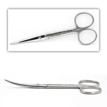
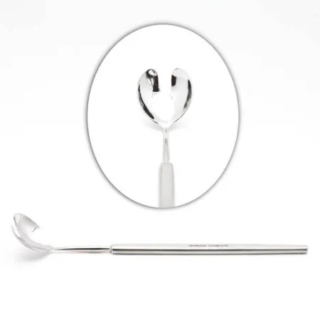
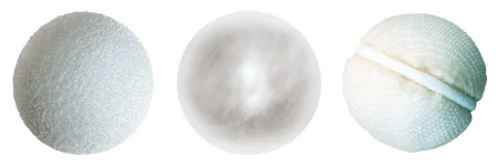
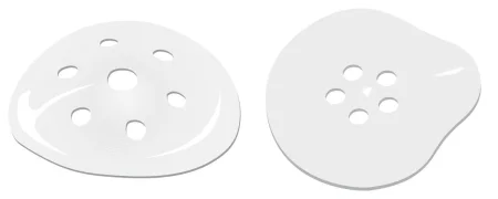
Key steps (enucleation):
- A speculum. The conjunctiva is cut all the way around the cornea, as close to the limbus as possible to save it for the closure.
- The four rectus muscles are looped with sutures if they will be sewn to the implant, then cut from the globe, leaving a stump of the medial rectus to hold.
- Blunt curved scissors free the globe from Tenon capsule, and the surgeon turns the eye by the medial rectus stump.
- The surgeon slides a large curved hemostat behind the eye and clamps the optic nerve for about 60 seconds; then the enucleation scissors cut the nerve, and the oblique muscles are cut as the eye comes out.
- The socket is packed with saline-soaked sponges and pressed to stop the bleeding. Keep track of how many go in, so every one comes out.
- The implant goes into the cone of muscles, the muscles are sewn to it if planned, and Tenon capsule and the conjunctiva are closed in separate layers over it.
- A conformer goes in behind the lids, antibiotic ointment is applied, and a pressure dressing goes on.
Evisceration: the surgeon cuts in at the limbus and may remove the cornea, scoops out the eye's contents with a spoon, and scrapes the choroid from the inside of the shell with curettes. Two relieving cuts in the sclera ease its tension, the implant is placed inside the scleral shell, and the sclera and conjunctiva are closed over it.
Specimen: the eye, or its contents, received in a small specimen cup or basin.
Wound class: I, clean, for an elective removal. After an injury, Class III when the wound is fresh, and Class IV when it is old with dead tissue or infected.
After surgery: bleeding and infection are the main risks. Teach the patient to protect the remaining eye, with protective glasses for sports and yard work.
Recovering eyes from a deceased donor
Chapter 18 teaches eye donation; here is the team's part. Every death and every imminent death is reported to the organ procurement organization; a specially trained person, such as a procurement organization nurse, asks the family, never other staff, and the next of kin sign the consent. Most people can donate, whatever their age, blood type or eye color, even with poor sight or cataracts. After death, the donor's head is raised on a pillow to limit swelling near the eyes, and saline-soaked gauze covers the eyes while the body waits in the morgue. The procurement team recovers the eyes within several hours of death.
some references still say the attending physician must ask the family, as some states' required request laws direct.
The recovery is done much like an enucleation, with the same sterile field, drapes and instruments. Each eye is placed cornea up in a sterile eye cage or on gauze in a sterile jar labeled right or left eye, and the sockets are packed with cotton and the lids closed. The corneas are placed in a storage medium such as Optisol-GS, and each sealed jar is labeled with the donor's name or number, the time of death, the time of recovery and the date, then carried on crushed ice to the eye bank. Treat the tissue as the gift it is.
16Tear ducts
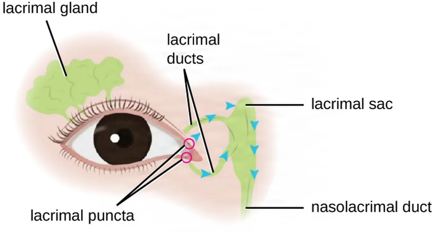
Probing, dilation and stents
What and why: a blocked tear drain makes tears overflow (epiphora) and can lead to infection of the lacrimal sac. Children can be born with a blocked nasolacrimal duct, which oculoplastic surgeons treat.
Before surgery: usually topical and local anesthesia. A baby 6 months old or younger may have only numbing drops, wrapped snugly with the bony points padded; an older child usually has general anesthesia. Drapes are optional.
Setup: punctal dilators, a set of lacrimal probes, a lacrimal cannula on a syringe of saline, and a silicone stent or intubation set. Bowman probes come in sets of six, double-ended, each end a different size, with a flat plate in the middle to hold.
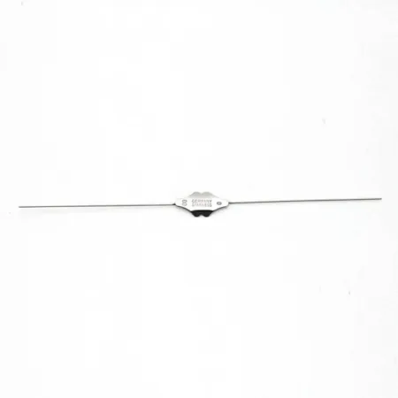
Key steps:
- A punctal dilator widens the opening at the inner end of the lid.
- A probe is passed through the punctum, the canaliculus, the lacrimal sac and the nasolacrimal duct into the nose, working up through the sizes.
- Saline flushed through the duct and suctioned from the nose shows that it is open.
- For a stent, a silicone tube on a fine metal rod or leader is passed through the punctum and down into the nose, where it is caught with a hook and drawn out, and left in place to lower the chance that the duct closes again.
- Fluorescein dye may be used to test that the duct is open, and antibiotic drops may go in at the end.
Wound class: the references give none for probing, so your facility's policy decides, from the CDC's definitions.
Dacryocystorhinostomy (DCR)
What and why: a DCR makes a new path for tears from the lacrimal sac straight into the nose, bypassing a nasolacrimal duct blocked by scarring after infection, a stone or an injury. Dacryocystitis, an infection of the lacrimal sac, shows as a painful, red swelling at the inner corner of the eye, sometimes with pus from the punctum; it is most common in adults over 40. The operation is done open, through a small skin incision beside the nose, or endoscopically through the nose, often by an ear, nose and throat surgeon.
Before surgery: usually general anesthesia, because the case can be long and bloody, sometimes with lowered blood pressure to reduce bleeding. The surgeon packs the nose with cottonoids soaked in a topical anesthetic and vasoconstrictor, such as cocaine or oxymetazoline, and injects lidocaine with epinephrine. Cocaine is a topical drug only, never injected, and a Schedule II controlled substance. The head of the table may be raised.
Setup: a lacrimal or DCR set with a Freer elevator, Kerrison rongeurs, a nasal speculum and bayonet forceps, a power drill with burs, bipolar, Frazier suction, lacrimal probes, a silicone intubation set, gelatin sponge with thrombin, iodoform packing, and a nasal endoscope for an endoscopic case.
Key steps (open DCR):
- An incision beside the inner corner of the eye with a #15 blade; the orbicularis muscle is spread to the bone. Have cotton-tipped applicators ready to sponge, and suction going.
- The periosteum is lifted with a Freer elevator, and an opening is made through the thin lacrimal bone and widened with a Kerrison rongeur or a drill. Clean the bone bits from the rongeur's tip with a moist sponge between bites.
- A probe is passed through a punctum into the sac, the sac is opened over it, and the nasal lining is opened. Have suction ready when the sac is opened.
- The flaps of the sac and the nasal lining are sewn together to make the new channel.
- Silicone tubes are passed through the upper and lower puncta, through the new opening and out of the nose, where the ends are tied and trimmed so they sit inside the nostril.
- The muscle and skin are closed, ointment goes on the incision, and a gauze pad is taped under the nose.
Wound class: II, clean-contaminated, by the CDC's definitions, because the nasal cavity, part of the respiratory tract, is opened; one reference lists it as Class I.
After surgery: the silicone tubes stay in for up to 4 to 8 weeks, then are removed; one reference removes them at about 2 weeks. Ice and keeping the head raised help the swelling. Bleeding and infection are the risks, and the open approach leaves a scar.
17After eye surgery
Wound classes
No eye operation opens the respiratory, digestive, genital or urinary tract except a dacryocystorhinostomy, which opens the lining of the nose, so most eye surgery is Class I, clean. Trauma and infection change that, as chapter 11 teaches.
| Operation | Wound class |
|---|---|
| Cataract extraction, keratoplasty, iridotomy and iridectomy, glaucoma surgery, vitrectomy, scleral buckle, pneumatic retinopexy, strabismus surgery, entropion and ectropion repair, pterygium excision | I, clean |
| Elective enucleation or evisceration | I |
| Enucleation after an injury; corneal, conjunctival and eyelid lacerations | III when fresh; IV when old with dead tissue, or infected (one reference also lists II for a lid laceration) |
| Dacryocystorhinostomy | II, because the nose is entered (one reference lists I) |
| Chalazion excision | I by the CDC's definitions (a chronic granuloma; no tract entered), as facility policy decides; one reference lists III |
The table rests on the CDC's definitions and the classes the references give for each operation.
Infection and inflammation inside the eye
- Endophthalmitis is an infection inside the eye, a dangerous complication. Povidone-iodine drops in the eye before surgery and antibiotics injected into the front chamber at the end help prevent it.
- Toxic anterior segment syndrome (TASS), a sterile inflammation inside the front of the eye from something toxic that got in, is taught in chapter 27 with the instrument cleaning that prevents it. At the eye case, your part is the drugs: only preservative-free drugs and solutions go into the eye, intraocular drugs stay apart and labeled, and no ointment goes on after cataract surgery.
Going home
Most eye patients go home the same day. Before discharge, the patient and caregiver learn to put in the drops without touching the tip to the eye, to wear the shield, not to rub or squeeze the eye, to avoid straining, heavy lifting and bending as the surgeon directs, and to call at once for sudden pain, sudden loss of vision, light sensitivity, a severe headache, nausea and vomiting, or swelling and redness. Sight may be blurry for a few days, so they take care not to fall.
keep the drugs that go inside the eye apart, preservative-free and labeled; keep the cornea moist; and never let anything jar the eye while an instrument is in it.
Lo que te llevas de este capítulo
- Eye surgery is microsurgery on an often awake patient: keep the room quiet, pass instruments into the surgeon's hand in position of use so the surgeon never looks up from the microscope, keep a fixed order on the Mayo, and keep the cornea moist with BSS.
- Prep with dilute povidone-iodine, never CHG or alcohol near the eye. Confirm the correct eye at the time-out, including the time-out before a block, and treat the intraocular lens like a drug: say its model and power aloud as you pass it.
- Eye drugs look alike and one error can blind. Label every one at once, keep intraocular drugs preservative-free and apart, and know the classes: mydriatics dilate, cycloplegics dilate and stop focusing, miotics constrict, viscoelastics hold the chamber open and must be washed out.
- In phacoemulsification the lens comes out in pieces and the capsule stays to hold the new lens; keep an anterior vitrectomy setup and a second lens ready in case the capsule tears.
- Keratoplasty replaces the cornea with donor tissue prepared on its own separate table; glaucoma surgery (trabeculectomy, trabeculoplasty, tube shunts, iridotomy) protects remaining sight, and mitomycin and 5-FU are hazardous drugs.
- Retinal detachments are repaired by vitrectomy, scleral buckling or pneumatic retinopexy; a gas bubble means no nitrous oxide, a wristband, head positioning and no flying.
- Strabismus surgery moves or shortens a muscle; pulling on it can slow the heart, so the surgeon lets go. Lid surgery fixes lids that turn in or out, removes chalazia and repairs lacerations with exact alignment of the margin.
- Enucleation removes the globe, evisceration its contents and exenteration the whole orbit; a sphere and conformer fill the socket. A DCR opens a new tear path into the nose and is Class II; most other eye cases are Class I.
Ponte a prueba5 preguntas rápidas
Gratis para todos. Elige una respuesta para cada una y luego revísalas todas juntas.
En el quirófanoDónde vas a usar esto
Todo el banco de preguntas del capítulo en series de 10, con la respuesta explicada después de cada una.
Parte del programa para educadoresLas palabras del capítulo en tarjetas de estudio: las que fallas vuelven antes.
Parte del programa para educadoresLo asigna tu docente. Cada grupo saca sus propias 25 preguntas del banco del capítulo.
Parte del programa para educadores
Sugerir una corrección
¿Algo está mal, confuso o desactualizado en este capítulo? Avísanos y lo corregimos para todos. Tu correo solo se usa para responderte.