The nervous system and the senses
The brain, spinal cord and nerves that run the body, and the eyes and ears that take in the world, with what each one means at the surgical field.
1Why it matters
Your nervous system runs your body. It takes in information from inside and outside the body, makes sense of it, and sends out commands to muscles and glands. It controls movement, feeling, thought, memory and emotion, and it sets your heart rate and your breathing without any thought from you.
The senses belong to the same system. The eyes, ears, nose and tongue are sense organs that turn light, sound and chemicals into nerve signals the brain can read.
You meet this system in almost every case.
- In brain and spine surgery, you pass instruments through the layers that protect the brain and spinal cord, and you help keep the fluid that bathes them from leaking out.
- In positioning, the patient is asleep and cannot tell anyone that a nerve is being pressed or stretched. A badly placed arm or leg can injure a nerve, so the whole team watches for it.
- In the field, important nerves run right beside many operations, such as the facial nerve through the parotid gland and the nerve to the vocal cords in the neck.
- In eye and ear surgery, the team works with delicate microsurgical instruments, and ear operations are done under the operating microscope or through an endoscope, so you need to know what each structure is and what it does.
This chapter starts with the nerve cell and the plan of the whole system, then moves through the brain, its coverings and its fluid, the cranial and spinal nerves, and finally the senses, the eye and the ear. Other chapters teach what it leaves out: nervous tissue and the nerve impulse at the cell level in chapter 2; the bones of the skull and spine in chapter 3; the pituitary gland and hypothalamus as glands in chapter 9; anesthesia, nerve blocks and paralyzing drugs in chapter 13; positioning injuries in chapter 31; and the eye, ear and neurosurgery operations in chapters 35, 36 and 42.
2The plan of the nervous system
Central and peripheral
The nervous system has two main parts, divided by where they are.
- The central nervous system (CNS) is the brain and the spinal cord. The brain sits inside the skull, in the cranial cavity, and the spinal cord runs down the vertebral canal of the spine. Bone protects both.
- The peripheral nervous system (PNS) is everything outside the brain and spinal cord: the 12 pairs of cranial nerves that leave the brain, the 31 pairs of spinal nerves that leave the spinal cord, and the ganglia, small clusters of nerve cell bodies outside the CNS.
The system can also be divided by what it does.
- The somatic nervous system handles what you feel consciously and the skeletal muscles you move on purpose.
- The autonomic nervous system (ANS) runs the body's automatic work, such as the heart rate, breathing, digestion and sweating, through heart muscle, smooth muscle and glands. Its two divisions, the sympathetic and the parasympathetic, come later in this chapter.
The neuron
Nervous tissue has two kinds of cells: neurons, which carry the signals, and glial cells, which support them. Chapter 2 introduced both. Here are the parts of a neuron you need to name.
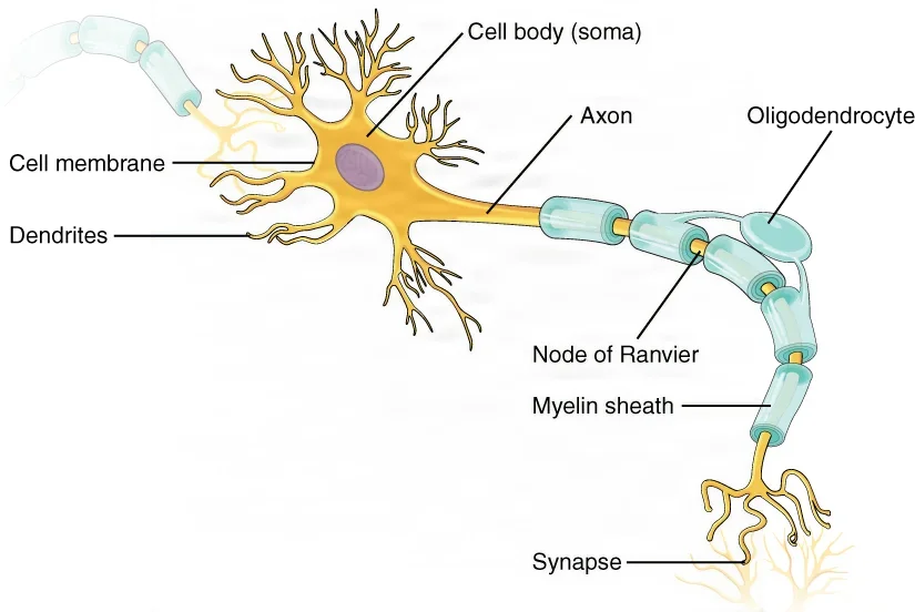
- Cell body: the main part of the neuron, also called the soma. It holds the nucleus and most of the cell's working parts.
- Dendrites: short, branching extensions that pick up signals from other neurons and carry them toward the cell body.
- Axon: one long fiber that carries the signal away from the cell body to the next cell. An axon can branch to reach many cells, and some axons are about a meter long.
- Myelin: a fatty white wrapping around many axons. It insulates the axon the way plastic covers a wire, and it makes the signal travel much faster, jumping from one gap in the wrapping to the next. The gaps are called nodes of Ranvier.
- Schwann cells: the cells that make myelin around axons in the PNS. Each one wraps a single stretch of one axon. In the brain and spinal cord, other cells, the oligodendrocytes, make the myelin instead.
- Neurilemma: the outer layer of a Schwann cell, which holds its nucleus and cytoplasm, lying on the outside of the myelin. Schwann cells are needed for a cut peripheral nerve to grow back.
A signal moves through a neuron in one direction only: into the dendrites, across the cell body and out along the axon.
Schwann cells matter in surgery, too. A vestibular schwannoma, also called an acoustic neuroma, is a benign tumor that grows from the sheath of the nerve for hearing and balance, deep inside the skull. Removing it can put the facial nerve beside it at risk.
Glial cells and the words for the CNS
Glial cells, also called glia or neuroglia, make up most of the brain and spinal cord. Four kinds live in the CNS:
- Astrocytes, star-shaped cells and the most common kind. They support neurons, keep the fluid around them balanced, and help form the blood-brain barrier, which keeps many substances in the blood out of the brain.
- Ependymal cells, which line the ventricles, the fluid-filled spaces inside the brain, and make cerebrospinal fluid.
- Oligodendrocytes, which make myelin in the CNS.
- Microglia, small cells that clean up debris and germs, much like the body's white blood cells do elsewhere.
Glial cells matter in surgery because gliomas, among the more common brain tumors, are believed to grow from them.
The CNS has its own words for its tissue.
- Gray matter is tissue made mostly of nerve cell bodies and dendrites, with few myelinated axons. The outer layer of the brain, the cortex, is gray matter.
- White matter is tissue made mostly of axons, white because of their fatty myelin.
- A nucleus, in the CNS, is a cluster of nerve cell bodies that work together. The same kind of cluster outside the CNS is called a ganglion (plural, ganglia).
- A pathway, or tract, is a bundle of axons inside the CNS that carries signals from one region to another. The same kind of bundle outside the CNS is called a nerve.
| What it is | Name in the CNS | Name in the PNS |
|---|---|---|
| A group of nerve cell bodies (gray matter) | Nucleus | Ganglion |
| A bundle of axons (white matter) | Tract, or pathway | Nerve |
The optic nerve shows the difference. The same axons are called the optic nerve from the eye to the optic chiasm, where the two optic nerves meet, and the optic tract once they are inside the brain.
3The autonomic nervous system
The ANS works without your thinking about it, keeping the inside of the body steady. Most organs get nerves from both of its divisions, which push in opposite directions, like a gas pedal and a brake.
- The sympathetic division readies the body for stress or danger, the "fight or flight" response. Its nerves leave the spinal cord in the chest and upper lumbar (lower back) regions, so it is also called the thoracolumbar division.
- The parasympathetic division runs the body at rest, the "rest and digest" response, and saves energy. Its nerves leave the brain stem, through four of the cranial nerves, and the sacral part of the spinal cord, so it is also called the craniosacral division. The vagus nerve carries it to the organs of the chest and upper abdomen.
| Organ | Sympathetic (fight or flight) | Parasympathetic (rest and digest) |
|---|---|---|
| Heart | Beats faster | Beats slower |
| Airways | Widen, so more air gets in | Narrow |
| Blood vessels | Most narrow and blood pressure rises; blood is sent to the brain, heart, lungs and muscles, away from the gut and skin | Widen and blood pressure falls; more blood flows to the gut |
| Pupils | Widen (dilate) | Narrow (constrict), through the oculomotor nerve |
| Digestion and saliva | Slow down | Speed up, with more saliva |
Sympathetic stimulation also makes the sweat glands work harder.
The two divisions use different chemical messengers. Acetylcholine carries the signal at every parasympathetic ending, and norepinephrine at most sympathetic endings. Drugs that copy or block these messengers are taught in chapter 12.
You may see the brake applied in surgery. In eye muscle surgery, pulling on the muscles can set off a parasympathetic (vagal) response that slows the heart. When that happens, the surgeon briefly lets go of the muscle.
4The synapse
A synapse is the junction where a neuron passes its signal on to the next cell, which may be another neuron, a muscle fiber or a gland. At a chemical synapse, the kind described here, the two cells do not touch.
How a signal crosses the synapse:
- The signal reaches the end of the axon. There, tiny sacs called vesicles hold a chemical messenger, a neurotransmitter.
- The vesicles release the neurotransmitter into the narrow gap between the cells, the synaptic cleft.
- The neurotransmitter drifts across the gap and fits into receptors on the next cell like a key in a lock. Each neurotransmitter fits only its own receptors.
- Depending on the receptor, the next cell is either stirred to fire or calmed.
- The neurotransmitter is then cleared away so the signal stops. Some are broken down by enzymes: acetylcholinesterase breaks down acetylcholine. Others are pumped back into the axon's end, or into a nearby glial cell, to be used again, which is called reuptake.
The neuron sending the signal is the presynaptic neuron, and the cell receiving it is the postsynaptic cell.
| Neurotransmitter | Where it works | How it is cleared |
|---|---|---|
| Acetylcholine | Nerve to skeletal muscle, every parasympathetic ending, and parts of the brain | Broken down by acetylcholinesterase |
| Norepinephrine and epinephrine | Most sympathetic endings; the adrenal gland also releases both into the blood as hormones | Taken back into the axon's end |
| Dopamine | Brain systems, including the basal ganglia, which help control movement | Taken back into the axon's end |
| Glutamate, GABA and glycine | In the CNS: glutamate stirs the next cell to fire; GABA and glycine calm it | Pumped back into the axon's end or a glial cell |
Because each neurotransmitter has its own receptors, a drug can block one kind of signal and leave the rest alone. A neuromuscular blocking drug, a paralytic, stops the signal from nerve to muscle but does nothing to pain. A patient who has a paralytic without enough anesthesia can feel pain and cannot move to show it. Chapter 13 teaches these drugs.
5The brain
The adult brain has four main regions.
| Region | Where it is | Main jobs |
|---|---|---|
| Cerebrum | The large upper part, in two hemispheres | Thought, memory, language, planned movement, and conscious touch, sight and hearing |
| Diencephalon (thalamus and hypothalamus) | Deep in the middle, under the cerebrum, around the third ventricle | Relays the senses to the cortex; controls the ANS, body temperature, thirst, hunger and many hormones |
| Brain stem (midbrain, pons and medulla oblongata) | The stalk that joins the brain to the spinal cord | Heart rate, breathing and blood pressure; wakefulness; carries signals between the brain and the cord; holds the centers of most cranial nerves |
| Cerebellum | At the back, behind the brain stem and under the cerebrum | Coordination, balance and posture |
The cerebrum
The cerebrum is by far the largest part of the brain.
- Hemispheres: a deep groove, the longitudinal fissure, splits the cerebrum into right and left hemispheres. Underneath, they stay linked by the corpus callosum, a thick band of white matter that lets the two sides talk.
- Crossed control: each hemisphere moves, and feels for, the opposite side of the body. Damage to the motor area on the left side of the brain weakens the right side of the body.
- Cortex: the outer layer, the cerebral cortex, is a thin sheet of gray matter folded into ridges called gyri (singular, gyrus) and grooves called sulci (singular, sulcus). Deeper and larger grooves are called fissures. The folds fit far more cortex into the skull. White matter lies beneath the cortex.
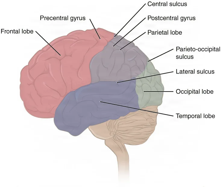
Each hemisphere has four lobes, named for the skull bones that cover them.
| Lobe | Where it is | Main jobs |
|---|---|---|
| Frontal | The front, ahead of the central sulcus | Planned movement (the precentral gyrus is the main motor area), producing speech, personality, judgment and reasoning |
| Parietal | The top, behind the central sulcus | Touch, pain, pressure and body position (the postcentral gyrus is the main sensory area) |
| Temporal | The side, below the lateral sulcus | Hearing, memory and understanding language |
| Occipital | The back | Vision |
Two grooves are landmarks that surgeons use: the central sulcus (fissure of Rolando), between the frontal and parietal lobes, and the lateral sulcus (sylvian fissure), which separates the temporal lobe from the lobes above it.
Deep inside the cerebrum, under the white matter, lie two more groups of structures.
- The basal ganglia, also called the basal nuclei, are clusters of gray matter, including the caudate, the putamen and the globus pallidus, that help start wanted movements and hold back unwanted ones. In Parkinson's disease, the cells that feed them dopamine die, and movement becomes slow and stiff, often with a tremor.
- The limbic system is a group of structures along the inner wall of each hemisphere, including the hippocampus and the amygdala, that handles emotion, memory and behavior. It works closely with the hypothalamus.
The diencephalon
The diencephalon sits deep in the center of the brain, around the third ventricle. Its two main parts are the thalamus and the hypothalamus.
- The thalamus is the relay station for the senses. Every sense except smell passes through it on the way to the cortex.
- The hypothalamus lies below the thalamus. It runs the ANS and keeps the body in balance: temperature, fluid balance, hunger and much of the hormone system. The pituitary gland hangs from it on a thin stalk. Chapter 9 teaches both as glands.
The brain stem
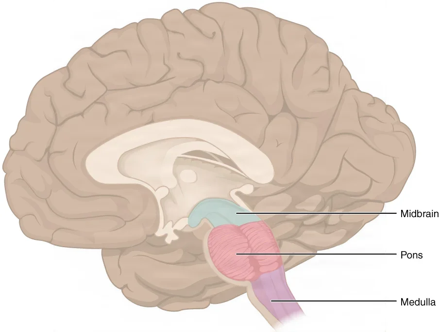
The brain stem joins the brain to the spinal cord. It has three parts, from top to bottom:
- Midbrain, also called the mesencephalon: a short section under the diencephalon. It holds centers for eye movement and for reflexes to what you see and hear. The cerebral aqueduct, a channel for cerebrospinal fluid, runs through it.
- Pons, Latin for bridge: a bulge of white matter that links the cerebellum to the rest of the brain. With the medulla, it helps control breathing and the heart. Cranial nerves V to VIII attach here.
- Medulla oblongata: the lowest part, which joins the spinal cord at the foramen magnum, the large hole in the base of the skull. It holds the centers that control the heart rate, blood pressure and breathing.
All but two of the cranial nerves connect to the brain stem. Damage to the brain stem is often life-threatening, because it controls breathing and consciousness.
The cerebellum
The cerebellum, the "little brain", sits at the back of the skull behind the brain stem. It compares the movements the cerebrum orders with feedback from the muscles, joints and inner ear, and corrects them, so movement is smooth and the body stays balanced. It has two hemispheres joined by a middle part, the vermis.
6The meninges
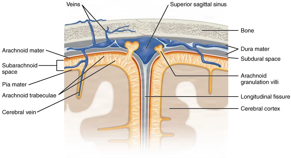
Three membranes, the meninges, wrap the brain and spinal cord and protect them. From the outside in:
- Dura mater, Latin for "tough mother": a thick, strong, fibrous layer that lines the inside of the skull and the spinal canal. In the skull it has two layers. Between them run the dural venous sinuses, large channels that drain blood from the brain toward the jugular veins.
- Arachnoid mater: a thin, delicate layer named for its spider-web threads, which reach down to the surface of the brain.
- Pia mater, "tender mother": a very thin layer that hugs the brain, following it into every groove, and carries a rich network of small blood vessels.
The dura folds inward in places, dividing the inside of the skull into compartments:
- The falx cerebri, a sickle-shaped fold that dips into the longitudinal fissure between the two hemispheres.
- The tentorium cerebelli, a tent-like shelf between the cerebrum and the cerebellum. Surgeons call the space above it supratentorial and the space below it infratentorial.
- The falx cerebelli, a small fold between the two halves of the cerebellum.
The meninges create three spaces, each named for where it sits.
| Space | Where it is | Why it matters |
|---|---|---|
| Epidural space | Outside the dura | In the skull, a torn middle meningeal artery can bleed here, an epidural hematoma; in the spine, epidural anesthesia is given here |
| Subdural space | Between the dura and the arachnoid | Veins cross it on their way to the sinuses; a tear after a head injury bleeds here, a subdural hematoma |
| Subarachnoid space | Between the arachnoid and the pia | Filled with cerebrospinal fluid, with the large vessels and nerves of the brain's surface; spinal anesthesia is injected into the fluid here |
The meninges at a craniotomy
A craniotomy is an operation that opens the skull to reach the brain. The surgeon passes through these layers in order, and closes them in reverse.
- Scalp: skin, a dense layer full of blood vessels, the galea (a tough fibrous sheet), and the pericranium, the bone's outer covering. The scalp bleeds freely, so have the ESU and plenty of sponges ready. The surgeon clips the cut edges with Raney clips and turns the scalp flap back in the loose, nearly bloodless layer under the galea.
- Bone: the surgeon drills burr holes and cuts a bone flap between them. Irrigate the drill bit to keep heat off the tissue underneath, have bone wax ready for bleeding bone edges, and keep the bone flap safe on the back table, in saline or antibiotic solution if your surgeon asks.
- Dura: the surgeon lifts it with a dura hook, nicks it with a #15 blade and opens it with scissors. Opened dura shrinks as it dries, so it is kept moist under wet strips.
- Arachnoid and pia: these thin layers lie on the brain itself.
At closing, the dura is sewn with a running stitch of fine suture, often 4-0 or 3-0 polypropylene or nylon. A sealant or fibrin glue may go over the stitch line to make it watertight, and a graft patches a gap too big to close.
The closure must be watertight because a gap lets cerebrospinal fluid leak out, and a leak raises the risk of meningitis, an infection of the meninges.
A few more facts help you follow the case.
- The brain itself feels no pain. The scalp, the arteries outside the skull and parts of the dura do.
- The venous sinuses run along the edges of the dural folds. A torn sinus bleeds heavily and can pull air into the vein, an air embolism.
- The skull is a closed box. Bleeding, swelling or a tumor adds to what is inside it and raises the intracranial pressure (ICP), which can damage the brain for good.
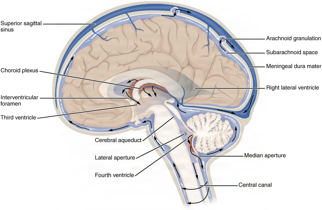
From the outside in: scalp, bone, dura, arachnoid, pia. The dura is tough, the arachnoid is a web, and the pia hugs the brain. Cerebrospinal fluid flows in the subarachnoid space, between the arachnoid and the pia.
7The ventricles and cerebrospinal fluid
Cerebrospinal fluid (CSF) is a clear, watery fluid filtered from the blood. It is mostly water, with small molecules and salts. It fills the ventricles inside the brain and bathes the brain and spinal cord from the subarachnoid space.
What CSF does:
- It cushions the brain and spinal cord against blows and helps hold up the weight of the brain.
- It carries wastes away from the nervous tissue.
- Its volume shifts to help keep the pressure inside the skull steady.
The ventricles
The ventricles are four connected spaces inside the brain.
| Ventricle | Where it is | Where its CSF goes next |
|---|---|---|
| Two lateral ventricles | One deep in each cerebral hemisphere | To the third ventricle, through the interventricular foramen (foramen of Monro) |
| Third ventricle | In the center, between the left and right halves of the diencephalon | To the fourth ventricle, through the cerebral aqueduct (aqueduct of Sylvius), which runs through the midbrain |
| Fourth ventricle | Between the pons and medulla in front and the cerebellum behind | Out to the subarachnoid space through three openings, the two lateral foramina of Luschka and the median foramen of Magendie, and down into the central canal of the spinal cord |
How CSF is made and where it goes
- Made: a choroid plexus in each ventricle, a tuft of tiny blood vessels covered by ependymal cells, filters blood into CSF. Most of it is made in the lateral ventricles.
- Flows: from the lateral ventricles through the foramina of Monro to the third ventricle, through the aqueduct of Sylvius to the fourth ventricle, and out through the foramina of Luschka and Magendie into the subarachnoid space. There it bathes the brain and runs down around the spinal cord.
- Absorbed: arachnoid granulations, small pouches of the arachnoid that push into the dural venous sinuses, mostly the superior sagittal sinus, pass the CSF back into the blood.
An adult has about 150 mL of CSF at any moment, about 25 mL of it in the ventricles. The body makes roughly 450 to 500 mL a day, so the whole supply is replaced about three times a day.
When the flow goes wrong
- Hydrocephalus: CSF builds up and the ventricles swell, because its flow is blocked or it is not absorbed, and the pressure in the skull rises. A common treatment is a ventriculoperitoneal (VP) shunt: a catheter from a ventricle, through a valve, runs under the skin to the peritoneal cavity of the abdomen, where the fluid drains.
- Lumbar puncture: the spinal cord ends in the upper lumbar spine, so a needle placed lower in the back can draw CSF from the subarachnoid space without touching the cord.
- CSF leaks: a fracture of the base of the skull can let CSF drip from the nose (rhinorrhea) or the ear (otorrhea). A leak that does not stop on its own may need surgery to repair the dura.
8The cranial nerves
The cranial nerves are 12 pairs of nerves attached to the brain. They are numbered with Roman numerals, I to XII, from the front of the brain to the back. Most serve the head and neck, but the vagus nerve reaches down to the organs of the chest and abdomen.
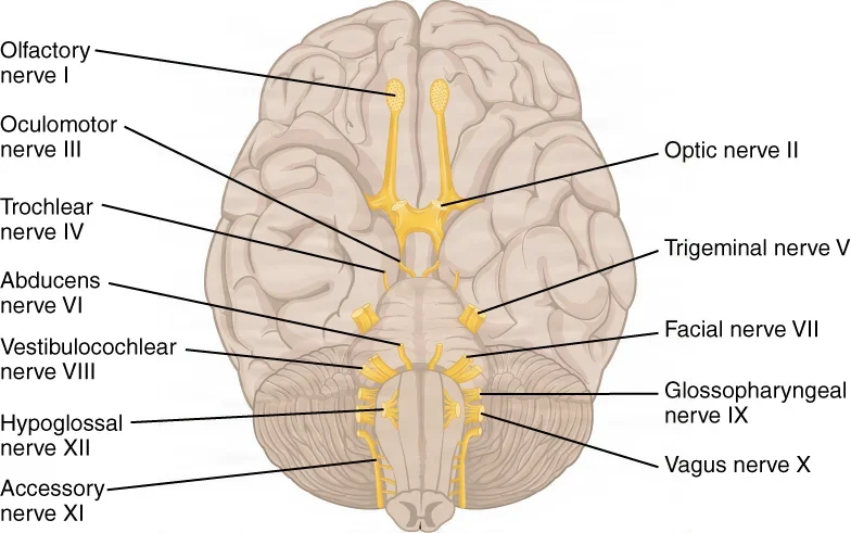
Each cranial nerve is sensory (it carries feeling or a special sense to the brain), motor (it carries commands out to muscles or glands), or both. Three are sensory, five are motor and four are both.
Students have always used memory sentences for these, where the first letter of each word stands for one nerve. For the names in order, try "Oh Once One Takes The Anatomy Final, Very Good Vacations Are Heavenly." For the types, try "Some Say Marry Money But My Brother Says Big Brains Matter More," where S is sensory, M is motor and B is both.
| Nerve | Type | What it does | Where you meet it |
|---|---|---|---|
| I, olfactory | Sensory | Smell | Its fibers pass up through the roof of the nose to the olfactory bulbs under the frontal lobes; a blow to the head can tear them and take away smell |
| II, optic | Sensory | Vision | The two optic nerves meet at the optic chiasm, just above the pituitary gland; a pituitary tumor can press on it and narrow the field of vision |
| III, oculomotor | Motor | Moves the eyeball through four of the six eye muscles (the superior, inferior and medial rectus and the inferior oblique), lifts the upper eyelid, and works the iris muscle that narrows the pupil | Supplies the medial rectus, a muscle a surgeon may move back (a recession) to straighten the eyes |
| IV, trochlear | Motor | Moves the eyeball at an angle through the superior oblique muscle | Works with III and VI so both eyes aim at the same point |
| V, trigeminal | Both | Carries feeling from the face, forehead, eyes, teeth and mouth; through its mandibular branch, moves the jaw for chewing and tightens the tympanic membrane | Has three branches, ophthalmic, maxillary and mandibular; trigeminal neuralgia causes bursts of severe stabbing face pain |
| VI, abducens | Motor | Moves the eye outward, away from the nose, through the lateral rectus muscle | Its muscle is one a surgeon may shorten or move to correct strabismus, eyes that do not line up |
| VII, facial | Both | Moves the muscles of facial expression; drives two of the salivary glands (the submandibular and sublingual), the glands of the nose and mouth lining and the tear gland; carries taste from the front two-thirds of the tongue, and a little feeling from the outer ear | Runs through the middle ear and through the parotid gland, where it splits into branches; at risk in ear surgery and parotidectomy |
| VIII, vestibulocochlear (also called auditory or acoustic) | Sensory | Hearing (its cochlear part) and balance, the sense of the head's position and movement (its vestibular part) | An acoustic neuroma grows on it where it enters the internal auditory meatus, the canal it runs through between the inner ear and the brain stem |
| IX, glossopharyngeal | Both | Moves the stylopharyngeus, a muscle of the throat; sends parasympathetic fibers to the parotid salivary gland; senses blood pressure in the carotid sinus and blood chemistry in the carotid body; carries feeling from the throat, the middle ear and the back of the tongue, and taste from the back third of the tongue | When blood pressure rises, its signals from the carotid sinus help slow the heart |
| X, vagus | Both | Moves the muscles of the soft palate, throat and larynx (voice box); sends parasympathetic fibers to the trachea and the organs of the chest and abdomen, slowing the heart and moving digestion along; carries feeling from the throat, larynx and the organs of the chest and abdomen, signals from the pressure and chemical sensors of the aortic arch and aortic body, and feeling from part of the outer ear and ear canal | Its recurrent laryngeal branch moves the vocal cords, and injuring it in neck surgery paralyzes a vocal cord; the lead of a vagus nerve stimulator wraps around the left vagus in the neck |
| XI, accessory (spinal accessory) | Motor | Moves the trapezius and sternocleidomastoid, which shrug the shoulders and turn the head | Can be joined to a damaged facial nerve to bring movement back to the face |
| XII, hypoglossal | Motor | Moves the tongue | Can also be joined to a damaged facial nerve for the same reason |
The Core Curriculum calls VIII's balance job proprioception, a word that in its wider sense covers posture, movement and balance, with the inner ear's balance receptors counted among the proprioceptors.
The Core Curriculum and some surgical technology textbooks also give XI the muscles of the larynx and pharynx, through a part of XI that joins the vagus; this table follows the references that count those fibers as part of the vagus.
Cranial nerves at the surgical field
- The facial nerve leaves the skull just behind the parotid gland and fans out through the gland into five branches: temporal, zygomatic, buccal, marginal mandibular and cervical. Surgeons describe the gland as a superficial lobe and a deep lobe, divided by the nerve. Damage weakens or paralyzes one side of the face, so a nerve monitor is kept ready; it warns the team when the nerve is being handled.
- The recurrent laryngeal nerve, a branch of the vagus, works the muscles inside the larynx that move the vocal cords. During a thyroidectomy, the surgeon finds this nerve and protects it, sometimes with the help of a nerve monitor, because an injured nerve leaves a vocal cord paralyzed.
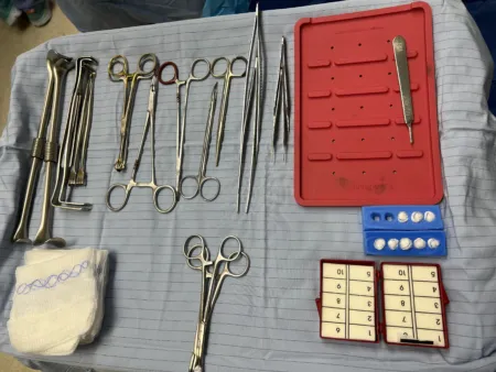
- The vagus nerve can be stimulated to treat epilepsy. A vagus nerve stimulator is a small generator placed under the skin below the collarbone, with a lead whose coils wrap around the left vagus nerve in the neck. The left side is used because the right vagus has more control over the heart's pacemaker, so stimulating it is more likely to slow the heart dangerously. The generator's battery wears down with use. When it nears the end of its life, the generator is replaced through its pocket under the skin, and the lead on the nerve is left alone.
- The optic nerves and chiasm lie just above the pituitary gland, so a pituitary tumor can press on the crossing fibers and take away the outer parts of the field of vision.
9The spinal cord and spinal nerves
The spinal cord
The spinal cord carries on from the medulla at the foramen magnum and runs down the vertebral canal, inside the protecting ring of the vertebrae. It has two jobs:
- A highway: its white matter carries sensory signals up to the brain in ascending tracts and motor commands down from the brain in descending tracts.
- A reflex center: some reflexes, such as pulling a hand away from something hot, are handled by circuits in the cord itself.
Inside, the arrangement is the reverse of the brain's, where the gray matter is on the outside. In the cord, gray matter forms a butterfly or H shape in the center, with white matter around it. A narrow central canal, filled with CSF, runs down the middle.
- The posterior (dorsal) horns of the gray matter handle incoming sensation.
- The anterior (ventral) horns hold the motor neurons that send commands to skeletal muscles.
- The lateral horns, found only in the thoracic, upper lumbar and sacral cord, hold the cell bodies of autonomic neurons.
External features
The Core Curriculum names five features you can see on the outside of the cord.
- Vertebral segments (regions): the cord is divided into cervical, thoracic, lumbar and sacral regions, named for the part of the spine where their nerves exit. It gives off 31 pairs of spinal nerves: 8 pairs in the neck (cervical), 12 in the chest (thoracic), 5 in the lower back (lumbar), 5 sacral, and a single coccygeal pair. There are 8 cervical nerves but only 7 cervical vertebrae, because the first nerve leaves above the first vertebra, between it and the skull.
- Nerve roots: each spinal nerve joins the cord by two roots. The dorsal (posterior) root carries sensation in and has a swelling, the dorsal root ganglion, that holds the cell bodies of the sensory neurons. The ventral (anterior) root carries motor commands out. The two roots join into one mixed nerve, which leaves the spine through an intervertebral foramen, the opening between two vertebrae.
- Conus medullaris: the tapered lower end of the cord, at about the first or second lumbar vertebra in an adult. A thin thread of pia mater, the filum terminale, runs on from it to anchor the cord below.
- Cauda equina: the bundle of lumbar and sacral nerve roots that hang down in the canal below the end of the cord, named for its look, a horse's tail. It forms because the spine keeps growing after the cord stops.
- Sympathetic trunk and ganglia: a chain of ganglia that runs down alongside the vertebral column. It receives fibers from the lateral horns of the thoracic and upper lumbar cord and sends sympathetic signals out to the organs.
The meninges go on below the end of the cord, making a sac of dura and arachnoid that reaches down to about the second or third sacral vertebra. That is why a lumbar puncture or spinal anesthetic is given low in the back, into the fluid around the cauda equina, well below the cord itself.
Each spinal nerve's sensory root serves a band of skin called a dermatome. A map of the dermatomes helps the care team find which nerve root is damaged.
The spine in surgery
A herniated disc can bulge into the canal and press on a nerve root as it leaves the spine. In a laminectomy, the surgeon removes part of the lamina, the back of a vertebra's bony arch, often with disc material, to take the pressure off the nerves or the cord.
The dura lies just beneath the lamina and its ligament, right where the surgeon is working. A tear in it leaks CSF, and the surgeon repairs it right away when possible, because a leak raises the risk of infection.
10The spinal nerve plexuses
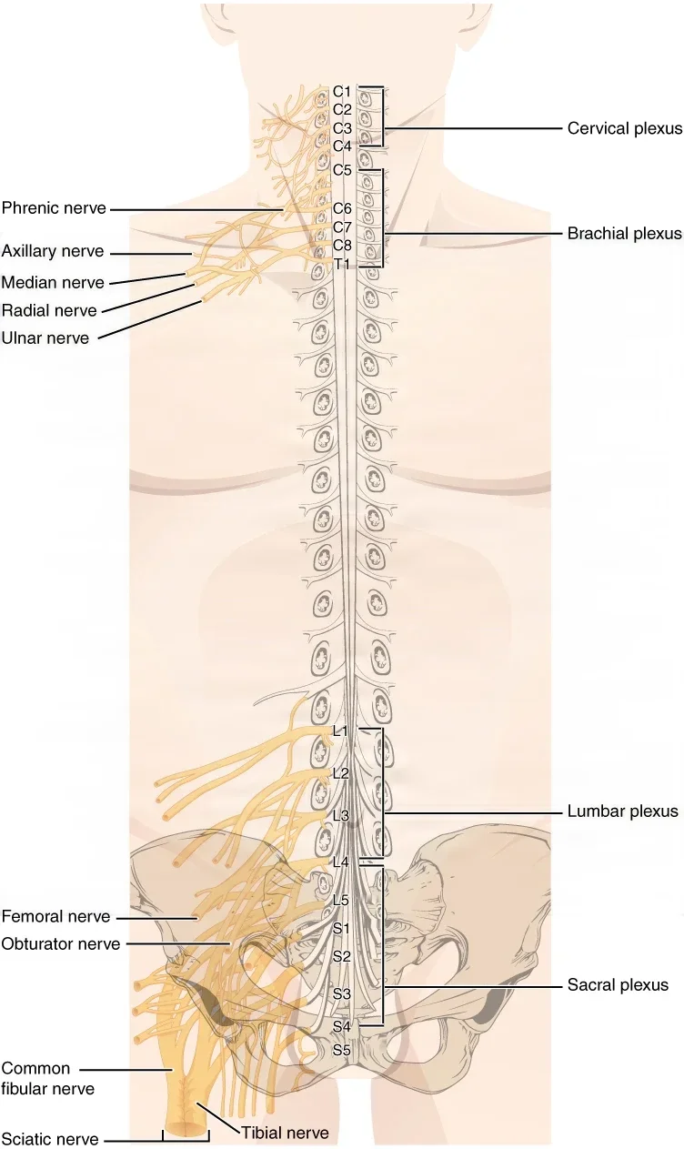
At four places along the spine, the spinal nerves do not run straight to their targets. Their fibers mix and regroup in networks called plexuses (singular, plexus), and new, named nerves come out the other side. The thoracic nerves T2 to T11 do not form a plexus; they run between the ribs as the intercostal nerves.
| Plexus | Formed from | Named nerves | Supplies |
|---|---|---|---|
| Cervical | C1 to C4 | Phrenic nerve, from C3, C4 and C5 | The muscles of the neck and shoulder and the skin of parts of the head, neck and chest; the phrenic nerve works the diaphragm, the main muscle of breathing |
| Brachial | C5 to T1 | Axillary, radial, median and ulnar nerves | The shoulder, arm and hand |
| Lumbosacral: lumbar and sacral plexuses | L1 to L4, and L4 to S4 | Femoral nerve (lumbar); sciatic nerve (sacral) | The pelvis and the front of the thigh (lumbar); the back of the thigh, the leg and the foot (sacral) |
Some references count the cervical plexus from C1 to C5, the brachial plexus from C4 to T1 or the sacral plexus from L4 to S3; the table gives the levels most surgical technology references use.
The Core Curriculum names six of these nerves.
- Phrenic nerve: carries signals from the neck down to the diaphragm. Because it starts so high, from C3 to C5, a patient whose spinal cord is cut above C3 cannot breathe on their own.
- Axillary nerve: a nerve from the back (posterior) cord of the brachial plexus, the same cord that goes on as the radial nerve. It moves the deltoid and teres minor muscles and carries feeling from the skin over the shoulder.
- Radial nerve: a large nerve of the brachial plexus that carries both feeling and movement for the arm.
- Median nerve: runs down the middle of the forearm and through the carpal tunnel at the wrist into the hand.
- Ulnar nerve: runs down the inner side of the arm and passes behind the elbow, in the cubital tunnel, on its way to the hand.
- Sciatic nerve: the largest nerve in the body. It crosses the hip, serves the back of the leg, and divides into the tibial nerve and the common peroneal (fibular) nerve, which together serve the leg and foot.
Nerves on the operating table
The patient is asleep and cannot tell anyone that a nerve is being stretched or pinched. Nerves are injured by traction (stretch) and by pressure, and the most common to be hurt are the ulnar nerve, the common peroneal nerve and the brachial plexus. The scrub helps the team prevent it.
- Brachial plexus: stretched when an arm on an armboard is opened out more than 90 degrees from the body, and by shoulder braces in the head-down (Trendelenburg) position.
- Ulnar nerve: pressed by arm straps, table attachments or a badly placed arm at the side. Padding the elbow and placing the arm correctly prevent it.
- Common peroneal nerve: pressed against the stirrups in the lithotomy position, where it can cause foot drop. Padding the stirrups and keeping the lower legs off the stirrup posts prevent it.
Chapter 31 teaches each position and its padding in detail.
Some operations free a nerve that is being squeezed.
- Carpal tunnel release: in carpal tunnel syndrome, the median nerve is pressed inside the carpal tunnel, the narrow passage at the wrist under the transverse carpal ligament, causing numbness and tingling in the hand. The surgeon divides the ligament to open the tunnel and give the nerve room.
- Ulnar nerve transposition: the ulnar nerve is freed from its groove behind the elbow, where it is trapped, and moved to the front of the elbow, under a flap of fascia that keeps it from slipping back.
If an arm slips off an armboard, a leg rests against a stirrup post, or the patient's position shifts during the case, say so at once. A pinched or stretched nerve gives no warning under anesthesia, and the patient may wake with a numb hand or a dropped foot.
11The senses
The kinds of senses
A sense starts with a receptor, a cell or nerve ending that turns light, sound, a chemical, touch or stretch into a nerve signal. The brain then turns those signals into a perception that means something.
The Core Curriculum lists six senses.
| Sense | What it detects | Where its receptors are |
|---|---|---|
| Visual (sight) | Light | The retina of the eye |
| Auditory (hearing) | Sound | The cochlea of the inner ear |
| Gustatory (taste) | Chemicals in food and drink | Taste buds, mostly on the tongue |
| Olfactory (smell) | Chemicals in the air | The roof of the nasal cavity |
| Touch | Pressure, vibration, light touch, pain and temperature | The skin, and also muscles, tendons, joints and organ walls |
| Proprioception | The position and movement of the body's parts | Muscles, tendons and joints |
Balance is a sense too, and its receptors sit in the inner ear beside the hearing organ.
Senses are also sorted as special senses, which have an organ of their own (the eye, the inner ear, the tongue and the nose), and general senses, whose receptors are spread through the body, such as touch, pain, temperature and proprioception.
Taste and smell
Taste, or gustation, starts in taste buds, which sit in small bumps on the tongue called papillae. They sense five tastes: sweet, salty, sour, bitter and umami (savory). The facial nerve carries taste from the front two-thirds of the tongue, the glossopharyngeal nerve from the back third, and the vagus from the very back, near the throat.
Smell, or olfaction, starts in a small patch of lining high in the nasal cavity. The axons of its receptor neurons pass up through tiny holes in the cribriform plate of the ethmoid bone to the olfactory bulbs under the frontal lobes. Smell is the only sense that does not pass through the thalamus, and it links closely to memory and emotion. Much of what we call flavor is really smell, which is why food tastes bland to someone who has lost it.
Touch and proprioception
Touch is a group of senses. Different receptors in the skin pick up light touch, pressure, vibration, stretch, pain and temperature.
Proprioception is the sense of where your body parts are and how they are moving, without looking. Stretch receptors in the muscles and tendons, called muscle spindles and Golgi tendon organs, and receptors in the joint capsules report it to the brain. The parietal lobe and the cerebellum use it to guide movement and balance.
12The eye
The orbit
The eyeball sits in the orbit, a cone-shaped bony socket that is wide at the front and narrow at the back. At the back, the optic canal lets the optic nerve pass through to the brain.
Seven bones form the walls of each orbit:
- the frontal bone, which forms the roof
- the zygomatic bone (cheekbone), which forms the outer wall and the outer part of the floor
- the maxilla, the upper jaw bone, and the small palatine bone, which add to the floor
- the ethmoid and lacrimal bones, which form the inner wall, nearest the nose
- the sphenoid bone, which forms the back of the orbit, where the optic canal opens
The eyelids
The eyelids, or palpebrae (singular, palpebra), protect the eye from injury and light, and every blink spreads tears over the cornea. The gap between the open lids is the palpebral fissure, and the corners where the lids meet are the canthi.
- Tarsal plate: a firm plate of dense fibrous tissue that stiffens each lid.
- Meibomian glands: oil glands inside the tarsal plates that open along the lid edges, about 30 in the upper lid and 20 in the lower. Their oil coats the tears so they do not evaporate. A blocked one can swell into a chalazion, a lump that is usually removed through the inner side of the lid.
- Eyelashes: grow from the lid edges and catch dust and particles before they reach the eye.
Two muscles work the lids: the orbicularis oculi closes them, and the levator palpebrae superioris, supplied by the oculomotor nerve, lifts the upper lid.
The extraocular muscles
Six extraocular muscles move each eye. Most of them start from a ring of tendon at the back of the orbit, and all of them attach to the sclera, the white of the eye.
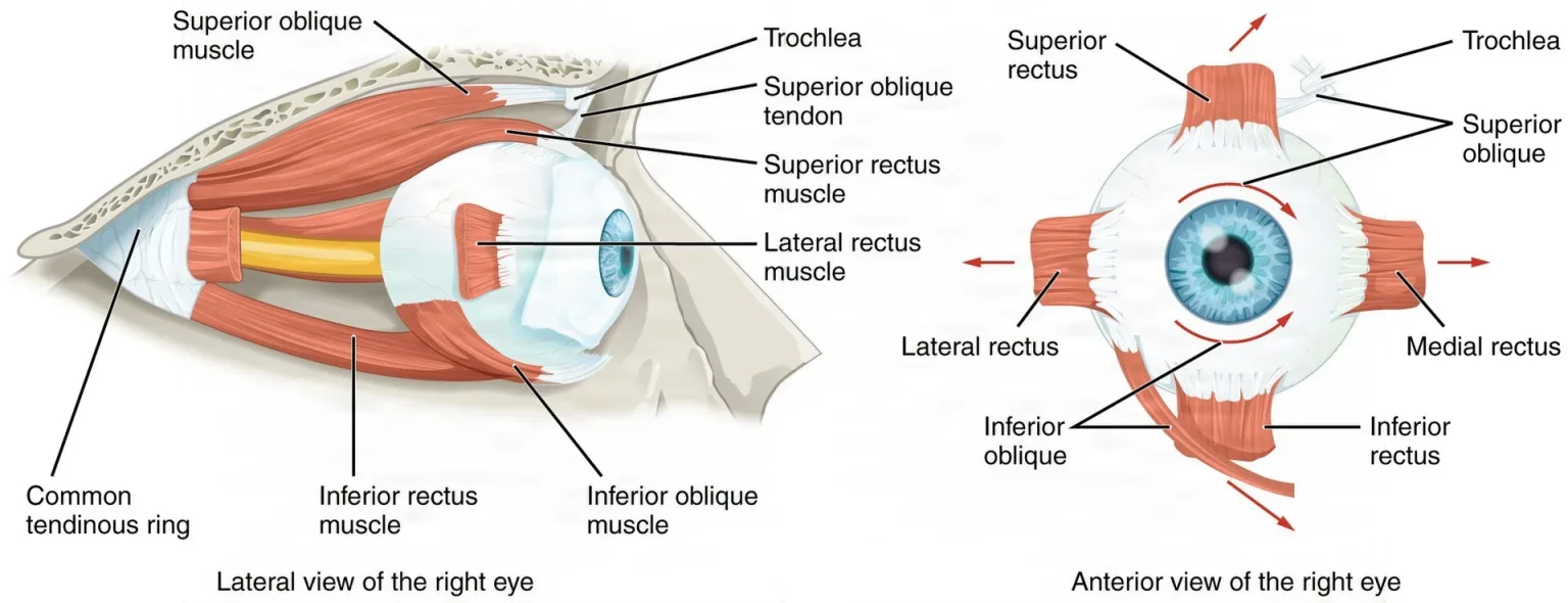
| Muscle | Turns the eye | Nerve |
|---|---|---|
| Superior rectus | Up | Oculomotor (III) |
| Inferior rectus | Down | Oculomotor (III) |
| Medial rectus | In, toward the nose | Oculomotor (III) |
| Lateral rectus | Out, away from the nose | Abducens (VI) |
| Superior oblique | Rotates the eye; its tendon loops through a pulley of cartilage, the trochlea | Trochlear (IV) |
| Inferior oblique | Rotates the eye the other way; it is the only one that starts from the floor of the orbit | Oculomotor (III) |
A short way to remember the nerves: the lateral rectus is VI, the superior oblique is IV, and all the rest are III.
In strabismus, the eyes do not line up because their muscles do not work together. The surgeon corrects it by detaching an eye muscle and reattaching it: a recession moves the attachment back to weaken the muscle, and a resection removes a piece of the muscle and reattaches the shortened end where it was, to strengthen it. This is the surgery where pulling on a muscle can slow the heart.
The conjunctiva and the lacrimal apparatus
The conjunctiva is a thin, clear mucous membrane. Its palpebral part lines the inside of the eyelids, and it folds back as the bulbar part over the white of the eye, up to the edge of the cornea, which is called the limbus. Inflammation of it is called conjunctivitis.
The lacrimal apparatus makes tears and drains them away. Tears follow this path.
- The lacrimal gland, in the upper outer part of the orbit, makes tears.
- The lacrimal ducts, small ducts from the gland, carry the tears onto the surface of the eye.
- Blinking sweeps the tears across the eye toward its inner corner.
- The puncta (singular, punctum), tiny openings on the inner end of each lid's edge, collect the tears.
- The lacrimal canals, also called canaliculi, carry them to the nasolacrimal sac, also called the lacrimal sac.
- The nasolacrimal duct drains the sac into the nose. That is why your nose runs when you cry.
Tears keep the cornea moist and clean, carry it nutrients and antibodies (it has no blood vessels of its own), and contain enzymes that break down bacteria.
The globe
The globe is the eyeball. Its wall has three layers, called tunics.
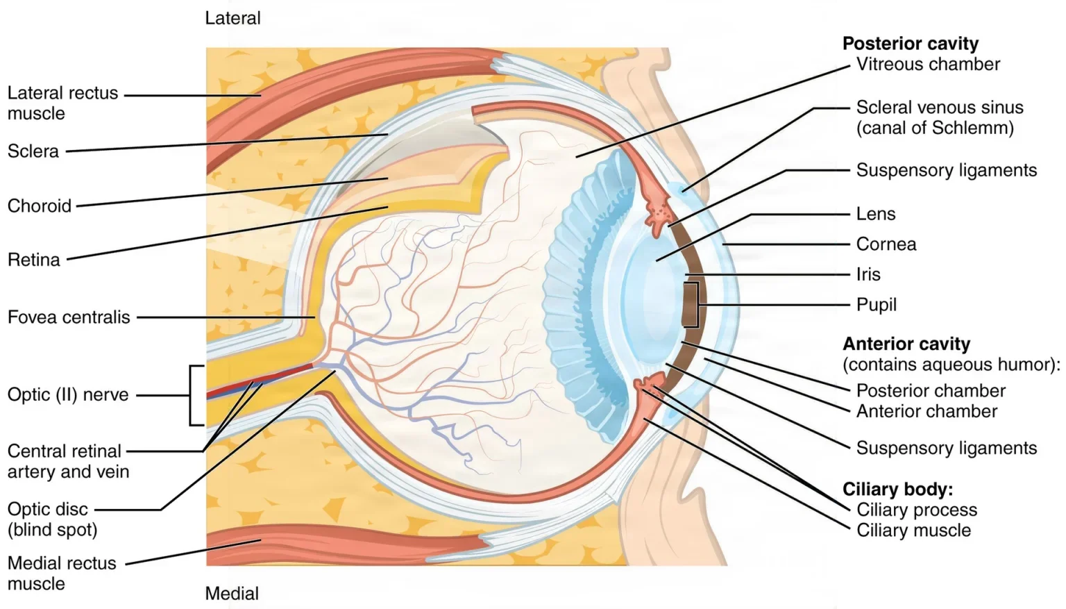
| Tunic | Its parts | What they do |
|---|---|---|
| Fibrous tunic (outer) | Sclera and cornea | The sclera, the tough white coat, covers most of the eyeball, holds its shape and anchors the eye muscles. The cornea, the clear window at the front, lets light in and bends it; it has no blood vessels, and it meets the sclera at the limbus |
| Vascular tunic (middle) | Choroid, ciliary body, iris and pupil | The choroid is a dark layer full of blood vessels that feeds the eye. The ciliary body is a ring of muscle that changes the shape of the lens and makes aqueous humor. The iris is the colored ring of muscle around the pupil, the opening that lets light in |
| Nervous tunic (inner) | Retina | The retina senses light and sends the signals to the brain through the optic nerve |
The lens is a clear, flexible disc that bulges on both sides. It sits just behind the iris and pupil, inside a thin capsule, and hangs from the ciliary body by fine fibers called zonules, or suspensory ligaments.
Two eye operations work on these parts.
- Corneal transplant: in a penetrating keratoplasty, the surgeon replaces the full thickness of a damaged cornea with a donor cornea to restore vision. The cornea's inner layer of cells pumps fluid out to keep it clear, and it does not grow back once damaged.
- Cataract surgery: a cataract is a clouding of the lens. In phacoemulsification, the surgeon enters through a small incision at the limbus, breaks the lens into tiny pieces with a vibrating probe, and suctions them out, leaving the capsule in place to hold an artificial intraocular lens (IOL).
Chambers and fluids
The inside of the eyeball is divided into two cavities.
- The anterior cavity, in front of the lens, is split by the iris into two chambers. The anterior chamber is the space from the back of the cornea to the iris, and the posterior chamber lies between the iris and the lens. Both are filled with aqueous humor.
- The posterior cavity, behind the lens, is filled with vitreous humor, a clear jelly that is 99% water. It fills about 80% of the eyeball and holds it in shape from inside.
Aqueous humor is a clear, watery fluid made by the ciliary body. It feeds the lens and the cornea, which have no blood vessels. It flows from the posterior chamber through the pupil into the anterior chamber, then drains through a small channel, the canal of Schlemm, into the veins. The balance between how much is made and how much drains sets the pressure inside the eye, the intraocular pressure (IOP).
In most types of glaucoma, the aqueous humor does not drain well, so the IOP rises. Over time the pressure starves the optic nerve and can lead to blindness. A trabeculectomy creates a new channel so the fluid can drain from the anterior chamber.
In a vitrectomy, the surgeon removes all or part of the vitreous, for example when blood has clouded it or to repair a detached retina. In a pars plana vitrectomy, small openings are made through the pars plana, a band of the eye's wall just in front of where the retina attaches, and the vitreous may be replaced with fluid, air or gas.
The optic nerve, cranial nerve II, leaves the back of the eye at the optic disc. It is a bundle of more than a million nerve fibers, and the central retinal artery and vein travel inside it to feed the retina.
How the eye sees
The Core Curriculum calls this photoreception: turning light into sight.
- Refractive media: light is bent, or refracted, as it passes through the cornea, the aqueous humor, the lens and the vitreous humor, so it comes to a sharp focus on the retina. The cornea does about two-thirds of the bending and the lens about a third.
- Light regulation: the iris narrows the pupil in bright light and widens it in dim light, to control how much light gets in. Narrowing is a parasympathetic job, carried by the oculomotor nerve; widening is a sympathetic one.
- Accommodation: to focus on something close, the ciliary muscle changes the lens to a rounder shape. With age, usually from about age 40, the lens stiffens and close focus fails, which is called presbyopia.
- Photoreceptors: the retina has two kinds of light-sensing cells. Rods work in dim light but do not see color. Cones need brighter light and give color vision, through three types tuned to red, green and blue light.
- Macula lutea: a small yellowish area in the center of the retina, used for sharp central vision.
- Fovea centralis: the small pit at the center of the macula. It holds only cones and gives the sharpest vision of all.
- Optic disc: the spot where the optic nerve leaves the eye. It has no photoreceptors, so it makes a small blind spot.
- Brain pathways: the signals run back along the optic nerve to the optic chiasm. There, the fibers from the inner (nose) half of each retina cross to the other side. The optic tract then carries them to the thalamus, which relays them to the visual cortex in the occipital lobe. So each side of the brain sees the opposite half of what is in front of you.
Let anything press on a patient's closed eye. Pressure on the eye can scratch the cornea or starve the retina of blood. For a prone patient, use a headrest that leaves the eyes and nose free, and tape the eyes gently shut instead of padding them.
13The ear
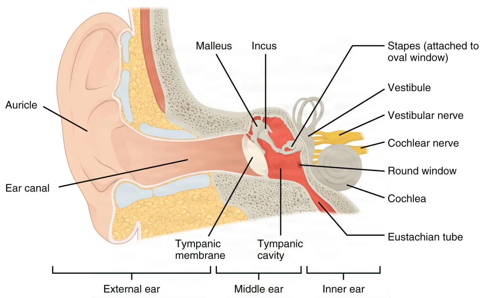
The ear has three parts, from the outside in: the external ear, the middle ear and the inner ear. Together they give us hearing and balance.
The external ear
- Auricle, or pinna: the curved flap of skin-covered cartilage on the side of the head. Its curves catch sound waves and steer them into the ear canal.
- Tragus: the small flap of skin-covered cartilage just in front of the opening of the ear canal.
- External auditory canal: an S-shaped tunnel about 2.5 cm (1 inch) long in an adult. Glands in its outer part make cerumen, or earwax, which traps dirt and keeps germs down.
- Tympanic membrane, or eardrum: a thin membrane across the end of the canal that vibrates when sound strikes it. It has three layers: skin on the outside, a fibrous middle layer for strength, and a mucous membrane on the inside. It is the border between the external and middle ear.
The middle ear
The middle ear is a small, air-filled space inside the temporal bone, lined with mucous membrane.
- Ossicles: three tiny bones that form a chain across the space. The malleus (hammer) is attached to the eardrum, the incus (anvil) is in the middle, and the stapes (stirrup) is innermost, with its footplate in the oval window. Their joints work as levers that make the vibration stronger.
- Oval window: the opening into the inner ear where the footplate of the stapes sits. Vibration passes through it into the fluid of the inner ear.
- Round window: a second opening into the inner ear, closed by a thin membrane that bulges out and in as the fluid inside moves.
- Eustachian tube, also called the auditory tube: a tube from the middle ear to the nasopharynx, at the back of the nose. It opens when you swallow, yawn or sneeze, which evens out the air pressure on the two sides of the eardrum. It is also a path for germs from the throat into the middle ear, which causes otitis media, a middle ear infection.
- Mastoid sinus: the mastoid air cells, small air pockets in the mastoid part of the temporal bone behind the ear. They open into the middle ear through a passage called the antrum, so a middle ear infection can spread into them, which is called mastoiditis.
When fluid keeps collecting behind the eardrum, the surgeon may make a tiny cut in the eardrum, a myringotomy, and place a small tympanostomy tube in it to let air into the middle ear.
Two tiny muscles protect the inner ear from loud sound: the tensor tympani pulls the eardrum tight, and the stapedius steadies the stapes. The facial nerve runs through the wall of the middle ear on its way out of the skull.
Much ear surgery happens in the middle ear.
- Tympanoplasty repairs the eardrum and, when needed, the chain of ossicles. It is done for a hole in the eardrum that will not heal, or for damage from long-lasting infection or a cholesteatoma.
- A cholesteatoma is a growth of skin cells trapped in the middle ear. It slowly destroys bone, and it can wreck the ossicles, cause hearing loss and even paralyze the facial nerve. A mastoidectomy removes the diseased mastoid air cells, and it is the way to clear a cholesteatoma that has spread into the mastoid.
Use chlorhexidine (CHG) in or around the ear. It is ototoxic: if it drains into the middle ear through the incision or a hole in the eardrum, it can damage the ear. Before an ear prep, a sterile cotton ball goes in the ear canal, and the circulator checks the solution: an iodine prep without alcohol, such as povidone-iodine, may be used on the outer ear, but not on the structures inside it, once the patient is checked for iodine sensitivity.
The AST skin prep standard counts iodine (iodophor) preps as ototoxic too, and it rules out both CHG and iodophors for eye and facial preps, with cotton balls placed in the ears.
The inner ear
The inner ear lies deep in the petrous part of the temporal bone. It is a maze of tunnels called the labyrinth, which comes in two layers.
- The bony labyrinth is the set of channels in the bone, filled with a fluid called perilymph.
- The membranous labyrinth is a set of membrane tubes inside the bony one, filled with a second fluid, endolymph.
The labyrinth has three parts.
- The cochlea, a spiral shaped like a snail shell, is for hearing. Its central duct holds the organ of Corti, a strip of hair cells that sits on the basilar membrane.
- The vestibule, the middle chamber, holds two small sacs, the utricle and the saccule, that sense the position of the head.
- The three semicircular canals, loops set at right angles to one another, sense the head turning.
The nerve from the cochlea and the nerve from the vestibule run side by side to the brain stem as one nerve, the vestibulocochlear nerve, cranial nerve VIII.
How we hear
The Core Curriculum follows sound through four steps.
- Sound wave reception: sound is a wave of pressure in the air. The auricle gathers it and sends it down the canal to the eardrum, which starts to vibrate.
- Conduction through bone: the eardrum moves the malleus, the malleus moves the incus, and the incus moves the stapes, which passes the vibration, now stronger, to the oval window. Sound can also reach the cochlea another way, by making the bones of the skull vibrate, which skips the outer and middle ear. Hearing tests compare this bone conduction with normal air conduction to tell where a hearing loss comes from.
- Fluid conduction: the stapes rocking in the oval window makes waves in the fluid of the cochlea, and the round window bulges to make room. The waves move the basilar membrane, high-pitched sounds near the base of the cochlea and low-pitched sounds near its tip.
- Nerve conduction: as the basilar membrane moves, the hair cells of the organ of Corti bend and turn the motion into nerve signals. The cochlear nerve carries them to the brain stem, and on to the hearing area of the temporal lobe.
The human ear hears pitches from about 20 to 20,000 cycles a second (hertz).
Hearing loss comes in two main kinds. In conductive loss, sound cannot get through the external or middle ear, because of wax, a hole in the eardrum or stiff ossicles. In sensorineural loss, the damage is in the cochlea, the hearing nerve or the brain.
When the cochlea's hair cells no longer work, a cochlear implant can bring back some hearing. A processor behind the ear picks up sound and sends it to a receiver under the skin, which passes it to electrodes threaded into the cochlea. The electrodes stimulate the hearing nerve directly.
How we keep our balance
Balance, or equilibrium, comes from the vestibule and the semicircular canals, which work with the eyes and with proprioception.
- Vestibule: in the utricle and the saccule, patches of hair cells sit under a layer of gel topped with tiny crystals. When the head tilts, gravity slides the heavy gel and bends the hairs, which tells the brain where the head is and when it starts moving in a straight line.
- Semicircular canals: each canal has a swelling at its base, the ampulla, holding a crest of hair cells. When the head turns, the fluid in the canals lags behind and bends the hairs, which signals the turn. Because the three canals lie at right angles, together they sense turning in any direction.
- Vestibular nerve: carries the balance signals to the brain stem and the cerebellum, alongside the cochlear nerve in cranial nerve VIII.
A problem in the inner ear can cause vertigo, a feeling that the room is spinning, and it can come with hearing loss and ringing in the ears, as in Ménière disease.
Sound travels air, bone, fluid, nerve: the auricle and canal, the eardrum and the three ossicles, the fluid of the cochlea, then the cochlear nerve. Balance comes from the other part of the inner ear, the vestibule and semicircular canals.
Lo que te llevas de este capítulo
- The CNS is the brain and spinal cord; the PNS is the 12 pairs of cranial nerves, the 31 pairs of spinal nerves and the ganglia. The sympathetic division (fight or flight) speeds the heart and widens the pupils; the parasympathetic division (rest and digest), carried mostly by the vagus nerve, slows the heart and drives digestion.
- A neuron takes signals in through its dendrites and sends them out along its axon. Myelin, made by Schwann cells in the PNS and oligodendrocytes in the CNS, speeds the signal. At the synapse, a neurotransmitter crosses the gap and is then cleared, as acetylcholinesterase clears acetylcholine. A paralytic stops movement but not pain.
- The cerebrum (four lobes, each side running the opposite side of the body) thinks and moves; the thalamus relays the senses and the hypothalamus controls the ANS; the brain stem (midbrain, pons, medulla) holds the centers for the heart rate and breathing; the cerebellum coordinates.
- The meninges, from the outside in, are the dura, arachnoid and pia. CSF fills the subarachnoid space. At a craniotomy the surgeon opens scalp, bone and dura, and closes the dura watertight to prevent a CSF leak.
- CSF is made by the choroid plexus and flows from the lateral ventricles through the foramina of Monro to the third ventricle, down the aqueduct of Sylvius to the fourth, and out through the foramina of Luschka and Magendie, to be absorbed by the arachnoid granulations. A blockage causes hydrocephalus, often treated with a VP shunt.
- Know the 12 cranial nerves by number, name, type and job. The facial nerve runs through the parotid gland and the middle ear; the vagus serves the chest and abdomen, and its recurrent laryngeal branch moves the vocal cords.
- The plexuses form the named nerves: phrenic (cervical), axillary, radial, median and ulnar (brachial), and sciatic (lumbosacral). Under anesthesia, the ulnar nerve, the common peroneal nerve and the brachial plexus are the nerves most often hurt by pressure and stretch.
- In the eye, light passes through the cornea, aqueous humor, lens and vitreous humor to the retina, and the signal goes out through the optic nerve to the occipital lobe. In the ear, sound passes through the eardrum, the ossicles and the cochlear fluid to the cochlear nerve. Never use chlorhexidine in or near the ear.
Ponte a prueba5 preguntas rápidas
Gratis para todos. Elige una respuesta para cada una y luego revísalas todas juntas.
En el quirófanoDónde vas a usar esto
Todo el banco de preguntas del capítulo en series de 10, con la respuesta explicada después de cada una.
Parte del programa para educadoresLas palabras del capítulo en tarjetas de estudio: las que fallas vuelven antes.
Parte del programa para educadoresLo asigna tu docente. Cada grupo saca sus propias 25 preguntas del banco del capítulo.
Parte del programa para educadores
Sugerir una corrección
¿Algo está mal, confuso o desactualizado en este capítulo? Avísanos y lo corregimos para todos. Tu correo solo se usa para responderte.