Skin, bones and muscles
The layers a surgeon cuts through, the bones and joints that frame every case, and the muscles that move them, with the words the team uses for each.
1Why it matters
Many operations start by cutting through skin, then fat, then the tough fascia over muscle, and many go on to work on bone, a joint or a tendon. To follow the surgeon through those layers, you need to know what each one is, what it does, and how it behaves when it is cut, held, grafted or repaired.
The skeleton is the frame the whole team works around. You hand the instruments that lift the membrane off a bone, shape bone and hold it. You help prepare grafts of skin, bone and tendon. The surgeon asks for "more abduction" or "internal rotation" and expects the leg to move the right way, because a vague word can injure the patient.
This chapter covers three body systems: the skin (the integumentary system), the skeleton and its joints, and the skeletal muscles. Other chapters teach what this one leaves out: word parts in chapter 1; tissue types and how a muscle cell contracts in chapter 2; the nerves that run the muscles and the spinal cord in chapter 4; wound healing and burns in chapter 11; fracture types and their treatment in chapter 39; skin prep and positioning in chapter 31; bone wax and stopping bleeding in chapter 29; orthopedic instruments and power tools in chapter 24; and the procedures themselves, plastic surgery in chapter 38, orthopedic surgery in chapter 39 and spine surgery in chapter 42.
2The skin
The skin and its accessory structures (hair, nails and glands) make up the integumentary system. Together they are the largest organ system in the body: the skin covers about 1.5 to 2 square meters and makes up about 16 percent of body weight.
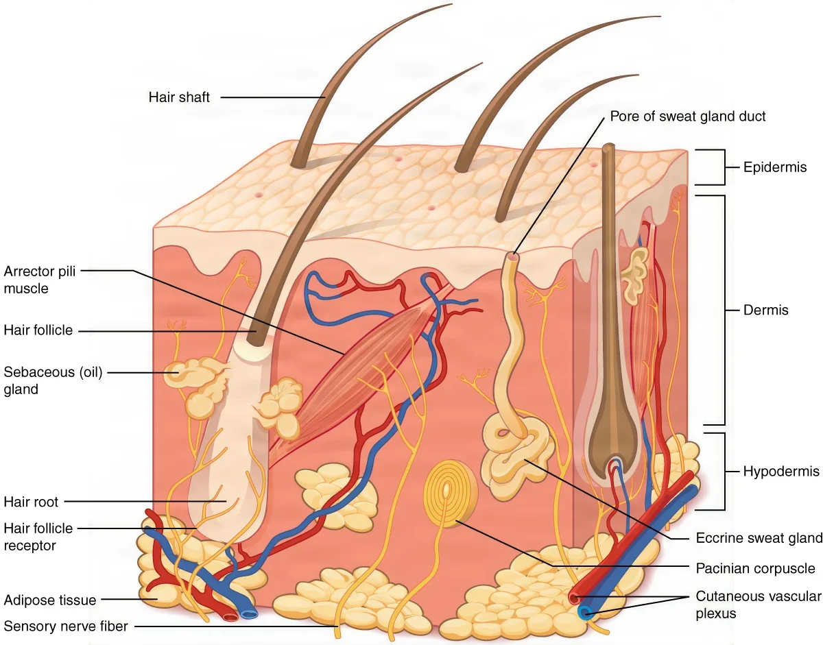
The skin has two layers. The epidermis is the thin outer layer of packed epithelial cells. The dermis below it is a thicker layer of connective tissue that holds the blood vessels, hair follicles, glands and nerve endings. Under the dermis lies the hypodermis, also called the subcutaneous layer, made mostly of fat and loose connective tissue.
What the skin does
The Core Curriculum names six jobs of the skin.
- Protection: the skin is a barrier against wind, water, sunlight, grit, chemicals and microbes. It is the body's first line of defense. Sweat even carries a natural antibiotic that keeps germs on the surface in check.
- Preventing fluid loss: keratin and water-repelling fats in the outer layer keep the body's water in.
- Regulating body temperature: when the body is warm, sweat glands release sweat that cools the skin as it evaporates, and blood vessels in the dermis widen to give off heat. When the body is cold, those vessels narrow to keep heat in.
- Sensing the outside world: nerve endings in the skin detect touch, pressure, vibration, temperature and pain, so the body can react to its surroundings.
- Excretion: sweat carries off water, salts and small amounts of wastes such as urea.
- Absorption: substances on the skin can pass through it into the blood. Some medicines are given this way as skin patches, and some chemicals can harm a person by the same route.
The skin also makes vitamin D when sunlight strikes it, and the body needs vitamin D to absorb calcium for healthy bones.
Every incision breaks the barrier. Once the skin is open, germs have a way in, which is why the surgical site is prepped and the field kept sterile (chapters 26 and 31).
The epidermis
The epidermis is made of stacked layers of flat epithelial cells, called strata (one layer is a stratum). It has no blood vessels of its own; it gets its nourishment from the dermis below. Most skin has four layers. The thick skin of the palms and soles has a fifth.
From deep to surface, the layers are:
- Stratum basale (also called the stratum germinativum): the deepest layer, a single row of stem cells that divide all the time. Every keratinocyte, the cell that makes up all the layers above, starts here. This layer also holds the melanocytes, the cells that make melanin, the pigment that colors skin and hair and protects the living cells from ultraviolet (UV) light. Merkel cells, which sense touch, sit here too.
- Stratum spinosum: 8 to 10 rows of cells pushed up from below. These cells begin to make keratin, the tough protein that makes skin, hair and nails hard and water resistant, and a fatty substance that helps waterproof the skin. Immune cells called Langerhans cells patrol this layer and swallow bacteria.
- Stratum granulosum: three to five rows of flattening cells packed with keratin. Here the cells die.
- Stratum lucidum: a thin, clear layer of dead cells, found only in the thick skin of the palms, the soles and the fingers and toes.
- Stratum corneum: the outermost layer, usually 15 to 30 rows of dead, flat cells full of keratin. It keeps out microbes, slows water loss and takes the wear of daily life. Its cells are shed regularly, and the whole layer is replaced about every 4 weeks.
In short, new cells are born at the bottom, get pushed upward, fill with keratin, die, and flake off the top.
The dermis
The dermis is the skin's working core. It is connective tissue woven from collagen fibers, which give strength, and elastin fibers, which let the skin stretch and spring back. It holds the blood and lymph vessels, nerves, hair follicles and glands. It has two layers:
- Papillary layer: the thin top layer of loose connective tissue. Finger-like bumps called dermal papillae push up into the epidermis and lock the two layers together; on the fingertips they form fingerprints. This layer is rich in small blood vessels, and it holds touch receptors and defense cells that fight germs that get through the epidermis.
- Reticular layer: the thick deeper layer of dense connective tissue with a strong mesh of collagen and elastin. It has a rich blood and nerve supply and holds most of the glands and hair follicles.
The dermis feeds the epidermis and sends it nerve supply, and its vessels carry the oxygen and the heat exchange the skin depends on.
Structures in the dermis
The Core Curriculum names the structures that live in or start from the dermis: glands, hair follicles, nails and sensory nerve endings.
- Eccrine sweat glands: coiled glands deep in the dermis, found all over the body and especially on the palms, soles and forehead. Their watery sweat cools the body.
- Apocrine sweat glands: larger glands in the armpits and groin that empty into hair follicles. Their thicker sweat has no smell until skin bacteria break it down.
- Sebaceous glands: oil glands, most of them opening into hair follicles. Their oil, called sebum, keeps the skin and hair soft and waterproof, and its fatty acids fight bacteria.
- Hair follicles: a hair follicle is the sac in the dermis that a hair grows from. The hair root sits in the follicle and ends in the hair bulb, where cells divide to lengthen the hair; the shaft above the skin is dead keratin. A tiny smooth muscle, the arrector pili, attaches to each follicle and makes the hair stand up (goose bumps) to trap warm air.
- Nails: a nail is a hard plate of dead, keratin-filled cells that grows forward from the nail root over the nail bed. It protects the fingertip and toe tip. The bed's rich blood supply makes the nail look pink, except at the pale half-moon (lunula) near the cuticle.
- Sensory nerve endings: different receptors sense different things. Meissner corpuscles in the papillary layer feel light touch, Pacinian corpuscles deeper down feel vibration, and free nerve endings sense pain and temperature. Hair follicles are wrapped in nerve endings too.
The hypodermis
The hypodermis, or subcutaneous tissue, lies directly under the dermis. Strictly it is not part of the skin, but it anchors the skin to the fascia, the tough fibrous tissue over the muscles and bones beneath. It is loose connective tissue and adipose (fat) tissue with a good blood supply.
Its fat stores energy, insulates the body against heat loss and cushions the structures beneath it.
How thick it is varies a great deal from person to person and from place to place. On the abdomen, the subcutaneous fat ranges from about a quarter inch to more than 8 inches.
The skin at the surgical field
When the surgeon makes an incision on the abdomen, the knife crosses the epidermis and dermis, then the subcutaneous fat, then the fascia, then muscle, and finally the peritoneum, the lining of the abdominal cavity. Chapter 2 explains how each of these tissues heals and holds sutures, and chapter 29 teaches incisions.
A few facts about the skin explain what you see at the field.
- The epidermis has no blood vessels, while the dermis is full of them. That is why a shallow scrape barely bleeds, but a cut into the dermis does.
- Some of the skin's own germs live below its surface, so scrubbing cannot remove them all, and skin can never be made sterile. A scrub or a prep lowers their number, and sterile gloves cover the hands of the team.
- An older patient's epidermis is thinner, the dermis repairs itself more slowly and the fat under it thins out. Older skin is fragile and easily irritated, so handle it gently, and lift the patient into position instead of pulling or dragging.
Skin grafts
A graft is tissue moved from one place to another to replace tissue lost to injury or disease. The place it is taken from is the donor site, and the place it goes is the recipient site. A graft cut from the patient's own body is an autograft; one from another person, usually through a tissue bank, is an allograft; and one from an animal, such as pig skin, is a xenograft.
Skin grafts are named by how much of the skin's thickness they take:
- A split-thickness skin graft (STSG) takes the epidermis and only part of the dermis. How much of the dermis it takes varies.
- A full-thickness skin graft (FTSG) takes the epidermis and the whole dermis. The surgeon trims any fat off its underside before suturing it in place.
The difference matters at both sites. An STSG donor site heals on its own, because new epithelium grows from the dermis left behind. An FTSG takes all of the dermis, so nothing is left to regrow, and its donor site is sutured closed.
At the recipient site, the graft has been cut off from its blood supply, so it lives only if new vessels grow into it from the wound bed. A split-thickness graft picks up a blood supply quickly, but it may shrink as it heals, is less tough, and looks and feels different from normal skin. A full-thickness graft shrinks very little, holds up to wear, can be used over joints that bend, and gives an acceptable look.
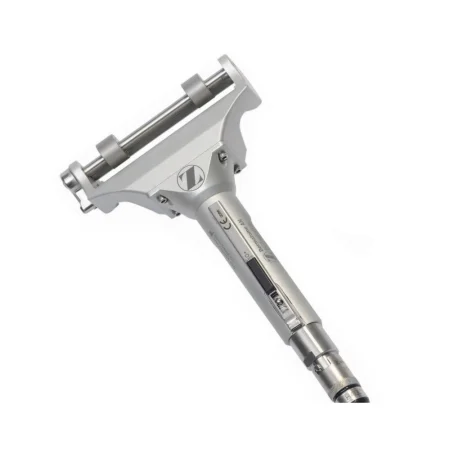
A large STSG is cut with a dermatome, a powered instrument with an oscillating blade housed in an adjustable head; a small lever on its side, marked in tenths of a millimeter, sets the thickness of the graft. The donor site is usually a broad, fleshy area, and it is lubricated so the dermatome glides. If you assemble the dermatome, check that the right blade and guard are seated and secured exactly as the maker directs: a loose blade or guard can injure the donor site or tear the graft. Each site, donor and recipient, gets its own prep and its own drapes.
An STSG is often run through a graft mesher, which cuts small diamond-shaped holes in it so the sheet opens into a net. Meshing keeps blood and serum from collecting under the graft, and lets the graft stretch to cover a larger or uneven area. The ratio of skin to holes is marked on the carrier plate, so use the plate the surgeon asks for.
Split-thickness means epidermis plus part of the dermis, and the donor site regrows on its own. Full-thickness means epidermis plus all of the dermis, and the donor site has to be closed.
3The skeleton
The skeleton is the body's frame. It is built of bones, held together at joints by ligaments, with cartilage where bones need to bend or glide. An adult has 206 bones. A newborn has more, about 270 to 300, because some of them fuse together as a child grows.
The axial and appendicular skeleton
The skeleton has two divisions.
- The axial skeleton is the central, upright axis of the body: the skull, the vertebral column and the rib cage, 80 bones in all. It protects the brain, spinal cord, heart and lungs.
- The appendicular skeleton is the limbs, plus the girdles that attach each limb to the axial skeleton: 126 bones.
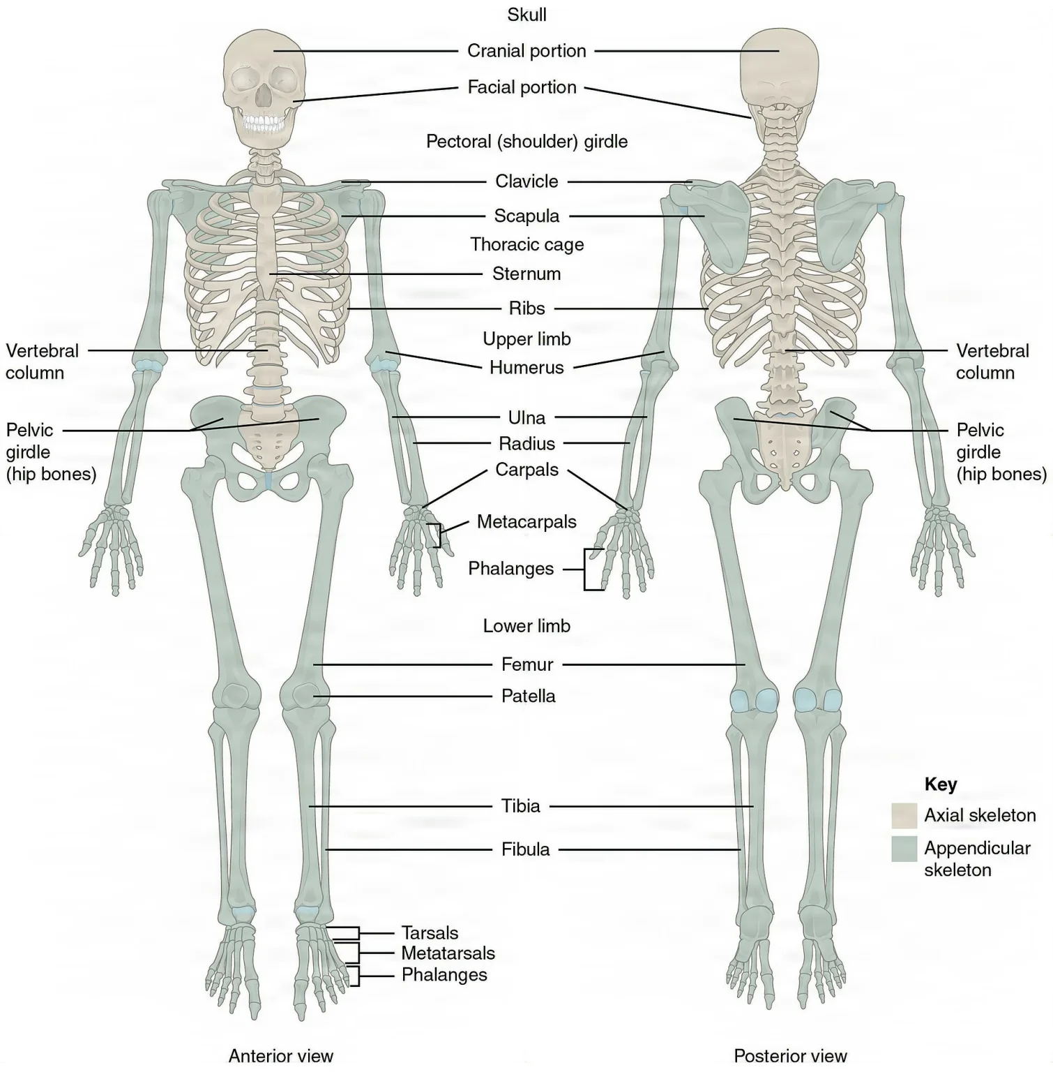
In anatomy, the arm is only the part between the shoulder and the elbow, and the leg is only the part between the knee and the ankle. The forearm, the thigh, the hand and the foot each have their own names.
| Region | Main bones |
|---|---|
| Skull (axial) | 22 bones. The cranium, or brain case, has 8: the frontal bone, two parietal bones, two temporal bones, the occipital, the sphenoid and the ethmoid. The other 14 are facial bones, including the maxilla (upper jaw), the zygomatic (cheek) and nasal bones, and the mandible (lower jaw), the only skull bone that moves. |
| Hyoid and ear bones (axial) | The horseshoe-shaped hyoid in the upper neck, the only bone that joins no other bone, and three tiny ossicles in each middle ear |
| Vertebral column (axial) | 24 vertebrae (7 cervical, 12 thoracic, 5 lumbar), plus the sacrum and the coccyx |
| Thoracic cage (axial) | The sternum (breastbone: manubrium, body and xiphoid process) and 12 pairs of ribs. Ribs 1 to 7, the true ribs, join the sternum by their own cartilage; ribs 8 to 12 are false ribs, and the last two pairs, 11 and 12, are floating ribs with no attachment in front. |
| Pectoral (shoulder) girdle | The clavicle (collarbone) and the scapula (shoulder blade), which attach each upper limb to the trunk |
| Upper limb, 30 bones each | The humerus in the arm; the ulna (on the little-finger side) and the radius (on the thumb side) in the forearm; 8 carpals in the wrist; 5 metacarpals in the palm; and 14 phalanges in the fingers and thumb |
| Pelvic girdle | One hip bone (coxal bone) on each side, formed when three bones, the ilium, ischium and pubis, fuse into one as growth ends. The two hip bones, the sacrum and the coccyx make up the pelvis. |
| Lower limb, 30 bones each | The femur in the thigh, the longest bone in the body; the patella (kneecap); the tibia (shinbone), which bears the weight, and the slender fibula in the leg; 7 tarsals in the ankle and back of the foot, including the calcaneus (heel bone); 5 metatarsals; and 14 phalanges in the toes |
What bones do
The Core Curriculum lists six functions of bone.
- Support: bones hold the body upright and carry its weight against the pull of gravity, and the bones of the lower limbs and lower back are built thick for the load.
- Framework: the skeleton gives the body its shape and gives the soft tissues something to hang on.
- Protection: the skull surrounds the brain, the vertebrae surround the spinal cord, and the ribs guard the heart and lungs.
- Attachment for muscles: muscles pull on bones through their tendons. The bones act as levers and the joints as the pivot points: a muscle that crosses a joint moves the bone when it contracts.
- Blood cell production: red bone marrow makes red blood cells, white blood cells and platelets. This is called hematopoiesis.
- Storage of calcium: bone is the body's store of calcium and phosphorus. Calcium is released into the blood when the body needs it, because calcium is needed for muscles to contract, for nerves to signal, for the heart to beat and for blood to clot. Hormones keep the blood level steady by moving calcium into and out of bone.
Bone also stores fat. Yellow bone marrow holds fat tissue, which the body can use for energy.
Types of bones
Bones are sorted into five types by their shape, and each shape suits a job.
| Type | Shape | Job | Examples |
|---|---|---|---|
| Long | Longer than wide, like a tube | Act as levers that muscles move | Femur, tibia, fibula, humerus, radius, ulna, metacarpals, metatarsals, phalanges |
| Short | About as wide and thick as long, like a cube | Give stability and support with a little motion | Carpals (wrist) and tarsals (ankle) |
| Flat | Thin and often curved | Protect organs and give muscles a place to attach | Cranial bones, sternum, ribs, scapula |
| Irregular | Complex shapes that fit no other group | Protect organs | Vertebrae and many facial bones |
| Sesamoid | Small and round, like a sesame seed, formed inside a tendon | Protect the tendon where it presses over a joint | The patella, the largest sesamoid bone and the only one everyone has |
"Long" describes the shape, not the size: the small bones of the fingers are long bones too.
Inside a long bone
A long bone shows every part a bone can have.
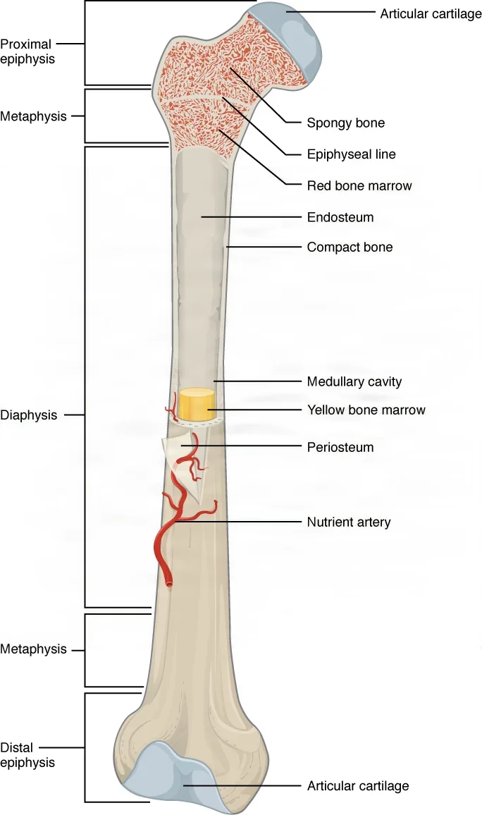
- Diaphysis: the shaft, the long tube between the two ends. Its walls are hard compact bone.
- Medullary cavity (medullary canal): the hollow center of the shaft, filled with yellow marrow in an adult.
- Epiphysis (plural, epiphyses): the wider end at each side of the bone, filled with spongy bone and red marrow. The epiphyses form the joints.
- Epiphyseal plate (growth plate): a layer of cartilage between the shaft and each end in a growing bone, where the bone grows in length. When growth stops in early adulthood, bone replaces the cartilage, and only a thin epiphyseal line is left. A break through the plate in a child can delay the bone's growth.
- Articular cartilage: a thin, smooth cap of cartilage over the ends where the bone meets another bone at a joint. It cuts friction and absorbs shock.
- Periosteum: a tough fibrous membrane that covers the outside of the bone everywhere except the articular cartilage. It carries blood vessels and nerves that feed the bone, holds bone-forming cells, and gives tendons and ligaments a place to attach.
- Endosteum: a thin membrane lining the medullary cavity, where bone growth, repair and remodeling take place.
- Cortical bone and cancellous bone: the two kinds of bone tissue, described next.
Blood vessels reach the inside of the bone through small holes in the shaft, each called a nutrient foramen.
The periosteum matters at the field. It is tough and tends to shred or tear when a saw, rongeur or cutting instrument meets it, so the surgeon first strips it off the bone surface with a periosteal elevator before cutting or reshaping the bone. A periosteal elevator is used in nearly every open orthopedic case.
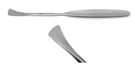
The Freer elevator also lifts periosteum off bone, and it is used to retract in confined spaces as well.
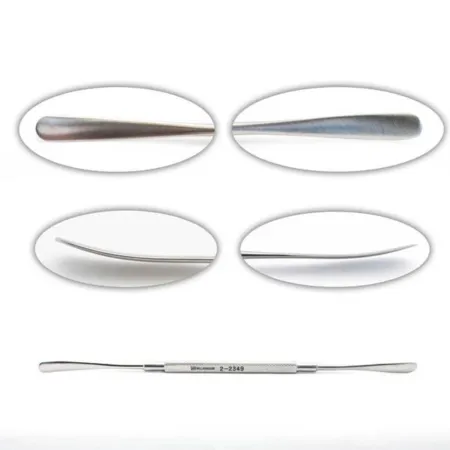
Periosteum covers the bone; endosteum lines its hollow center. The periosteum comes off with an elevator before bone is cut.
Bone tissue: cortical and cancellous
Bone tissue is a hard matrix of collagen fibers covered in calcium crystals. The crystals make bone hard and strong, and the collagen keeps it from being brittle. There are two kinds.
- Cortical bone, also called compact bone, is the dense, heavy bone of the outer shell. It lies just under the periosteum and forms the walls of the shaft of a long bone, where it gives support and protection and withstands the forces that press on the bone.
- Cancellous bone, also called spongy or trabecular bone, is a lighter network of thin bony struts called trabeculae, lined up along the lines of stress. The spaces between the struts hold marrow. It fills the ends of long bones and the inside of flat bones, the vertebrae and the pelvis.
Cancellous bone in the iliac crest, the ends of long bones and the sternum holds red marrow, and the iliac crest is a common place to take a bone graft.
Under a microscope, compact bone is built of tiny columns called osteons, or Haversian systems. Each osteon is a set of rings of hard matrix around a central (Haversian) canal that carries blood vessels, nerves and lymph vessels. Bone cells live in small spaces between the rings and pass nutrients to each other through fine channels.
Bone cells
Bone is living tissue that is built, kept and broken down all through life by its cells.
| Cell | What it does | Where it is |
|---|---|---|
| Osteogenic cell | A stem cell, the only bone cell that divides; it becomes an osteoblast | Deep layers of the periosteum and the marrow |
| Osteoblast | Builds new bone: lays down collagen and calcium matrix | Growing parts of bone, including the periosteum and endosteum |
| Osteocyte | A former osteoblast walled in by the bone it made; keeps the matrix healthy; the most common bone cell | Inside the matrix, in a small space called a lacuna |
| Osteoclast | Breaks bone down (resorption) to reshape it, repair it or release calcium; a large cell with many nuclei that comes from white blood cells | Bone surfaces, at old, injured or unneeded bone |
A simple way to remember them: osteoblasts build, osteoclasts chew. In healthy bone the two work in balance.
How bone forms and grows
Bone formation is called ossification. In an embryo the skeleton starts as sheets of membrane and models made of cartilage, and ossification replaces them with bone by two pathways.
- Intramembranous bone formation: bone forms directly from sheets of membrane tissue. Most cranial bones, the flat bones of the face and the clavicles form this way.
- Endochondral bone formation: bone replaces a model made of hyaline cartilage. The long bones and the bones of the skull's base form this way. The cartilage does not turn into bone; it is broken down and bone is laid in its place.
A long bone grows in length at its epiphyseal plates: new cartilage grows on the side toward the end, and bone replaces it on the side toward the shaft. Growth stops when the plate is all bone. Bones also grow wider, as osteoblasts add bone under the periosteum while osteoclasts widen the hollow center.
Bone keeps rebuilding itself for life. About 5 to 10 percent of the skeleton is broken down and rebuilt each year, and bone thickens where it is stressed. That is why regular exercise strengthens bone, while a limb held in a cast loses bone.
How a broken bone mends
When a bone breaks, it heals through a callus, a mass of new tissue that bone later replaces. The Core Curriculum names five stages of bone healing, and their timing overlaps:
- Inflammation: torn vessels bleed and a clot, the fracture hematoma, forms at the break within hours. This stage lasts about 2 days. Bone cells at the break die because their blood supply is cut.
- Cellular proliferation: from about the second day, cells called macrophages clean up the debris, a mesh of fibrin seals the broken edges, and new capillaries grow into that mesh.
- Callus formation: within days, a callus of cartilage and new bone forms. An internal callus fills the gap between the bone ends, and an external callus forms a collar around the outside of the break that steadies it. This stage lasts 3 to 4 weeks.
- Ossification: from about the second or third week, the callus hardens into bone as minerals are laid down, firmly joining the ends. This can take 3 to 4 months.
- Remodeling: osteoclasts remove extra and dead bone and osteoblasts lay down compact bone, until the bone takes back its shape and strength.
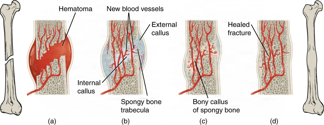
Healing needs the bone ends lined up and held still. If the pieces move, the hematoma is torn apart again and healing is delayed. Bringing the pieces back into line is called reduction, and holding them there is called fixation. In an open reduction and internal fixation (ORIF), the surgeon exposes the break and holds it with implants such as plates and screws. One break of the distal radius at the wrist, the Colles fracture, is usually caused by a fall on an outstretched hand. Fracture types and their treatment are taught in chapter 39.
Cartilage
Cartilage is a firm but flexible connective tissue. Its cells, chondrocytes, sit in small spaces in its matrix, and a membrane called the perichondrium wraps it. Chapter 2 introduced it as a tissue; here is how it works in the skeleton.
Its main characteristics:
- It has no blood vessels. Nutrients have to soak in from around it, so damaged cartilage heals very slowly and often poorly.
- It is smooth and slippery where bones glide, and it bends and springs back where the body needs flexibility.
There are three types, each in its own places:
- Hyaline cartilage, the most common type, smooth and glassy. It forms the articular cartilage at the ends of bones, the cartilage that joins the ribs to the sternum, the nose, and the growth plates.
- Fibrocartilage, a tough type, packed with thick collagen bundles. It forms the intervertebral discs, the pubic symphysis and the menisci of the knee.
- Elastic cartilage, which bends and springs back, as in the outer ear.
The Core Curriculum names two fibrocartilage structures you will meet in joint surgery:
- Labrum: a lip of fibrocartilage around the rim of a socket. The glenoid labrum deepens the shallow socket of the shoulder, and the acetabular labrum rims the socket of the hip. The shoulder's labrum also anchors ligaments that steady the joint.
- Meniscus (plural, menisci): a C-shaped pad of fibrocartilage. Each knee has two, the medial and lateral menisci, sitting between the rounded ends of the femur and the flat top of the tibia. They cushion the knee, spread its load and steady it.
Parts of each meniscus have no blood supply and heal poorly, so a torn meniscus often cannot repair itself. A meniscus tear is the most common knee injury, and the medial meniscus tears far more often than the lateral one, partly because it is anchored to a ligament and the joint capsule. In an arthroscopic meniscectomy, the surgeon removes the torn part, or most of the meniscus, through small incisions; even a complete meniscectomy leaves the rim to help carry load and steady the knee.
Bone markings
The bumps, ridges, holes and grooves on bones are called bone markings or landmarks. Each has a job: a tendon or ligament attaches there, a joint forms there, or a vessel or nerve passes through. Surgeons name them to say exactly where they are working, as in "the iliac crest" or "an osteotomy (bone cut) through the femoral neck". The Core Curriculum sorts them into three groups.
| Marking | What it is | Example |
|---|---|---|
| Articulating surfaces | Where a bone meets another bone at a joint | Knee joint |
| Head | A large, rounded end | Head of the femur |
| Condyle | A rounded, knuckle-like surface at a joint | Condyles of the femur at the knee |
| Facet | A small, flat surface | Facets where vertebrae meet each other and the ribs |
| Projections | Raised parts where tendons and ligaments attach; the stronger the pull, the bigger the projection | Processes of a vertebra |
| Process | Any bony projection | Spinous process of a vertebra, xiphoid process |
| Spine | A sharp, narrow projection | Ischial spine |
| Tubercle | A small, rounded projection | Greater tubercle of the humerus |
| Tuberosity | A larger, roughened projection | Ischial tuberosity, deltoid tuberosity, tibial tuberosity |
| Trochanter | A bony projection at the top of the femur, at the base of its neck: the large greater trochanter on the outer side and the small lesser trochanter on the inner side | Greater and lesser trochanters; the greater trochanter, where muscles that move the hip attach, can be felt at the side of the upper thigh |
| Crest | A ridge of bone | Iliac crest, the top edge of the hip bone you feel at your waist |
| Depressions and openings | Hollows, grooves and holes, many for vessels and nerves | Openings in the skull |
| Fossa | A shallow depression | Mandibular fossa, iliac fossa |
| Sulcus | A groove | Sigmoid sulcus of the temporal bone |
| Foramen | A hole through a bone, usually for vessels or nerves | Foramen magnum in the occipital bone; the intervertebral foramina between vertebrae |
| Canal, meatus | A canal is a passage through bone; a meatus is the opening into a canal | The auditory (ear) canal, and the external auditory meatus that opens into it |
| Sinus | An air-filled space in a bone | Paranasal sinuses |
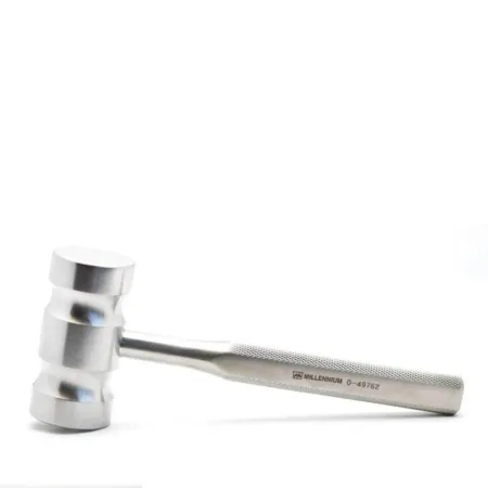
The iliac crest is a common source of a bone graft for spinal fusion. The surgeon cuts down to the crest, strips its periosteum with an elevator, then takes a plug of bone with a special cutter or shaves slivers off with an osteotome struck with a mallet. Bone left bleeding at the site is managed as chapter 29 describes. Bone graft substitutes are becoming more popular, because the harvest site is very painful after surgery.
4The vertebral column
The vertebral column, or spine, runs from the skull to the tailbone. It holds up the head and trunk, protects the spinal cord, and gives the back muscles their anchor. It starts as 33 vertebrae, which become 24 separate vertebrae (singular, vertebra) plus the sacrum and the coccyx.
Regions and curves
The Core Curriculum names five regions.
| Region | Vertebrae | What sets them apart |
|---|---|---|
| Cervical (neck) | 7, C1 to C7 | Smallest, because they carry the least weight. Each has a hole in its transverse process for an artery to the brain, and most have a forked spinous process. C1, the atlas, has no body and holds up the skull; C2, the axis, has a peg called the dens that the atlas turns around when you shake your head "no". |
| Thoracic (chest) | 12, T1 to T12 | Larger, with facets where the ribs attach and long spinous processes that slope down and overlap |
| Lumbar (lower back) | 5, L1 to L5 | Largest and thickest bodies, because they carry the most weight, with short, blunt spinous processes |
| Sacrum | 5 vertebrae fused into one triangular bone | Forms the back wall of the pelvis and joins the hip bones at the sacroiliac joints; holes on its front and back let the sacral nerves out |
| Coccyx (tailbone) | Usually 4 small vertebrae fused together | Joins the bottom of the sacrum; bears no weight when you stand, but may take some when you sit |
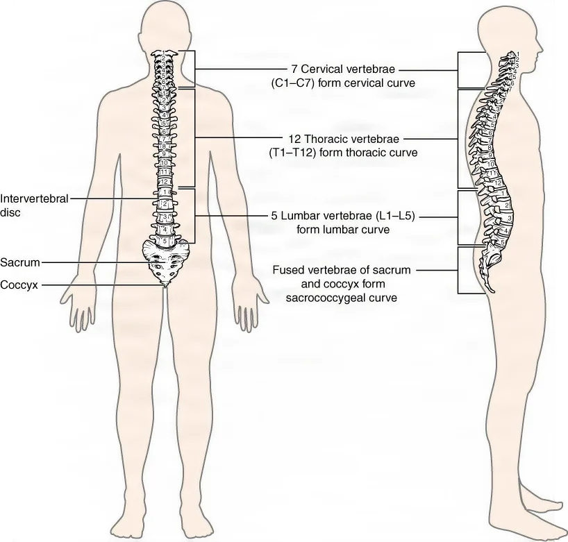
Seen from the side, the adult spine has four curves, which make it stronger, more flexible and better at absorbing shock. The thoracic and sacrococcygeal curves are present before birth. The cervical curve forms as a baby learns to hold up its head, and the lumbar curve as a child starts to stand and walk. An abnormal sideways curve with twisting is scoliosis; an excessive rounding of the upper back is kyphosis; and an excessive inward curve of the lower back is lordosis.
Parts of a vertebra
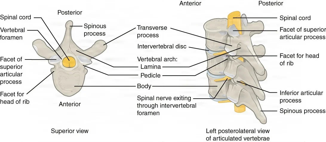
Vertebrae differ by region, but a typical one has the same parts:
- Body: the thick, round front part that carries the weight. The bodies get bigger toward the bottom of the spine.
- Vertebral arch: the ring of bone behind the body. It is made of two pedicles, which attach to the body, and two laminae (singular, lamina), which meet in the back to form the roof of the arch.
- Vertebral foramen: the large hole between the body and the arch. Stacked together, these holes form the vertebral (spinal) canal, which holds the spinal cord.
- Processes: seven bony projections arise from the arch: two transverse processes that stick out to the sides, one spinous process that points backward (the bumps you feel down the middle of the back), and four articular processes, two up and two down, that form small gliding joints with the vertebrae above and below. The transverse and spinous processes anchor muscles.
- Intervertebral foramen: the gap between two neighboring vertebrae, beside the pedicles, where a spinal nerve leaves the spine.
Intervertebral discs
Between the bodies of the vertebrae sit the intervertebral discs, pads of fibrocartilage that bind the vertebrae together, cushion them under load and let the spine bend. They are thinnest in the neck and thickest in the lower back, which carries the most weight.
Each disc has two parts:
- Annulus fibrosus (also spelled anulus fibrosus): the tough outer ring of fibrous tissue, anchored to the edges of the vertebral bodies.
- Nucleus pulposus: the soft, gel-like center. Its high water content resists compression. With age it dries out, so the discs thin and the spine stiffens.
If the annulus weakens or tears, the nucleus can bulge out backward. This is a herniated disc ("slipped" or "ruptured" disc), and it can press on a spinal nerve as the nerve leaves through the intervertebral foramen, causing pain and weakness. It happens most often at L4 to L5 or L5 to S1, where it can cause sciatica, pain that runs from the lower back down the leg.
Spinal ligaments
Ligaments run the length of the spine and tie the vertebrae together.
- Anterior longitudinal ligament: runs down the front of the vertebral bodies and stops the spine from bending too far backward. Whiplash can tear it.
- Posterior longitudinal ligament: runs down the back of the vertebral bodies, inside the canal in front of the spinal cord.
- Ligamentum flavum: short, yellow, stretchy ligaments joining the laminae of neighboring vertebrae, behind the spinal cord.
- Supraspinous ligament: joins the tips of the spinous processes; in the neck it widens into the nuchal ligament.
The spine at the surgical field
Spine surgery reaches the spine from the back or the front, and the parts above tell you what is being removed.
- A laminectomy reaches the spinal canal from the back. Through a midline incision, the surgeon goes through the fascia, packs the large back muscles away from the bone with periosteal elevators and sponges, removes the spinous process with a large rongeur, and then removes the lamina bit by bit with Kerrison rongeurs. The ligamentum flavum is then opened or removed to reach the disc. Keep the rongeur tips clean as each bite is presented, and save the bone pieces as a specimen.
- An anterior cervical discectomy and fusion (ACDF) reaches the neck spine from the front. The incision passes through the platysma muscle, and the surgeon works along the edge of the sternocleidomastoid muscle, between the carotid artery on one side and the trachea and esophagus on the other. The anterior longitudinal ligament is opened, the disc is removed piece by piece, and the space is filled with bone graft so the two vertebrae fuse.
5Joints
A joint, or articulation, is any place where two bones, or a bone and cartilage, meet. The way a joint is built decides how much it can move, and how much it moves decides how stable it is: the freest joints are the least stable.
Joints by how much they move
The Core Curriculum sorts joints by movement.
- Synarthrotic joints (synarthrosis): immovable or nearly so. Their strength protects what lies beneath, as in the sutures between the skull bones, which are joined by fibrous tissue.
- Amphiarthrotic joints (amphiarthrosis): slightly movable, with the bones joined by cartilage. Examples are the joints between vertebral bodies, joined by the discs, and the pubic symphysis in the front of the pelvis.
- Diarthrotic joints (diarthrosis): freely movable. All of them are synovial joints, such as the shoulder, hip, elbow and knee, and most are in the limbs.
Joints are also sorted by what holds the bones together: fibrous joints are joined by dense connective tissue, cartilaginous joints by cartilage, and synovial joints meet inside a fluid-filled space.
The synovial joint
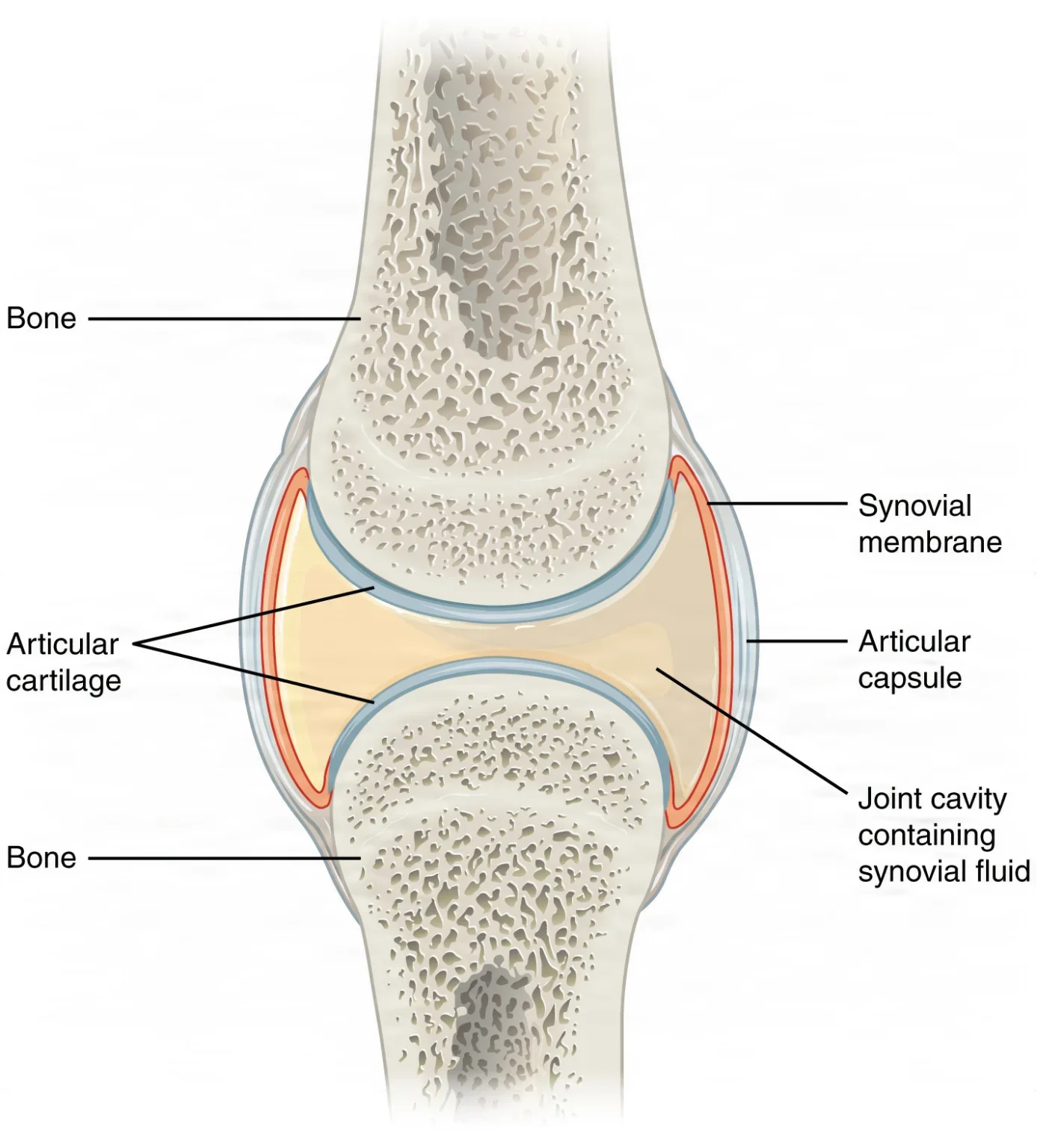
No joint type is more common in the body than the synovial joint. Its bone ends are not joined to each other; they meet inside a closed space, the joint cavity.
Its parts:
- Capsule (articular capsule): a sleeve of fibrous connective tissue attached to both bones just beyond their joint surfaces, enclosing the joint cavity.
- Synovial membrane: a thin lining on the inside of the capsule that makes synovial fluid, a thick, slippery fluid that lubricates the joint and feeds its articular cartilage, which has no blood vessels.
- Articular cartilage: the smooth hyaline cartilage on each bone end, which lets the bones glide without wearing each other down.
- Ligaments: strong bands of dense connective tissue that tie bone to bone, allow normal movement and stop movement that would injure the joint. An external (extrinsic, or extra-articular) ligament lies outside the capsule, such as the collateral ligaments on the sides of the knee. An intra-articular (intracapsular) ligament lies inside the capsule, such as the cruciate ligaments in the center of the knee. Some ligaments are thickenings in the capsule wall itself.
- Bursae (singular, bursa): small sacs of lubricating fluid that sit where skin, a tendon, a ligament or a muscle would rub against bone or against each other, usually near a joint. Examples are the bursa over the kneecap, the one at the tip of the elbow, and the subacromial bursa that lets the rotator cuff tendons glide under the shoulder's bony roof. A tendon sheath works the same way, wrapping a tendon where it crosses a joint.
Some synovial joints also hold a pad of fibrocartilage between the bones: a small articular disc, as in the jaw joint, or a larger meniscus, as in the knee.
Neither tendons nor ligaments have much blood supply, so, like cartilage, they heal slowly.
Types of synovial joints
Synovial joints are named for the shape of the surfaces that meet, and the shape decides the motion.
| Type | How it moves | Examples |
|---|---|---|
| Hinge | Bends and straightens in one plane, like a door hinge | Elbow, knee, ankle, finger and toe joints |
| Pivot | One bone rotates around another | Atlas turning on the axis (shaking the head "no"); the radius turning on the ulna near the elbow |
| Condyloid | An oval end in a shallow socket: bends, straightens and moves side to side | The wrist joint and the knuckles |
| Saddle | Two saddle-shaped surfaces: moves in two planes | The joint at the base of the thumb, which lets the thumb touch the fingertips |
| Plane (gliding) | Flat surfaces slide over each other | Between the carpal bones, the tarsal bones and the articular processes of the vertebrae |
| Ball-and-socket | A rounded head in a cup: moves in every plane and rotates; the most movable joint | Shoulder and hip |
The shoulder's socket, the glenoid cavity, is shallow, so the shoulder moves more freely than any other joint but leans on its muscles, especially the rotator cuff, to stay in place. The hip's socket, the acetabulum, is deep, with strong ligaments, so it trades movement for stability and weight bearing.
Joints at the surgical field
- Arthroscopy: in arthroscopic surgery, the scope and instruments pass through the capsule into the joint cavity through small incisions. Synovial fluid runs out when a capsule is injured or opened.
- Arthroplasty: surgery to repair or replace a joint. When articular cartilage wears thin, as in osteoarthritis, more pressure falls on the bone beneath it, the bone surface turns rough, and the joint becomes painful and stiff. In a total hip arthroplasty, the surgeon opens the capsule, dislocates the hip, cuts through the femoral neck to remove the femoral head, prepares the acetabulum, and replaces both sides with implants.
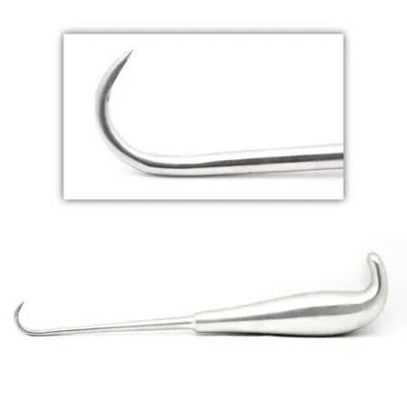
A bone hook slipped under the femoral neck helps lift the head of the femur out of the acetabulum when the hip is dislocated. Because bone does not yield to a retractor the way soft tissue does, hooks and bone clamps are used to grasp and move it.
Landmarks of the joints surgeons repair
Orthopedic surgery (chapter 39) names these parts of the limb joints again and again. A neck is a narrow bridge of bone, and cruciate means cross shaped.
- Shoulder: the head of the humerus sits in the glenoid, a shallow socket on the scapula, at the glenohumeral joint, and a rim of cartilage, the labrum, deepens the socket a little. The clavicle meets the acromion of the scapula at the acromioclavicular (AC) joint and the sternum at the sternoclavicular joint. Just below the humerus's two tuberosities is its surgical neck, where most fractures of the upper humerus happen.
- Elbow and forearm: the olecranon, the point of the elbow, is the upper end of the ulna, and the radial head meets the humerus beside it; the radius turns around the ulna to turn the palm up and down.
- Wrist: the scaphoid, which links the two rows of carpal bones, is the carpal bone most often broken, and the lower end of the radius forms most of the wrist joint.
- Knee: the rounded condyles of the femur rest on the flat top of the tibia, the tibial plateau, and the two cruciate ligaments cross in the middle of the joint.
- Ankle: the lower ends of the tibia (the medial malleolus) and the fibula (the lateral malleolus) form a slot, the mortise, that holds the top of the talus; below it, the subtalar joint with the calcaneus turns the foot in and out.
6Muscles
What muscle does
Skeletal muscle has three main jobs.
- Movement: muscles pull on bones to move the body, and some move other structures, such as the skin of the face, the tongue and the eyes.
- Posture: muscles of the spine, trunk and pelvis hold the body upright and balanced, working in turns so no group tires out.
- Heat production: working muscle makes heat, which helps keep body temperature steady.
There are three types of muscle tissue, which chapter 2 describes cell by cell:
- Skeletal muscle: striped (striated) fibers attached to bones, under voluntary control. It is the muscle this section is about.
- Cardiac muscle: striated muscle found only in the heart, involuntary, contracting on its own rhythm.
- Smooth muscle: unstriped, involuntary muscle in the walls of hollow organs, airways and blood vessels.
The words for a skeletal muscle
The Core Curriculum names six terms every scrub uses.
- Origin: the end of a muscle attached to the bone that stays still when the muscle contracts.
- Insertion: the end attached to the bone that moves. When a muscle contracts, it pulls the insertion toward the origin.
- Muscle belly: the thick, fleshy middle of a muscle, between its two ends. You can see the belly of your biceps bulge when you bend your elbow.
- Tendon: a cord of dense connective tissue that anchors a muscle to bone. Its collagen blends into the muscle at one end and into the periosteum at the other, so the pull of the muscle reaches the bone. A broad, flat tendon is called an aponeurosis.
- Fascia: sheets of tough connective tissue, mostly collagen, that wrap, support and separate muscles and other structures. Each muscle has its own sheath, and deep fascia divides the thigh and leg into compartments of muscles.
- Bursa: a fluid-filled sac that keeps a muscle or tendon from rubbing on bone, described with the joints above.
Not every skeletal muscle pulls on bone. The muscles of facial expression insert into the skin, so they move the skin, and the diaphragm changes the size of the chest without moving a bone.
Muscles work in teams
A single movement uses several muscles, each with a role. The Core Curriculum names four.
- Agonist, or prime mover: the main muscle that makes a movement. When you lift a cup, the biceps brachii is the prime mover that bends the elbow.
- Synergist: a muscle that helps the prime mover. The brachialis, deep to the biceps, helps bend the elbow.
- Fixator: a synergist that holds a bone steady, often at the prime mover's origin, so the movement happens where it should.
- Antagonist: a muscle with the opposite action, which relaxes while the prime mover works. Antagonists also hold a limb in position and slow a fast movement so it stops under control.
The roles switch with the movement. To straighten the knee, the quadriceps on the front of the thigh are the agonists and the hamstrings on the back are the antagonists. To bend the knee, the hamstrings become the agonists. The biceps and triceps of the arm, and the finger flexors and extensors of the forearm, are paired the same way.
The origin stays put and the insertion moves. The agonist does the job, the synergist helps, the fixator steadies, and the antagonist does the opposite.
The actions of muscles
Body movements are named in pairs of opposites, and they are always described from anatomical position: standing, arms at the sides, palms facing forward. The surgeon uses these words to ask for a limb to be moved, as in "more internal rotation" or "increase the abduction", and less precise words can lead to an injury.
| Action | What it does | Example |
|---|---|---|
| Flexion | Bends a joint, making the angle between the bones smaller; also bending the neck or trunk forward | Bending the elbow or the knee |
| Extension | Straightens a joint, making the angle larger; also bending the trunk back or returning upright | Straightening the elbow or the knee |
| Hyperextension | Extension beyond the normal range, which injures the joint | Whiplash of the neck |
| Abduction | Moves a limb away from the midline of the body; spreads the fingers or toes | Raising the arm out to the side |
| Adduction | Moves a limb toward or across the midline; brings the fingers or toes together | Lowering the arm back to the side |
| Medial rotation (internal rotation) | Turns the front of the arm or thigh in toward the midline, at the shoulder or hip | Turning the thigh so the knee points inward |
| Lateral rotation (external rotation) | Turns the front of the arm or thigh out, away from the midline | Turning the thigh so the knee points outward |
| Circumduction | Moves the end of a limb in a circle by flexion, abduction, extension and adduction in turn | Circling the arm at the shoulder |
| Supination | Turns the forearm so the palm faces forward, or up; the radius and ulna lie side by side | The palm up, as when carrying a bowl of soup |
| Pronation | Turns the forearm so the palm faces backward, or down; the radius crosses over the ulna | The palm down on a table |
| Dorsiflexion | At the ankle, lifts the top of the foot toward the shin | Lifting the toes off the floor while the heel stays down |
| Plantar flexion | At the ankle, points the toes down and lifts the heel | Standing on tiptoe |
| Inversion | Turns the sole of the foot in, toward the midline | The soles facing each other |
| Eversion | Turns the sole of the foot out, away from the midline | The soles facing outward |
| Elevation | Moves the scapula or the mandible up | Shrugging; closing the mouth |
| Depression | Moves the scapula or the mandible down | Lowering the shoulders; opening the mouth |
| Retraction | Moves the scapula or the mandible back | Pulling the shoulder blades together; pulling the chin back |
| Protraction | Moves the scapula or the mandible forward | Reaching forward to push; jutting the chin out |
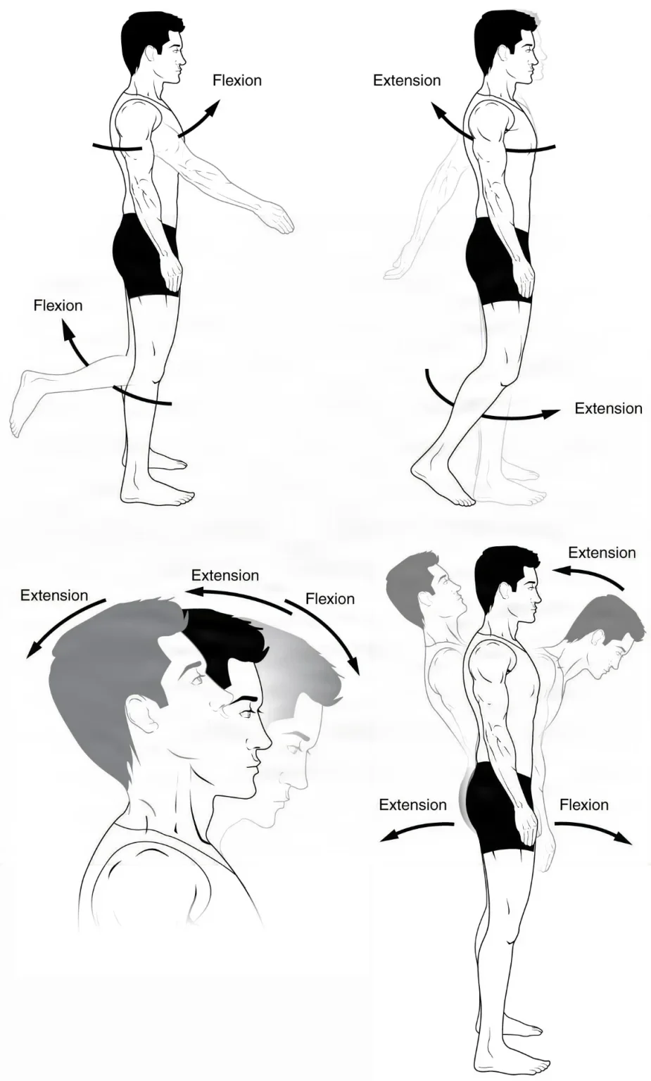
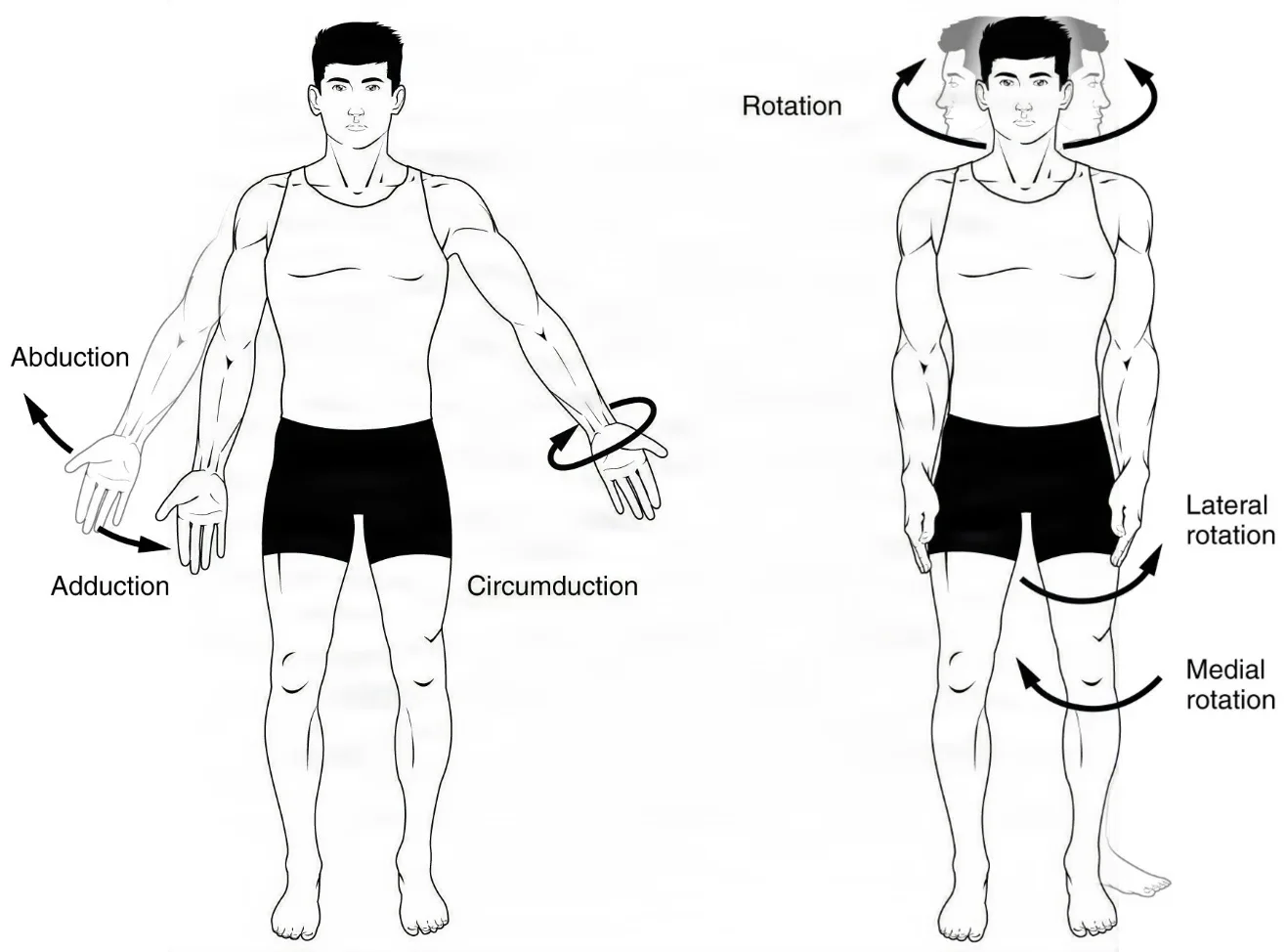
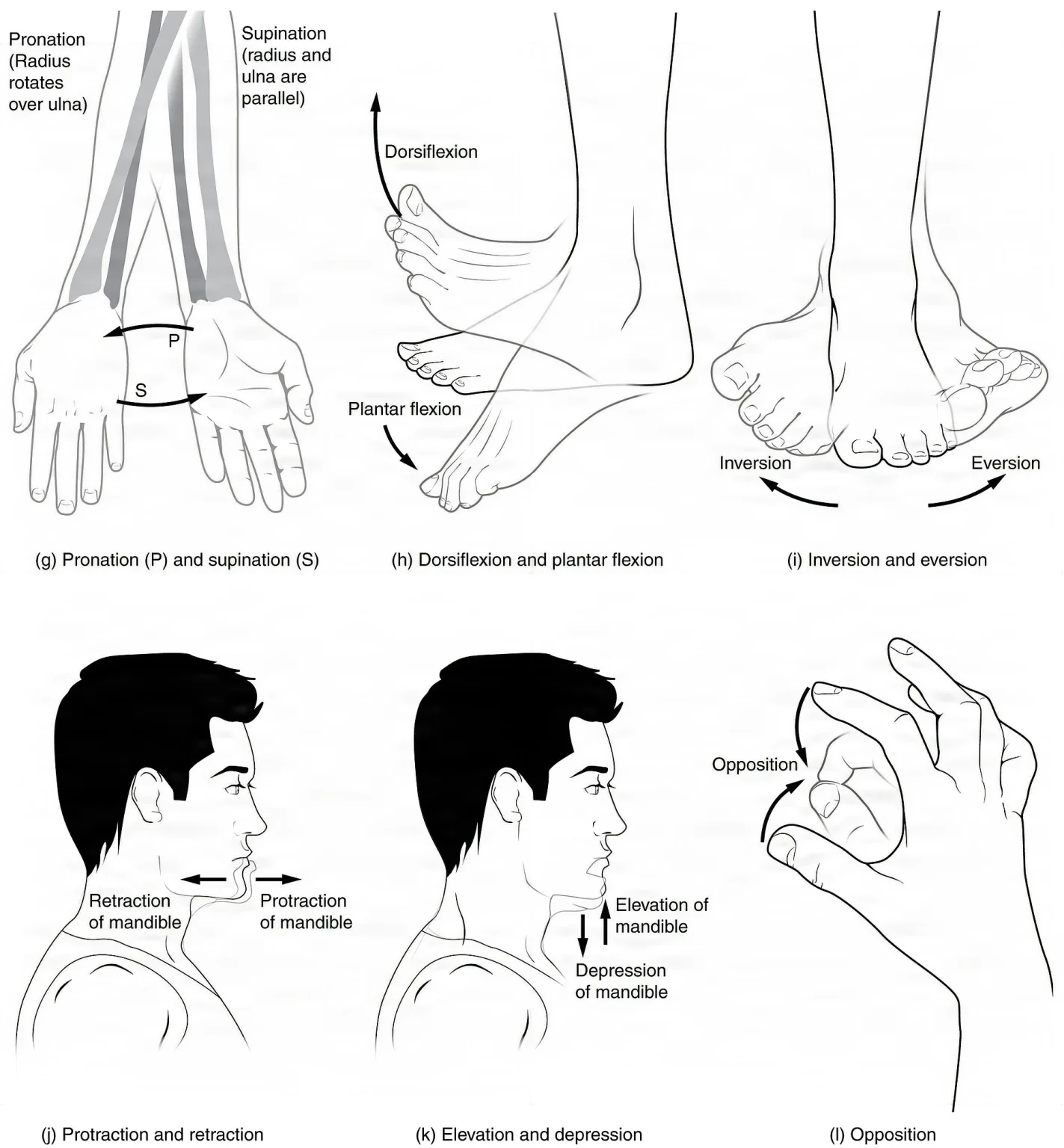
A memory trick: you hold soup in a supinated palm. Abduction takes a limb away, and adduction adds it back to the body.
You will use these words every time a patient is positioned. For a supine patient, AST directs that the arms be abducted less than 90 degrees, and preferably no more than 60, on padded armboards, with the palms up and the fingers extended. Once a patient is anesthetized, they cannot tell the team that a joint hurts, so the team must protect the skin, joints and muscles for them. Chapter 31 teaches positioning.
Move an anesthetized patient's joint past its normal range of motion, whether you are positioning the patient or holding a limb for the surgeon. The patient cannot tell you it hurts, so the team must protect the joint for them.
The word retraction has a second meaning in the OR: holding tissue back with a retractor to expose the field. The surgeon's request will tell you which one is meant.
How muscles get their names
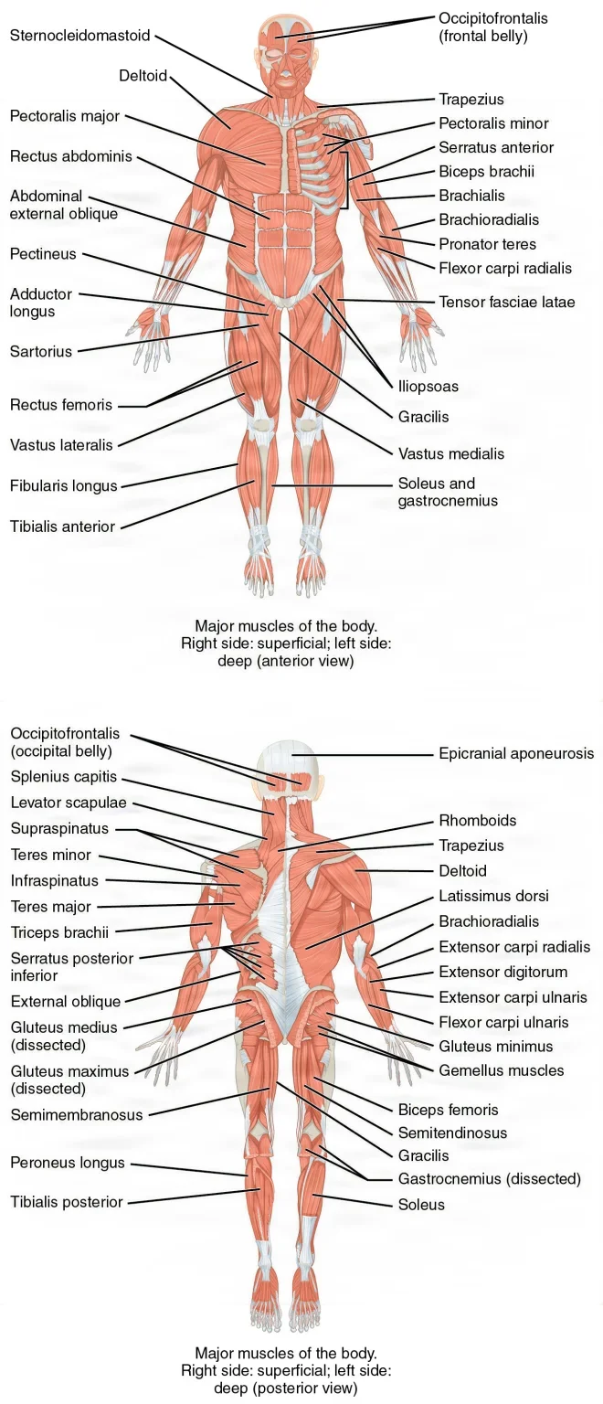
Muscle names look hard, but most of them describe the muscle, so if you can read the name, you already know something about it. Muscles are named for:
- Location: the frontalis lies over the frontal bone; the tibialis anterior lies on the front of the tibia.
- Shape: the deltoid is triangular, like the Greek letter delta; the trapezius is shaped like a trapezoid.
- Size: maximus is largest, medius is middle and minimus is smallest, as in the three gluteal muscles; longus is long and brevis is short.
- Direction of the fibers: rectus means straight and oblique means slanted, as in the rectus abdominis and the external oblique.
- Number of heads or origins: the biceps has two, the triceps three, and the quadriceps is a group of four.
- Attachments: the sternocleidomastoid runs from the sternum and clavicle (cleido) to the mastoid process. When a muscle is named for its attachments, the origin comes first.
- Action: a flexor bends, an extensor straightens, an abductor moves away from the midline and an adductor moves toward it.
Many names carry two clues at once. The flexor carpi ulnaris, for example, flexes the hand at the wrist, where the carpal bones are, and lies on the ulna side of the forearm.
The muscles you will meet most at the field
The full Core Curriculum list is in the tables that follow. These are the muscles you will hear named most, and why.
The abdominal wall. Four pairs of muscles form the front and sides of the abdomen, protecting organs where there is no bone and helping to breathe and to bear down. The two rectus abdominis muscles run straight up and down on either side of the midline, from the pubis to the ribs, each wrapped in a fibrous rectus sheath. The sheaths meet in the midline as the linea alba, a white band of fibrous tissue with almost no blood vessels. On each flank, three flat muscles lie in layers with their fibers crossing like the layers of plywood: the external oblique outermost, its fibers running down and toward the midline; the internal oblique beneath it, its fibers at right angles to those above; and the transversus abdominis deepest, its fibers running straight across.
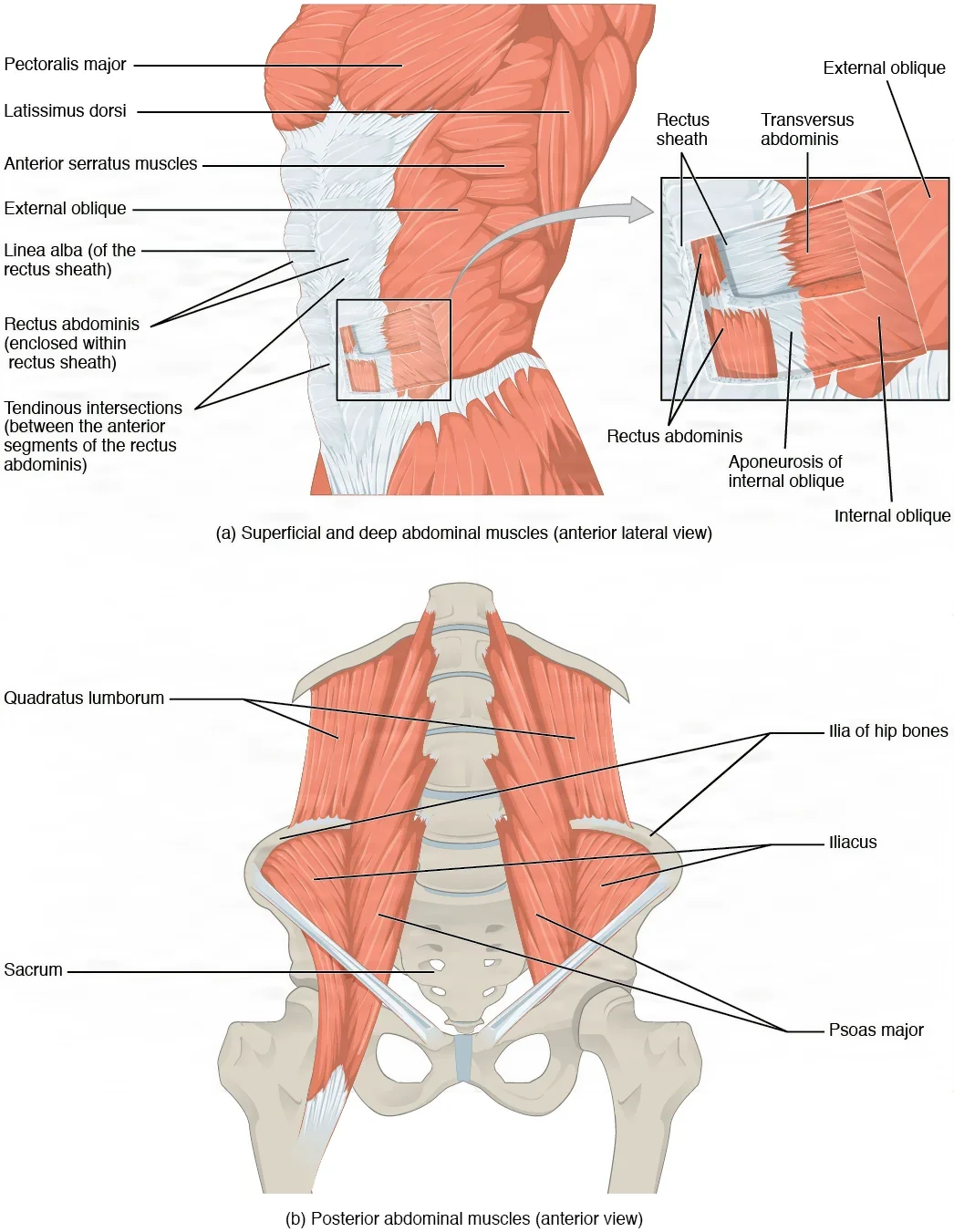
The incision decides which layers are cut. A midline incision goes through the linea alba, which bleeds little because it has so few vessels. A McBurney incision for the appendix splits the oblique and transverse muscles along their fibers rather than cutting across them. A Pfannenstiel incision runs low and across, following the skin's natural folds, through the rectus fascia, and the rectus muscles are then separated in the midline. The fascia carries the strength of the closed wound, so it is closed with strong suture, often size 0 or 2-0. Chapter 29 teaches incisions, and chapter 25 teaches closure.
The neck. The platysma is a thin sheet of muscle just under the skin of the front of the neck. It is cut in operations on the front of the neck, such as thyroid surgery and an ACDF, and its edges are sewn back together at the close. Below it, the thin strap muscles (the sternohyoid and sternothyroid) are separated in the midline to reach the structures beneath, then sewn back together at the close. The sternocleidomastoid, running at an angle from the sternum and clavicle up to behind the ear, is a key landmark: in an ACDF, the surgeon works along its front edge.
Under general anesthesia, most skeletal muscles relax, including the tongue, which can fall back and block the airway. That is one reason the anesthesia provider secures the airway with a breathing tube.
The back and chest. The erector spinae muscles run in thick columns along both sides of the spine. In spine surgery from the back, they are pushed off the vertebrae with periosteal elevators and sponges and held aside to expose the bone. In a thoracotomy, the surgeon reaches the lung between two ribs by cutting the intercostal muscles along the upper border of the lower rib, because the nerve, artery and vein of each rib space run along the lower border of the rib above.
The shoulder. Four muscles from the scapula, the supraspinatus, infraspinatus, teres minor and subscapularis, end in tendons that fuse with the shoulder's capsule to form the rotator cuff. Because the shoulder's socket is so shallow, the cuff is what mainly holds the head of the humerus in place. A cuff tear usually happens where the supraspinatus tendon attaches to the humerus. In a rotator cuff repair, the torn tendon is fixed back to the humerus with sutures or suture anchors, and small bony outgrowths (spurs) may be removed so they do not pinch the cuff.
The wrist. On the front of the wrist, the carpal bones form a U-shaped trough, and a strong band of fibrous tissue, the flexor retinaculum, spans its top to make the carpal tunnel. The central part of this band is the transverse carpal ligament. The flexor tendons of the fingers slide through the tunnel with the median nerve. When the nerve is squeezed, the patient has carpal tunnel syndrome, and in a carpal tunnel release, the surgeon divides the transverse carpal ligament with a #15 blade to give the nerve room.
The thigh and knee. The four quadriceps muscles on the front of the thigh share one tendon that wraps around the patella, the body's largest sesamoid bone, and continues below it as the patellar ligament to the tibial tuberosity. The three hamstrings on the back of the thigh bend the knee. The central third of the patellar ligament with a plug of bone at each end, the semitendinosus and gracilis tendons (called a hamstring graft), or the end of the quadriceps tendon with a block of bone from the patella can each serve as the graft that rebuilds a torn anterior cruciate ligament (ACL).
The calf. The gastrocnemius and the soleus, the large muscles of the calf, join into the Achilles (calcaneal) tendon, the strong tendon that attaches to the heel bone and points the foot down. It can rupture, especially in sports with jumping. In a repair, the surgeon brings the torn ends together with heavy nonabsorbable suture, tying it with the knee bent and the foot pointed down (plantar flexed) to take tension off the repair, and may reinforce it with a graft of the plantaris tendon, harvested with a tendon stripper.
Tendons and ligaments are commonly repaired with nonabsorbable sutures and suture anchors, and because they have a poor blood supply, they heal slowly.
The diaphragm. The diaphragm is the dome-shaped sheet of skeletal muscle that separates the chest from the abdomen. When it contracts, it flattens, the chest gets bigger and air flows into the lungs. The esophagus, the aorta and the inferior vena cava pass through openings in it.
7The major muscles by region
These tables follow the Core Curriculum's list from the face to the foot. Learn the location and main action of each; the names often tell you both.
Face and head
| Muscle | Where it is | Main action |
|---|---|---|
| Muscles of facial expression (such as the orbicularis oris, orbicularis oculi and buccinator) | Under the skin of the face; they start on the skull and end in the skin | Move the skin to make expressions: the orbicularis oris closes and moves the lips, the orbicularis oculi closes the eye, and the buccinator compresses the cheek |
| Scalp muscle: occipitofrontalis | A front belly over the forehead and a back belly over the occipital bone, joined across the top of the skull by a flat tendon, the epicranial aponeurosis (galea) | Moves the scalp and raises the eyebrows |
| Temporalis (a muscle of mastication, or chewing) | Fan-shaped, over the temple, from the side of the skull to the mandible | Closes the jaw and pulls it back |
| Masseter (a muscle of mastication) | In the cheek, from the zygomatic arch to the angle of the mandible | The main chewing muscle: raises the mandible to close the mouth |
| Medial and lateral pterygoids (muscles of mastication) | Deep, from the sphenoid bone to the mandible | Help chew: open and close the jaw, push it forward and move it side to side |
| Extrinsic tongue muscles (genioglossus, styloglossus, palatoglossus, hyoglossus) | Start outside the tongue, on the mandible, the styloid process, the soft palate and the hyoid, and insert into it | Move the whole tongue forward, back, up and down, to help chew, swallow and speak |
| Intrinsic tongue muscles | Start and end inside the tongue | Change the tongue's shape, curling or flattening it |
Neck
| Muscle | Where it is | Main action |
|---|---|---|
| Platysma | A thin sheet just under the skin of the front of the neck, from the upper chest to the lower face | A muscle of facial expression: helps lower the jaw and widens the neck |
| Sternocleidomastoid | From the sternum and clavicle up to the mastoid process behind the ear; divides each side of the neck into front and back triangles | One side turns the head toward the opposite side and tilts it; both together flex the head |
| Scalenes (anterior, middle and posterior) | The side of the neck, from the cervical vertebrae down to the first two ribs | Flex, tilt and turn the head and neck, and raise the top two ribs in deep breathing |
| Sternohyoid | A thin strap muscle from the sternum up to the hyoid bone, below the hyoid | Pulls the hyoid down |
| Sternothyroid | A strap muscle from the sternum up to the thyroid cartilage of the larynx | Pulls the larynx down |
| Thyrohyoid | A strap muscle from the thyroid cartilage up to the hyoid | Pulls the hyoid down, or raises the larynx |
| Omohyoid | A strap muscle below the hyoid with two bellies, an upper and a lower | Pulls the hyoid down |
Back
| Muscle | Where it is | Main action |
|---|---|---|
| Erector spinae (three columns: iliocostalis, longissimus and spinalis) | Along each side of the spine, from the pelvis and sacrum up to the ribs, vertebrae and skull; most of the muscle of the back | The main extensor of the spine; also controls bending and twisting and holds the lumbar curve |
| Latissimus dorsi | A broad, flat, triangular muscle over the lower back, fused into a wide aponeurosis, ending on the humerus | Adducts, extends and medially rotates the arm |
| Trapezius | A broad sheet over the upper back and the back of the neck, from the skull and spine out to the clavicle and scapula | Raises, pulls back and rotates the scapula, as in shrugging, and helps tilt the head back |
Shoulder and arm
| Muscle | Where it is | Main action |
|---|---|---|
| Deltoid | The thick, triangular muscle that rounds the shoulder, ending on the deltoid tuberosity of the humerus | The main abductor of the arm; its front and back parts also flex, extend and rotate it |
| Rotator cuff: supraspinatus | On the back of the scapula, above its spine | Abducts the arm |
| Rotator cuff: infraspinatus | On the back of the scapula, below its spine | Laterally rotates the arm |
| Rotator cuff: teres minor | On the back of the scapula, just above the teres major | Laterally rotates and extends the arm |
| Rotator cuff: subscapularis | On the front of the scapula | Medially rotates the arm |
| Biceps brachii | The front of the arm, with two heads; crosses both the shoulder and the elbow | Flexes the elbow, and helps supinate the forearm and flex the arm at the shoulder |
| Triceps brachii | The back of the arm, with three heads, ending on the olecranon at the tip of the elbow | Extends the elbow; the antagonist of the biceps |
Forearm, hand and fingers
| Muscle | Where it is | Main action |
|---|---|---|
| Flexor group | The front (anterior compartment) of the forearm; most start on the humerus near the elbow, and their tendons cross the front of the wrist | Flex the wrist and the fingers |
| Extensor group | The back (posterior compartment) of the forearm, from the humerus, radius and ulna; their tendons cross the back of the wrist | Extend the wrist and the fingers, and move the thumb |
| Flexor carpi group: flexor carpi radialis and flexor carpi ulnaris | Front of the forearm, on the thumb side and the little-finger side | Flex the hand at the wrist; the radialis also abducts it and the ulnaris adducts it |
| Extensor carpi group: extensor carpi radialis longus, extensor carpi radialis brevis and extensor carpi ulnaris | Back of the forearm | Extend the hand at the wrist; the radialis muscles also abduct it and the ulnaris adducts it |
| Flexor digitorum group: flexor digitorum superficialis and flexor digitorum profundus | Front of the forearm, the superficialis over the deeper profundus; long tendons run to the fingers | Flex the fingers and help flex the wrist |
| Extensor digitorum | Back of the forearm, with tendons over the back of the hand to the fingers | Extends the fingers and the wrist |
| Intrinsic muscles of the hand (thenar, hypothenar and intermediate groups) | Start and end within the hand: at the base of the thumb, at the base of the little finger and in the middle of the palm | Fine movements of the fingers and thumb, such as bringing the thumb across to touch a fingertip |
Chest wall and diaphragm
| Muscle | Where it is | Main action |
|---|---|---|
| External intercostals | Between the ribs, the outer set; 11 pairs | Raise the ribs to breathe in |
| Internal intercostals | Between the ribs, just under the external set; 11 pairs | Draw the ribs together to breathe out |
| Pectoralis major | The thick, fan-shaped muscle over the upper chest, from the clavicle, sternum and rib cartilages to the humerus | Adducts and medially rotates the arm |
| Pectoralis minor | Under the pectoralis major, from the 3rd to 5th ribs to the coracoid process of the scapula | Pulls the shoulder forward and down, and helps lift the ribs when breathing in |
| Diaphragm | A dome between the chest and the abdomen; muscle around the edges, a central tendon in the middle, attached to the xiphoid process, the lower six ribs and the lumbar vertebrae | The main muscle of breathing: contracts and flattens to draw air into the lungs; helps the abdominal muscles bear down |
Abdominal wall
| Muscle | Where it is | Main action |
|---|---|---|
| External oblique | The outermost of the three flank layers; its fibers run down and toward the midline | Compresses the abdomen, and bends and twists the trunk |
| Internal oblique | The middle flank layer; its fibers run at right angles to the external oblique's | Compresses the abdomen, and bends and twists the trunk |
| Transversus abdominis | The deepest flank layer; its fibers run straight across the abdomen | Compresses the abdomen |
| Rectus abdominis | A pair of long, straight muscles on either side of the midline, from the pubis up to the xiphoid process of the sternum and the cartilages of ribs 5 to 7, inside the rectus sheaths, crossed by bands of tendon | Flexes the spine (sitting up) and tightens the abdominal wall |
Thigh
| Muscle | Where it is | Main action |
|---|---|---|
| Adductor group (adductor longus, brevis and magnus, with the gracilis and pectineus) | The inner (medial) compartment of the thigh | Adduct the thigh; some also flex it, extend it or rotate it |
| Biceps femoris | The back of the thigh, on the outer side; one of the three hamstrings, with the semitendinosus and semimembranosus | Flexes the knee and extends the thigh at the hip |
| Gluteus maximus | The buttock; the largest of the three gluteal muscles | Extends the thigh at the hip, and helps abduct and laterally rotate it |
| Gluteus medius | Deep to the gluteus maximus | Abducts the thigh at the hip |
| Gluteus minimus | The smallest gluteal, deep to the gluteus medius | Helps abduct the thigh |
| Iliopsoas (the psoas major and the iliacus) | The psoas starts along the last thoracic vertebra and the lumbar vertebrae, and the iliacus inside the pelvis; they join to end on the lesser trochanter of the femur | Flexes the thigh at the hip, and flexes the trunk |
| Quadriceps femoris (the rectus femoris and three vasti: lateralis, intermedius and medialis) | The front of the thigh; their shared tendon holds the patella and continues as the patellar ligament to the tibial tuberosity | Extends the knee |
| Sartorius | A long, narrow strap from the front of the iliac crest (anterior superior iliac spine) across the thigh to the inner side of the tibia | Flexes the knee and flexes, abducts and laterally rotates the thigh, as when sitting cross-legged |
Leg
| Muscle | Where it is | Main action |
|---|---|---|
| Extensors (the anterior compartment: tibialis anterior, extensor hallucis longus and extensor digitorum longus) | The front of the leg, beside the tibia; their tendons are held at the ankle by bands of connective tissue | Raise the front of the foot (dorsiflexion) and extend the toes |
| Gastrocnemius | The most superficial calf muscle, with two heads, one from each condyle (medial and lateral) of the femur, ending in the Achilles tendon | Plantar flexes the foot, and helps flex the knee |
| Soleus | A wide, flat muscle deep to the gastrocnemius, from the tibia and fibula to the Achilles tendon | Plantar flexes the foot |
| Plantaris | A small muscle running at an angle between the gastrocnemius and soleus; some people do not have one | Weakly plantar flexes the foot and flexes the knee; joins the gastrocnemius and soleus on the Achilles tendon, and its tendon is used as a graft |
| Peroneus longus (fibularis longus) | The outer (lateral) leg, on the fibula | Everts the foot and plantar flexes it |
| Peroneus brevis (fibularis brevis) | The outer leg, on the lower fibula, ending on the base of the fifth metatarsal | Everts and plantar flexes the foot |
Foot and toes
| Muscle | Where it is | Main action |
|---|---|---|
| Extensor digitorum group (extensor digitorum longus in the leg and extensor digitorum brevis on the top of the foot) | The front of the leg and the top (dorsum) of the foot | Extend the toes and help raise the front of the foot |
| Flexor digitorum group (flexor digitorum longus in the deep calf, with flexors in the sole) | The deep back of the leg and the sole of the foot | Flex the toes |
| Peroneus longus | The outer leg; its tendon crosses under the sole to the base of the big toe's metatarsal | Everts the foot and plantar flexes it |
| Tibialis anterior | The front of the leg, along the outer side of the tibia, with its tendon to the inner side of the foot | Dorsiflexes and inverts the foot |
Lo que te llevas de este capítulo
- The skin has two layers, the epidermis (no blood vessels, renewed from its deepest layer) and the dermis (vessels, nerves, glands and hair follicles), over the fatty hypodermis. It protects, keeps fluid in, controls temperature, senses, excretes and absorbs.
- A split-thickness skin graft takes the epidermis and part of the dermis, and its donor site regrows; a full-thickness graft takes all the dermis, and its donor site is closed.
- The axial skeleton (skull, spine, rib cage) protects and supports; the appendicular skeleton (girdles and limbs) is built for standing, walking and handling objects. Bones support, frame, protect, anchor muscles, make blood cells and store calcium.
- A long bone has a shaft (diaphysis) of cortical bone around a medullary cavity, spongy ends (epiphyses) capped by articular cartilage, and a periosteum that the surgeon strips with an elevator before cutting bone.
- Osteoblasts build bone, osteocytes keep it, and osteoclasts break it down. A broken bone heals through a callus that hardens into bone and then remodels.
- The spine has 7 cervical, 12 thoracic and 5 lumbar vertebrae plus the sacrum and coccyx. A laminectomy removes the lamina at the back of the arch; a disc has a tough annulus around a soft nucleus.
- Joints are immovable, slightly movable or freely movable; a freely movable synovial joint has a capsule, a synovial membrane and fluid, articular cartilage, ligaments and often bursae.
- Muscles pull their insertion toward their origin through tendons; the agonist moves, the antagonist opposes. Know the paired movement words, because the surgeon uses them to position and move the patient.
Ponte a prueba5 preguntas rápidas
Gratis para todos. Elige una respuesta para cada una y luego revísalas todas juntas.
En el quirófanoDónde vas a usar esto
Todo el banco de preguntas del capítulo en series de 10, con la respuesta explicada después de cada una.
Parte del programa para educadoresLas palabras del capítulo en tarjetas de estudio: las que fallas vuelven antes.
Parte del programa para educadoresLo asigna tu docente. Cada grupo saca sus propias 25 preguntas del banco del capítulo.
Parte del programa para educadores
Sugerir una corrección
¿Algo está mal, confuso o desactualizado en este capítulo? Avísanos y lo corregimos para todos. Tu correo solo se usa para responderte.