The body: cells, tissues and organs
How the body is built, from one cell to whole systems, and the words the surgical team uses to say exactly where things are.
1Why it matters
Anatomy is the study of the body's structures, the parts you can see and touch and the parts you need a microscope to see. Physiology is the study of how those parts work. The two go together, because the shape of a part fits the job it does.
At the surgical field you use both every day. You hear the surgeon ask for something "deep" or "lateral". You read a scan cut in a plane. You help open a body cavity, and you know what lining you will meet. You hand off a specimen whose label names the site and the side. You choose a toothed or a smooth instrument because of the tissue it will hold. And you help protect the body's balance, which anesthesia and a cold room both upset.
This chapter builds the map. It starts with the words for direction and place, then goes small, to the cell, and builds back up through tissues and organs to whole systems. Other chapters teach what this one leaves out: building words from their parts in chapter 1; each body system in chapters 3 to 9 (skin, bone and muscle in 3, nerves and the senses in 4, the heart, blood and lymph in 5, breathing in 6, digestion in 7, the kidneys and reproduction in 8, hormones in 9); germs, infection and the immune system in chapter 10; inflammation, healing, tumors and cancer in chapter 11; paralyzing drugs in chapter 13; incisions and specimen handling in chapter 29; and surgical positions in chapter 31.
2How the body is built
The body is organized in levels. Each level is built from the one below it, and each one is more complex.
- Chemical: atoms join to make molecules, such as water, proteins and sugars. Molecules are the building blocks of every structure in the body.
- Cell: the smallest unit that can live and work on its own. Almost everything the body does happens in cells or starts in them.
- Tissue: a group of similar cells that work together on one job.
- Organ: a distinct structure made of two or more kinds of tissue, with one or more specific jobs.
- Organ system: a group of organs that work together on a major job for the body.
- Organism: the whole living person, in whom every cell, tissue, organ and system works together to keep the person alive and healthy.
The body has trillions of cells and at least 200 different cell types. They differ a great deal in shape and job, but they fall into four main kinds: epithelial cells, connective tissue cells, muscle cells and neurons, the nerve cells. Those four kinds of cells make up the four tissue types, which are epithelial, connective, muscle and nervous tissue.
The AST Core Curriculum names 13 body systems. Some textbooks group them differently and count 11. Sorting organs into systems is not exact, because many organs work in more than one system. This course follows the Core Curriculum's list.
| System | Taught in |
|---|---|
| Integumentary (skin, hair and nails) | Chapter 3 |
| Skeletal | Chapter 3 |
| Muscular | Chapter 3 |
| Nervous | Chapter 4 |
| Sensory | Chapter 4 |
| Circulatory | Chapter 5 |
| Lymphatic | Chapter 5 |
| Respiratory | Chapter 6 |
| Digestive | Chapter 7 |
| Genitourinary | Chapter 8 |
| Reproductive | Chapter 8 |
| Endocrine | Chapter 9 |
| Immune | Chapter 10 |
3Anatomical position and the words for direction
Anatomical position
To describe the body without confusion, everyone pictures it in the same standard pose, called anatomical position: standing upright, feet flat and pointing forward, arms at the sides, palms facing forward. Every direction is described as if the body were in this pose, however the patient is actually lying. So the palm side of the wrist is always the front of the wrist, even when the hand rests palm down on an arm board.
A patient lying face up is supine, and a patient lying face down is prone. You will help place patients in these and other positions; chapter 31 teaches them.
Right and left always mean the patient's right and left, never yours. When you stand face to face with a patient, the patient's right side is on your left.
Directional terms
Directional terms tell where one part is compared with another. They come in pairs of opposites.
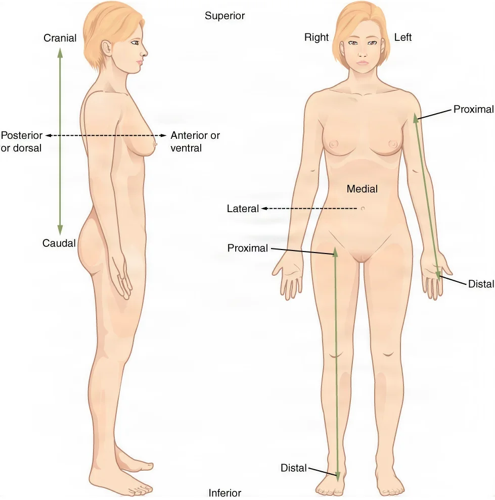
| Term | Means | Example |
|---|---|---|
| Superior (also cranial or cephalad) | Toward the head, or higher | The lungs are superior to the diaphragm. |
| Inferior (also caudal) | Away from the head, or lower | The pelvic cavity is inferior to the abdominal cavity. |
| Anterior (also ventral) | Toward the front | The abdominal wall is anterior to the intestines. |
| Posterior (also dorsal) | Toward the back | The kidneys lie posterior to the abdominal organs, behind the peritoneum. |
| Medial | Toward the midline of the body | The rectus muscles lie medial to the three flank muscles of the abdominal wall. |
| Lateral | Away from the midline, toward the side | The lumbar regions of the abdomen are lateral to the umbilical region. |
| Proximal | In a limb, closer to where it joins the trunk | A tourniquet cuff goes on the limb proximal to the surgical site. |
| Distal | In a limb, farther from where it joins the trunk | The hand lies at the distal end of the forearm. |
| Superficial | Closer to the body's surface | The fat under the skin is superficial to the fascia. |
| Deep | Farther from the body's surface | The peritoneum is the deepest layer of the abdominal wall. |
You will hear these words all day in surgery. The Trendelenburg position, which tilts the head down, moves the abdominal and pelvic organs cephalad, toward the head, so the surgeon can see the pelvis.
On paper, the words protect the patient. The surgical site is marked so there is no doubt about where the incision goes, which helps prevent wrong-site surgery. A specimen label names the site and the side, left or right. When a surgeon places a marking stitch or dye on a specimen to show which edge was superior, lateral or medial, the scrub keeps those marks intact on the field, so the pathologist can tell which edge is which.
If you are not sure which side a specimen came from, or which edge a marking stitch shows, ask the surgeon before the specimen leaves the field. The label should name the side, and any suture tag and where it was placed.
Body planes
A plane is an imaginary flat surface that passes through the body. A cut along a plane is called a section. Scans such as CT give pictures of the body in sections, and you can read them only if you know which plane they were cut in.
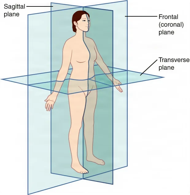
- Sagittal plane: a vertical plane that splits the body or an organ into a left part and a right part.
- Midsagittal plane: the sagittal plane that runs exactly down the middle, making equal right and left halves. It is also called the median plane. A sagittal plane off to one side, making unequal parts, is a parasagittal plane.
- Frontal plane, also called the coronal plane: a vertical plane that divides the body into front (anterior) and back (posterior) parts.
- Transverse plane: a horizontal plane that divides the body into upper and lower parts. It makes cross sections, and it is also called the axial plane.
- Oblique plane: a plane that crosses the body at an angle, neither horizontal nor vertical.
You will see these planes in the OR. A CT scan of the head is shown as a stack of transverse slices. A spine surgeon describes scoliosis, a sideways curve of the spine, as a curve in the frontal plane, and kyphosis as a curve in the sagittal plane. To correct them with rods, the surgeon bends the rods to shape and rotates them.
4The abdomen: quadrants and regions
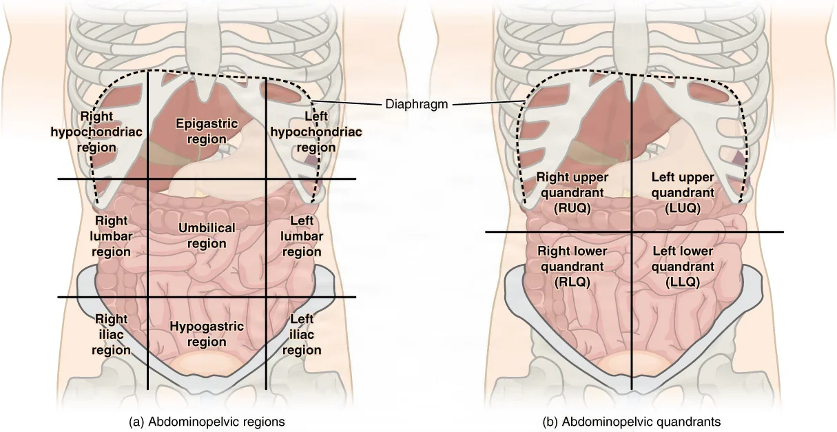
To describe where pain, a mass or an organ sits, the team divides the abdomen into four quadrants or nine regions. The quadrants are the simpler and more common way.
The four quadrants
One vertical line and one horizontal line cross at the umbilicus, the navel, and make four quadrants.
| Quadrant | Main contents |
|---|---|
| Right upper quadrant (RUQ) | Gallbladder, right lobe of the liver, parts of the small and large intestines |
| Left upper quadrant (LUQ) | Stomach, pancreas, spleen, left lobe of the liver, parts of the small and large intestines |
| Right lower quadrant (RLQ) | Appendix, right ureter, right ovary and fallopian tube, parts of the small and large intestines |
| Left lower quadrant (LLQ) | Left ureter, left ovary and fallopian tube, parts of the small and large intestines |
The quadrants show up in the OR. The appendix sits in the right lower quadrant, and a McBurney incision for an open appendectomy is made there, splitting the oblique muscle rather than cutting it. In laparoscopic surgery, port sites are named by quadrant too, such as a left upper quadrant port.
The nine regions
For a more exact location, two vertical lines and two horizontal lines make a grid of nine regions, like a tic-tac-toe board. The vertical lines drop from the middle of each collarbone. One horizontal line runs just below the ribs and the other just above the pelvis.
| Region | Where it is | Some contents |
|---|---|---|
| Right hypochondriac | Top row, right, under the ribs | Right lobe of the liver, gallbladder, right kidney |
| Epigastric | Top row, middle | Stomach, liver, pancreas, lower end of the esophagus |
| Left hypochondriac | Top row, left, under the ribs | Stomach, spleen, left kidney |
| Right lumbar (right flank) | Middle row, right | Ascending colon, right kidney, part of the small intestine |
| Umbilical | Middle row, center, around the navel | Transverse colon, small intestine |
| Left lumbar (left flank) | Middle row, left | Descending colon, left kidney, part of the small intestine |
| Right iliac, or right inguinal (groin) | Bottom row, right | Cecum, appendix, right ovary and fallopian tube |
| Hypogastric (pubic or suprapubic) | Bottom row, middle | Urinary bladder, uterus, prostate, rectum, sigmoid colon |
| Left iliac, or left inguinal (groin) | Bottom row, left | Sigmoid and descending colon, left ovary and fallopian tube |
The names come from word parts. Hypo- means below and chondr means cartilage, so the hypochondriac regions lie below the rib cartilages. Epi- means upon and gastr means stomach, so the epigastric region lies over the stomach. You may also hear the groin regions called inguinal, the lumbar regions called the flanks, and the hypogastric region called pubic or suprapubic.
5Body cavities and their linings
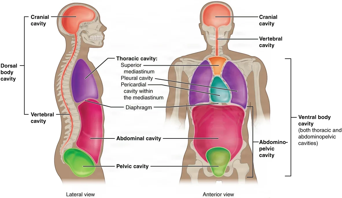
The body's organs sit in compartments called cavities, which hold and protect them. The two largest are the dorsal cavity at the back and the ventral cavity at the front. Each is divided into smaller cavities.
The dorsal cavity
- Cranial cavity: inside the skull. It holds the brain.
- Spinal cavity, also called the vertebral cavity: inside the vertebral column. It holds the spinal cord.
The two connect without a break, just as the brain and spinal cord do. Bone protects them, and so does cerebrospinal fluid, a clear fluid that cushions the brain and cord.
The ventral cavity
- Thoracic cavity: the chest, inside the rib cage. It holds the lungs, each in its own pleural cavity, and the heart. The heart sits in the mediastinum, the central space between the two lungs, along with the great vessels, the trachea and the esophagus.
- Diaphragm: the dome of muscle under the lungs. It is the floor of the chest and walls it off from the abdomen and pelvis below.
- Abdominopelvic cavity: the largest cavity in the body. No wall divides it, but it is described in two parts. The abdominal cavity holds most of the digestive organs, such as the stomach, liver, gallbladder and intestines. The pelvic cavity below holds the bladder, part of the large intestine and the internal reproductive organs.
The ventral cavity gives organs room to change size. The lungs fill, the heart beats and the stomach stretches without crowding their neighbors.
The serous membranes
The walls and organs of the ventral cavity are covered by thin membranes called serous membranes. Each one has two layers: the parietal layer lines the cavity wall, and the visceral layer covers the organs (the viscera). A thin film of slippery serous fluid between the layers cuts friction as the organs move.
- Pleura: surrounds each lung. The parietal pleura lines the chest wall, and the visceral pleura covers the lung.
- Pericardium: surrounds the heart.
- Peritoneum: lines the abdominal cavity and covers many abdominal organs. Folds of peritoneum connect the abdominal organs; one fold, the mesentery, attaches to the back wall of the abdomen and fans out to cover the small intestine.
Some organs lie behind the peritoneum, in the retroperitoneum: the kidneys, the adrenal glands and the ureters. A layer of peritoneum separates them from the abdominal organs in front.
After inflammation or infection, strands of scar tissue called adhesions can form between the peritoneum and the abdominal organs. During surgery the surgeon may have to release them, which is called lysis of adhesions.
Opening a cavity
Many operations are named for the cavity they open. A craniotomy opens the cranial cavity through the scalp and skull. A thoracotomy opens the thoracic cavity. A laparotomy opens the abdominal cavity, and an exploratory laparotomy is done to look inside when less invasive tests have not found the cause of a problem.
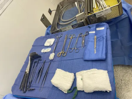
An open cavity also loses heat. In open chest and abdominal surgery, moisture evaporates from the exposed organs, and the longer the organs lie open in a cool room, the colder the patient can get. You will see why that matters later in this chapter.
6The cell
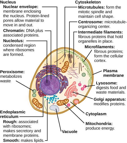
A cell is a tiny, fluid-filled package wrapped in a membrane. Inside, small working parts called organelles each do their own jobs, the way organs do for the whole body. Nearly every body cell carries the full set of the person's DNA.
The cell membrane
The cell membrane, also called the plasma membrane, wraps every cell. It keeps the cell's contents in and controls what passes in or out.
It is built of two layers of fat-like molecules called phospholipids. Each phospholipid has a head that is drawn to water and two tails that push water away. The heads face the watery fluid inside and outside the cell, and the tails tuck into the middle. Cholesterol and many proteins sit in this double layer. Some proteins are channels that let certain particles through. Others are receptors that catch chemical messages, and others are markers that tell the immune system the cell belongs to this person. Those markers are one reason a donated organ can be rejected.
The membrane is selectively permeable: it lets some things through and keeps others out. Small, uncharged molecules such as oxygen and carbon dioxide slip straight through. Sugar, charged particles called ions and most other water-loving substances need help to cross.
Passive transport moves substances without using the cell's energy.
- Diffusion: particles spread from where they are crowded to where they are scarce, until they are even. Oxygen diffuses into cells and carbon dioxide diffuses out.
- Facilitated diffusion: diffusion through a protein channel or carrier, for substances too big or too charged to cross alone, such as glucose and sodium.
- Osmosis: the diffusion of water across a membrane. Water moves toward the side with more dissolved particles.
- Filtration: pressure pushes fluid and the small particles in it through a membrane, as blood pressure pushes fluid out of the smallest blood vessels into the tissues.
Active transport uses energy, from a molecule called ATP, to move substances, usually from where there is little to where there is more.
- Pumps: the sodium-potassium pump pushes sodium out of the cell and pulls potassium in. Nerve cells run these pumps constantly.
- Endocytosis: the membrane folds around something outside the cell and pinches off to bring it in. Phagocytosis, "cell eating," takes in large particles such as bacteria; pinocytosis, "cell drinking," takes in fluid.
- Exocytosis: the reverse. A sac inside the cell joins the membrane and releases its contents outside, the way gland cells release enzymes and hormones.
Osmosis matters at the back table. A fluid with the same concentration of particles as the cell is isotonic, and cells keep their normal shape in it. In a fluid with more particles, a hypertonic one, cells lose water and shrivel. In a fluid with fewer particles, a hypotonic one, cells take in water, swell and can burst. Irrigation fluids are chosen to suit the body tissue they touch, and normal saline is isotonic. Plain distilled water is so hypotonic that, used to irrigate during a prostate resection, it can burst red blood cells.
Cytoplasm and the organelles
Everything between the cell membrane and the nucleus is the cytoplasm: a jelly-like fluid called cytosol, plus the organelles floating in it. A web of protein fibers, the cytoskeleton, holds the cell's shape and keeps the organelles in place.
| Organelle | What it is | What it does |
|---|---|---|
| Centrosome and centrioles | A centrosome is a pair of centrioles, short tubes that sit near the nucleus | Organize the protein tubes that pull chromosomes apart when the cell divides, and anchor cilia and flagella |
| Cilia | Short, hair-like extensions on the cell's surface | Beat constantly in rhythm to move fluid and particles, such as mucus up the airway away from the lungs, or an egg along a fallopian tube |
| Flagellum | A single long, whip-like tail | Moves the whole cell; in humans only the sperm cell has one |
| Rough endoplasmic reticulum (ER) | A network of membrane channels dotted with ribosomes, joined to the membrane around the nucleus | Builds and finishes proteins that will be sent out of the cell or placed in its membrane |
| Smooth endoplasmic reticulum | The same channels without ribosomes | Makes fats such as phospholipids and steroid hormones, stores calcium and breaks down some toxins |
| Golgi apparatus | Stacks of flat membrane sacs | Sorts, changes and packages products from the rough ER into vesicles and sends them where they go; makes lysosomes |
| Lysosomes | Sacs of digestive enzymes | Break down worn-out cell parts, and bacteria that white blood cells swallow |
| Mitochondria | Bean-shaped bodies with a folded inner membrane | Carry out cellular respiration, turning the energy in food into ATP; muscle and nerve cells have many |
| Ribosomes | Tiny bodies of ribosomal RNA and protein, free in the cytoplasm or on the rough ER | The place where proteins are built |
| Vacuoles | Membrane sacs | Hold water, secretions, enzymes or the remains of material the cell has taken in |
| Vesicles | Small, round sacs of membrane | Carry materials into the cell, around inside it and out of it |
The nucleus
The nucleus is the largest organelle and the control center of the cell, because it holds the instructions for making every protein. Most cells have one nucleus. Skeletal muscle cells have many, and mature red blood cells have none.
- Nuclear membrane, or nuclear envelope: a double membrane around the nucleus. Small openings called nuclear pores let RNA and proteins pass between the nucleus and the cytoplasm.
- Nucleolus: a dark spot inside the nucleus that makes the RNA used to build ribosomes.
- Chromatin and chromosomes: the DNA in the nucleus is wound around proteins into threads called chromatin. When the cell gets ready to divide, the chromatin coils up tightly into chromosomes so it can be moved safely. A human body cell has 46 chromosomes, holding about 22,000 genes.
Nucleic acids: DNA and RNA
DNA and RNA are nucleic acids, the molecules that store and use the body's genetic instructions.
DNA (deoxyribonucleic acid) is shaped like a long, twisted ladder, the double helix. Its side rails are sugar and phosphate. Each rung is a pair of bases, and the bases always pair the same way: adenine (A) with thymine (T), and cytosine (C) with guanine (G). The order of the bases is the genetic code. A gene is a stretch of DNA that holds the code for one protein, and each set of three bases stands for one amino acid, the building blocks of proteins.
Proteins do almost all of the cell's work. Many are enzymes, which speed up the cell's chemical reactions; others form the cell's structure.
RNA (ribonucleic acid) carries out DNA's instructions. It differs from DNA in three ways: it is usually a single strand, its sugar has one more oxygen atom, and it uses the base uracil (U) in place of thymine. Three kinds of RNA build proteins together.
- Messenger RNA (mRNA): copies the code of one gene inside the nucleus, a step called transcription, and carries the copy out to the cytoplasm.
- Ribosomal RNA (rRNA): joins with proteins to form the ribosome, the workbench where the protein is built.
- Transfer RNA (tRNA): reads the mRNA code three bases at a time and brings the matching amino acid to the ribosome. There each new amino acid is joined to the last, building a chain, a step called translation, until a stop signal ends the protein.
Cellular respiration: how cells get energy
Cells run on ATP (adenosine triphosphate). A cell stores energy by making ATP, then breaks ATP apart to release a small, controlled burst of energy where work needs doing, such as a muscle fiber contracting or a pump moving sodium. Cellular respiration is how cells make ATP from food. The sugar glucose is the body's most readily available fuel.
- Glycolysis: in the cytoplasm, one glucose molecule is split into two smaller molecules called pyruvate. This gives a net gain of two ATP, and it does not need oxygen.
- Anaerobic oxidation, without oxygen: when oxygen is short, the pyruvate is turned into lactic acid so glycolysis can keep going. This works for seconds to a few minutes. Hard-working muscles do it when oxygen cannot reach them fast enough, and red blood cells, which have no mitochondria, make all their ATP this way.
- Aerobic oxidation, with oxygen: inside the mitochondria, the pyruvate is broken down completely. Carbon dioxide is given off as waste, oxygen is used up and becomes water, and about 36 ATP are made from each glucose, far more than glycolysis alone. This is why you must keep breathing.
Brain cells need a steady, high supply of ATP. Without oxygen, brain damage is likely within about five minutes.
You will see this in surgery. A pneumatic tourniquet stops blood flow to a limb so the surgeon has a bloodless field. Tissue cut off from its blood flow runs short of oxygen, so its cells switch to anaerobic oxidation and lactic acid builds up. When the cuff comes down, that acid and other waste rush into the patient's circulation, and the effect grows with the length of tourniquet time. Because the harm grows with time, AST recommends keeping cuff time as short as possible. Its general recommendation for adults is an inflation time of 60 minutes on an arm and 90 minutes on a leg. A CST in the assistant circulator role helps watch the cuff's pressure and time.
Cell division
Most body cells divide to grow and to replace old cells, and billions divide every day. Cells of the skin and the lining of the digestive tract divide very often, because they are worn away constantly. A few kinds rarely or never divide, including most neurons and skeletal and cardiac muscle cells.
This shapes how tissue heals. Skeletal muscle can be repaired by special cells called satellite cells, but dead heart muscle is replaced by scar tissue.
DNA replication comes first. The two strands of the DNA ladder unzip, and each strand is used as a pattern to build a new partner strand. Each of the two new DNA molecules keeps one old strand and one new one. Enzymes check the new strands and fix most mistakes, because each new cell needs an exact copy.
The life of a cell is called the cell cycle. Most of the time a cell is in interphase: it grows, does its normal work and, before dividing, copies its DNA. Then comes mitosis, the division of the nucleus, in four stages:
- Prophase: the chromatin coils into visible chromosomes, each one now an X of two identical copies joined together. The nuclear membrane breaks down, and the two centrosomes move to opposite sides and send out a spindle of protein tubes.
- Metaphase: the chromosomes line up across the middle of the cell.
- Anaphase: the two copies of each chromosome are pulled apart to opposite ends of the cell.
- Telophase: a new nuclear membrane forms around each set of chromosomes, making two nuclei.
Last comes cytokinesis, when a ring of protein fibers pinches the cytoplasm in two. The result is two daughter cells, each with the same 46 chromosomes as the parent.
When the controls on cell division fail, cells keep dividing and can form a tumor. Chapter 11 teaches tumors and cancer.
Meiosis is a different kind of division that makes only eggs and sperm, the gametes. The DNA is copied once, then the cell divides twice, making four cells. Each one has only 23 chromosomes, half the usual number, and each is genetically different from the others. When a sperm and an egg join, the new cell has a full set again, one copy of each chromosome from each parent.
New technology: growing cells for surgery
A stem cell is an unspecialized cell that can keep dividing and, under the right conditions, become a specialized cell. Stem cells are grouped by how many cell types they can become:
- Totipotent: the first cells made after an egg is fertilized. They can become any cell, enough to build a whole person.
- Pluripotent: embryonic cells that can become any tissue, but not a whole person.
- Multipotent: cells that can become a few related cell types. The adult stem cells in bone marrow, which make all the blood cells, are multipotent, and so are stem cells in the skin and in umbilical cord blood.
Stem cells already help the body heal: scattered stem cells in connective tissue supply new cells to repair it after an injury. Researchers hope to use stem cells to repair damaged tissue: in cell-based therapy, stem cells are turned into specialized cells in a lab dish and injected into the patient, though many obstacles remain. Embryonic stem cells can become almost any cell, but the patient's immune system may reject them, and taking them raises ethical questions. A patient's own adult stem cells are not rejected, but they can become fewer cell types.
The Core Curriculum lists cell cloning of cartilage and skin as new technology. A clone is a group of cells that all come from one cell, and the term is used for cells grown in tissue culture. Two operations already put a patient's own skin or cartilage cells, multiplied in a lab, back into the patient:
- Skin: for a patient with large burns and too little healthy skin to graft, a small piece of the patient's skin can be sent to a lab, where its skin cells are grown into a sheet of new epidermis, called a cultured epithelial autograft.
- Cartilage: to repair a hole in the cartilage of a knee, a surgeon removes a small piece of the patient's cartilage through an arthroscope. Its cartilage cells, the chondrocytes, are grown in a lab, and usually 3 to 6 weeks later a second, open operation places them into the defect under a sutured patch. This is called autologous chondrocyte implantation.
7Tissues
What makes a tissue
Cells of the same kind, working side by side on the same job, make a tissue. The Core Curriculum points to four things that make a tissue:
- A collection of like cells: the cells share a shape and an origin and are arranged in an orderly pattern.
- A common, specialized job: such as covering, connecting, moving or signaling.
- Cell attachments: cells are joined by junctions. Tight junctions seal neighbors together so nothing leaks between them. Anchoring junctions, such as desmosomes, hold cells firmly together. Gap junctions are tiny tunnels that let ions pass from cell to cell, so a group of cells can work as one.
- Material between the cells: the extracellular matrix, made by the cells themselves, is a ground substance crossed by protein fibers. Epithelial tissue has almost none; connective tissue has a great deal.
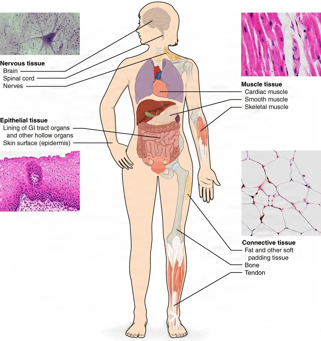
The body has four types of tissue: epithelial, connective, muscle and nervous.
Epithelial tissue
Epithelial tissue, or epithelium, is made of sheets of cells. It covers the outside of the body, lines the body's cavities, hollow organs and passageways, and forms much of the body's glandular tissue.
- Its cells are packed tightly with almost no matrix between them, held together by junctions.
- One surface faces the outside or the inside of a hollow organ, and the other rests on a thin basement membrane that joins it to the connective tissue below.
- It has no blood vessels of its own. Food and oxygen reach it by diffusion from the tissue underneath.
- Many epithelia replace damaged and dead cells quickly.
- It protects the body, controls what passes in, and secretes and absorbs. Everything that enters the body crosses an epithelium.
Epithelium is named by the shape of its cells and the number of layers. Squamous cells are flat and thin, cuboidal cells are as wide as they are tall, and columnar cells are taller than they are wide. Simple epithelium is one layer of cells; stratified epithelium has several layers and is named by the shape of the top layer.
| Type | Looks like | Found in | Good for |
|---|---|---|---|
| Simple squamous | One layer of flat cells | Air sacs of the lungs, capillaries, the lining of blood vessels (endothelium) and of serous membranes (mesothelium) | Fast passage of gases and fluids, and a smooth, slippery surface |
| Simple cuboidal | One layer of box-shaped cells | Kidney tubules and the ducts of glands | Secretion and absorption |
| Simple columnar | One layer of tall cells, some with cilia | Parts of the digestive tract; the fallopian tubes (with cilia) | Absorption and secretion; cilia move particles along |
| Pseudostratified columnar | One layer that looks like several, often with cilia | The respiratory tract | Moving mucus and trapped particles |
| Stratified squamous | Many layers, flat on top | The skin, topped with dead cells full of keratin; the lining of the mouth, without keratin | Protection from rubbing and wear |
| Stratified cuboidal and stratified columnar | Several layers | Some glands and ducts; uncommon in the body | Protection |
| Transitional | Layers whose top cells change shape as the tissue stretches | The ureters and urinary bladder | Stretching as the bladder fills |
Epithelium that covers or lines a surface is lining epithelium. Epithelium made mostly of cells that secrete is glandular epithelium. Goblet cells, single cells that make mucus, are mixed into some simple and pseudostratified columnar epithelium. Larger glands grow from epithelium too: exocrine glands release their products through ducts, and endocrine glands release hormones into the fluid around them, to be carried in the blood.
Connective tissue
Connective tissue binds the body's parts together, supports and protects them, carries materials around the body in blood and lymph, and stores energy as fat. Its cells are spread out in a large amount of matrix.
Connective tissue typically has three parts: cells, ground substance and fibers.
- Cells: the most common is the fibroblast, which makes the fibers and the ground substance. Fat cells (adipocytes) store fat. Immune cells such as macrophages and mast cells also move through connective tissue.
- Fibers: collagen fibers are strong and resist stretching, which gives tendons and ligaments their strength. Elastic fibers spring back after stretching, as skin does. Reticular fibers form a fine net that supports soft organs such as the liver and spleen.
- Ground substance: a clear, gel-like material that fills the space around the cells and fibers. In bone it is hardened with minerals.
| Type | What it is like | Where it is found |
|---|---|---|
| Loose areolar | A loose web of all three fiber types | Under most epithelium, between muscle fibers, around blood vessels, and holding organs in the abdomen |
| Adipose tissue | Fat cells with little matrix | Under the skin and around the kidneys; it stores energy, insulates and cushions |
| Dense regular | Collagen fibers packed in parallel bundles, strong in one direction | Tendons, which join muscle to bone, and ligaments, which join bone to bone |
| Dense irregular | Collagen fibers running in every direction, strong in all directions | The dermis of the skin |
| Cartilage | A firm, smooth matrix with cells in small spaces, and no blood vessels | The ends of bones in joints, the rib cage and nose (hyaline cartilage); the knee menisci and spinal discs (fibrocartilage); the outer ear (elastic cartilage) |
| Bone | The hardest connective tissue: collagen hardened with calcium phosphate, with a rich blood supply | The skeleton |
| Blood | Cells floating in a liquid matrix, the plasma | The blood vessels; red cells carry oxygen, white cells defend the body, and platelets help blood clot |
| Lymph | Fluid with white blood cells | The lymph vessels, which return extra tissue fluid to the blood |
The Core Curriculum also lists dense fibrous connective tissue. Fibrous tissue is another name for dense connective tissue, which is mostly collagen fibers and comes in the regular and irregular forms in the table. Fascia and tendon are examples.
Blood supply is one factor in how fast a tissue heals. Cartilage has no blood vessels and heals very slowly. Tendons have a poor blood supply and also heal slowly. Bone is rich in blood vessels and heals in a relatively short time.
Connective tissue also changes how you handle it. Fascia, tendon and cartilage are strong and slippery, and with few blood vessels to injure, they can be held with toothed clamps such as a Kocher. Fat makes instruments slippery, so you wipe them with a damp sponge, and the surgeon uses toothed forceps to hold fat while suturing.
Muscle tissue
Muscle tissue is excitable, which means it responds to a signal, and contractile, which means it can shorten and pull. It can also stretch and then spring back to its resting length. There are three types.
| Type | What it looks like | Control | Where it is and what it does |
|---|---|---|---|
| Skeletal | Long, cylinder-shaped fibers with stripes (striations) and many nuclei at the edges | Voluntary: you choose to move it | Attached to bones and around body openings such as the mouth and anus; moves the body, holds posture and makes heat |
| Cardiac | Short, branched, striated cells with one central nucleus, joined end to end by intercalated discs | Involuntary; it sets its own rhythm | The wall of the heart; pumps blood |
| Smooth, also called visceral | Spindle-shaped cells with one nucleus and no stripes | Involuntary | The walls of hollow organs, airways and arteries; moves food and secretions and controls blood flow |
Skeletal muscle makes up about 40 percent of body mass, and the heat of its contractions helps keep body temperature steady. Shivering is skeletal muscle contracting on its own to make heat.
At the field, surgeons separate or pull muscle aside along its fibers rather than cut across it whenever they can, because cut muscle is slow to regain its function. Muscle also holds sutures poorly.
Nervous tissue
Nervous tissue is excitable too. It receives information, sends electrical signals and passes messages to other cells. It has two kinds of cells: neurons and neuroglia.
A neuron has three main parts:
- the cell body, which holds the nucleus and most of the organelles
- dendrites, thin branches that receive signals and carry them toward the cell body
- one axon, a long fiber that carries the signal away from the cell body. Many axons are wrapped in myelin, an insulating layer made by neuroglia.
The axon ends at a synapse, a tiny gap between the neuron and the next cell, which may be another neuron, a muscle fiber or a gland. Chemicals called neurotransmitters carry the message across the gap.
Neurons are grouped by the direction their signals travel. Afferent, or sensory, neurons carry information from the body to the brain and spinal cord. Efferent, or motor, neurons carry commands away from the brain and spinal cord to muscles and glands. An easy way to remember: afferent signals arrive, efferent signals exit.
Neuroglia, or glial cells, support and protect the neurons. In fact, the brain and spinal cord are made mostly of neuroglia. There are six types:
- in the brain and spinal cord: astrocytes, the most common, which support neurons and help form the barrier between the blood and the brain; oligodendrocytes, which make myelin; microglia, which defend against infection; and ependymal cells, which make cerebrospinal fluid
- in the nerves of the body: satellite cells, which support neurons, and Schwann cells, which make myelin
How a muscle contracts
Each skeletal muscle fiber is packed with long strands called myofibrils, and each myofibril is a chain of tiny units called sarcomeres. The sarcomere is the smallest part of a muscle that contracts. It holds two kinds of protein filaments: thin filaments made of actin, and thick filaments made of myosin. Two more proteins, troponin and tropomyosin, sit on the actin and control when it can be grabbed. The neat, repeating pattern of these filaments is what gives skeletal and cardiac muscle their stripes.
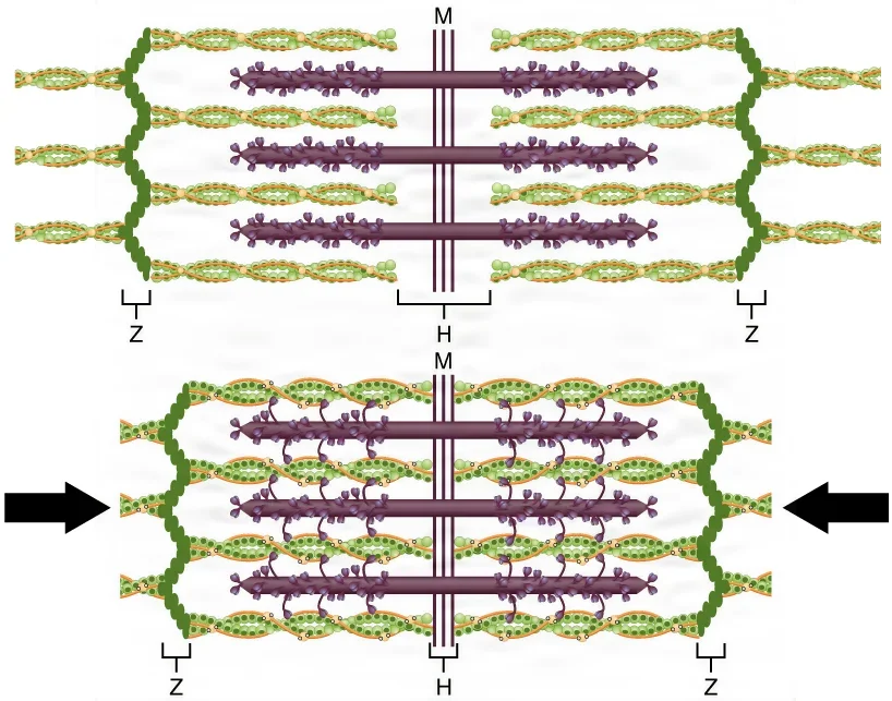
The sliding filament mechanism works like this:
- A signal from a motor neuron makes an electrical wave run along the muscle fiber.
- The wave makes the fiber's sarcoplasmic reticulum, a special kind of smooth ER, release stored calcium.
- Calcium binds to troponin, and tropomyosin slides off the binding sites on the actin.
- The heads of the myosin filaments grab the actin, forming cross-bridges, and pull the actin toward the center of the sarcomere.
- ATP lets each myosin head let go, reset and grab again farther along, like hands pulling in a rope one grip at a time. Without ATP, the heads cannot let go.
- The thin filaments slide past the thick ones, and the sarcomere shortens.
When the nerve signal stops, calcium is pumped back into the sarcoplasmic reticulum, tropomyosin covers the binding sites again, and the muscle relaxes. A muscle that runs out of ATP also stops contracting. Smooth muscle uses calcium in a different way, but every muscle needs ATP to keep contracting.
This chain of events has an OR emergency behind it. Malignant hyperthermia is a dangerous reaction to some anesthetic gases and the muscle relaxant succinylcholine. In 50 to 70 percent of families who carry it, the faulty gene is the one for the channel in the sarcoplasmic reticulum that releases calcium for contraction. Chapter 13 teaches how the team responds.
Neuromuscular excitability
Excitable cells, neurons and muscle fibers, carry signals as electricity. At rest, the inside of a nerve cell is negative compared with the outside, about minus 70 millivolts, and the sodium-potassium pump keeps it that way. This is the resting membrane potential.
A nerve impulse is a wave of electrical change that runs along the membrane:
- A stimulus opens channels in the membrane, and sodium ions rush in. The inside becomes less negative, then briefly positive. This is depolarization.
- Then potassium channels open, potassium ions flow out, and the inside turns negative again. This is repolarization.
- This quick swing, called an action potential, triggers the same change in the next patch of membrane, so the signal travels along the whole cell.
The neuromuscular junction is where a motor neuron meets a skeletal muscle fiber, and every skeletal muscle fiber has one. The nerve ending and the muscle do not touch; a tiny gap called the synaptic cleft lies between them.
- The nerve impulse reaches the end of the motor neuron, and the neuron releases the neurotransmitter acetylcholine (ACh).
- ACh crosses the cleft and binds to receptors on the muscle fiber, at the motor end plate.
- The receptors open channels, sodium enters, and the muscle fiber fires its own action potential, which starts contraction.
- An enzyme, acetylcholinesterase, breaks down the ACh almost at once, so the muscle does not keep contracting.
Skeletal muscle contracts only when a nerve tells it to. Heart and smooth muscle can also respond to hormones and local signals, and the heart sets its own beat.
This is why paralyzing drugs work as they do. Neuromuscular blocking agents act at this junction. They are given with general anesthesia to relax the skeletal muscles, and they have little effect on heart or smooth muscle, so the heart keeps beating. A drug that blocks acetylcholinesterase can help reverse some of them. Chapter 13 teaches these drugs.
A motor nerve signal, then acetylcholine, then calcium, then actin and myosin, powered by ATP. Break any link and the muscle cannot contract.
Membranes of the body
A tissue membrane is a thin sheet of tissue that covers the body, lines a cavity or passageway, or wraps an organ. There are two groups.
Epithelial membranes are epithelium resting on connective tissue:
- Mucous membranes line the passages that open to the outside: the digestive, respiratory, urinary and reproductive tracts. A coat of mucus protects them.
- Serous membranes line the closed cavities and cover the organs inside them: the pleura, the pericardium and the peritoneum. Their thin serous fluid cuts friction.
- The cutaneous membrane is the skin, a stratified squamous epithelium on connective tissue.
Connective tissue membranes are made only of connective tissue. One is the synovial membrane, which lines movable joints such as the shoulder and knee and makes synovial fluid, a slippery liquid that lets the bones move with little friction.
8Organs and organ systems
What makes an organ
An organ is a distinct structure built from two or more kinds of tissue that work as one unit to do one or more specific jobs. It is one level more complex than a tissue. A skeletal muscle is an organ: muscle fibers, blood vessels, nerve fibers and connective tissue wrapped together. The Core Curriculum uses four organs to show how tissues combine.
- Stomach: its wall has four layers. The inner lining, the mucosa, has an epithelium that makes protective mucus, with glands that make gastric juice. Under it, the submucosa is dense connective tissue carrying blood and lymph vessels. Next comes the muscularis, smooth muscle in three layers that churn and mix food. The outer layer, the serosa, is serous membrane.
- Heart: its wall has three layers. The epicardium outside is the visceral layer of the pericardium. The thick myocardium in the middle is cardiac muscle built on a framework of collagen, with its own blood vessels and nerves. The endocardium inside is endothelium, simple squamous epithelium, that lines the chambers and covers the valves.
- Brain: nervous tissue, neurons and neuroglia, protected by the meninges, layers of connective tissue whose tough outer layer is the dura mater.
- Nerves: bundles of axons, the long fibers of neurons, held together by layers of connective tissue and fed by blood vessels.
Compare the four and you see that each one mixes the tissue types in its own way. The heart and the stomach both have a muscle layer in their walls, but the heart's is cardiac muscle and the stomach's is smooth muscle. The brain and the nerves are built around nervous tissue, wrapped and protected by connective tissue.
What organ systems do together
Organ systems share the work of keeping every cell alive. The Core Curriculum names four things they do together.
Exchange of materials. Cells need oxygen and nutrients brought in and carbon dioxide and wastes carried away. Everything that enters the body crosses an epithelium, and exchange is fastest where the epithelium is thinnest. Gases pass through the simple squamous lining of the lungs' air sacs and the capillaries. Nutrients pass from the intestine into the blood. Fluid filters out of capillaries to feed the tissues, and the kidneys filter wastes out of the blood. Blood and lymph carry all of it around the body.
Homeostasis. Homeostasis is the body's steady inner balance. Body temperature, blood pressure, blood sugar and many other values each have a set point, with a narrow normal range around it. The set point for body temperature is about 37°C (98.6°F). When a value drifts too far, illness follows.
Regulation and feedback. The body keeps its balance mostly by negative feedback, which reverses a change. A negative feedback system has three parts:
- a sensor, or receptor, that measures a value
- a control center, often in the brain, that compares the value with the normal range
- an effector that acts to bring the value back, such as the sweat glands or the blood vessels of the skin
Body temperature is the classic example. When you are too warm, the brain widens the blood vessels in the skin to shed heat and turns on sweating. When you are too cold, it narrows those vessels to keep heat in the core and makes the muscles shiver, and hormones raise heat production in the cells. Blood sugar works the same way: when it rises, the pancreas releases insulin, cells take up the sugar, and the insulin stops as the level falls.
Positive feedback does the opposite: it pushes a change further until an end point is reached. It is normal only when there is a definite end. In childbirth, stretching of the cervix triggers the hormone oxytocin, which makes the uterus contract harder, which stretches the cervix more, until the baby is born. Blood clotting works the same way: each step releases more clotting substances until the bleeding vessel is sealed.
Response to the environment. The body senses changes outside it and inside it and adjusts, such as sweating on a hot day or moving away from danger.
Homeostasis in the OR
Keeping a patient warm is homeostasis you can help with. The body normally holds its core temperature within a very small range, run by the brain's temperature center. Surgery attacks that balance from every side.
- General anesthesia blunts the brain's temperature control. In planned (nonemergency) surgery, this effect of anesthesia is the main cause of unplanned hypothermia.
- Anesthesia also widens blood vessels, so warm blood moves from the core out to the skin and loses heat.
- The patient lies on a cold table, in a cool room, sometimes with cold fluids running in, and an open chest or abdomen loses heat as moisture evaporates.
- With a spinal or epidural block, the numb part of the body cannot shiver or narrow its blood vessels.
AST sets the line at 36°C: a core temperature below 36°C is hypothermia. An estimated more than half of surgical patients in the United States become hypothermic. Hypothermia is linked to slower wound healing, more surgical site infections, weaker clotting and more bleeding, and anesthetic drugs that last longer. That is why the team works to keep patients warm, with steps such as warmed fluids and forced-air warming.
The body heals best in balance. Anesthesia, a cold room and an open cavity all pull a patient away from it, and the surgical team's job is to help bring the patient back.
9When tissue growth goes wrong
Healthy tissues grow, shrink and repair themselves in a controlled way. The Core Curriculum names seven ways growth can go wrong. Most of the names end in -plasia, which means formation or development; the prefix tells you what kind. Hyper- means excessive, hypo- means deficient, a- means without, and dys- means bad.
| Term | Meaning | Where you may meet it |
|---|---|---|
| Atrophy | A tissue or organ shrinks or wastes away, from less use, poor blood supply, poor nutrition, pressure or hormone changes | Muscles waste when they are not used, and muscle loss with age is called sarcopenia; the adenoids normally shrink during the teen years |
| Hypertrophy | An organ grows larger because its cells grow larger, not more numerous | Muscle fibers thicken with exercise; enlarged adenoids can block the airway and ear tubes, a reason for an adenoidectomy |
| Hyperplasia | An organ or tissue grows because it makes more normal cells, with no cancer | Benign prostatic hypertrophy (BPH, also called benign prostatic hyperplasia) enlarges the prostate, which presses on the urethra and blocks urine flow |
| Hypoplasia | A tissue or organ is underdeveloped | In hypoplastic left heart syndrome, the left side of a baby's heart is underdeveloped and needs a series of operations |
| Aplasia | An organ or tissue fails to develop normally | In aplasia cutis congenita, a small patch of skin, usually on the scalp, fails to develop |
| Dysplasia | Tissue develops abnormally; the cells look wrong in size, shape and arrangement | Cervical dysplasia, which may be removed with a loop electrode (LEEP) or a cone biopsy |
| Anaplasia | Cells lose their specialized form and job and look primitive, a feature of most cancers | Anaplastic thyroid cancer is highly aggressive and spreads fast |
Hyperplasia and hypertrophy are easy to mix up. Both make an organ bigger: hyperplasia by adding cells, hypertrophy by enlarging the cells already there. BPH goes by both names: the Core Curriculum calls it benign prostatic hypertrophy, and other references call it benign prostatic hyperplasia. Both names mean the same disease.
BPH is often treated with a transurethral resection of the prostate (TURP), in which the surgeon removes the extra prostate tissue with a resectoscope passed up the urethra.
Dysplasia and anaplasia are also steps on the scale pathologists use to grade the cells of many tumors. Mildly dysplastic cells look only a little different from normal; severe dysplasia looks very abnormal; anaplastic cells are so primitive that it is hard to tell what tissue they came from. The more abnormal the cells, the worse the outlook.
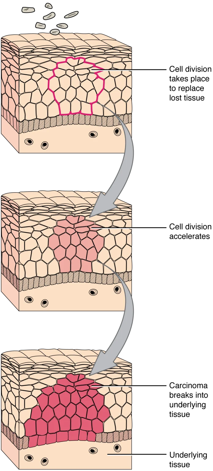
Cervical dysplasia is one reason for two procedures, which remove the abnormal tissue so a pathologist can examine it. A LEEP (loop electrosurgical excision procedure) removes the abnormal cells with a thin, electrified wire loop, and the loop's size depends on how much dysplasia there is and on the surgeon's preference. A cold knife cone biopsy cuts out a cone of tissue around the opening of the cervix with a scalpel and scissors. Before the biopsy, the surgeon may paint the cervix with Lugol solution, a mild iodine stain that shows where the dysplasia is. Do not confuse it with Monsel solution, another brown liquid on the same back table: it is a mixture containing acids, used to stop bleeding, and painted on by mistake it can damage the biopsy area.
Tumors, benign and malignant, and how cancer spreads are taught in chapter 11.
10Tissue layers at the field
Everything in this chapter comes together when the surgeon makes an incision. The abdominal wall is built of these layers, from the outside in:
- Skin: stratified squamous epithelium on top of the dermis, which is dense irregular connective tissue.
- Subcutaneous tissue: fatty tissue, adipose tissue mixed with loose areolar tissue, from a quarter inch to more than 8 inches thick.
- Fascia: tough connective tissue that covers the muscles. The layer over the front of the muscles is the anterior fascia.
- Muscle: skeletal muscle. Behind it lies another layer of fascia, the posterior fascia.
- Peritoneum: the serous membrane lining the abdominal cavity, just beneath the posterior fascia.
The surgeon closes the wound layer by layer, and each tissue behaves differently:
- Peritoneum is thin and heals fast. If the posterior fascia over it is closed well, it may not need sutures at all.
- Fascia is the body's main supporting soft tissue. It heals slowly and takes most of the stress on a healing wound, so it is closed securely with strong sutures.
- Muscle holds sutures poorly. It is usually split or pulled aside rather than cut, so it often needs no closure.
- Subcutaneous fat holds sutures poorly too. It is where most wound infections start, because fatty tissue has weak defenses.
- Skin is closed last. A fine stitch just under the skin can close it neatly, because the fascia is carrying the tension.
Healing speed follows the tissue too. Many epithelia replace their cells quickly; the lining of the bladder covers a suture line within about 5 days. Bone, with its rich blood supply, heals fairly quickly. Cartilage and tendons, with little or no blood supply, heal slowly. Fascia heals slowly as well.
Chapter 25 teaches sutures and how to choose them, and chapter 29 teaches incisions and how the scrub follows the surgeon through the layers.
Lo que te llevas de este capítulo
- Anatomy is structure and physiology is function. The body is built in levels: chemicals, cells, tissues, organs, organ systems and the whole person.
- Every direction is described as if the patient stood in anatomical position, and right and left are always the patient's own. Directional terms come in pairs: superior and inferior, anterior and posterior, medial and lateral, proximal and distal, superficial and deep.
- Sagittal planes divide right from left, frontal (coronal) planes divide front from back, and transverse planes divide top from bottom. The abdomen is mapped in four quadrants or nine regions.
- The dorsal cavity holds the brain and spinal cord; the ventral cavity holds the chest and abdominopelvic organs, lined by the pleura, pericardium and peritoneum.
- A cell is run by its nucleus and organelles. DNA holds the code, RNA builds proteins, mitochondria make ATP, and cells divide by mitosis; meiosis makes eggs and sperm.
- The four tissue types are epithelial, connective, muscle and nervous. A muscle contracts when a nerve releases acetylcholine, calcium frees actin, and myosin pulls it, powered by ATP.
- Organs are two or more tissues working as one. Organ systems exchange materials and keep homeostasis, mostly by negative feedback, which anesthesia and a cold OR disturb.
- Atrophy, hypertrophy, hyperplasia, hypoplasia, aplasia, dysplasia and anaplasia describe growth gone wrong, and each tissue layer of an incision heals and holds sutures differently.
Ponte a prueba5 preguntas rápidas
Gratis para todos. Elige una respuesta para cada una y luego revísalas todas juntas.
En el quirófanoDónde vas a usar esto
Todo el banco de preguntas del capítulo en series de 10, con la respuesta explicada después de cada una.
Parte del programa para educadoresLas palabras del capítulo en tarjetas de estudio: las que fallas vuelven antes.
Parte del programa para educadoresLo asigna tu docente. Cada grupo saca sus propias 25 preguntas del banco del capítulo.
Parte del programa para educadores
Sugerir una corrección
¿Algo está mal, confuso o desactualizado en este capítulo? Avísanos y lo corregimos para todos. Tu correo solo se usa para responderte.