Skin, bones and muscles
The layers a surgeon cuts through, the bones and joints that frame every case, and the muscles that move them, with the words the team uses for each.
1Why it matters
Many operations start by cutting through skin, then fat, then the tough fasciaFASH-ee-uhA sheet or band of tough connective tissue that covers and separates muscles and organs.fasci/o bandFull entry over muscle, and many go on to work on bone, a joint or a tendon. To follow the surgeon through those layers, you need to know what each one is, what it does, and how it behaves when it is cut, held, graftGRAFTTissue or a man-made material moved into place in surgery to replace or repair a damaged part.Full entry or repaired.
The skeleton is the frame the whole team works around. You hand the instruments that lift the membrane off a bone, shape bone and hold it. You help prepare grafts of skin, bone and tendon. The surgeon asks for "more abductionab-DUK-shunMoving a limb or part away from the midline of the body, such as raising an arm out to the side.ab- away fromduct to lead-ion actionFull entry " or "internal rotation" and expects the leg to move the right way, because a vague word can injure the patient.
This chapter covers three body systems: the skin (the integumentaryin-teg-yoo-MEN-tuh-reeRelating to the skin and the parts that grow from it, such as hair, nails and skin glands.integument covering (Latin)-ary relating toFull entry system), the skeleton and its joints, and the skeletal muscleSKEL-uh-tul MUS-ulMuscle attached to bones that a person moves by choice, such as the muscles of the arms and legs.Full entry . Other chapters teach what this one leaves out: word parts in chapter 1; tissue types and how a muscle cell contracts in chapter 2; the nerves that run the muscles and the spinal cord in chapter 4; wound healingwoond HEE-lingThe body's process of repairing damaged skin and tissue.Full entry and burns in chapter 11; fracture types and their treatment in chapter 39; skin prep and positioning in chapter 31; bone wax and stopping bleeding in chapter 29; orthopedicor-thuh-PEE-dikHaving to do with the bones, joints, muscles, ligaments and tendons, and the branch of medicine that treats them.orth/o straightped/o child (the field began with straightening children's bones)-ic relating toFull entry instruments and power tools in chapter 24; and the procedures themselves, plastic surgery in chapter 38, orthopedic surgeryor-thuh-PEE-dik SUR-juh-reeSurgery on the bones, joints, ligaments, tendons and muscles.ortho- straightped child (the specialty began with straightening children's bones)-ic relating toFull entry in chapter 39 and spine surgery in chapter 42.
2The skin
The skin and its accessory structures (hair, nails and glands) make up the integumentaryin-teg-yoo-MEN-tuh-reeRelating to the skin and the parts that grow from it, such as hair, nails and skin glands.integument covering (Latin)-ary relating toFull entry system. Together they are the largest organ system in the body: the skin covers about 1.5 to 2 square meters and makes up about 16 percent of body weight.
The skin has two layers. The epidermisep-ih-DER-misThe thin outer layer of the skin.epi- on, uponderm/o skinFull entry is the thin outer layer of packed epithelial cells. The dermisDER-misThe thick layer of skin under the epidermis that holds blood vessels, nerves, hair roots and glands.derm/o skinFull entry below it is a thicker layer of connective tissuekuh-NEK-tiv TISH-ooBody tissue that supports, binds together and protects other tissues and organs.Full entry that holds the blood vessels, hair folliclehair FOL-ih-kulA small tube-shaped pocket in the skin from which a hair grows.Full entry , glands and nerve endings. Under the dermis lies the subcutaneous tissuesub-kyoo-TAY-nee-us TISH-ooThe layer of fat and loose connective tissue under the skin that attaches the skin to muscle.sub- undercutane/o skin-ous relating toFull entry , also called the subcutaneoussub-kyoo-TAY-nee-usUnder the skin; in the fatty layer between the skin and the muscle.sub- undercutane/o skin-ous relating toFull entry layer, made mostly of fat and loose connective tissue.
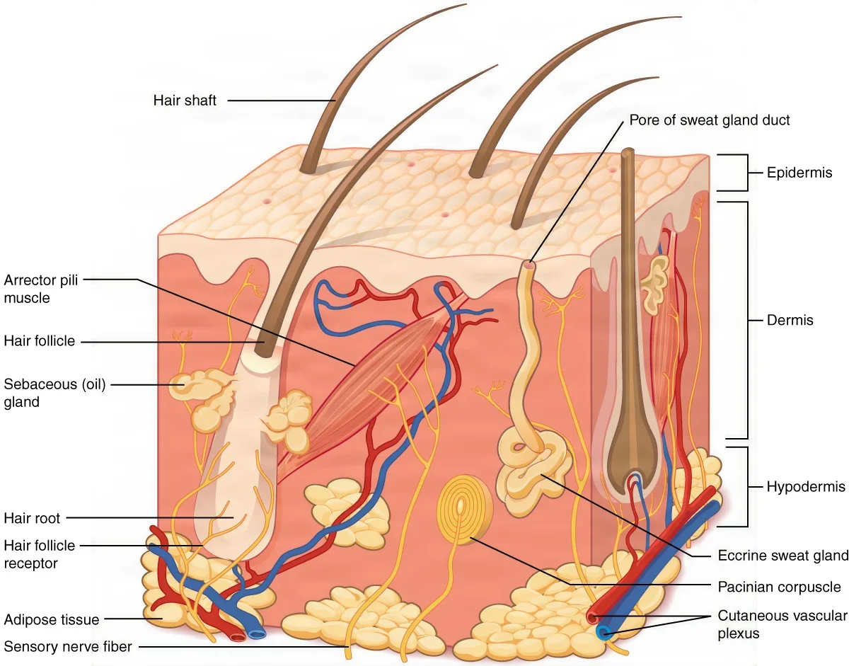
What the skin does
The Core Curriculum names six jobs of the skin.
- Protection: the skin is a barrier against wind, water, sunlight, grit, chemicals and microbes. It is the body's first line of defense. Sweat even carries a natural antibiotic that keeps germs on the surface in check.
- Preventing fluid loss: keratin and water-repelling fats in the outer layer keep the body's water in.
- Regulating body temperature: when the body is warm, sweat glands release sweat that cools the skin as it evaporates, and blood vessels in the dermis widen to give off heat. When the body is cold, those vessels narrow to keep heat in.
- Sensing the outside world: nerve endings in the skin detect touch, pressure, vibration, temperature and pain, so the body can react to its surroundings.
- excretionek-SKREE-shunThe removal of wastes, or of a drug and its breakdown products, from the body.Full entry : sweat carries off water, salts and small amounts of wastes such as ureayoo-REE-uhA waste product the liver makes when it breaks down protein, which the kidneys remove in urine.ur/o urineFull entry .
- absorptionub-ZORP-shunThe taking in of a substance, such as a drug or a nutrient, into the bloodstream.Full entry : substances on the skin can pass through it into the blood. Some medicines are given this way as skin patches, and some chemicals can harm a person by the same route.
The skin also makes vitamin D when sunlight strikes it, and the body needs vitamin D to absorb calcium for healthy bones.
Every incision breaks the barrier. Once the skin is open, germs have a way in, which is why the surgical site is prepped and the field kept sterile (chapters 26 and 31).
The epidermis
The epidermis is made of stacked layers of flat epithelial cells, called strata (one layer is a stratum). It has no blood vessels of its own; it gets its nourishment from the dermis below. Most skin has four layers. The thick skin of the palms and soles has a fifth.
From deep to surface, the layers are:
- Stratum basale (also called the stratum germinativum): the deepest layer, a single row of stem cellSTEM SELAn unspecialized cell that can divide to make more of itself and develop into specialized cells.Full entry that divide all the time. Every keratinocyte, the cell that makes up all the layers above, starts here. This layer also holds the melanocytemuh-LAN-uh-syteA cell in the bottom layer of the skin's outer layer that makes melanin, the pigment that gives skin and hair their color.melan/o black-cyte cellFull entry , the cells that make melaninMEL-uh-ninThe dark pigment that gives color to the skin, hair and eyes.melan/o black, dark-in substanceFull entry , the pigment that colors skin and hair and protects the living cells from ultraviolet (UV) light. Merkel cells, which sense touch, sit here too.
- Stratum spinosum: 8 to 10 rows of cells pushed up from below. These cells begin to make keratin, the tough protein that makes skin, hair and nails hard and water resistant, and a fatty substance that helps waterproof the skin. Immune cells called Langerhans cells patrol this layer and swallow bacteria.
- Stratum granulosum: three to five rows of flattening cells packed with keratin. Here the cells die.
- Stratum lucidum: a thin, clear layer of dead cells, found only in the thick skin of the palms, the soles and the fingers and toes.
- Stratum corneum: the outermost layer, usually 15 to 30 rows of dead, flat cells full of keratin. It keeps out microbes, slows water loss and takes the wear of daily life. Its cells are shed regularly, and the whole layer is replaced about every 4 weeks.
In short, new cells are born at the bottom, get pushed upward, fill with keratin, die, and flake off the top.
The dermis
The dermis is the skin's working core. It is connective tissue woven from collagenKOL-uh-jenThe main structural protein of the body, which gives strength to skin, bones, tendons, ligaments and scar tissue.coll/a glue-gen producingFull entry fibers, which give strength, and elastin fibers, which let the skin stretch and spring back. It holds the blood and lymphLIMFThe clear fluid that flows through the lymphatic vessels and carries white blood cells.Full entry vessels, nerves, hair follicles and glands. It has two layers:
- Papillary layer: the thin top layer of loose connective tissue. Finger-like bumps called dermal papillae push up into the epidermis and lock the two layers together; on the fingertips they form fingerprints. This layer is rich in small blood vessels, and it holds touch receptorrih-SEP-terA structure on or in a cell that a specific chemical, such as a hormone or drug, attaches to in order to act.Full entry and defense cells that fight germs that get through the epidermis.
- Reticular layer: the thick deeper layer of dense connective tissue with a strong mesh of collagen and elastin. It has a rich blood and nerve supply and holds most of the glands and hair follicles.
The dermis feeds the epidermis and sends it nerve supply, and its vessels carry the oxygen and the heat exchange the skin depends on.
Structures in the dermis
The Core Curriculum names the structures that live in or start from the dermis: glands, hair follicles, nails and sensory nerveSEN-suh-ree NURVA nerve that carries signals about touch, pain, temperature, position and the other senses toward the brain and spinal cord.Full entry endings.
- Eccrine sweat glands: coiled glands deep in the dermis, found all over the body and especially on the palms, soles and forehead. Their watery sweat cools the body.
- Apocrine sweat glands: larger glands in the armpits and groin that empty into hair follicles. Their thicker sweat has no smell until skin bacteria break it down.
- sebaceous glandsuh-BAY-shus glandA small gland in the skin that releases an oil (sebum) into the hair follicles to keep skin and hair soft.Full entry : oil glands, most of them opening into hair follicles. Their oil, called sebum, keeps the skin and hair soft and waterproof, and its fatty acidFAT-ee AS-idA building block of fats: a chain of carbon atoms that the body uses for energy and to build cells.Full entry fight bacteria.
- Hair follicles: a hair follicle is the sac in the dermis that a hair grows from. The hair root sits in the follicleFOL-ih-kulA small sac or cavity in the body, such as the pocket a hair grows from or the sac in the ovary that holds an egg.Full entry and ends in the hair bulb, where cells divide to lengthen the hair; the shaft above the skin is dead keratin. A tiny smooth musclesmooth MUS-ulMuscle that works without conscious control and lines hollow organs such as the stomach, intestines, bladder, airways and blood vessels.Full entry , the arrector pili, attaches to each follicle and makes the hair stand up (goose bumps) to trap warm air.
- Nails: a nail is a hard plate of dead, keratin-filled cells that grows forward from the nail root over the nail bed. It protects the fingertip and toe tip. The bed's rich blood supply makes the nail look pink, except at the pale half-moon (lunula) near the cuticle.
- Sensory nerve endings: different receptors sense different things. Meissner corpuscles in the papillary layer feel light touch, Pacinian corpuscles deeper down feel vibration, and free nerve endings sense pain and temperature. Hair follicles are wrapped in nerve endings too.
The hypodermis
The hypodermis, or subcutaneous tissue, lies directly under the dermis. Strictly it is not part of the skin, but it anchors the skin to the fasciaFASH-ee-uhA sheet or band of tough connective tissue that covers and separates muscles and organs.fasci/o bandFull entry , the tough fibrousFY-brusMade of or containing tough threads (fibers), especially of connective tissue.fibr/o fiber-ous full of, relating toFull entry tissue over the muscles and bones beneath. It is loose connective tissue and adipose tissueAD-ih-pohs TISH-ooBody fat: connective tissue made mostly of fat cells that store energy.adip/o fat-ose full ofFull entry (fat) tissue with a good blood supply.
Its fat stores energy, insulates the body against heat loss and cushions the structures beneath it.
How thick it is varies a great deal from person to person and from place to place. On the abdomen, the subcutaneous fat ranges from about a quarter inch to more than 8 inches.
The skin at the surgical field
When the surgeon makes an incision on the abdomen, the knife crosses the epidermis and dermis, then the subcutaneous fat, then the fascia, then muscle, and finally the peritoneumpair-ih-toh-NEE-umThe thin membrane that lines the belly cavity and covers most of the organs inside it.peri- aroundton stretched-eum a structureFull entry , the lining of the abdominal cavity. Chapter 2 explains how each of these tissues heals and holds sutureSOO-cherA stitch, or the thread used to stitch, that holds the edges of a wound or incision together.Full entry , and chapter 29 teaches incisions.
A few facts about the skin explain what you see at the field.
- The epidermis has no blood vessels, while the dermis is full of them. That is why a shallow scrape barely bleeds, but a cut into the dermis does.
- Some of the skin's own germs live below its surface, so scrubbing cannot remove them all, and skin can never be made sterile. A scrub or a prep lowers their number, and sterile gloveSTAIR-ul gluvA glove that has been sterilized and packed so it can touch a sterile field or an open body area without bringing in germs.Full entry cover the hands of the team.
- An older patient's epidermis is thinner, the dermis repairs itself more slowly and the fat under it thins out. Older skin is fragile and easily irritated, so handle it gently, and lift the patient into position instead of pulling or dragging.
Skin grafts
A graftGRAFTTissue or a man-made material moved into place in surgery to replace or repair a damaged part.Full entry is tissue moved from one place to another to replace tissue lost to injury or disease. The place it is taken from is the donor site, and the place it goes is the recipient site. A graft cut from the patient's own body is an autograft; one from another person, usually through a tissue bank, is an allograft; and one from an animal, such as pig skin, is a xenograft.
Skin grafts are named by how much of the skin's thickness they take:
- A split-thickness skin graft (STSG) takes the epidermis and only part of the dermis. How much of the dermis it takes varies.
- A full-thicknessful-THIK-nisGoing through every layer of the skin, or every layer of another tissue such as the wall of an organ.Full entry skin graft (FTSG) takes the epidermis and the whole dermis. The surgeon trims any fat off its underside before suturing it in place.
The difference matters at both sites. An STSG donor site heals on its own, because new epitheliumep-ih-THEE-lee-umThe tissue that covers the body's surfaces and lines its organs, vessels and cavities.Full entry grows from the dermis left behind. An FTSG takes all of the dermis, so nothing is left to regrow, and its donor site is sutured closed.
At the recipient site, the graft has been cut off from its blood supply, so it lives only if new vessels grow into it from the wound bed. A split-thickness graft picks up a blood supply quickly, but it may shrink as it heals, is less tough, and looks and feels different from normal skin. A full-thickness graft shrinks very little, holds up to wear, can be used over joints that bend, and gives an acceptable look.
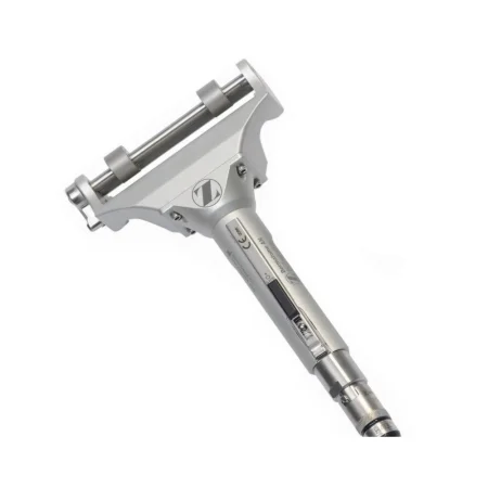
A large STSG is cut with a dermatome, a powered instrument with an oscillating blade housed in an adjustable head; a small lever on its side, marked in tenths of a millimeter, sets the thickness of the graft. The donor site is usually a broad, fleshy area, and it is lubricated so the dermatome glides. If you assemble the dermatome, check that the right blade and guard are seated and secured exactly as the maker directs: a loose blade or guard can injure the donor site or tear the graft. Each site, donor and recipient, gets its own prep and its own drapes.
An STSG is often run through a graft mesher, which cuts small diamond-shaped holes in it so the sheet opens into a net. Meshing keeps blood and serumSEER-umThe clear, yellow liquid left from blood after it clots and the cells are removed.Full entry from collecting under the graft, and lets the graft stretch to cover a larger or uneven area. The ratio of skin to holes is marked on the carrier plate, so use the plate the surgeon asks for.
Split-thickness means epidermis plus part of the dermis, and the donor site regrows on its own. Full-thickness means epidermis plus all of the dermis, and the donor site has to be closed.
3The skeleton
The skeleton is the body's frame. It is built of bones, held together at joints by ligaments, with cartilageKAR-tih-lijFirm, smooth, rubbery tissue that cushions joints and gives shape to parts such as the ear, nose and windpipe.chondr/o cartilageFull entry where bones need to bend or glide. An adult has 206 bones. A newborn has more, about 270 to 300, because some of them fuse together as a child grows.
The axial and appendicular skeleton
The skeleton has two divisions.
- The axial skeleton is the central, upright axis of the body: the skull, the vertebral columnVER-tuh-brul KOL-umThe spine: the column of small bones (vertebrae) that runs down the back and surrounds the spinal cord.vertebr joint, vertebra-al relating toFull entry and the rib cage, 80 bones in all. It protects the brain, spinal cord, heart and lungs.
- The appendicular skeleton is the limbs, plus the girdles that attach each limb to the axial skeleton: 126 bones.
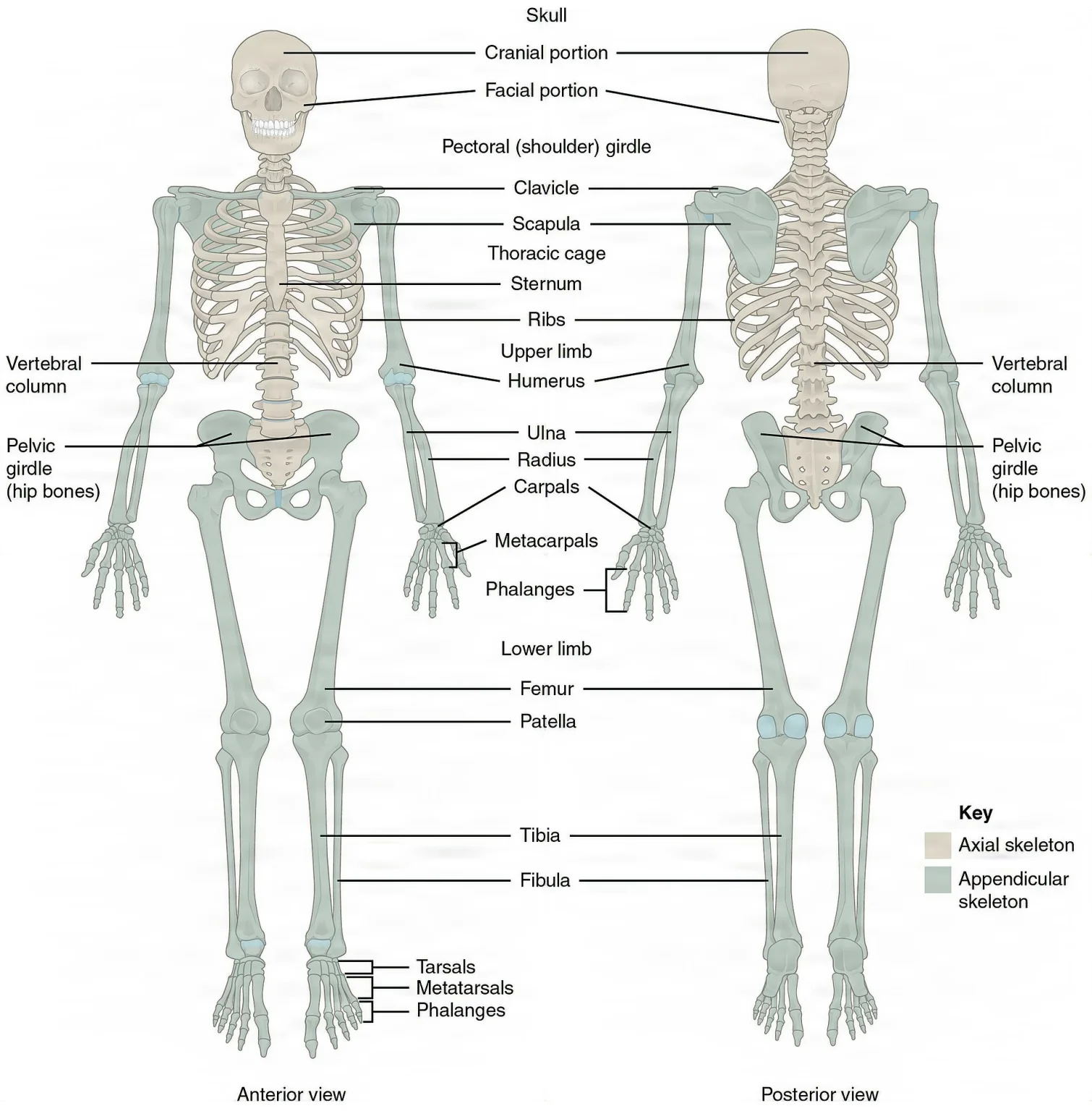
In anatomy, the arm is only the part between the shoulder and the elbow, and the leg is only the part between the knee and the ankle. The forearm, the thigh, the hand and the foot each have their own names.
| Region | Main bones |
|---|---|
| Skull (axial) | 22 bones. The craniumKRAY-nee-umThe part of the skull that encloses and protects the brain.crani/o skullFull entry , or brain case, has 8: the frontalFRUN-tulRelating to the front of the body, or to the forehead and the bone or brain area behind it.Full entry bone, two parietalpuh-RY-uh-tulRelating to the wall of a body cavity; also names a lobe of the brain and two bones of the skull.pariet/o wall-al relating toFull entry bones, two temporal bones, the occipitalok-SIP-ih-tulRelating to the back of the head.Full entry , the sphenoid and the ethmoid. The other 14 are facial bones, including the maxilla (upper jaw), the zygomatic (cheek) and nasal bones, and the mandibleMAN-dih-bulThe lower jawbone, the only bone of the skull that moves.Full entry (lower jaw), the only skull bone that moves. |
| Hyoid and ear bones (axial) | The horseshoe-shaped hyoid in the upper neck, the only bone that joins no other bone, and three tiny ossicles in each middle ear |
| Vertebral column (axial) | 24 vertebrae (7 cervicalSER-vih-kulRelating to the neck, or to the cervix, the lower end of the uterus.cervic/o neck, cervix-al relating toFull entry , 12 thoracicthuh-RAS-ikRelating to the chest, or to the middle part of the spine.thorac/o chest-ic relating toFull entry , 5 lumbarLUM-barHaving to do with the lower back, the part of the spine between the ribs and the pelvis.Full entry ), plus the sacrumSAY-krumThe large, triangle-shaped bone at the base of the spine, between the two hip bones.sacr/o sacrumFull entry and the coccyxKOK-siksThe small, triangle-shaped bone at the very bottom of the spine; the tailbone.Full entry |
| Thoracic cage (axial) | The sternum (breastbone: manubrium, body and xiphoid processZY-foyd PROS-esThe small, pointed tip at the lower end of the breastbone.Full entry ) and 12 pairs of ribs. Ribs 1 to 7, the true ribs, join the sternum by their own cartilage; ribs 8 to 12 are false ribs, and the last two pairs, 11 and 12, are floating ribs with no attachmentuh-TACH-mentThe deep emotional bond between an infant and a parent or caregiver.Full entry in front. |
| Pectoral (shoulder) girdle | The clavicleKLAV-ih-kulThe long, slightly S-shaped bone that runs from the top of the breastbone to the shoulder; the collarbone.clavicul/o collarboneFull entry (collarbone) and the scapulaSKAP-yuh-luhThe flat, triangle-shaped bone at the back of each shoulder; the shoulder blade.scapul/o shoulder bladeFull entry (shoulder blade), which attach each upper limb to the trunk |
| Upper limb, 30 bones each | The humerusHYOO-mer-usThe long bone of the upper arm, from the shoulder to the elbow.Full entry in the arm; the ulna (on the little-finger side) and the radius (on the thumb side) in the forearm; 8 carpals in the wrist; 5 metacarpals in the palm; and 14 phalanges in the fingers and thumb |
| Pelvic girdle | One hip bone (coxal bone) on each side, formed when three bones, the iliumIL-ee-umThe large, wing-shaped upper part of each hip bone.Full entry , ischium and pubisPYOO-bisThe front part of each hip bone, low in the pelvis; the two meet in the middle at the pubic symphysis.pub/o pubisFull entry , fuse into one as growth ends. The two hip bones, the sacrum and the coccyx make up the pelvis. |
| Lower limb, 30 bones each | The femur in the thigh, the longest bone in the body; the patella (kneecap); the tibiaTIB-ee-uhThe large inner bone of the lower leg, between the knee and the ankle; the shinbone.tibi/o tibiaFull entry (shinbone), which bears the weight, and the slender fibulaFIB-yuh-luhThe thin outer bone of the lower leg, beside the larger tibia.Full entry in the leg; 7 tarsals in the ankle and back of the foot, including the calcaneus (heel bone); 5 metatarsals; and 14 phalanges in the toes |
What bones do
The Core Curriculum lists six functions of bone.
- Support: bones hold the body upright and carry its weight against the pull of gravity, and the bones of the lower limbs and lower back are built thick for the load.
- Framework: the skeleton gives the body its shape and gives the soft tissues something to hang on.
- Protection: the skull surrounds the brain, the vertebrae surround the spinal cord, and the ribs guard the heart and lungs.
- Attachment for muscles: muscles pull on bones through their tendons. The bones act as levers and the joints as the pivot points: a muscle that crosses a joint moves the bone when it contracts.
- Blood cell production: red bone marrowbone MAIR-ohThe soft, spongy tissue inside bones that makes blood cells.Full entry makes red blood cells, white blood cells and plateletPLAYT-litA tiny cell fragment in the blood that sticks together at a wound to help form a clot.Full entry . This is called hematopoiesis.
- Storage of calcium: bone is the body's store of calcium and phosphorusFOS-fer-usA mineral in the blood and bones, measured as phosphate, that works with calcium to build bone and supply energy to cells.Full entry . Calcium is released into the blood when the body needs it, because calcium is needed for muscles to contract, for nerves to signal, for the heart to beat and for blood to clot. Hormones keep the blood level steady by moving calcium into and out of bone.
Bone also stores fat. Yellow bone marrow holds fat tissue, which the body can use for energy.
Types of bones
Bones are sorted into five types by their shape, and each shape suits a job.
| Type | Shape | Job | Examples |
|---|---|---|---|
| Long | Longer than wide, like a tube | Act as levers that muscles move | Femur, tibia, fibula, humerus, radius, ulna, metacarpals, metatarsals, phalanges |
| Short | About as wide and thick as long, like a cube | Give stability and support with a little motion | Carpals (wrist) and tarsals (ankle) |
| Flat | Thin and often curved | Protect organs and give muscles a place to attach | cranialKRAY-nee-ulRelating to the skull, or toward the head end of the body.crani/o skull-al relating toFull entry bones, sternum, ribs, scapula |
| Irregular | Complex shapes that fit no other group | Protect organs | Vertebrae and many facial bones |
| Sesamoid | Small and round, like a sesame seed, formed inside a tendon | Protect the tendon where it presses over a joint | The patella, the largest sesamoid bone and the only one everyone has |
"Long" describes the shape, not the size: the small bones of the fingers are long bones too.
Inside a long bone
A long bone shows every part a bone can have.
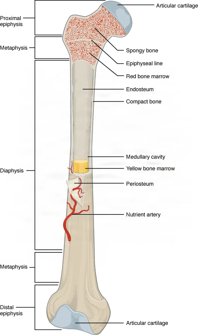
- Diaphysis: the shaft, the long tube between the two ends. Its walls are hard compact bone.
- medullamuh-DUL-uhThe inner part of an organ; most often the medulla oblongata, the lowest part of the brainstem.Full entry cavity (medullary canal): the hollow center of the shaft, filled with yellow marrow in an adult.
- Epiphysis (plural, epiphyses): the wider end at each side of the bone, filled with spongy bone and red marrow. The epiphyses form the joints.
- Epiphyseal plate (growth plate): a layer of cartilage between the shaft and each end in a growing bone, where the bone grows in length. When growth stops in early adulthood, bone replaces the cartilage, and only a thin epiphyseal line is left. A break through the plate in a child can delay the bone's growth.
- Articular cartilage: a thin, smooth cap of cartilage over the ends where the bone meets another bone at a joint. It cuts frictionFRIK-shunThe rubbing of one surface against another, such as skin against bed sheets.Full entry and absorptionub-ZORP-shunThe taking in of a substance, such as a drug or a nutrient, into the bloodstream.Full entry shockshokA life-threatening state in which blood flow is too low to bring enough oxygen to the body's tissues.Full entry .
- Periosteum: a tough fibrousFY-brusMade of or containing tough threads (fibers), especially of connective tissue.fibr/o fiber-ous full of, relating toFull entry membrane that covers the outside of the bone everywhere except the articular cartilage. It carries blood vessels and nerves that feed the bone, holds bone-forming cells, and gives tendons and ligaments a place to attach.
- Endosteum: a thin membrane lining the medullary cavity, where bone growth, repair and remodeling take place.
- cortexKOR-teksThe outer layer of an organ, especially the folded gray outer layer of the brain.cortic/o cortex, outer layerFull entry bone and cancellous bone: the two kinds of bone tissue, described next.
Blood vessels reach the inside of the bone through small holes in the shaft, each called a nutrient foramen.
The periosteum matters at the field. It is tough and tends to shred or tear when a saw, rongeurron-ZHURA strong surgical instrument with sharp, cup-shaped jaws used to bite away small pieces of bone.Full entry or cutting instrument meets it, so the surgeon first strips it off the bone surface with a periosteal elevator before cutting or reshaping the bone. A periosteal elevator is used in nearly every open orthopedicor-thuh-PEE-dikHaving to do with the bones, joints, muscles, ligaments and tendons, and the branch of medicine that treats them.orth/o straightped/o child (the field began with straightening children's bones)-ic relating toFull entry case.
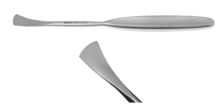
The Freer elevator also lifts periosteum off bone, and it is used to retract in confined spaces as well.
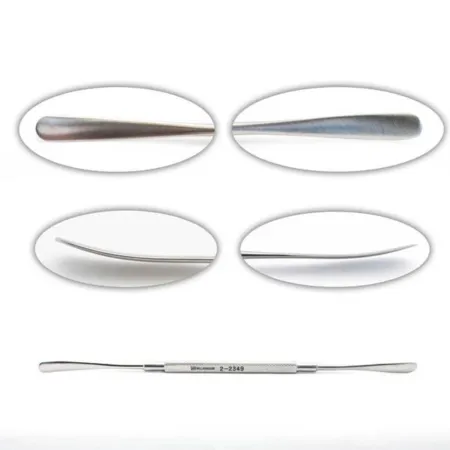
Periosteum covers the bone; endosteum lines its hollow center. The periosteum comes off with an elevator before bone is cut.
Bone tissue: cortical and cancellous
Bone tissue is a hard matrix of collagenKOL-uh-jenThe main structural protein of the body, which gives strength to skin, bones, tendons, ligaments and scar tissue.coll/a glue-gen producingFull entry fibers covered in calcium crystals. The crystals make bone hard and strong, and the collagen keeps it from being brittle. There are two kinds.
- Cortical bone, also called compact bone, is the dense, heavy bone of the outer shell. It lies just under the periosteum and forms the walls of the shaft of a long bone, where it gives support and protection and withstands the forces that press on the bone.
- Cancellous bone, also called spongy or trabecular bone, is a lighter network of thin bony struts called trabeculae, lined up along the lines of stress. The spaces between the struts hold marrow. It fills the ends of long bones and the inside of flat bones, the vertebrae and the pelvis.
Cancellous bone in the iliacIL-ee-akHaving to do with the ilium, the large, wing-shaped upper part of the hip bone.Full entry crest, the ends of long bones and the sternum holds red marrow, and the iliac crest is a common place to take a bone graftGRAFTTissue or a man-made material moved into place in surgery to replace or repair a damaged part.Full entry .
Under a microscope, compact bone is built of tiny columns called osteons, or Haversian systems. Each osteon is a set of rings of hard matrix around a central (Haversian) canal that carries blood vessels, nerves and lymphLIMFThe clear fluid that flows through the lymphatic vessels and carries white blood cells.Full entry vessels. Bone cells live in small spaces between the rings and pass nutrients to each other through fine channels.
Bone cells
Bone is living tissue that is built, kept and broken down all through life by its cells.
| Cell | What it does | Where it is |
|---|---|---|
| Osteogenic cell | A stem cellSTEM SELAn unspecialized cell that can divide to make more of itself and develop into specialized cells.Full entry , the only bone cell that divides; it becomes an osteoblast | Deep layers of the periosteum and the marrow |
| Osteoblast | Builds new bone: lays down collagen and calcium matrix | Growing parts of bone, including the periosteum and endosteum |
| Osteocyte | A former osteoblast walled in by the bone it made; keeps the matrix healthy; the most common bone cell | Inside the matrix, in a small space called a lacuna |
| osteoclastOS-tee-oh-klastA bone cell that breaks down and removes old bone tissue.oste/o bone-clast something that breaksFull entry | Breaks bone down (resorption) to reshape it, repair it or release calcium; a large cell with many nuclei that comes from white blood cells | Bone surfaces, at old, injured or unneeded bone |
A simple way to remember them: osteoblasts build, osteoclasts chew. In healthy bone the two work in balance.
How bone forms and grows
Bone formation is called ossification. In an embryo the skeleton starts as sheets of membrane and models made of cartilage, and ossification replaces them with bone by two pathways.
- Intramembranous bone formation: bone forms directly from sheets of membrane tissue. Most cranial bones, the flat bones of the face and the clavicles form this way.
- Endochondral bone formation: bone replaces a model made of hyaline cartilage. The long bones and the bones of the skull's base form this way. The cartilage does not turn into bone; it is broken down and bone is laid in its place.
A long bone grows in length at its epiphyseal plates: new cartilage grows on the side toward the end, and bone replaces it on the side toward the shaft. Growth stops when the plate is all bone. Bones also grow wider, as osteoblasts add bone under the periosteum while osteoclasts widen the hollow center.
Bone keeps rebuilding itself for life. About 5 to 10 percent of the skeleton is broken down and rebuilt each year, and bone thickens where it is stressed. That is why regular exercise strengthens bone, while a limb held in a cast loses bone.
How a broken bone mends
When a bone breaks, it heals through a callusKAL-usA thick, hard patch of skin formed by repeated rubbing or pressure; also, the new bone that forms as a fracture heals.Full entry , a mass of new tissue that bone later replaces. The Core Curriculum names five stages of bone healing, and their timing overlaps:
- Inflammation: torn vessels bleed and a clot, the fracture hematomahee-muh-TOH-muhA collection of blood, often clotted, that has leaked out of the blood vessels into the tissue.hem/o blood (here as hemat-)-oma swelling or massFull entry , forms at the break within hours. This stage lasts about 2 days. Bone cells at the break die because their blood supply is cut.
- Cellular proliferation: from about the second day, cells called macrophageMAK-roh-fayjA large white blood cell in the tissues that swallows and digests germs, dead cells and debris.macro- large-phage eaterFull entry clean up the debris, a mesh of fibrin seals the broken edges, and new capillaryKAP-ih-lair-eeOne of the tiniest blood vessels, joining the smallest arteries to the smallest veins.Full entry grow into that mesh.
- Callus formation: within days, a callus of cartilage and new bone forms. An internal callus fills the gap between the bone ends, and an external callus forms a collar around the outside of the break that steadies it. This stage lasts 3 to 4 weeks.
- Ossification: from about the second or third week, the callus hardens into bone as minerals are laid down, firmly joining the ends. This can take 3 to 4 months.
- Remodeling: osteoclasts remove extra and dead bone and osteoblasts lay down compact bone, until the bone takes back its shape and strength.
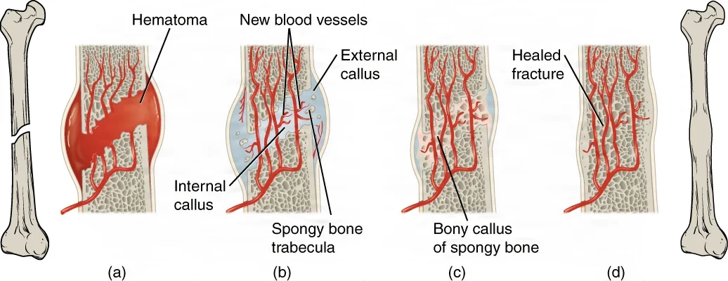
Healing needs the bone ends lined up and held still. If the pieces move, the hematoma is torn apart again and healing is delayed. Bringing the pieces back into line is called reduction, and holding them there is called fixation. In an ORIFOR-if, or said as lettersOpen reduction internal fixation: surgery that opens the skin to line up a broken bone and hold it with plates, screws or rods.Full entry (ORIF), the surgeon exposes the break and holds it with implants such as plates and screws. One break of the distal radius at the wrist, the Colles fracture, is usually caused by a fall on an outstretched hand. Fracture types and their treatment are taught in chapter 39.
Cartilage
Cartilage is a firm but flexible connective tissuekuh-NEK-tiv TISH-ooBody tissue that supports, binds together and protects other tissues and organs.Full entry . Its cells, chondrocytes, sit in small spaces in its matrix, and a membrane called the perichondrium wraps it. Chapter 2 introduced it as a tissue; here is how it works in the skeleton.
Its main characteristics:
- It has no blood vessels. Nutrients have to soak in from around it, so damaged cartilage heals very slowly and often poorly.
- It is smooth and slippery where bones glide, and it bends and springs back where the body needs flexibility.
There are three types, each in its own places:
- Hyaline cartilage, the most common type, smooth and glassy. It forms the articular cartilage at the ends of bones, the cartilage that joins the ribs to the sternum, the nose, and the growth plates.
- Fibrocartilage, a tough type, packed with thick collagen bundles. It forms the intervertebral discs, the pubic symphysisSIM-fih-sisA joint where two bones are joined firmly by a pad of cartilage that allows only a little movement.Full entry and the menisci of the knee.
- Elastic cartilage, which bends and springs back, as in the outer ear.
The Core Curriculum names two fibrocartilage structures you will meet in joint surgery:
- Labrum: a lip of fibrocartilage around the rim of a socket. The glenoid labrum deepens the shallow socket of the shoulder, and the acetabular labrum rims the socket of the hip. The shoulder's labrum also anchors ligaments that steady the joint.
- Meniscus (plural, menisci): a C-shaped pad of fibrocartilage. Each knee has two, the medial and lateral menisci, sitting between the rounded ends of the femur and the flat top of the tibia. They cushion the knee, spread its load and steady it.
Parts of each meniscus have no blood supply and heal poorly, so a torn meniscus often cannot repair itself. A meniscus tear is the most common knee injury, and the medial meniscus tears far more often than the lateral one, partly because it is anchored to a ligament and the joint capsule. In an arthroscopic meniscectomy, the surgeon removes the torn part, or most of the meniscus, through small incisions; even a complete meniscectomy leaves the rim to help carry load and steady the knee.
Bone markings
The bumps, ridges, holes and grooves on bones are called bone markings or landmarks. Each has a job: a tendon or ligament attaches there, a joint forms there, or a vessel or nerve passes through. Surgeons name them to say exactly where they are working, as in "the iliac crest" or "an osteotomy (bone cut) through the femoralFEM-er-ulRelating to the thigh or the femur, the thigh bone.Full entry neck". The Core Curriculum sorts them into three groups.
| Marking | What it is | Example |
|---|---|---|
| Articulating surfaces | Where a bone meets another bone at a joint | Knee joint |
| Head | A large, rounded end | Head of the femur |
| Condyle | A rounded, knuckle-like surface at a joint | Condyles of the femur at the knee |
| Facet | A small, flat surface | Facets where vertebrae meet each other and the ribs |
| Projections | Raised parts where tendons and ligaments attach; the stronger the pull, the bigger the projection | Processes of a vertebra |
| Process | Any bony projection | Spinous process of a vertebra, xiphoid process |
| Spine | A sharp, narrow projection | Ischial spine |
| Tubercle | A small, rounded projection | Greater tubercle of the humerus |
| Tuberosity | A larger, roughened projection | Ischial tuberosity, deltoidDEL-toydThe thick, triangle-shaped muscle that covers the shoulder and lifts the arm out to the side.Full entry tuberosity, tibial tuberosity |
| Trochanter | A bony projection at the top of the femur, at the base of its neck: the large greater trochanter on the outer side and the small lesser trochanter on the inner side | Greater and lesser trochanters; the greater trochanter, where muscles that move the hip attach, can be felt at the side of the upper thigh |
| Crest | A ridge of bone | Iliac crest, the top edge of the hip bone you feel at your waist |
| Depressions and openings | Hollows, grooves and holes, many for vessels and nerves | Openings in the skull |
| fossaFOS-uhA shallow hollow or dip in a bone or on the body's surface.Full entry | A shallow depression | Mandibular fossa, iliac fossa |
| Sulcus | A groove | sigmoidSIG-moydShaped like the letter S; most often short for the sigmoid colon, the S-shaped last part of the colon before the rectum.Full entry sulcus of the temporal bone |
| Foramen | A hole through a bone, usually for vessels or nerves | Foramen magnum in the occipital bone; the intervertebral foramina between vertebrae |
| Canal, meatusmee-AY-tusAn opening or passage in the body, such as the urinary opening or the ear canal.Full entry | A canal is a passage through bone; a meatus is the opening into a canal | The auditory (ear) canal, and the external auditory meatus that opens into it |
| sinusSY-nusA hollow space in the body, most often one of the air-filled spaces in the skull bones around the nose.Full entry | An air-filled space in a bone | Paranasal sinuses |
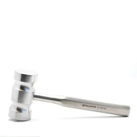
The iliac crest is a common source of a bone graft for spinal fusion. The surgeon cuts down to the crest, strips its periosteum with an elevator, then takes a plug of bone with a special cutter or shaves slivers off with an osteotome struck with a mallet. Bone left bleeding at the site is managed as chapter 29 describes. Bone graft substitutes are becoming more popular, because the harvest site is very painful after surgery.
4The vertebral column
The vertebral columnVER-tuh-brul KOL-umThe spine: the column of small bones (vertebrae) that runs down the back and surrounds the spinal cord.vertebr joint, vertebra-al relating toFull entry , or spine, runs from the skull to the tailbone. It holds up the head and trunk, protects the spinal cord, and gives the back muscles their anchor. It starts as 33 vertebrae, which become 24 separate vertebrae (singular, vertebra) plus the sacrumSAY-krumThe large, triangle-shaped bone at the base of the spine, between the two hip bones.sacr/o sacrumFull entry and the coccyxKOK-siksThe small, triangle-shaped bone at the very bottom of the spine; the tailbone.Full entry .
Regions and curves
The Core Curriculum names five regions.
| Region | Vertebrae | What sets them apart |
|---|---|---|
| cervicalSER-vih-kulRelating to the neck, or to the cervix, the lower end of the uterus.cervic/o neck, cervix-al relating toFull entry (neck) | 7, C1 to C7 | Smallest, because they carry the least weight. Each has a hole in its transversetrans-VERSLying across something, at a right angle to its length.Full entry process for an artery to the brain, and most have a forked spinous process. C1, the atlas, has no body and holds up the skull; C2, the axis, has a peg called the dens that the atlas turns around when you shake your head "no". |
| thoracicthuh-RAS-ikRelating to the chest, or to the middle part of the spine.thorac/o chest-ic relating toFull entry (chest) | 12, T1 to T12 | Larger, with facets where the ribs attach and long spinous processes that slope down and overlap |
| lumbarLUM-barHaving to do with the lower back, the part of the spine between the ribs and the pelvis.Full entry (lower back) | 5, L1 to L5 | Largest and thickest bodies, because they carry the most weight, with short, blunt spinous processes |
| Sacrum | 5 vertebrae fused into one triangular bone | Forms the back wall of the pelvis and joins the hip bones at the sacroiliac joints; holes on its front and back let the sacralSAY-krulHaving to do with the sacrum, the triangle-shaped bone at the base of the spine between the hip bones.Full entry nerves out |
| Coccyx (tailbone) | Usually 4 small vertebrae fused together | Joins the bottom of the sacrum; bears no weight when you stand, but may take some when you sit |
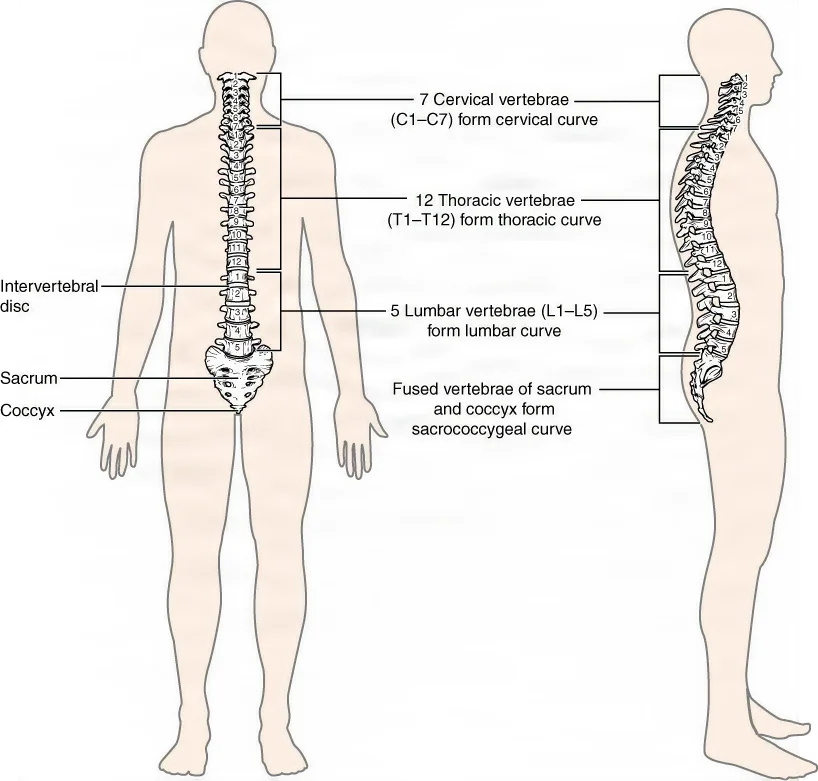
Seen from the side, the adult spine has four curves, which make it stronger, more flexible and better at absorbing shockshokA life-threatening state in which blood flow is too low to bring enough oxygen to the body's tissues.Full entry . The thoracic and sacrococcygeal curves are present before birth. The cervical curve forms as a baby learns to hold up its head, and the lumbar curve as a child starts to stand and walk. An abnormal sideways curve with twisting is scoliosisskoh-lee-OH-sisA sideways curve of the spine.scoli/o crooked, curved-osis abnormal conditionFull entry ; an excessive rounding of the upper back is kyphosisky-FOH-sisAn outward curve of the upper spine that makes the back look rounded or hunched.kyph/o hump-osis conditionFull entry ; and an excessive inward curve of the lower back is lordosis.
Parts of a vertebra
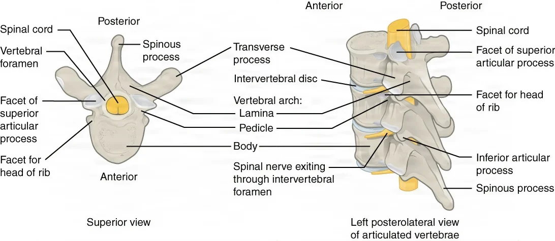
Vertebrae differ by region, but a typical one has the same parts:
- Body: the thick, round front part that carries the weight. The bodies get bigger toward the bottom of the spine.
- Vertebral arch: the ring of bone behind the body. It is made of two pediclePED-ih-kulA narrow stem or stalk that attaches a structure, such as a tumor or a skin flap, to the body.Full entry , which attach to the body, and two laminae (singular, lamina), which meet in the back to form the roof of the arch.
- Vertebral foramen: the large hole between the body and the arch. Stacked together, these holes form the vertebral (spinal) canal, which holds the spinal cord.
- Processes: seven bony projections arise from the arch: two transverse processes that stick out to the sides, one spinous process that points backward (the bumps you feel down the middle of the back), and four articular processes, two up and two down, that form small gliding joints with the vertebrae above and below. The transverse and spinous processes anchor muscles.
- Intervertebral foramen: the gap between two neighboring vertebrae, beside the pedicles, where a spinal nerveSPY-nul nervOne of the 31 pairs of nerves that branch from the spinal cord to carry signals to and from the body.Full entry leaves the spine.
Intervertebral discs
Between the bodies of the vertebrae sit the intervertebral discs, pads of fibrocartilage that bind the vertebrae together, cushion them under load and let the spine bend. They are thinnest in the neck and thickest in the lower back, which carries the most weight.
Each disc has two parts:
- Annulus fibrosus (also spelled anulus fibrosus): the tough outer ring of fibrousFY-brusMade of or containing tough threads (fibers), especially of connective tissue.fibr/o fiber-ous full of, relating toFull entry tissue, anchored to the edges of the vertebral bodies.
- Nucleus pulposus: the soft, gel-like center. Its high water content resists compression. With age it dries out, so the discs thin and the spine stiffens.
If the annulus weakens or tears, the nucleus can bulge out backward. This is a herniated disc ("slipped" or "ruptured" disc), and it can press on a spinal nerve as the nerve leaves through the intervertebral foramen, causing pain and weakness. It happens most often at L4 to L5 or L5 to S1, where it can cause sciatica, pain that runs from the lower back down the leg.
Spinal ligaments
Ligaments run the length of the spine and tie the vertebrae together.
- Anterior longitudinal ligament: runs down the front of the vertebral bodies and stops the spine from bending too far backward. Whiplash can tear it.
- Posterior longitudinal ligament: runs down the back of the vertebral bodies, inside the canal in front of the spinal cord.
- Ligamentum flavum: short, yellow, stretchy ligaments joining the laminae of neighboring vertebrae, behind the spinal cord.
- Supraspinous ligament: joins the tips of the spinous processes; in the neck it widens into the nuchal ligament.
The spine at the surgical field
Spine surgery reaches the spine from the back or the front, and the parts above tell you what is being removed.
- A laminectomy reaches the spinal canal from the back. Through a midlineMID-lineAn imaginary line down the center of the body; also, a type of IV catheter placed in a vein of the upper arm.Full entry incision, the surgeon goes through the fasciaFASH-ee-uhA sheet or band of tough connective tissue that covers and separates muscles and organs.fasci/o bandFull entry , packs the large back muscles away from the bone with periosteal elevators and sponges, removes the spinous process with a large rongeurron-ZHURA strong surgical instrument with sharp, cup-shaped jaws used to bite away small pieces of bone.Full entry , and then removes the lamina bit by bit with Kerrison rongeurs. The ligamentum flavum is then opened or removed to reach the disc. Keep the rongeur tips clean as each bite is presented, and save the bone pieces as a specimen.
- An anterior cervical discectomy and fusion (ACDF) reaches the neck spine from the front. The incision passes through the platysma muscle, and the surgeon works along the edge of the sternocleidomastoid muscle, between the carotidkuh-ROT-idHaving to do with the carotid arteries, the two large arteries on each side of the neck that carry blood to the head and brain.Full entry artery on one side and the tracheaTRAY-kee-uhThe windpipe: the tube that carries air from the throat to the lungs.Full entry and esophagusih-SOF-uh-gusThe muscular tube that carries food and liquid from the throat to the stomach.esophag/o esophagusFull entry on the other. The anterior longitudinal ligament is opened, the disc is removed piece by piece, and the space is filled with bone graftGRAFTTissue or a man-made material moved into place in surgery to replace or repair a damaged part.Full entry so the two vertebrae fuse.
5Joints
A joint, or articulation, is any place where two bones, or a bone and cartilageKAR-tih-lijFirm, smooth, rubbery tissue that cushions joints and gives shape to parts such as the ear, nose and windpipe.chondr/o cartilageFull entry , meet. The way a joint is built decides how much it can move, and how much it moves decides how stable it is: the freest joints are the least stable.
Joints by how much they move
The Core Curriculum sorts joints by movement.
- Synarthrotic joints (synarthrosis): immovable or nearly so. Their strength protects what lies beneath, as in the sutureSOO-cherA stitch, or the thread used to stitch, that holds the edges of a wound or incision together.Full entry between the skull bones, which are joined by fibrousFY-brusMade of or containing tough threads (fibers), especially of connective tissue.fibr/o fiber-ous full of, relating toFull entry tissue.
- Amphiarthrotic joints (amphiarthrosis): slightly movable, with the bones joined by cartilage. Examples are the joints between vertebral bodies, joined by the discs, and the pubic symphysisSIM-fih-sisA joint where two bones are joined firmly by a pad of cartilage that allows only a little movement.Full entry in the front of the pelvis.
- Diarthrotic joints (diarthrosis): freely movable. All of them are synovial jointsih-NOH-vee-ul JOYNTA freely movable joint in which the bone ends sit in a capsule filled with lubricating fluid.syn- togetherov egg (the fluid looks like egg white)-al relating toFull entry , such as the shoulder, hip, elbow and knee, and most are in the limbs.
Joints are also sorted by what holds the bones together: fibrous joints are joined by dense connective tissuekuh-NEK-tiv TISH-ooBody tissue that supports, binds together and protects other tissues and organs.Full entry , cartilaginous joints by cartilage, and synovial joints meet inside a fluid-filled space.
The synovial joint
No joint type is more common in the body than the synovial joint. Its bone ends are not joined to each other; they meet inside a closed space, the joint cavity.
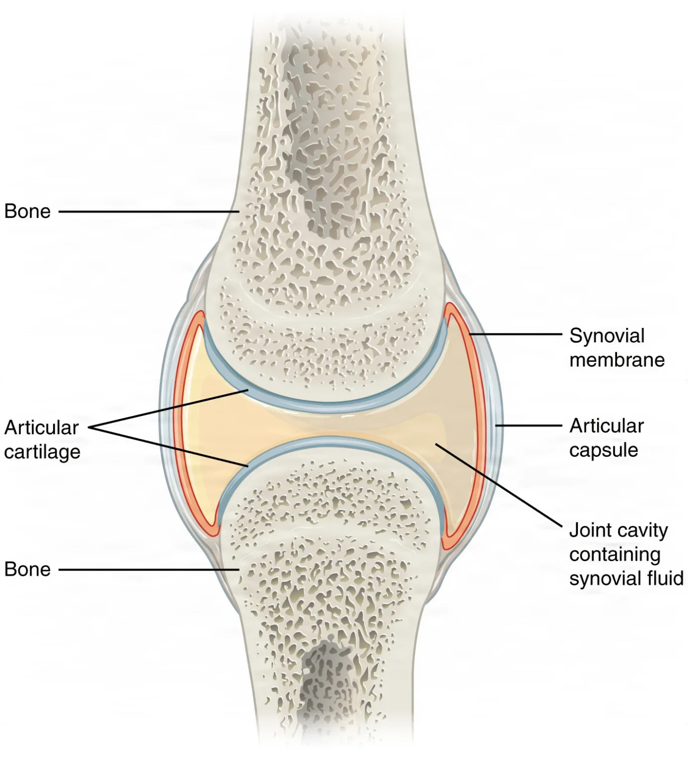
Its parts:
- Capsule (articular capsule): a sleeve of fibrous connective tissue attached to both bones just beyond their joint surfaces, enclosing the joint cavity.
- Synovial membrane: a thin lining on the inside of the capsule that makes synovial fluidsih-NOH-vee-ul FLOO-idThe thick, slippery fluid inside movable joints that lets the bones move smoothly.Full entry , a thick, slippery fluid that lubricates the joint and feeds its articular cartilage, which has no blood vessels.
- Articular cartilage: the smooth hyaline cartilage on each bone end, which lets the bones glide without wearing each other down.
- Ligaments: strong bands of dense connective tissue that tie bone to bone, allow normal movement and stop movement that would injure the joint. An external (extrinsic, or extra-articular) ligament lies outside the capsule, such as the collateral ligaments on the sides of the knee. An intra-articular (intracapsular) ligament lies inside the capsule, such as the cruciate ligaments in the center of the knee. Some ligaments are thickenings in the capsule wall itself.
- Bursae (singular, bursa): small sacs of lubricating fluid that sit where skin, a tendon, a ligament or a muscle would rub against bone or against each other, usually near a joint. Examples are the bursa over the kneecap, the one at the tip of the elbow, and the subacromial bursa that lets the rotator cuff tendons glide under the shoulder's bony roof. A tendon sheathSHEETHA covering that wraps around a structure; in procedures, a short hollow tube left in a blood vessel to pass catheters through.Full entry works the same way, wrapping a tendon where it crosses a joint.
Some synovial joints also hold a pad of fibrocartilage between the bones: a small articular disc, as in the jaw joint, or a larger meniscus, as in the knee.
Neither tendons nor ligaments have much blood supply, so, like cartilage, they heal slowly.
Types of synovial joints
Synovial joints are named for the shape of the surfaces that meet, and the shape decides the motion.
| Type | How it moves | Examples |
|---|---|---|
| Hinge | Bends and straightens in one plane, like a door hinge | Elbow, knee, ankle, finger and toe joints |
| Pivot | One bone rotates around another | Atlas turning on the axis (shaking the head "no"); the radius turning on the ulna near the elbow |
| Condyloid | An oval end in a shallow socket: bends, straightens and moves side to side | The wrist joint and the knuckles |
| Saddle | Two saddle-shaped surfaces: moves in two planes | The joint at the base of the thumb, which lets the thumb touch the fingertips |
| Plane (gliding) | Flat surfaces slide over each other | Between the carpal bones, the tarsal bones and the articular processes of the vertebrae |
| Ball-and-socket | A rounded head in a cup: moves in every plane and rotates; the most movable joint | Shoulder and hip |
The shoulder's socket, the glenoid cavity, is shallow, so the shoulder moves more freely than any other joint but leans on its muscles, especially the rotator cuff, to stay in place. The hip's socket, the acetabulum, is deep, with strong ligaments, so it trades movement for stability and weight bearing.
Joints at the surgical field
- Arthroscopy: in arthroscopic surgery, the scope and instruments pass through the capsule into the joint cavity through small incisions. Synovial fluid runs out when a capsule is injured or opened.
- arthroplastyAR-throh-plas-teeSurgery to repair or replace a joint, most often the hip or knee.arthr/o joint-plasty surgical repairFull entry : surgery to repair or replace a joint. When articular cartilage wears thin, as in osteoarthritisos-tee-oh-ar-THRY-tisA long-term joint disease in which the cartilage that cushions the ends of the bones wears away.oste/o bonearthr/o joint-itis inflammationFull entry , more pressure falls on the bone beneath it, the bone surface turns rough, and the joint becomes painful and stiff. In a total hip replacementTOH-tul HIP rih-PLAYS-mentSurgery that replaces the worn or damaged ball and socket of the hip joint with artificial parts; also called total hip arthroplasty.Full entry , the surgeon opens the capsule, dislocates the hip, cuts through the femoralFEM-er-ulRelating to the thigh or the femur, the thigh bone.Full entry neck to remove the femoral head, prepares the acetabulum, and replaces both sides with implants.
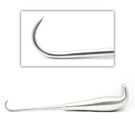
A bone hook slipped under the femoral neck helps lift the head of the femur out of the acetabulum when the hip is dislocationdis-loh-KAY-shunAn injury in which the ends of bones that meet at a joint are forced out of their normal position.Full entry . Because bone does not yield to a retractorrih-TRAK-terA surgical instrument that holds back tissue or organs so the surgeon can see and reach the operative site.Full entry the way soft tissue does, hooks and bone clamps are used to grasp and move it.
Landmarks of the joints surgeons repair
orthopedic surgeryor-thuh-PEE-dik SUR-juh-reeSurgery on the bones, joints, ligaments, tendons and muscles.ortho- straightped child (the specialty began with straightening children's bones)-ic relating toFull entry (chapter 39) names these parts of the limb joints again and again. A neck is a narrow bridge of bone, and cruciate means cross shaped.
- Shoulder: the head of the humerusHYOO-mer-usThe long bone of the upper arm, from the shoulder to the elbow.Full entry sits in the glenoid, a shallow socket on the scapulaSKAP-yuh-luhThe flat, triangle-shaped bone at the back of each shoulder; the shoulder blade.scapul/o shoulder bladeFull entry , at the glenohumeral joint, and a rim of cartilage, the labrum, deepens the socket a little. The clavicleKLAV-ih-kulThe long, slightly S-shaped bone that runs from the top of the breastbone to the shoulder; the collarbone.clavicul/o collarboneFull entry meets the acromionuh-KROH-mee-onThe bony tip of the shoulder blade that forms the top of the shoulder.Full entry of the scapula at the acromioclavicular (AC) joint and the sternum at the sternoclavicular joint. Just below the humerus's two tuberosities is its surgical neck, where most fractures of the upper humerus happen.
- Elbow and forearm: the olecranon, the point of the elbow, is the upper end of the ulna, and the radial head meets the humerus beside it; the radius turns around the ulna to turn the palm up and down.
- Wrist: the scaphoid, which links the two rows of carpal bones, is the carpal bone most often broken, and the lower end of the radius forms most of the wrist joint.
- Knee: the rounded condyles of the femur rest on the flat top of the tibiaTIB-ee-uhThe large inner bone of the lower leg, between the knee and the ankle; the shinbone.tibi/o tibiaFull entry , the tibial plateau, and the two cruciate ligaments cross in the middle of the joint.
- Ankle: the lower ends of the tibia (the medial malleolus) and the fibulaFIB-yuh-luhThe thin outer bone of the lower leg, beside the larger tibia.Full entry (the lateral malleolus) form a slot, the mortise, that holds the top of the talus; below it, the subtalar joint with the calcaneus turns the foot in and out.
6Muscles
What muscle does
skeletal muscleSKEL-uh-tul MUS-ulMuscle attached to bones that a person moves by choice, such as the muscles of the arms and legs.Full entry has three main jobs.
- Movement: muscles pull on bones to move the body, and some move other structures, such as the skin of the face, the tongue and the eyes.
- Posture: muscles of the spine, trunk and pelvis hold the body upright and balanced, working in turns so no group tires out.
- Heat production: working muscle makes heat, which helps keep body temperature steady.
There are three types of muscle tissue, which chapter 2 describes cell by cell:
- Skeletal muscle: striped (striated) fibers attached to bones, under voluntary control. It is the muscle this section is about.
- cardiac muscleKAR-dee-ak MUS-ulThe muscle tissue that makes up the walls of the heart.cardi/o heart-ac relating toFull entry : striated muscle found only in the heart, involuntary, contracting on its own rhythm.
- smooth musclesmooth MUS-ulMuscle that works without conscious control and lines hollow organs such as the stomach, intestines, bladder, airways and blood vessels.Full entry : unstriped, involuntary musclein-VOL-un-tair-ee MUS-ulMuscle that works without a person's conscious control, such as smooth muscle and heart muscle.Full entry in the walls of hollow organs, airways and blood vessels.
The words for a skeletal muscle
The Core Curriculum names six terms every scrub uses.
- Origin: the end of a muscle attached to the bone that stays still when the muscle contracts.
- Insertion: the end attached to the bone that moves. When a muscle contracts, it pulls the insertion toward the origin.
- Muscle belly: the thick, fleshy middle of a muscle, between its two ends. You can see the belly of your biceps bulge when you bend your elbow.
- Tendon: a cord of dense connective tissuekuh-NEK-tiv TISH-ooBody tissue that supports, binds together and protects other tissues and organs.Full entry that anchors a muscle to bone. Its collagenKOL-uh-jenThe main structural protein of the body, which gives strength to skin, bones, tendons, ligaments and scar tissue.coll/a glue-gen producingFull entry blends into the muscle at one end and into the periosteum at the other, so the pull of the muscle reaches the bone. A broad, flat tendon is called an aponeurosis.
- fasciaFASH-ee-uhA sheet or band of tough connective tissue that covers and separates muscles and organs.fasci/o bandFull entry : sheets of tough connective tissue, mostly collagen, that wrap, support and separate muscles and other structures. Each muscle has its own sheathSHEETHA covering that wraps around a structure; in procedures, a short hollow tube left in a blood vessel to pass catheters through.Full entry , and deep fascia divides the thigh and leg into compartments of muscles.
- Bursa: a fluid-filled sac that keeps a muscle or tendon from rubbing on bone, described with the joints above.
Not every skeletal muscle pulls on bone. The muscles of facial expression insert into the skin, so they move the skin, and the diaphragmDY-uh-framThe dome-shaped sheet of muscle under the lungs that is the main muscle of breathing; also the flat side of a stethoscope.Full entry changes the size of the chest without moving a bone.
Muscles work in teams
A single movement uses several muscles, each with a role. The Core Curriculum names four.
- agonistAG-uh-nistA drug or body chemical that attaches to a receptor on a cell and switches it on.Full entry , or prime mover: the main muscle that makes a movement. When you lift a cup, the biceps brachii is the prime mover that bends the elbow.
- Synergist: a muscle that helps the prime mover. The brachialis, deep to the biceps, helps bend the elbow.
- Fixator: a synergist that holds a bone steady, often at the prime mover's origin, so the movement happens where it should.
- antagonistan-TAG-uh-nistA drug that attaches to a receptor and blocks it, so the body's own chemicals or other drugs cannot act there.ant- againstagon struggle-ist one whoFull entry : a muscle with the opposite action, which relaxes while the prime mover works. Antagonists also hold a limb in position and slow a fast movement so it stops under control.
The roles switch with the movement. To straighten the knee, the quadriceps on the front of the thigh are the agonists and the hamstrings on the back are the antagonists. To bend the knee, the hamstrings become the agonists. The biceps and triceps of the arm, and the finger flexors and extensors of the forearm, are paired the same way.
The origin stays put and the insertion moves. The agonist does the job, the synergist helps, the fixator steadies, and the antagonist does the opposite.
The actions of muscles
Body movements are named in pairs of opposites, and they are always described from anatomical position: standing, arms at the sides, palms facing forward. The surgeon uses these words to ask for a limb to be moved, as in "more internal rotation" or "increase the abductionab-DUK-shunMoving a limb or part away from the midline of the body, such as raising an arm out to the side.ab- away fromduct to lead-ion actionFull entry ", and less precise words can lead to an injury.
| Action | What it does | Example |
|---|---|---|
| Flexion | Bends a joint, making the angle between the bones smaller; also bending the neck or trunk forward | Bending the elbow or the knee |
| Extension | Straightens a joint, making the angle larger; also bending the trunk back or returning upright | Straightening the elbow or the knee |
| Hyperextension | Extension beyond the normal range, which injures the joint | Whiplash of the neck |
| Abduction | Moves a limb away from the midlineMID-lineAn imaginary line down the center of the body; also, a type of IV catheter placed in a vein of the upper arm.Full entry of the body; spreads the fingers or toes | Raising the arm out to the side |
| adductionuh-DUK-shunMoving a limb or part toward the midline of the body, such as bringing an arm back down to the side.ad- towardduct to lead-ion actionFull entry | Moves a limb toward or across the midline; brings the fingers or toes together | Lowering the arm back to the side |
| Medial rotation (internal rotation) | Turns the front of the arm or thigh in toward the midline, at the shoulder or hip | Turning the thigh so the knee points inward |
| Lateral rotation (external rotation) | Turns the front of the arm or thigh out, away from the midline | Turning the thigh so the knee points outward |
| Circumduction | Moves the end of a limb in a circle by flexion, abduction, extension and adduction in turn | Circling the arm at the shoulder |
| supinationsoo-pih-NAY-shunTurning the forearm so the palm faces up or forward; for the foot, rolling it so the weight is on the outer edge.Full entry | Turns the forearm so the palm faces forward, or up; the radius and ulna lie side by side | The palm up, as when carrying a bowl of soup |
| pronationproh-NAY-shunTurning the forearm so the palm faces down or backward; for the foot, rolling it so the weight is on the inner edge.Full entry | Turns the forearm so the palm faces backward, or down; the radius crosses over the ulna | The palm down on a table |
| Dorsiflexion | At the ankle, lifts the top of the foot toward the shin | Lifting the toes off the floor while the heel stays down |
| plantarPLAN-terHaving to do with the sole of the foot.Full entry flexion | At the ankle, points the toes down and lifts the heel | Standing on tiptoe |
| inversionin-VER-zhunA turning inward or upside down; for the foot, turning the sole in toward the other foot.Full entry | Turns the sole of the foot in, toward the midline | The soles facing each other |
| Eversion | Turns the sole of the foot out, away from the midline | The soles facing outward |
| Elevation | Moves the scapulaSKAP-yuh-luhThe flat, triangle-shaped bone at the back of each shoulder; the shoulder blade.scapul/o shoulder bladeFull entry or the mandibleMAN-dih-bulThe lower jawbone, the only bone of the skull that moves.Full entry up | Shrugging; closing the mouth |
| Depression | Moves the scapula or the mandible down | Lowering the shoulders; opening the mouth |
| retractionrih-TRAK-shunA drawing back or pulling in of tissue; in breathing, the skin sinking in between, below or above the ribs with each breath.Full entry | Moves the scapula or the mandible back | Pulling the shoulder blades together; pulling the chin back |
| Protraction | Moves the scapula or the mandible forward | Reaching forward to push; jutting the chin out |
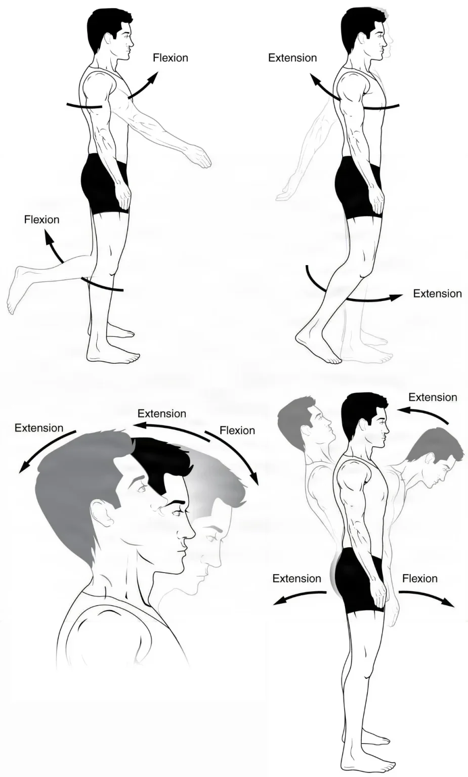
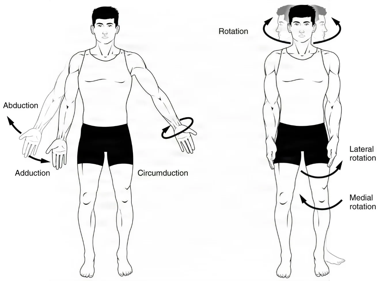
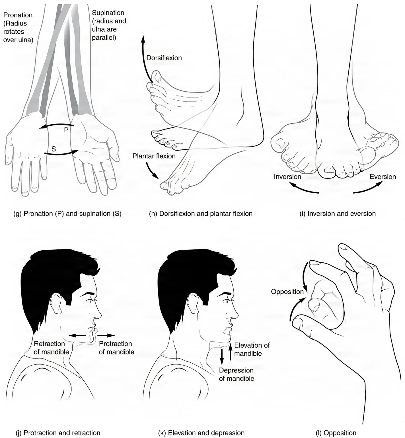
A memory trick: you hold soup in a supinated palm. Abduction takes a limb away, and adduction adds it back to the body.
You will use these words every time a patient is positioned. For a supine patient, AST directs that the arms be abducted less than 90 degrees, and preferably no more than 60, on padded armboards, with the palms up and the fingers extended. Once a patient is anesthetized, they cannot tell the team that a joint hurts, so the team must protect the skin, joints and muscles for them. Chapter 31 teaches positioning.
Move an anesthetized patient's joint past its normal range of motion, whether you are positioning the patient or holding a limb for the surgeon. The patient cannot tell you it hurts, so the team must protect the joint for them.
The word retraction has a second meaning in the OR: holding tissue back with a retractorrih-TRAK-terA surgical instrument that holds back tissue or organs so the surgeon can see and reach the operative site.Full entry to expose the field. The surgeon's request will tell you which one is meant.
How muscles get their names
Muscle names look hard, but most of them describe the muscle, so if you can read the name, you already know something about it. Muscles are named for:
- Location: the frontalis lies over the frontalFRUN-tulRelating to the front of the body, or to the forehead and the bone or brain area behind it.Full entry bone; the tibialis anterior lies on the front of the tibiaTIB-ee-uhThe large inner bone of the lower leg, between the knee and the ankle; the shinbone.tibi/o tibiaFull entry .
- Shape: the deltoidDEL-toydThe thick, triangle-shaped muscle that covers the shoulder and lifts the arm out to the side.Full entry is triangular, like the Greek letter delta; the trapezius is shaped like a trapezoid.
- Size: maximus is largest, medius is middle and minimus is smallest, as in the three gluteal muscles; longus is long and brevis is short.
- Direction of the fibers: rectusREK-tusLatin for straight; the name of several straight muscles, such as the rectus abdominis of the abdomen and the rectus femoris of the thigh.Full entry means straight and oblique means slanted, as in the rectus abdominis and the external oblique.
- Number of heads or origins: the biceps has two, the triceps three, and the quadriceps is a group of four.
- Attachments: the sternocleidomastoid runs from the sternum and clavicleKLAV-ih-kulThe long, slightly S-shaped bone that runs from the top of the breastbone to the shoulder; the collarbone.clavicul/o collarboneFull entry (cleido) to the mastoid process. When a muscle is named for its attachments, the origin comes first.
- Action: a flexor bends, an extensor straightens, an abductor moves away from the midline and an adductor moves toward it.
Many names carry two clues at once. The flexor carpi ulnaris, for example, flexes the hand at the wrist, where the carpal bones are, and lies on the ulna side of the forearm.
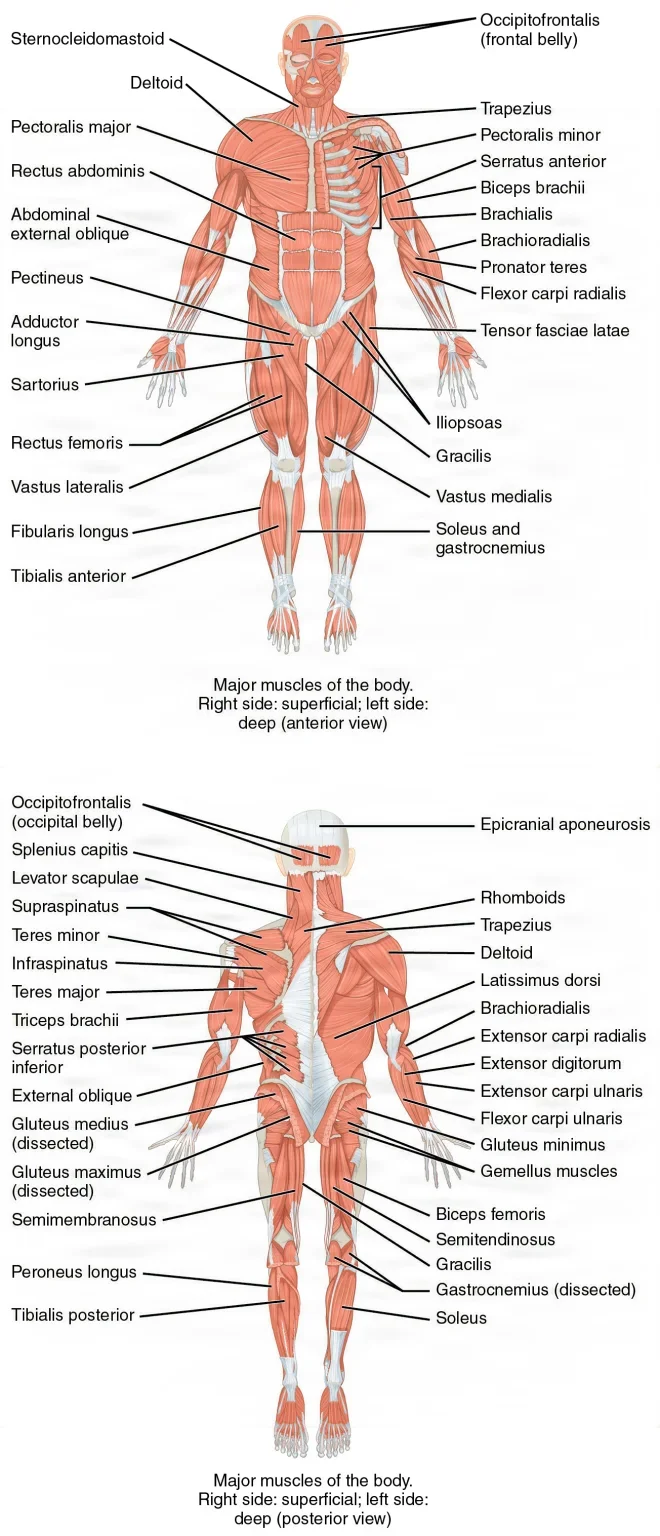
The muscles you will meet most at the field
The full Core Curriculum list is in the tables that follow. These are the muscles you will hear named most, and why.
The abdominal wall. Four pairs of muscles form the front and sides of the abdomen, protecting organs where there is no bone and helping to breathe and to bear down. The two rectus abdominis muscles run straight up and down on either side of the midline, from the pubisPYOO-bisThe front part of each hip bone, low in the pelvis; the two meet in the middle at the pubic symphysis.pub/o pubisFull entry to the ribs, each wrapped in a fibrousFY-brusMade of or containing tough threads (fibers), especially of connective tissue.fibr/o fiber-ous full of, relating toFull entry rectus sheath. The sheaths meet in the midline as the linea alba, a white band of fibrous tissue with almost no blood vessels. On each flank, three flat muscles lie in layers with their fibers crossing like the layers of plywood: the external oblique outermost, its fibers running down and toward the midline; the internal oblique beneath it, its fibers at right angles to those above; and the transversus abdominis deepest, its fibers running straight across.
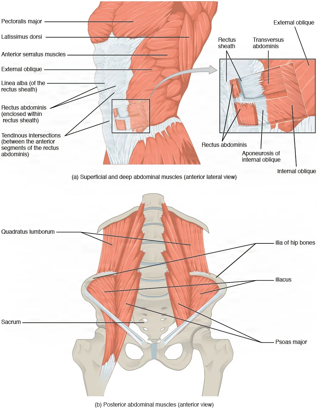
The incision decides which layers are cut. A midline incision goes through the linea alba, which bleeds little because it has so few vessels. A McBurney incision for the appendixuh-PEN-diksA small, finger-shaped pouch attached to the first part of the large intestine, in the lower right abdomen.Full entry splits the oblique and transversetrans-VERSLying across something, at a right angle to its length.Full entry muscles along their fibers rather than cutting across them. A Pfannenstiel incision runs low and across, following the skin's natural folds, through the rectus fascia, and the rectus muscles are then separated in the midline. The fascia carries the strength of the closed wound, so it is closed with strong sutureSOO-cherA stitch, or the thread used to stitch, that holds the edges of a wound or incision together.Full entry , often size 0 or 2-0. Chapter 29 teaches incisions, and chapter 25 teaches closure.
The neck. The platysma is a thin sheet of muscle just under the skin of the front of the neck. It is cut in operations on the front of the neck, such as thyroid surgery and an ACDF, and its edges are sewn back together at the close. Below it, the thin strap muscles (the sternohyoid and sternothyroid) are separated in the midline to reach the structures beneath, then sewn back together at the close. The sternocleidomastoid, running at an angle from the sternum and clavicle up to behind the ear, is a key landmark: in an ACDF, the surgeon works along its front edge.
Under general anesthesiaJEN-er-ul an-es-THEE-zhuhDrug-induced loss of consciousness and feeling over the whole body, so a person feels nothing and is not aware during surgery.an- withoutesthesi/o feeling, sensation-ia conditionFull entry , most skeletal muscles relax, including the tongue, which can fall back and block the airway. That is one reason the anesthesia provideran-us-THEE-zhuh pruh-VY-derThe licensed professional who gives anesthesia and watches over the client's breathing and vital functions during a procedure.Full entry secures the airway with a breathing tube.
The back and chest. The erector spinae muscles run in thick columns along both sides of the spine. In spine surgery from the back, they are pushed off the vertebrae with periosteal elevators and sponges and held aside to expose the bone. In a thoracotomythor-uh-KOT-uh-meeA surgical cut through the chest wall to reach the lungs, heart or other organs in the chest.thorac/o chest-tomy cutting intoFull entry , the surgeon reaches the lung between two ribs by cutting the intercostalin-ter-KOS-tulBetween the ribs.inter- betweencost/o rib-al relating toFull entry muscles along the upper border of the lower rib, because the nerve, artery and vein of each rib space run along the lower border of the rib above.
The shoulder. Four muscles from the scapula, the supraspinatus, infraspinatus, teres minor and subscapularis, end in tendons that fuse with the shoulder's capsule to form the rotator cuff. Because the shoulder's socket is so shallow, the cuff is what mainly holds the head of the humerusHYOO-mer-usThe long bone of the upper arm, from the shoulder to the elbow.Full entry in place. A cuff tear usually happens where the supraspinatus tendon attaches to the humerus. In a rotator cuff repair, the torn tendon is fixed back to the humerus with sutures or suture anchors, and small bony outgrowths (spurs) may be removed so they do not pinch the cuff.
The wrist. On the front of the wrist, the carpal bones form a U-shaped trough, and a strong band of fibrous tissue, the flexor retinaculum, spans its top to make the carpal tunnel. The central part of this band is the transverse carpal ligament. The flexor tendons of the fingers slide through the tunnel with the median nerve. When the nerve is squeezed, the patient has carpal tunnel syndrome, and in a carpal tunnel release, the surgeon divides the transverse carpal ligament with a #15 blade to give the nerve room.
The thigh and knee. The four quadriceps muscles on the front of the thigh share one tendon that wraps around the patella, the body's largest sesamoid bone, and continues below it as the patellar ligament to the tibial tuberosity. The three hamstrings on the back of the thigh bend the knee. The central third of the patellar ligament with a plug of bone at each end, the semitendinosus and gracilis tendons (called a hamstring graftGRAFTTissue or a man-made material moved into place in surgery to replace or repair a damaged part.Full entry ), or the end of the quadriceps tendon with a block of bone from the patella can each serve as the graft that rebuilds a torn anterior cruciate ligament (ACL).
The calf. The gastrocnemius and the soleus, the large muscles of the calf, join into the Achilles tendonuh-KIL-eez TEN-dunThe thick tendon at the back of the ankle that joins the calf muscles to the heel bone.Full entry (calcaneal) tendon, the strong tendon that attaches to the heel bone and points the foot down. It can rupture, especially in sports with jumping. In a repair, the surgeon brings the torn ends together with heavy nonabsorbable suture, tying it with the knee bent and the foot pointed down (plantar flexed) to take tension off the repair, and may reinforceree-in-FORSTo repeat and support teaching or care that has already been started, so the client remembers and follows it.Full entry it with a graft of the plantaris tendon, harvested with a tendon stripper.
Tendons and ligaments are commonly repaired with nonabsorbable sutures and suture anchors, and because they have a poor blood supply, they heal slowly.
The diaphragm. The diaphragm is the dome-shaped sheet of skeletal muscle that separates the chest from the abdomen. When it contracts, it flattens, the chest gets bigger and air flows into the lungs. The esophagusih-SOF-uh-gusThe muscular tube that carries food and liquid from the throat to the stomach.esophag/o esophagusFull entry , the aorta and the inferior vena cavain-FEER-ee-er VEE-nuh KAY-vuhThe large vein that carries blood from the lower body back to the right atrium of the heart.inferior lowervena veincava hollowFull entry pass through openings in it.
7The major muscles by region
These tables follow the Core Curriculum's list from the face to the foot. Learn the location and main action of each; the names often tell you both.
Face and head
| Muscle | Where it is | Main action |
|---|---|---|
| Muscles of facial expression (such as the orbicularis oris, orbicularis oculi and buccinator) | Under the skin of the face; they start on the skull and end in the skin | Move the skin to make expressions: the orbicularis oris closes and moves the lips, the orbicularis oculi closes the eye, and the buccinator compresses the cheek |
| Scalp muscle: occipitofrontalis | A front belly over the forehead and a back belly over the occipitalok-SIP-ih-tulRelating to the back of the head.Full entry bone, joined across the top of the skull by a flat tendon, the epicranial aponeurosis (galea) | Moves the scalp and raises the eyebrows |
| Temporalis (a muscle of mastication, or chewing) | Fan-shaped, over the temple, from the side of the skull to the mandibleMAN-dih-bulThe lower jawbone, the only bone of the skull that moves.Full entry | Closes the jaw and pulls it back |
| Masseter (a muscle of mastication) | In the cheek, from the zygomatic arch to the angle of the mandible | The main chewing muscle: raises the mandible to close the mouth |
| Medial and lateral pterygoids (muscles of mastication) | Deep, from the sphenoid bone to the mandible | Help chew: open and close the jaw, push it forward and move it side to side |
| Extrinsic tongue muscles (genioglossus, styloglossus, palatoglossus, hyoglossus) | Start outside the tongue, on the mandible, the styloid process, the soft palateSOFT PAL-itThe soft, muscular back part of the roof of the mouth, with no bone in it.Full entry and the hyoid, and insert into it | Move the whole tongue forward, back, up and down, to help chew, swallow and speak |
| intrinsicin-TRIN-zikComing from inside, or belonging naturally to something itself.Full entry tongue muscles | Start and end inside the tongue | Change the tongue's shape, curling or flattening it |
Neck
| Muscle | Where it is | Main action |
|---|---|---|
| Platysma | A thin sheet just under the skin of the front of the neck, from the upper chest to the lower face | A muscle of facial expression: helps lower the jaw and widens the neck |
| Sternocleidomastoid | From the sternum and clavicleKLAV-ih-kulThe long, slightly S-shaped bone that runs from the top of the breastbone to the shoulder; the collarbone.clavicul/o collarboneFull entry up to the mastoid process behind the ear; divides each side of the neck into front and back triangles | One side turns the head toward the opposite side and tilts it; both together flex the head |
| Scalenes (anterior, middle and posterior) | The side of the neck, from the cervicalSER-vih-kulRelating to the neck, or to the cervix, the lower end of the uterus.cervic/o neck, cervix-al relating toFull entry vertebrae down to the first two ribs | Flex, tilt and turn the head and neck, and raise the top two ribs in deep breathing |
| Sternohyoid | A thin strap muscle from the sternum up to the hyoid bone, below the hyoid | Pulls the hyoid down |
| Sternothyroid | A strap muscle from the sternum up to the thyroid cartilageKAR-tih-lijFirm, smooth, rubbery tissue that cushions joints and gives shape to parts such as the ear, nose and windpipe.chondr/o cartilageFull entry of the larynxLAIR-inksThe voice box: the part of the airway in the neck, above the windpipe, that holds the vocal cords.laryng/o larynxFull entry | Pulls the larynx down |
| Thyrohyoid | A strap muscle from the thyroid cartilage up to the hyoid | Pulls the hyoid down, or raises the larynx |
| Omohyoid | A strap muscle below the hyoid with two bellies, an upper and a lower | Pulls the hyoid down |
Back
| Muscle | Where it is | Main action |
|---|---|---|
| Erector spinae (three columns: iliocostalis, longissimus and spinalis) | Along each side of the spine, from the pelvis and sacrumSAY-krumThe large, triangle-shaped bone at the base of the spine, between the two hip bones.sacr/o sacrumFull entry up to the ribs, vertebrae and skull; most of the muscle of the back | The main extensor of the spine; also controls bending and twisting and holds the lumbarLUM-barHaving to do with the lower back, the part of the spine between the ribs and the pelvis.Full entry curve |
| Latissimus dorsi | A broad, flat, triangular muscle over the lower back, fused into a wide aponeurosis, ending on the humerusHYOO-mer-usThe long bone of the upper arm, from the shoulder to the elbow.Full entry | adductionuh-DUK-shunMoving a limb or part toward the midline of the body, such as bringing an arm back down to the side.ad- towardduct to lead-ion actionFull entry , extends and medially rotates the arm |
| Trapezius | A broad sheet over the upper back and the back of the neck, from the skull and spine out to the clavicle and scapulaSKAP-yuh-luhThe flat, triangle-shaped bone at the back of each shoulder; the shoulder blade.scapul/o shoulder bladeFull entry | Raises, pulls back and rotates the scapula, as in shrugging, and helps tilt the head back |
Shoulder and arm
| Muscle | Where it is | Main action |
|---|---|---|
| deltoidDEL-toydThe thick, triangle-shaped muscle that covers the shoulder and lifts the arm out to the side.Full entry | The thick, triangular muscle that rounds the shoulder, ending on the deltoid tuberosity of the humerus | The main abductor of the arm; its front and back parts also flex, extend and rotate it |
| Rotator cuff: supraspinatus | On the back of the scapula, above its spine | abductionab-DUK-shunMoving a limb or part away from the midline of the body, such as raising an arm out to the side.ab- away fromduct to lead-ion actionFull entry the arm |
| Rotator cuff: infraspinatus | On the back of the scapula, below its spine | Laterally rotates the arm |
| Rotator cuff: teres minor | On the back of the scapula, just above the teres major | Laterally rotates and extends the arm |
| Rotator cuff: subscapularis | On the front of the scapula | Medially rotates the arm |
| Biceps brachii | The front of the arm, with two heads; crosses both the shoulder and the elbow | Flexes the elbow, and helps supinationsoo-pih-NAY-shunTurning the forearm so the palm faces up or forward; for the foot, rolling it so the weight is on the outer edge.Full entry the forearm and flex the arm at the shoulder |
| Triceps brachii | The back of the arm, with three heads, ending on the olecranon at the tip of the elbow | Extends the elbow; the antagonistan-TAG-uh-nistA drug that attaches to a receptor and blocks it, so the body's own chemicals or other drugs cannot act there.ant- againstagon struggle-ist one whoFull entry of the biceps |
Forearm, hand and fingers
| Muscle | Where it is | Main action |
|---|---|---|
| Flexor group | The front (anterior compartment) of the forearm; most start on the humerus near the elbow, and their tendons cross the front of the wrist | Flex the wrist and the fingers |
| Extensor group | The back (posterior compartment) of the forearm, from the humerus, radius and ulna; their tendons cross the back of the wrist | Extend the wrist and the fingers, and move the thumb |
| Flexor carpi group: flexor carpi radialis and flexor carpi ulnaris | Front of the forearm, on the thumb side and the little-finger side | Flex the hand at the wrist; the radialis also abducts it and the ulnaris adducts it |
| Extensor carpi group: extensor carpi radialis longus, extensor carpi radialis brevis and extensor carpi ulnaris | Back of the forearm | Extend the hand at the wrist; the radialis muscles also abduct it and the ulnaris adducts it |
| Flexor digitorum group: flexor digitorum superficialis and flexor digitorum profundus | Front of the forearm, the superficialis over the deeper profundus; long tendons run to the fingers | Flex the fingers and help flex the wrist |
| Extensor digitorum | Back of the forearm, with tendons over the back of the hand to the fingers | Extends the fingers and the wrist |
| Intrinsic muscles of the hand (thenar, hypothenar and intermediate groups) | Start and end within the hand: at the base of the thumb, at the base of the little finger and in the middle of the palm | Fine movements of the fingers and thumb, such as bringing the thumb across to touch a fingertip |
Chest wall and diaphragm
| Muscle | Where it is | Main action |
|---|---|---|
| External intercostalin-ter-KOS-tulBetween the ribs.inter- betweencost/o rib-al relating toFull entry | Between the ribs, the outer set; 11 pairs | Raise the ribs to breathe in |
| Internal intercostals | Between the ribs, just under the external set; 11 pairs | Draw the ribs together to breathe out |
| Pectoralis major | The thick, fan-shaped muscle over the upper chest, from the clavicle, sternum and rib cartilages to the humerus | Adducts and medially rotates the arm |
| Pectoralis minor | Under the pectoralis major, from the 3rd to 5th ribs to the coracoid process of the scapula | Pulls the shoulder forward and down, and helps lift the ribs when breathing in |
| diaphragmDY-uh-framThe dome-shaped sheet of muscle under the lungs that is the main muscle of breathing; also the flat side of a stethoscope.Full entry | A dome between the chest and the abdomen; muscle around the edges, a central tendon in the middle, attached to the xiphoid processZY-foyd PROS-esThe small, pointed tip at the lower end of the breastbone.Full entry , the lower six ribs and the lumbar vertebrae | The main muscle of breathing: contracts and flattens to draw air into the lungs; helps the abdominal muscles bear down |
Abdominal wall
| Muscle | Where it is | Main action |
|---|---|---|
| External oblique | The outermost of the three flank layers; its fibers run down and toward the midlineMID-lineAn imaginary line down the center of the body; also, a type of IV catheter placed in a vein of the upper arm.Full entry | Compresses the abdomen, and bends and twists the trunk |
| Internal oblique | The middle flank layer; its fibers run at right angles to the external oblique's | Compresses the abdomen, and bends and twists the trunk |
| Transversus abdominis | The deepest flank layer; its fibers run straight across the abdomen | Compresses the abdomen |
| rectusREK-tusLatin for straight; the name of several straight muscles, such as the rectus abdominis of the abdomen and the rectus femoris of the thigh.Full entry abdominis | A pair of long, straight muscles on either side of the midline, from the pubisPYOO-bisThe front part of each hip bone, low in the pelvis; the two meet in the middle at the pubic symphysis.pub/o pubisFull entry up to the xiphoid process of the sternum and the cartilages of ribs 5 to 7, inside the rectus sheathSHEETHA covering that wraps around a structure; in procedures, a short hollow tube left in a blood vessel to pass catheters through.Full entry , crossed by bands of tendon | Flexes the spine (sitting up) and tightens the abdominal wall |
Thigh
| Muscle | Where it is | Main action |
|---|---|---|
| Adductor group (adductor longus, brevis and magnus, with the gracilis and pectineus) | The inner (medial) compartment of the thigh | Adduct the thigh; some also flex it, extend it or rotate it |
| Biceps femoris | The back of the thigh, on the outer side; one of the three hamstrings, with the semitendinosus and semimembranosus | Flexes the knee and extends the thigh at the hip |
| Gluteus maximus | The buttock; the largest of the three gluteal muscles | Extends the thigh at the hip, and helps abduct and laterally rotate it |
| Gluteus medius | Deep to the gluteus maximus | Abducts the thigh at the hip |
| Gluteus minimus | The smallest gluteal, deep to the gluteus medius | Helps abduct the thigh |
| Iliopsoas (the psoas major and the iliacus) | The psoas starts along the last thoracicthuh-RAS-ikRelating to the chest, or to the middle part of the spine.thorac/o chest-ic relating toFull entry vertebra and the lumbar vertebrae, and the iliacus inside the pelvis; they join to end on the lesser trochanter of the femur | Flexes the thigh at the hip, and flexes the trunk |
| Quadriceps femoris (the rectus femoris and three vasti: lateralis, intermedius and medialis) | The front of the thigh; their shared tendon holds the patella and continues as the patellar ligament to the tibiaTIB-ee-uhThe large inner bone of the lower leg, between the knee and the ankle; the shinbone.tibi/o tibiaFull entry tuberosity | Extends the knee |
| Sartorius | A long, narrow strap from the front of the iliacIL-ee-akHaving to do with the ilium, the large, wing-shaped upper part of the hip bone.Full entry crest (anterior superior iliac spine) across the thigh to the inner side of the tibia | Flexes the knee and flexes, abducts and laterally rotates the thigh, as when sitting cross-legged |
Leg
| Muscle | Where it is | Main action |
|---|---|---|
| Extensors (the anterior compartment: tibialis anterior, extensor hallucis longus and extensor digitorum longus) | The front of the leg, beside the tibia; their tendons are held at the ankle by bands of connective tissuekuh-NEK-tiv TISH-ooBody tissue that supports, binds together and protects other tissues and organs.Full entry | Raise the front of the foot (dorsiflexion) and extend the toes |
| Gastrocnemius | The most superficial calf muscle, with two heads, one from each condyle (medial and lateral) of the femur, ending in the Achilles tendonuh-KIL-eez TEN-dunThe thick tendon at the back of the ankle that joins the calf muscles to the heel bone.Full entry | plantarPLAN-terHaving to do with the sole of the foot.Full entry flexes the foot, and helps flex the knee |
| Soleus | A wide, flat muscle deep to the gastrocnemius, from the tibia and fibulaFIB-yuh-luhThe thin outer bone of the lower leg, beside the larger tibia.Full entry to the Achilles tendon | Plantar flexes the foot |
| Plantaris | A small muscle running at an angle between the gastrocnemius and soleus; some people do not have one | Weakly plantar flexes the foot and flexes the knee; joins the gastrocnemius and soleus on the Achilles tendon, and its tendon is used as a graftGRAFTTissue or a man-made material moved into place in surgery to replace or repair a damaged part.Full entry |
| Peroneus longus (fibularis longus) | The outer (lateral) leg, on the fibula | Everts the foot and plantar flexes it |
| Peroneus brevis (fibularis brevis) | The outer leg, on the lower fibula, ending on the base of the fifth metatarsal | Everts and plantar flexes the foot |
Foot and toes
| Muscle | Where it is | Main action |
|---|---|---|
| Extensor digitorum group (extensor digitorum longus in the leg and extensor digitorum brevis on the top of the foot) | The front of the leg and the top (dorsum) of the foot | Extend the toes and help raise the front of the foot |
| Flexor digitorum group (flexor digitorum longus in the deep calf, with flexors in the sole) | The deep back of the leg and the sole of the foot | Flex the toes |
| Peroneus longus | The outer leg; its tendon crosses under the sole to the base of the big toe's metatarsal | Everts the foot and plantar flexes it |
| Tibialis anterior | The front of the leg, along the outer side of the tibia, with its tendon to the inner side of the foot | Dorsiflexes and inverts the foot |
What to take from this chapter
- The skin has two layers, the epidermis (no blood vessels, renewed from its deepest layer) and the dermis (vessels, nerves, glands and hair follicles), over the fatty hypodermis. It protects, keeps fluid in, controls temperature, senses, excretes and absorbs.
- A split-thickness skin graft takes the epidermis and part of the dermis, and its donor site regrows; a full-thickness graft takes all the dermis, and its donor site is closed.
- The axial skeleton (skull, spine, rib cage) protects and supports; the appendicular skeleton (girdles and limbs) is built for standing, walking and handling objects. Bones support, frame, protect, anchor muscles, make blood cells and store calcium.
- A long bone has a shaft (diaphysis) of cortical bone around a medullary cavity, spongy ends (epiphyses) capped by articular cartilage, and a periosteum that the surgeon strips with an elevator before cutting bone.
- Osteoblasts build bone, osteocytes keep it, and osteoclasts break it down. A broken bone heals through a callus that hardens into bone and then remodels.
- The spine has 7 cervical, 12 thoracic and 5 lumbar vertebrae plus the sacrum and coccyx. A laminectomy removes the lamina at the back of the arch; a disc has a tough annulus around a soft nucleus.
- Joints are immovable, slightly movable or freely movable; a freely movable synovial joint has a capsule, a synovial membrane and fluid, articular cartilage, ligaments and often bursae.
- Muscles pull their insertion toward their origin through tendons; the agonist moves, the antagonist opposes. Know the paired movement words, because the surgeon uses them to position and move the patient.
Check yourself5 quick questions
Free for everyone. Pick an answer for each, then check them all at once.
In the ORWhere you'll use this
The chapter's whole question pool in sets of 10, with the answer explained after each one.
Part of the educators programThe chapter's words on spaced cards: the ones you miss come back sooner.
Part of the educators programGiven by your class's teacher. Each class draws its own 25 from the chapter's pool.
Part of the educators program
Suggest a fix
Something wrong, unclear or out of date in this chapter? Tell us and we will fix it for everyone. Your email is only used to reply.