Breast Biopsy
This procedure involves the excision of a suspicious mass or a portion of breast tissue for histological examination. The surgical goal is to obtain an adequate tissue sample from the mammary parenchyma to diagnose or rule out pathology.
24 instruments to master.
Master this setup: test yourself on every instrument, earn the tier, hold the record.
No site record yet · target 8:58
Study flashcards
Card by card, grade yourself · 24 cards
Practice test
Answer, see why · all 25
Take the exam
25 questions · target 8:58
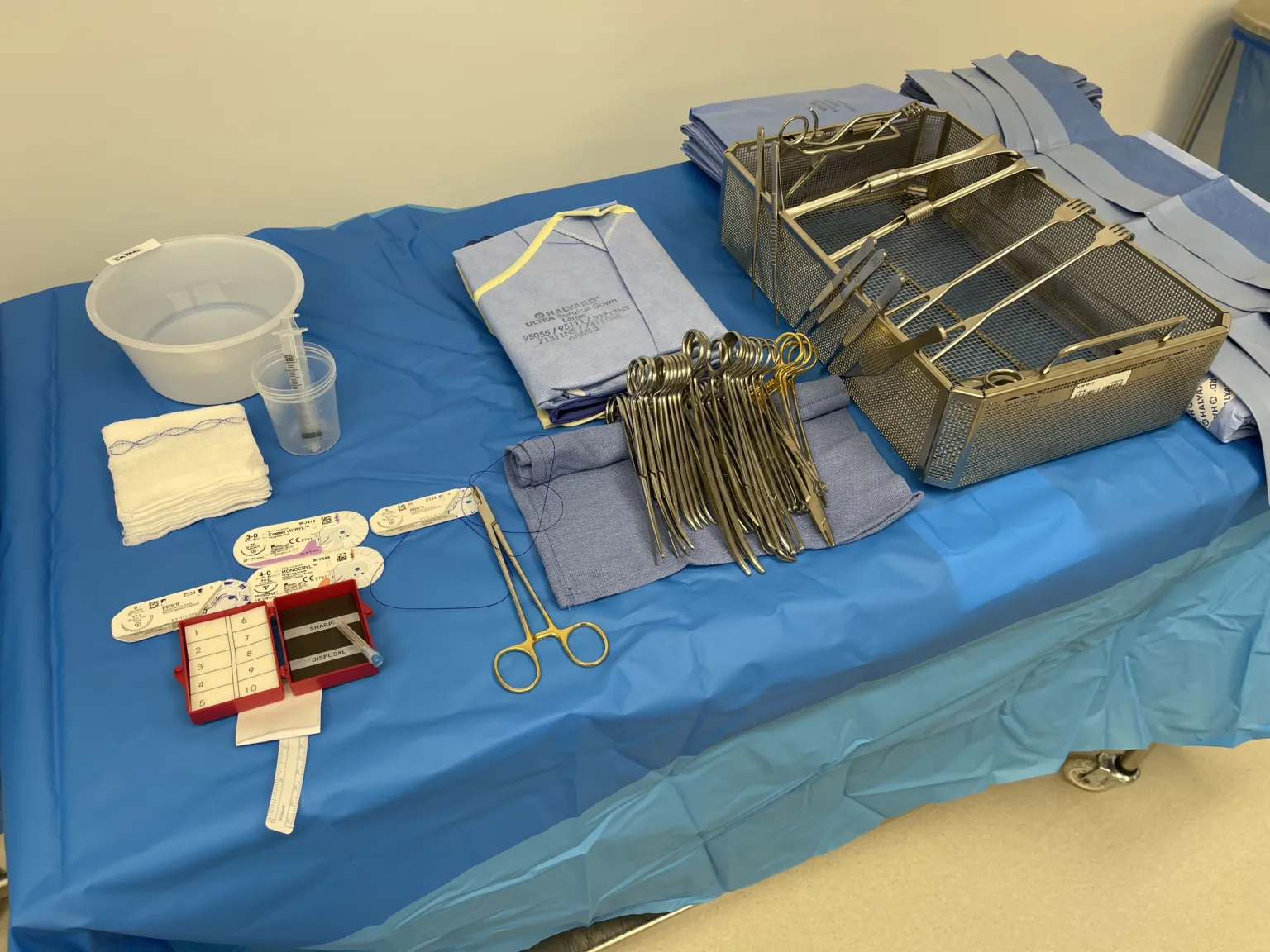
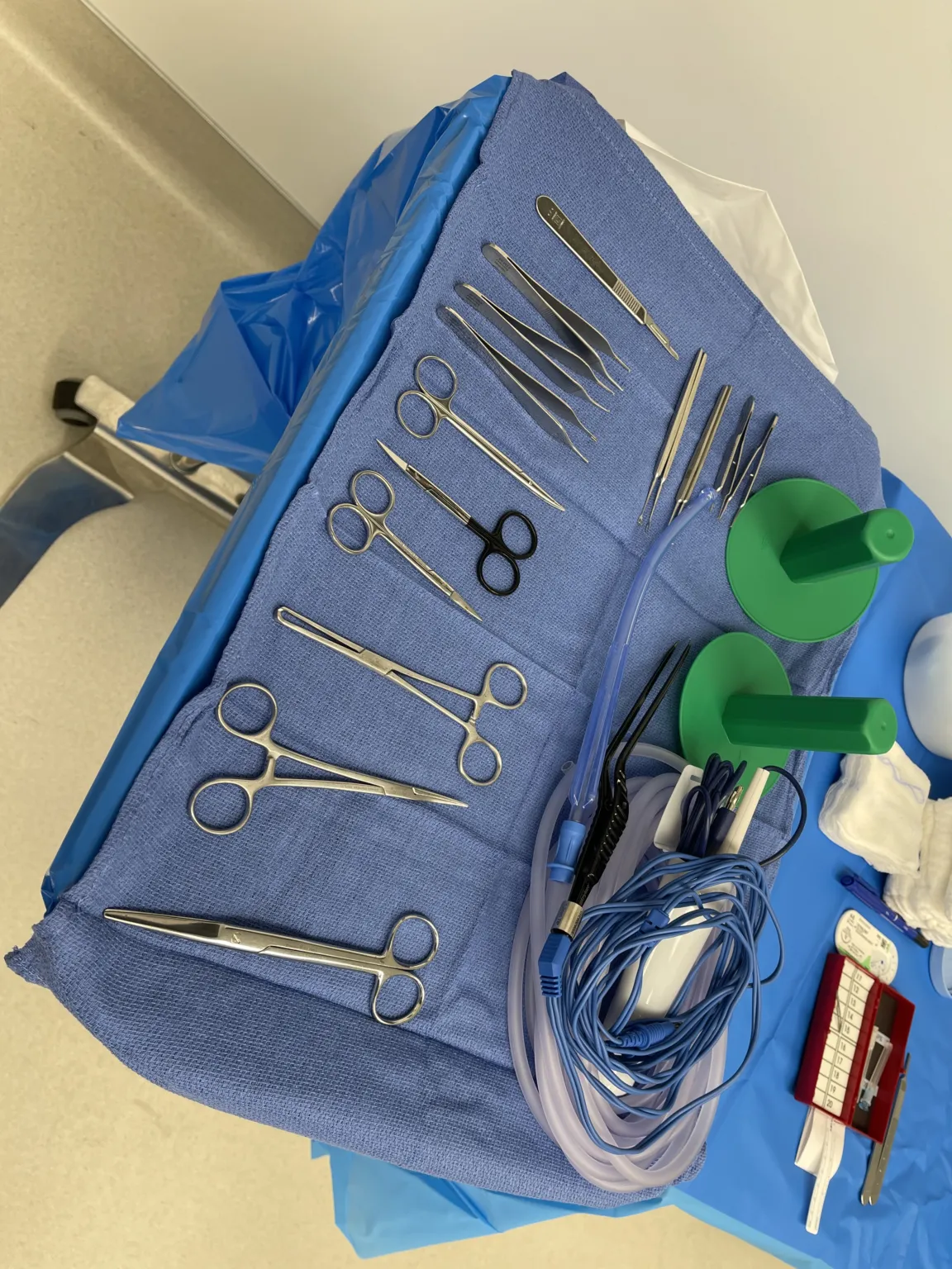
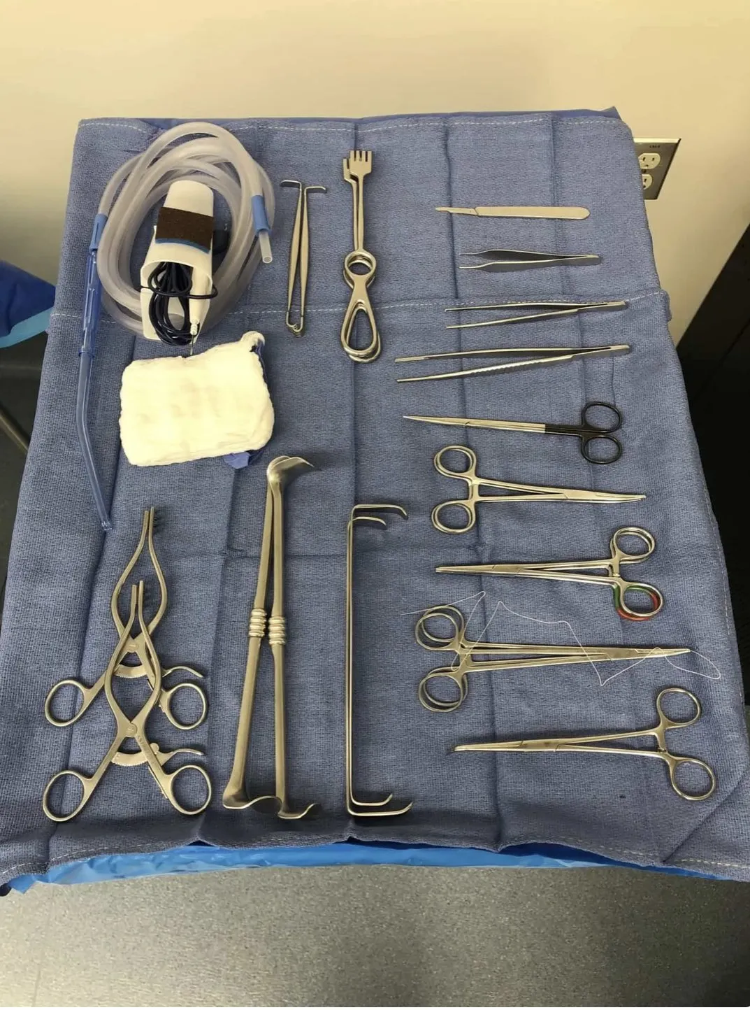
Surgical instruments
24 on this table. Tap a photo to zoom, the name for its card.
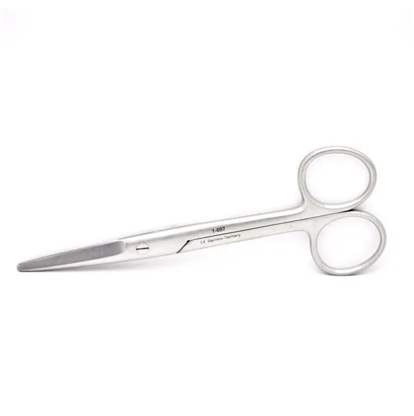
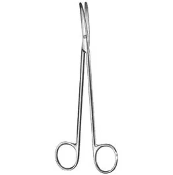
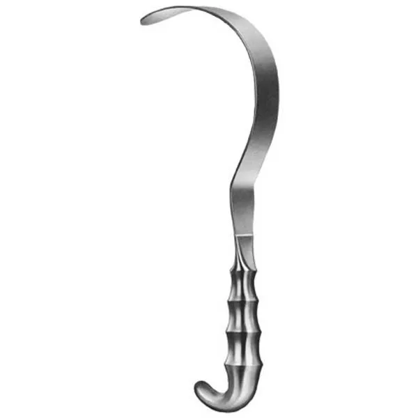
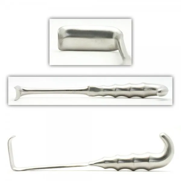
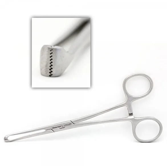
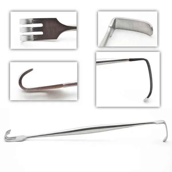
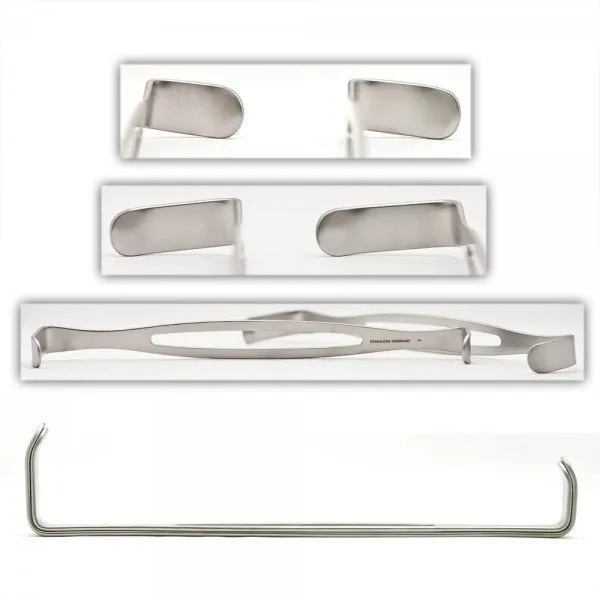
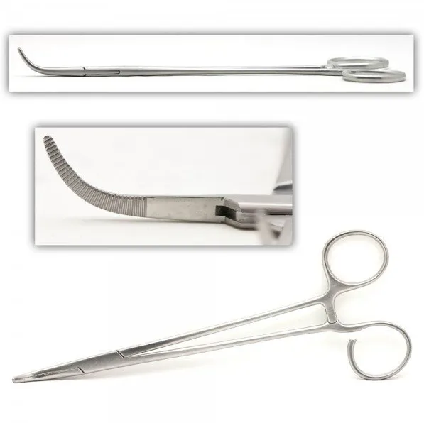
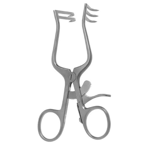
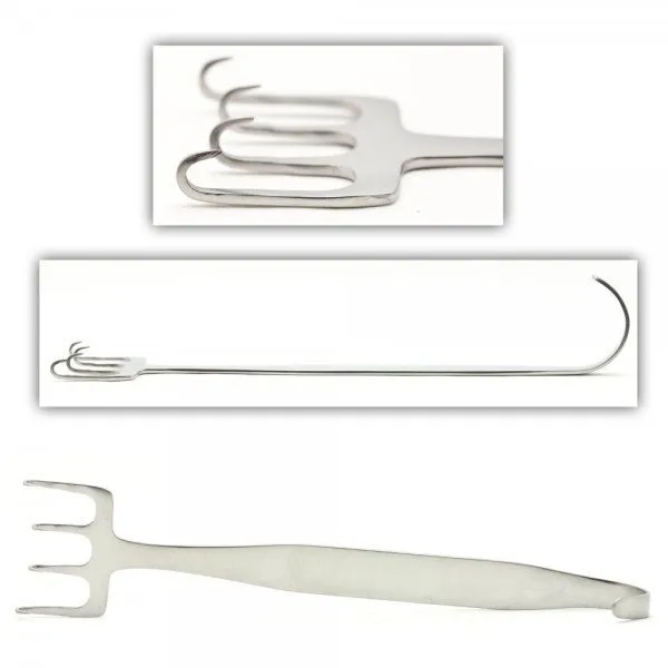
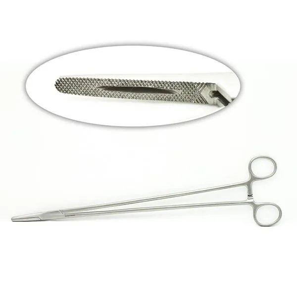
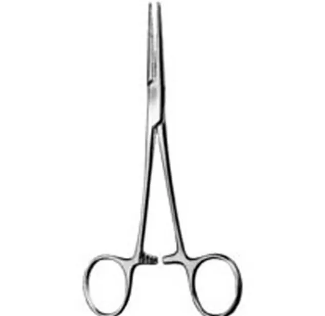
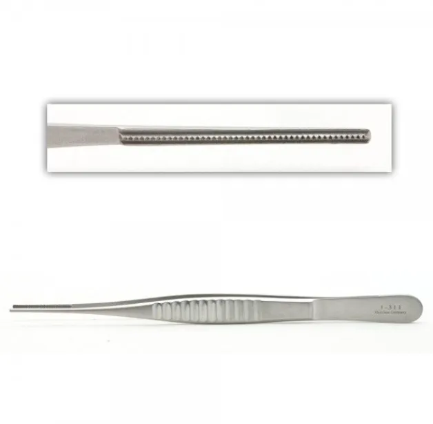
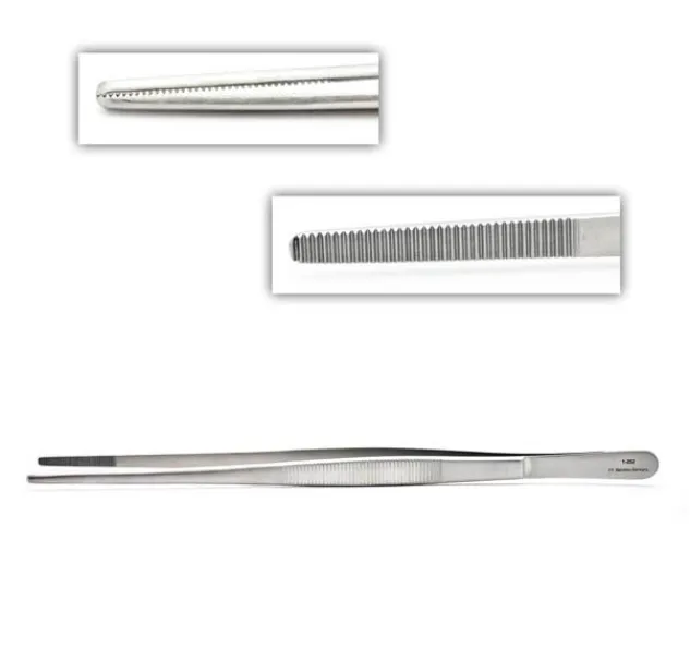
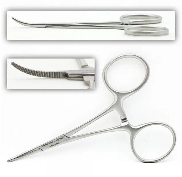
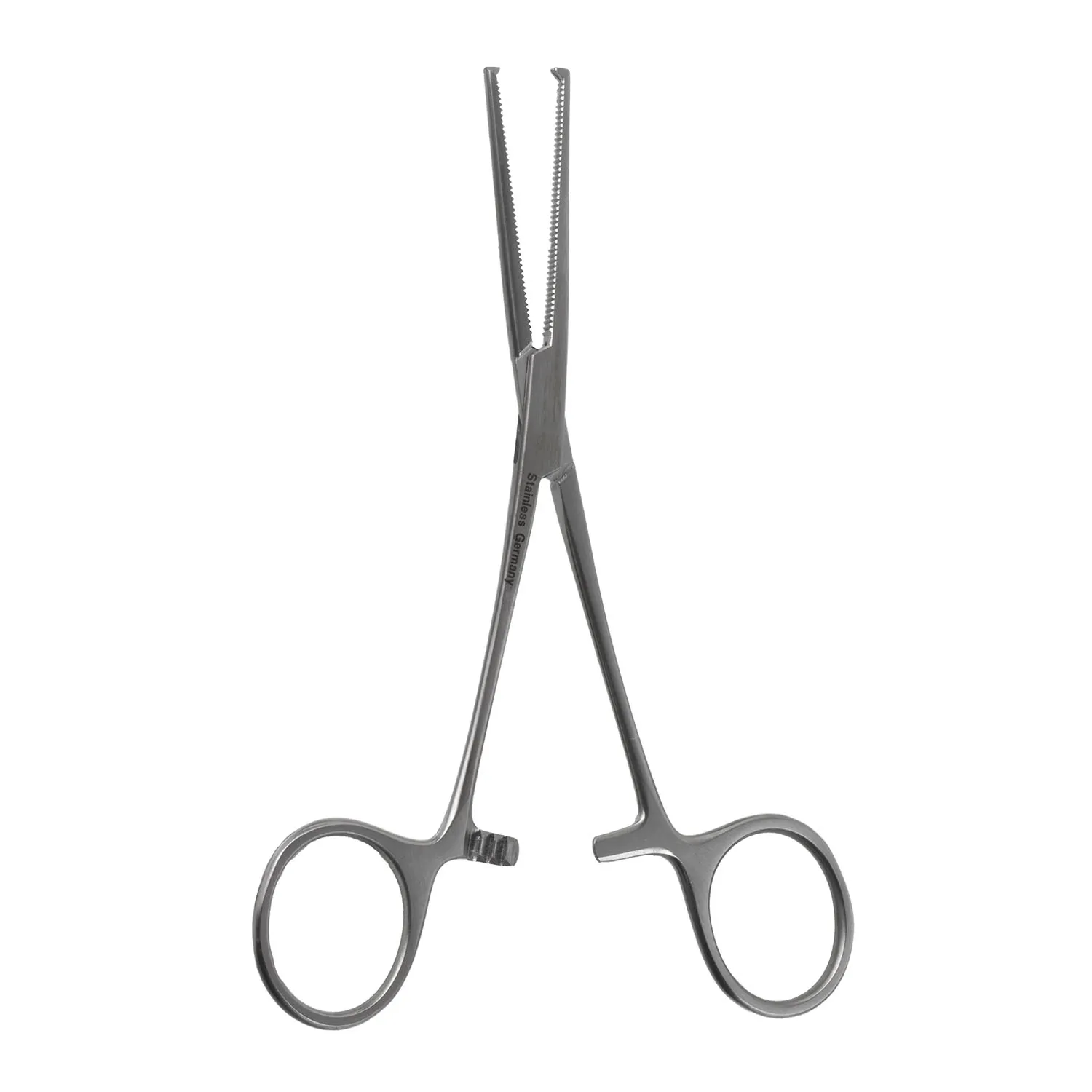
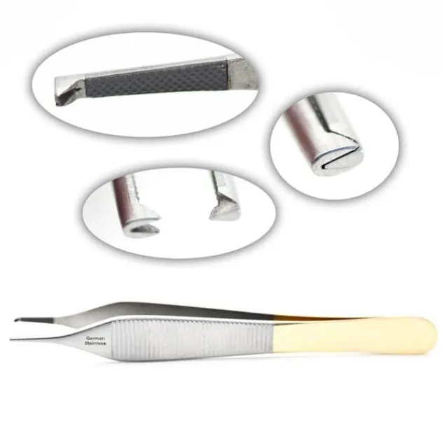
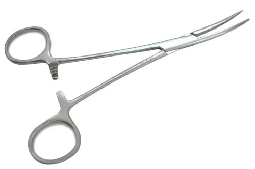
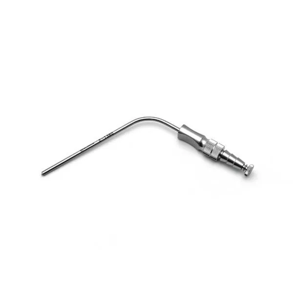
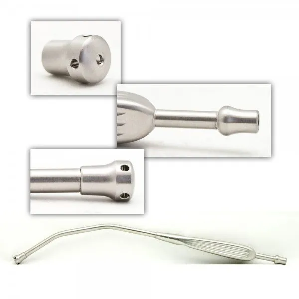
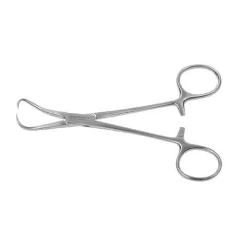
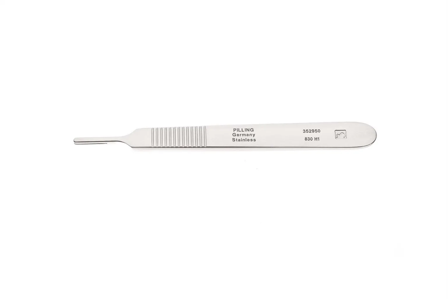
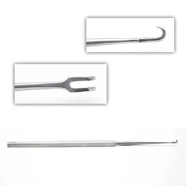
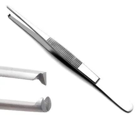
The case
Spark notes
The 30-second rundown. Scan this outside the room.
- Make incision with 15 or 10 blade
- Retract with senns, rakes, skin hooks or army-navys, Richardsons for larger incisions
- Dissect with cautery, DeBakey forcep, and Metzenbaum scissors
- Use Weitlaner to retract when lesion is in view
- Grasp lesion with Allis clamp and remove it
- Place making/orientation stitch with needle driver, adsons, and straight mayo scissors
- Send specimen to pathology and consider using a drain for larger incisions
Walkthrough
What the surgeon does, and what to have in your hand before they ask.
Incision
The doctor will make an incision near the location of the biopsy, using a 10 or 15 blade depending on the size of the incision needed.
Retraction
For small incisions, use senns, rakes, or skin hooks for retraction. For larger incisions, use army-navys or Richardsons.
Dissection
The doctor will dissect down to reach the lesion, using cautery, DeBakey forcep, and Metzenbaum scissors.
Retraction
Once dissected down, use a Weitlaner to retract the lesion into view.
Clamping
An Allis clamp may be used to grasp the lesion and remove it.
Stitching
After removing the lesion, a making/orientation stitch is usually placed using a needle driver, adsons, and straight mayo scissors.
Pathology
The specimen is then sent to pathology for analysis.
Drain
If the incision was bigger, a drain may also be used at the end.
Tips and tricks
Always make a note of the side and location of the specimen, as well as where the orientation stitch is placed, and double check specimen sheets.
Formalin is usually not used on breast biopsies.
Add your table
Every surgeon runs it a little differently. Submissions stay private, are reviewed by our team, and accepted contributions earn a credit in the contributor list.