Lumpectomy
This breast-conserving surgery involves the wide local excision of a primary tumor along with a surrounding margin of normal breast parenchyma. The goal is to remove the malignancy while preserving the majority of the breast tissue and appearance.
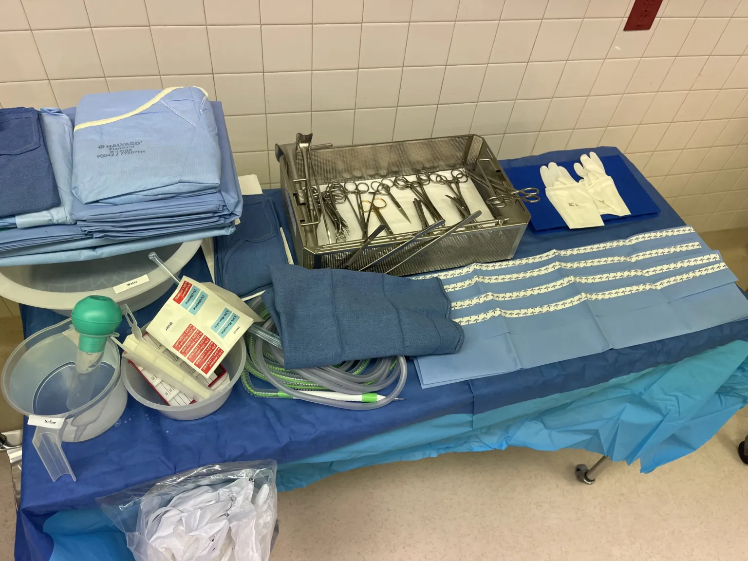
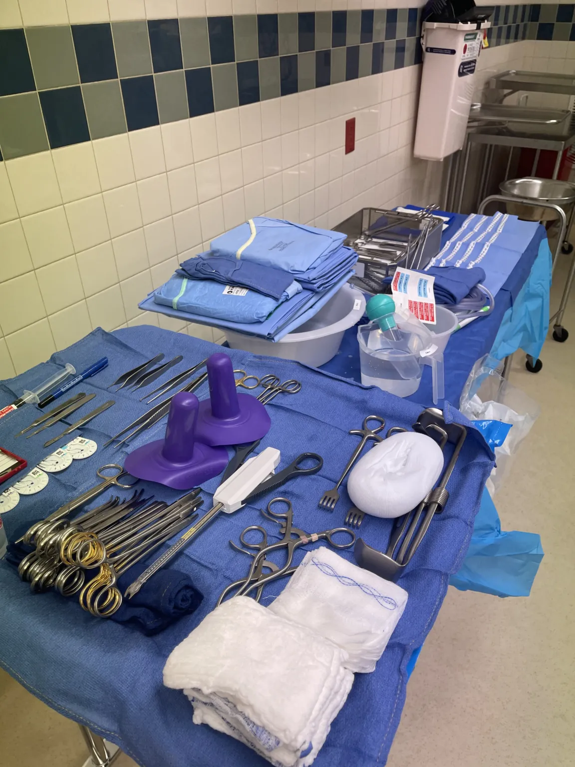
In 30 seconds
Scan this outside the room.
- Incision: Use a 15 blade loaded on a #3 knife handle. Senn retractorrih-TRAK-terA surgical instrument that holds back tissue or organs so the surgeon can see and reach the operative site.Full entry , skin hooks or army-navys may be used.
- dissectiondy-SEK-shunA tear in the inner layer of an artery wall that lets blood split the layers apart; also, the cutting apart of tissue in surgery.Full entry : Use Adson pickups, metzenbaum scissors, and a bovie to dissectdy-SEKT, or dih-SEKTTo cut apart or separate tissues carefully during surgery.Full entry the tissue. Remove the lump with an Allis clamp or kocher.
- Marking the lump: Mark with a directional stitch for orientation.
- Specimen handling: Quickly pass off the specimen to the circulator who will take it to pathologypuh-THOL-uh-jeeThe study of disease and its causes and effects, or the disease changes found in a person or tissue.path/o disease-logy study ofFull entry . Margins may be taken if needed.
The tray
21 instruments, by what they do. Tap a name for its card.
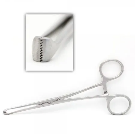 Allis ForcepsClamping
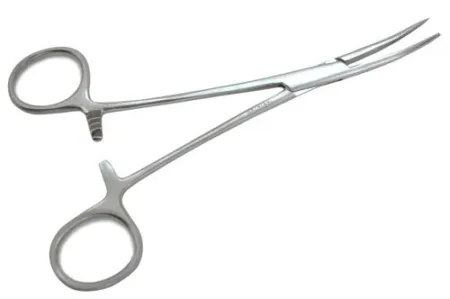 Curved HemostatsClamping
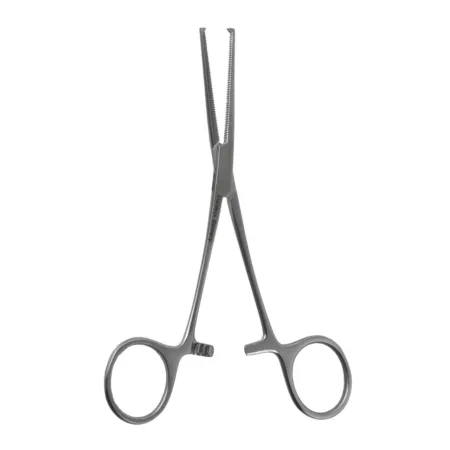 Kocher ForcepsClamping
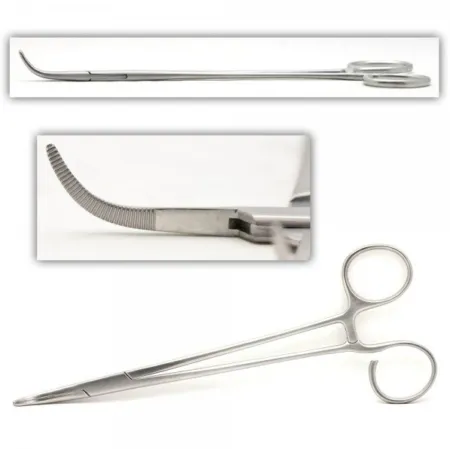 Tonsil ClampClamping
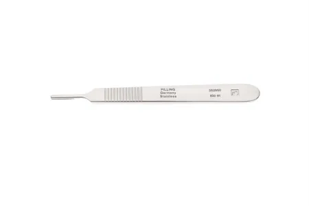 #3 Knife HandleCutting
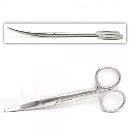 Curved Mayo ScissorCutting
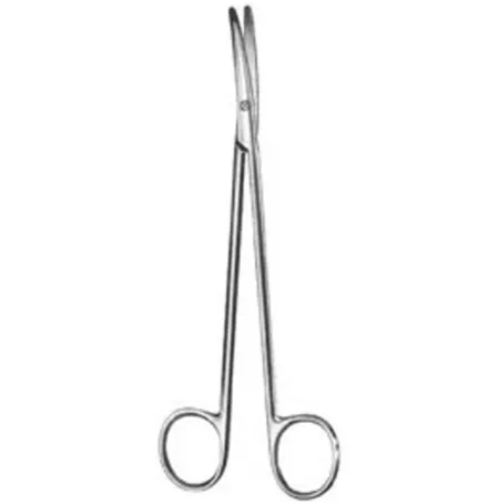 Metzenbaum ScissorCutting
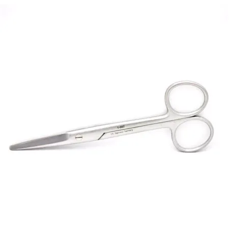 Straight Mayo ScissorCutting
All 21 instruments
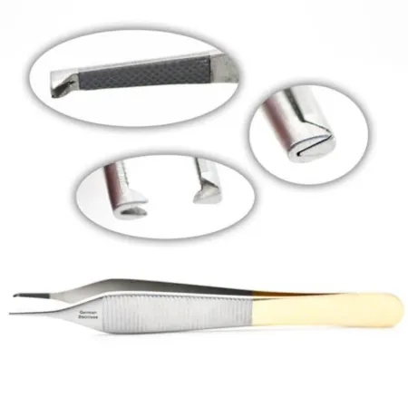 Adson ForcepsGrasping
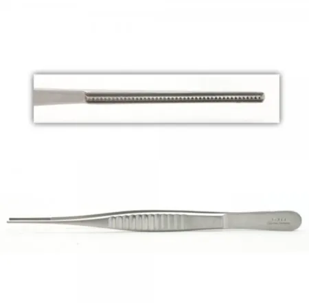 DeBakey ForcepsGrasping
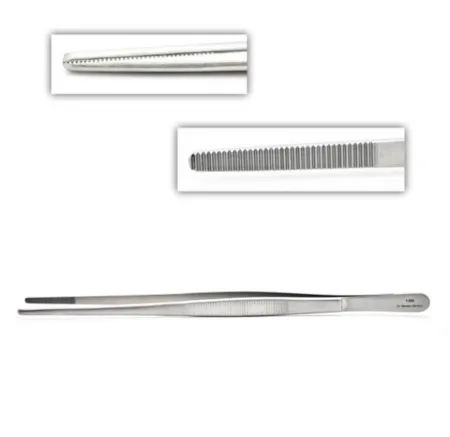 Non-Toothed PickupGrasping
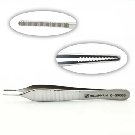 Smooth AdsonGrasping
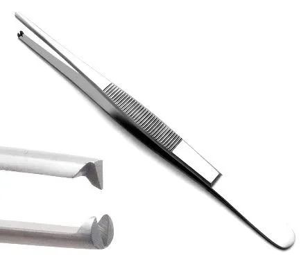 Toothed ForcepsGrasping
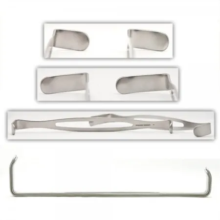 Army-Navy RetractorRetracting
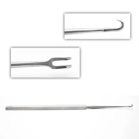 Freer Skin HookRetracting
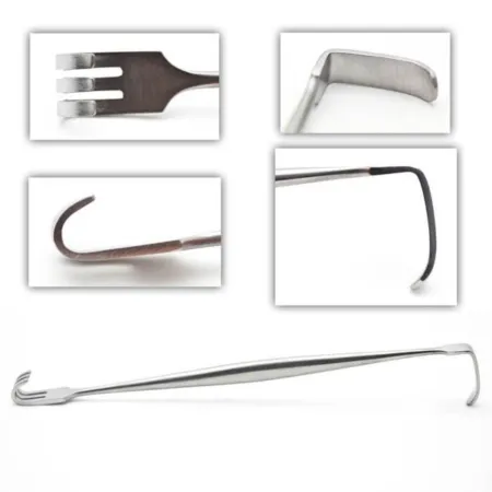 Senn RetractorRetracting
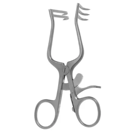 Weitlaner RetractorRetracting
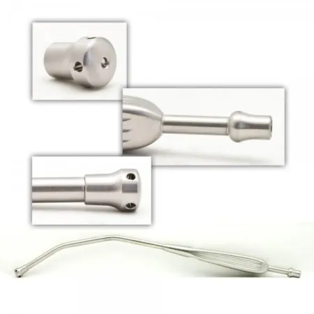 Yankauer SuctionSuctioning
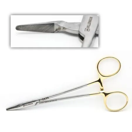 Crile Wood Needle HolderSuturing
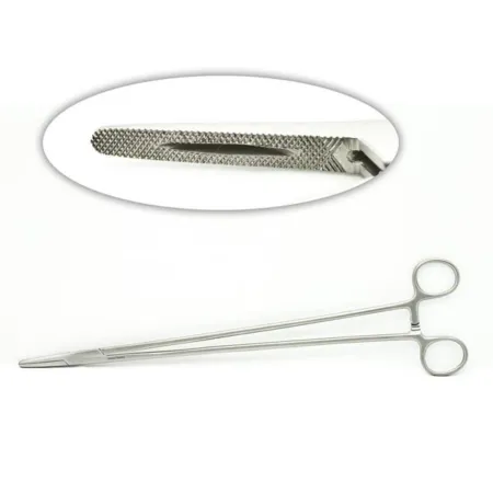 Mayo-Hegar Needle HolderSuturing
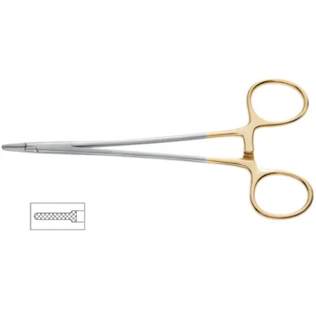 Ryder Needle HolderSuturing
Clamping & Occluding4
Cutting & Dissecting4
Grasping & Holding5
Retracting & Exposing4
Suctioning1
Suturing & Stapling3
Know this tray before you scrub in.
With Pro, for every setup in every specialty. Cancel any time.
Or scrub in free: every General setup and 5 questions a day.
Step by step
4 steps, one line each.
- 1
Incision
The surgeon will begin the Lumpectomy procedure by making an incision over the lump being removed. They will use a 15 blade loaded on a #3 knife handle to make the incision. The incision made during a Lumpectomy is typically small, so senn retractorrih-TRAK-terA surgical instrument that holds back tissue or organs so the surgeon can see and reach the operative site.Full entry or skin hooks may be used at the beginning to hold the incision open. If the tissue is deeper, army-navys might be used.
- 2
Dissection
After making the incision, the surgeon will use Adson pickups, metzenbaum scissors, and a bovie to dissectdy-SEKT, or dih-SEKTTo cut apart or separate tissues carefully during surgery.Full entry the tissue. These instruments are crucial during the Lumpectomy procedure as they are used to carefully separate the lump from surrounding tissues without damaging the tissue. The lump will be grasped by either an Allis clamp or kocher and removed from the incision.
- 3
Marking the lump
Once the lump is removed, the surgeon will mark it with a directional stitch to ensure that it is properly oriented. This stitch helps the pathologist to identify the location of the lump and determine if there are any cancerous cells present.
- 4
Specimen handling
After marking the lump, you will pass off the specimen to the circulator quickly. The circulator will then take it to the pathologypuh-THOL-uh-jeeThe study of disease and its causes and effects, or the disease changes found in a person or tissue.path/o disease-logy study ofFull entry lab for examination. If the pathologist determines that more tissue needs to be taken, margins may be taken to ensure that all cancerous cells are removed.
The case
A Lumpectomy is a surgical procedure that involves the removal of a breast lump and a small portion of surrounding tissue. It is a common treatment for breast cancer and is used to remove cancerous tissue while preserving as much of the healthy breast tissue as possible. The procedure is typically done under general anesthesia and can be performed on an outpatient basis.
Built by the techs who scrub it.
Every surgeon runs it a little differently. Send your table, or a fix for this page.
 Jerry GannReviewer
 Jessica CarrPhotos
The rest of this setup is Pro.
Everything unlocked above stays free. Pro opens the rest, on every setup: where new techs slip, what to load next at every step, the circulator’s side, the medicines, the specimen, wound class and trays on standby, what to grab if it goes wrong, and practice for every tray.
Free, for good. No card.
- All 2,882 instrument cards
- Every General setup in full
- 5 exam questions a day
- Your own preference cards
Pass your exam. Know every case.
Everything in Free, plus:
- All 260 setups in every specialty: back table, Mayo and every step
- Practice that never runs out and full timed exams for the CST, TS-C, CRCST, CSPDT and CSFA, with the why behind every answer
- Flashcards on any tray, timed to come back before you forget
- Every instrument card in full: the 3D model, cautions, inspection, how to pass it
Cancel any time from your account. Not worth it? Email us within 30 days of a charge for a refund, one per customer. Refund policy
Pro is also in the CSTSetup app, on the same account. Already have Pro? Log in
Send your table
Your Mayo, your back table, your surgeon’s way, or anything wrong on this page. Your email is only used to credit you. Submissions stay private and are reviewed by our team; accepted ones earn a Contributor credit here.