Robotic Partial Nephrectomy
The robot-assisted surgical removal of a localized tumor from the kidney while maintaining the integrity and function of the remaining healthy renal tissue.
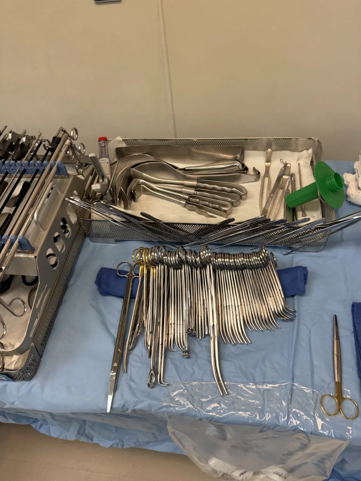
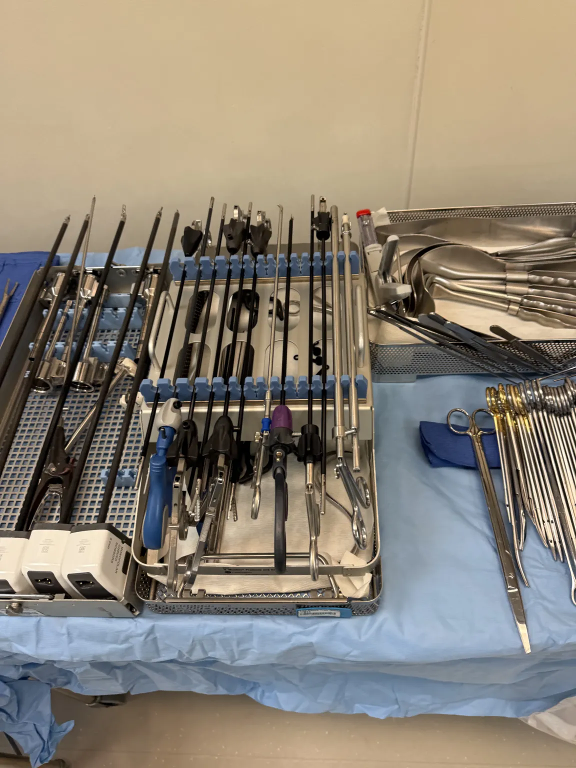
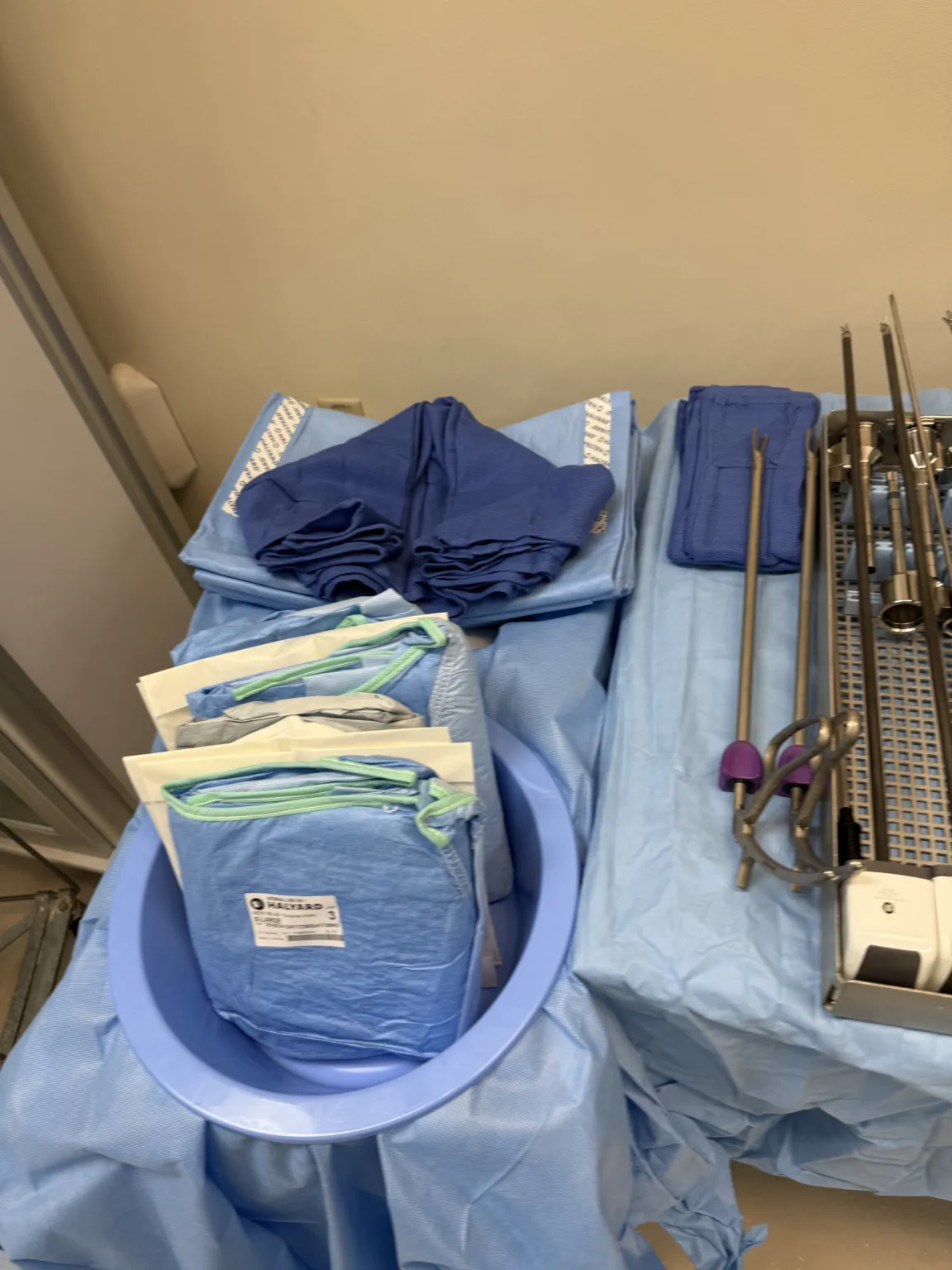
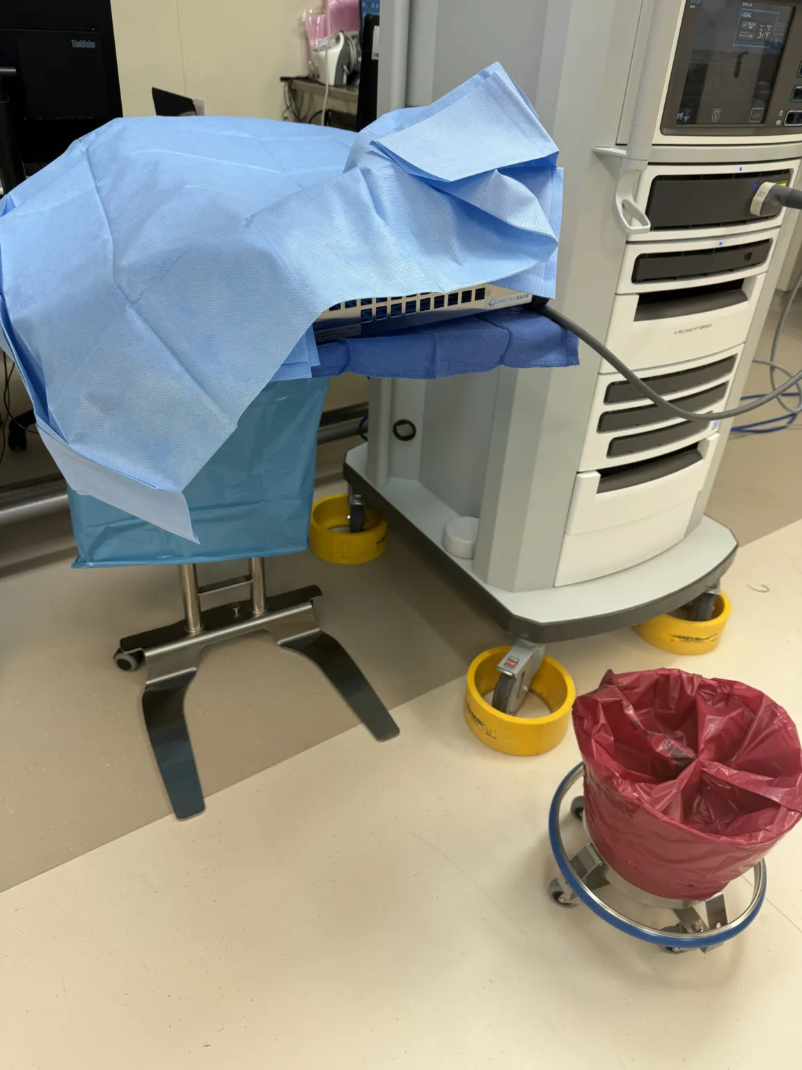
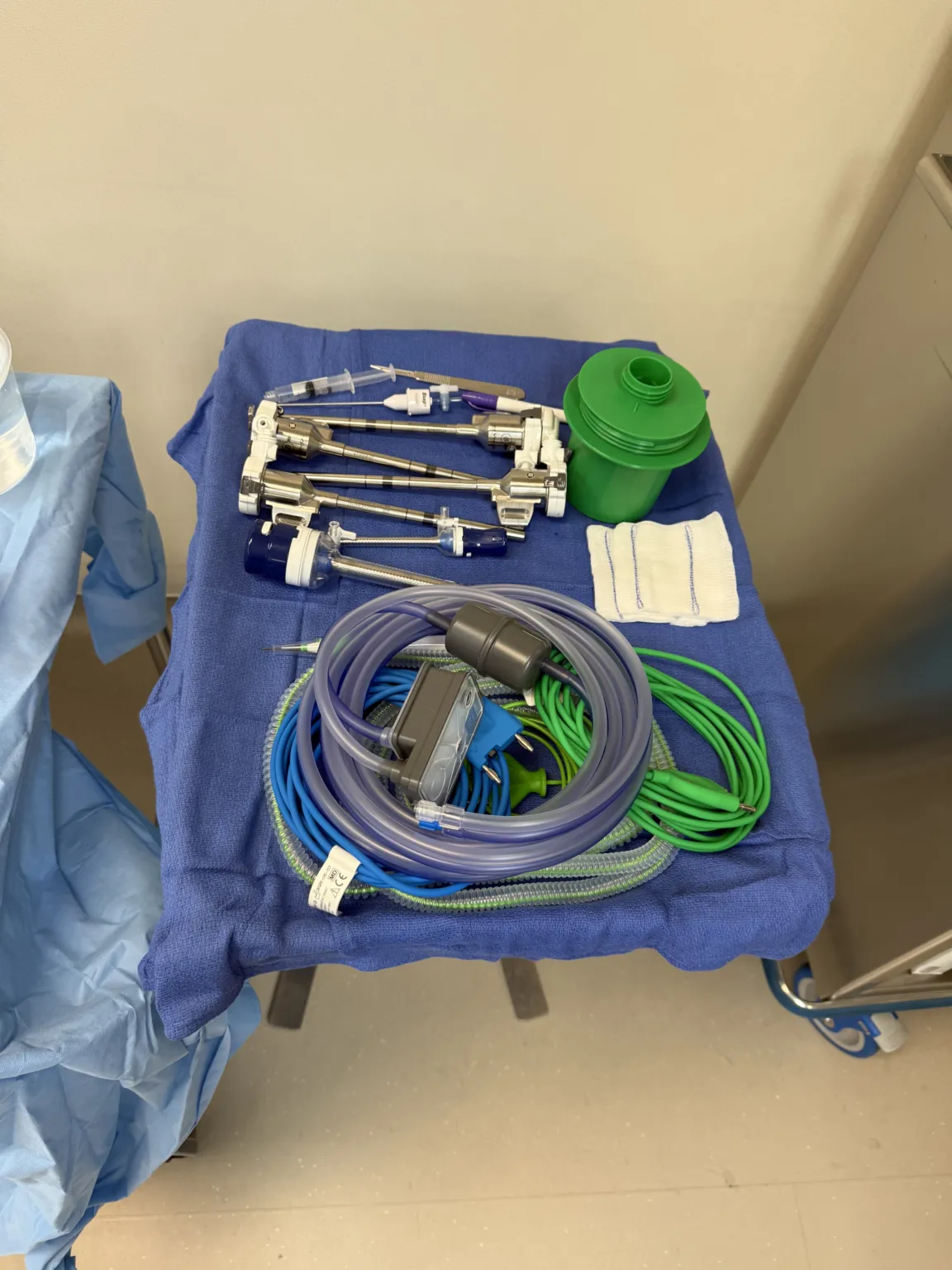
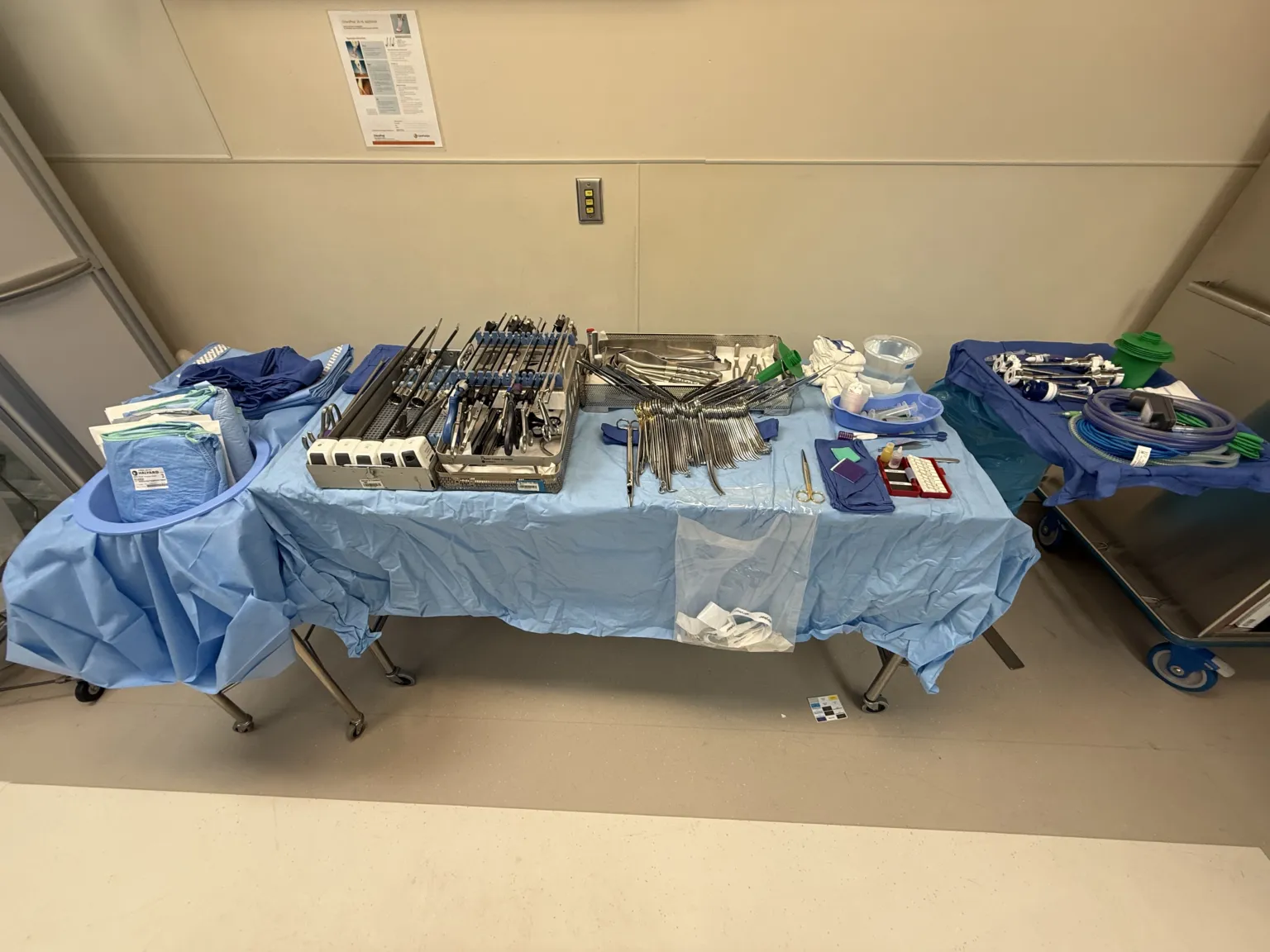
In 30 seconds
Scan this outside the room.
- Assemble robotic trocarTROH-karA sharp or bladed instrument inside a tube (cannula), used to pierce a body wall and leave the tube in place as a port.Full entry .
- Swap instruments as needed.
- Identify bleeders and control bleeding.
- Excise the tumor and use a robotic needle driver with sutureSOO-cherA stitch, or the thread used to stitch, that holds the edges of a wound or incision together.Full entry loaded to help reconstruct.
- Remove the specimen with a bag in the abdomen.
- Close incisions using a mayo-hegar needle driver, straight mayo scissors, and adsons.
The tray
9 instruments, by what they do. Tap a name for its card.
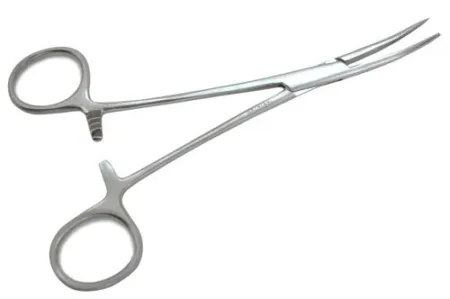 Curved HemostatsClamping
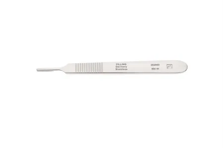 #3 Knife HandleCutting
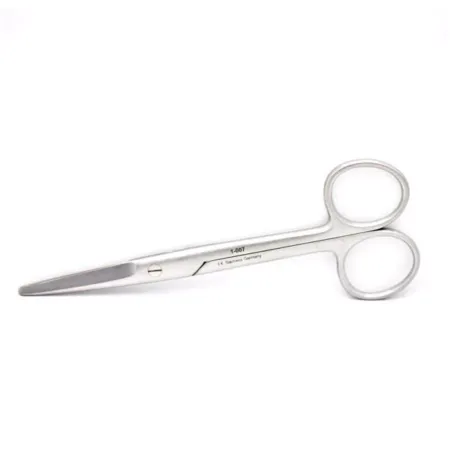 Straight Mayo ScissorCutting
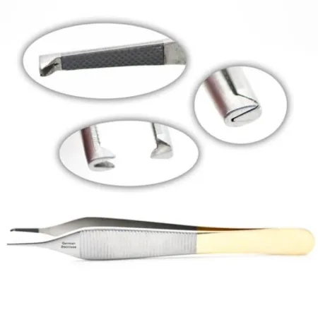 Adson ForcepsGrasping
 Camera HeadSpecialty
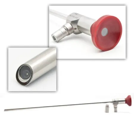 LaparoscopeSpecialty
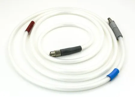 Light CordSpecialty
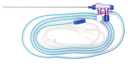 Suction IrrigatorSuctioning
All 9 instruments
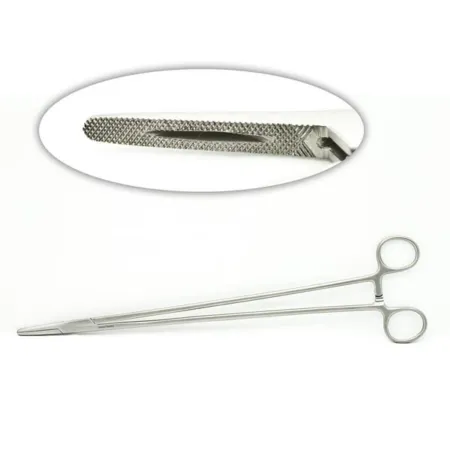
Clamping & Occluding1
Cutting & Dissecting2
Grasping & Holding1
Specialty3
Suctioning1
Suturing & Stapling1
Know this tray before you scrub in.
With Pro, for every setup in every specialty. Cancel any time.
Or scrub in free: every General setup and 5 questions a day.
The room
The equipment, the prep and drape, the suture and the medicines.
- Regular camera
- light cord
- and scope
The robot needs to be draped before the surgery begins, and all staff should be aware of how the robot works.
Step by step
6 steps, one line each.
- 1
Preparing for Surgery
Before the surgery begins, it is essential to assemble the robotic trocarTROH-karA sharp or bladed instrument inside a tube (cannula), used to pierce a body wall and leave the tube in place as a port.Full entry , which are kept in the robot sets. The surgeon will use a regular camera, light cord, and scope to make their initial incisions with either a 15 or an 11 blade attached to a #3 knife handle. Once the surgeon is satisfied with the port placements, they will break scrub and run the robot. At this point, the surgical technologistSUR-jih-kul tek-NOL-uh-jistA member of the operating room team who prepares the sterile field and instruments and hands them to the surgeon.Full entry will play a vital role in the procedure.
- 2
Swapping Instruments
During the surgery, the surgeon will require several instruments to perform the procedure. The laparoscope will be swapped out for the robotic scope. Additionally, grasping instruments, such as scissors and cautery, will be needed to mobilize the bowel, and any attachments on the liver will need to be dissectdy-SEKT, or dih-SEKTTo cut apart or separate tissues carefully during surgery.Full entry . The ureterYOOR-uh-terOne of the two tubes that carry urine from the kidneys down to the bladder.Full entry will be identified and lifted to create an area to locate the renal hilum to dissect. The tech will need to be aware of what instruments the surgeon needs and swap them out as necessary.
- 3
Identifying Bleeders
To control any bleeding during the surgery, laparoscopiclap-uh-roh-SKOP-ikDone through a few small cuts in the abdomen with a thin camera and long instruments, instead of one large opening.lapar/o abdomen-scopic viewingFull entry bulldog clamps may be used to clamp the renal hilum. Arteries and veins will also need to be clamped. During this step, it is important to ensure that all bleeders are identified and controlled to prevent complications.
- 4
Excising the Tumor
Once the bleeding is controlled, the surgeon will excise the tumor. A robotic needle driver with sutureSOO-cherA stitch, or the thread used to stitch, that holds the edges of a wound or incision together.Full entry loaded may be used to help reconstruct the excisionek-SIZH-unCutting out tissue, such as a growth or damaged area, during surgery.Full entry area. The tech will need to be prepared to provide the necessary instruments for this step.
- 5
Removing the Specimen
After the tumor has been excisionek-SIZH-unCutting out tissue, such as a growth or damaged area, during surgery.Full entry , the clamps can be taken off, and a bag will be placed in the abdomen to take out the specimen. The tech will need to assist in removing the specimen.
- 6
Closing the Incisions
Once the specimen has been removed, hemostasishee-moh-STAY-sisThe stopping of bleeding, either by the body's own clotting process or by medical or surgical methods.hem/o blood-stasis stopping, standing stillFull entry is ensured, and the incisions can be closed. Closing the incisions will require a mayo-hegar needle driver, straight mayo scissors, and adsons. The tech will need to provide these instruments as necessary.
The case
A Robotic Partial Nephrectomy is a minimally invasive surgical procedure that involves using a robotic system to remove part of a kidney affected by cancer while preserving the remaining healthy tissue. The surgery requires precise coordination between the surgical team, with the surgical tech playing a vital role in providing the necessary instruments during the procedure.
Da Vinci instruments will be needed for this surgery since it is robotic. Make sure to always have regular laparoscopic instruments close by in case the surgeons decides to not do the case robotically.
Built by the techs who scrub it.
Every surgeon runs it a little differently. Send your table, or a fix for this page.
 Diana Lei-alohaReviewer
 Anjanete PerezPhotos
The rest of this setup is Pro.
Everything unlocked above stays free. Pro opens the rest, on every setup: where new techs slip, what to load next at every step, the circulator’s side, the medicines, the specimen, wound class and trays on standby, what to grab if it goes wrong, and practice for every tray.
Free, for good. No card.
- All 2,882 instrument cards
- Every General setup in full
- 5 exam questions a day
- Your own preference cards
Pass your exam. Know every case.
Everything in Free, plus:
- All 260 setups in every specialty: back table, Mayo and every step
- Practice that never runs out and full timed exams for the CST, TS-C, CRCST, CSPDT and CSFA, with the why behind every answer
- Flashcards on any tray, timed to come back before you forget
- Every instrument card in full: the 3D model, cautions, inspection, how to pass it
Cancel any time from your account. Not worth it? Email us within 30 days of a charge for a refund, one per customer. Refund policy
Pro is also in the CSTSetup app, on the same account. Already have Pro? Log in
Send your table
Your Mayo, your back table, your surgeon’s way, or anything wrong on this page. Your email is only used to credit you. Submissions stay private and are reviewed by our team; accepted ones earn a Contributor credit here.