Anesthesia
How the team takes away pain and awareness for surgery, watches the patient through it, acts fast when anesthesia goes wrong, and hands the patient on to recovery.
1Why it matters
Anesthesia means "without sensation." Its goal in surgery is to let the patient get through the operation without pain while the body stays in a safe, balanced state, called homeostasis. Many of today's operations are possible only because of it.
Anesthesia is also one of the most dangerous parts of any operation. The drugs that take away pain and awareness also take away the reflexes that keep a person alive: breathing, swallowing, coughing, blinking and shivering. The anesthesia provider watches those functions continuously, and the whole team helps.
You have a part in it. You keep the room quiet at induction, hand the anesthesia provider what they need, apply cricoid pressure when asked, track every local anesthetic on your field, warm the patient, and know your job in a cardiac arrest, malignant hyperthermia or a severe allergic reaction. AST asks surgical technologists to know the signs of these emergencies and to act as another pair of eyes in the room.
This chapter starts with the words and the people of anesthesia, then the patient's assessment, the types of anesthesia and the drugs given before surgery. Then come the equipment and the monitors, the vital signs and their normal values, general anesthesia from induction to emergence, sedation, and regional and local anesthesia. It ends with your role, keeping the patient warm, the child under anesthesia, the emergencies, and the recovery room (PACU).
Other chapters teach what it leaves out: the neuromuscular junction, and how malignant hyperthermia starts in the muscle cell, in chapter 2; the autonomic nervous system in chapter 4; the heart's electrical system in chapter 5; the airway's anatomy in chapter 6; latex allergy and how the room is made latex-safe in chapter 10; shock as a disease process in chapter 11; how drugs work, their names, routes, dose math, blood products and safe handling on the field in chapter 12; the time-out, the counts and the chart review in chapter 28; dressings and moving the patient out of the room in chapter 30; the steps for taking vital signs, urinary catheters, positioning and the tourniquet cuff in chapter 31; the care of children as people in chapter 18; and the fire risk of oxygen in chapter 20.
2The words of anesthesia
Anesthesia works in two ways: it changes the patient's level of consciousness, or it stops the nerves from carrying sensation, or both. These words describe what is happening.
- Consciousness: being aware of the surroundings and able to respond to them, through sight, sound, touch, pressure, pain, heat and cold. It runs on a scale from fully awake to completely unresponsive.
- Sensation: being able to feel a stimulus, such as pain, heat, touch or sound.
- Analgesia: the absence of pain. Pain travels on its own nerve fibers, separate from those for pressure and temperature, and analgesic drugs interrupt those pain pathways.
- Amnesia: being unable to remember. Some anesthesia drugs cause it on purpose, so the patient recalls nothing of the events after the drug is given; memory returns once the drug is cleared.
- Sedation: a lowered level of awareness, on a scale from relaxed and fully responsive to unconscious. Central nervous system (CNS) depression is another way to say it.
- Unconsciousness: a complete inability to respond to the outside world. General anesthesia produces an unconsciousness that can be reversed. Coma is the deepest unconsciousness, with most brain activity stopped.
- Protective reflexes: the body's automatic responses to harm, such as coughing, gagging, swallowing, blinking, shivering and pulling away from pain. Deep unconsciousness takes them away.
- Apnea: no breathing at all.
- Anxiolytic: a drug that eases anxiety.
- Delirium: confusion and disorientation.
What a general anesthetic must do
A good general anesthetic gives the patient several things at once, and different drugs supply each part:
- Hypnosis: sleep, a loss of consciousness that can be reversed
- Analgesia: freedom from pain
- Amnesia: no memory of the operation
- Muscle relaxation: so the surgeon can retract the body wall and the airway tube can be placed
- Reflex control: the reflexes are dulled, some autonomic reflexes among them
- Homeostasis: the heart, lungs, temperature and fluids kept stable all the while
The anesthesia provider combines drugs to build these parts, because one drug given in a dose big enough to do all of them alone would be dangerous. This mix of IV drugs and inhaled agents, tailored to the patient and the operation, is called balanced anesthesia.
3The anesthesia team
The anesthesia provider (AP) is the general name for the licensed professional who gives the anesthetic and manages the patient through it. In the United States, anesthesia care is given by an anesthesiologist, by a certified registered nurse anesthetist, or by an anesthesiologist assistant under an anesthesiologist's supervision.
- Anesthesiologist: a physician who specializes in anesthesia and pain management.
- Certified registered nurse anesthetist (CRNA): an advanced-practice nurse licensed to give anesthesia. Nurse anesthesia programs are at the graduate level, and new students now graduate with a doctoral degree. In many small rural hospitals, a CRNA is the only anesthesia provider.
- Anesthesiologist assistant, also called a certified anesthesia assistant (CAA): a professional with a master's degree who works under an anesthesiologist's supervision. They take histories, place monitors, draw blood samples, give drugs and perform regional techniques, as state law allows.
- Anesthesia technician or certified anesthesia technologist: an allied health professional who assists the AP, keeping the anesthesia and monitoring equipment working, preparing drugs and supplies, and helping during the anesthetic.
The AP's job is to protect the patient's vital functions and control their consciousness and pain. They relax the muscles for general anesthesia, give sedation during regional anesthesia, tell the surgeon how the patient is responding (blood pressure, fluids, muscle relaxation, level of consciousness), respond to any emergency and support the patient emotionally. Their care begins when the patient arrives in the surgical suite and continues into the PACU, where they remain available for problems with the anesthesia and for pain.
The anesthesia team guards its work area the way you guard your back table. The drug and equipment carts by the head of the table, one usually for airways and others for drugs, syringes, needles and spinal sets, are for the anesthesia team only.
4Before anesthesia: the patient's assessment
Before surgery, the surgeon and the AP (or another qualified person, such as a nurse practitioner) assess the patient, usually a week to a few days ahead, often in a preadmission clinic. The aim is to find the patient's risks for anesthesia and to plan the type of anesthetic. It is also the patient's chance to ask questions, and good teaching here eases many fears about pain and waking up. In an emergency, the assessment happens just before the operation, sometimes in the holding area; at the very least, the baseline vital signs and the airway are checked.
The ASA physical status classes
The American Society of Anesthesiologists (ASA) sorts patients by how sick they are before surgery, which helps predict the risk of anesthesia. The AP assigns the class, but every team member should know what the classes mean. The disease that sets the class may or may not be the reason for the surgery.
| ASA class | The patient | Examples |
|---|---|---|
| ASA 1 | A normal, healthy patient | No disease at all |
| ASA 2 | Mild systemic disease, no real limit on daily activity | Well-controlled high blood pressure or diabetes, mild asthma, obesity, smoking |
| ASA 3 | Severe systemic disease that limits activity | Diabetes with complications, a past heart attack, poorly controlled high blood pressure, morbid obesity, kidney failure on dialysis |
| ASA 4 | Severe systemic disease that constantly threatens life | Severe heart failure, an acute heart attack, respiratory failure on a ventilator |
| ASA 5 | A dying patient not expected to survive 24 hours with or without the operation | A ruptured aneurysm, major multisystem trauma |
| ASA 6 | A brain-dead patient whose organs are being recovered for donation | Organ recovery |
An E is added to the class for any emergency operation, so a healthy patient having an emergency appendectomy is ASA 1E. One reference lists only classes 1 to 5.
What the assessment covers
- Medical conditions: heart disease, high blood pressure, lung disease such as asthma and COPD, diabetes and other gland disorders, kidney and liver disease, blood disorders, nerve and muscle diseases, and conditions of the joints and connective tissue all change the anesthetic plan.
- Medications and supplements: prescription and over-the-counter drugs and herbal remedies can block or strengthen the drugs used in surgery, so the AP tells the patient which to keep taking and which to stop. Heart, blood pressure, seizure and asthma drugs are usually taken with a sip of water. Aspirin, other anti-inflammatory drugs, blood thinners and many herbal supplements may be stopped days before. A patient on long-term steroids keeps taking the usual dose; whether an extra stress dose is added is decided for each patient, from the steroid dose, how long it has been taken, and the type and length of the operation (chapter 9).
- Allergies: drug, latex, tape and food allergies are recorded on every record and asked about again in the holding area.
- Substance use: alcohol, tobacco and recreational drugs, which the patient stops at least 12 hours before surgery.
- Past anesthesia: any problem the patient or a blood relative had with anesthesia, such as poor drug clearance, a difficult airway or malignant hyperthermia, is a warning.
- The airway: the AP looks for anything that could block the airway or make the breathing tube hard to place. Loose or cracked teeth can break off, missing teeth make a mask hard to seal, and dental work can be damaged. A large tongue, a thick neck, a stiff neck or jaw, a high body mass index and piercings in the mouth or nose add risk. The Mallampati score grades how much of the throat shows with the mouth open, from class I (soft palate, uvula and pillars all visible) to class IV (only the hard palate visible); the higher the class, the harder the intubation is likely to be.
- The bones and joints: joint replacements, old injuries and nerve damage limit how the patient can be positioned.
- The mind and nerves: a baseline of thinking, speech, gait, strength and feeling, so a change after surgery can be recognized. Many patients fear the anesthesia and pain more than the operation itself.
- Home: whether someone can care for the patient after discharge.
- Age, height and weight: drug doses are often figured by weight, and the very young and very old need special care.
- Tests: the ASA does not call for routine tests on every patient. Tests such as an ECG, a blood count, kidney function and electrolytes are ordered to check a known risk.
Getting ready the day before and the morning of surgery
The patient prepares at home as the anesthesia team directs: bathing and washing the hair, removing all makeup and nail polish so the AP can see the skin and nail beds, doing any bowel prep the surgeon orders, and stopping alcohol, tobacco and recreational drugs.
Fasting before surgery lowers the risk of aspiration, breathing stomach contents into the lungs, under sedation or general anesthesia. For healthy patients having planned surgery, the ASA's minimum fasting times are 2 hours after clear liquids, 4 hours after breast milk, 6 hours after infant formula, other milk or a light meal, and longer, about 8 hours, after a full or fatty meal. The anesthesia team sets the time for each patient, because a long fast can dehydrate children and older adults. Chapter 31 covers NPO in the holding area.
On arrival, the patient's identity, procedure, side and site, consents, allergies and any resuscitation orders are checked; dentures, hearing aids and other prostheses come out when possible; and jewelry, including body piercings, is removed. Chapters 28 and 31 teach these checks.
5Choosing the anesthetic
The patient, the AP and the surgeon choose the anesthetic together. The patient must give informed consent to the anesthesia, including its risks and the other choices, before surgery can begin.
The types of anesthesia
- General anesthesia: a reversible, drug-induced unconsciousness, with no pain, no awareness and no memory. It is given by inhalation, by vein (intravenously) or both.
- Regional anesthesia: a local anesthetic blocks the nerves to one region of the body, so it loses feeling while the patient stays conscious. Spinal, epidural and caudal blocks, nerve blocks and the IV (Bier) block are regional techniques.
- Local anesthesia: an anesthetic put on or injected into one small area, by topical application or infiltration, usually by the surgeon. No anesthesia provider is involved.
- Monitored anesthesia care (MAC): an anesthesia provider gives sedation and pain drugs, often with local anesthetic from the surgeon, watches the patient's vital functions throughout, and is ready to convert to general anesthesia.
- Moderate sedation (conscious sedation): drugs that dull awareness while the patient keeps their own airway and responds to voice or touch. It may be given by a specially trained registered nurse without an anesthesia provider.
Two more routes, rectal and intramuscular, are used mainly to put children to sleep (see The child under anesthesia).
What decides the choice
- The patient: age, allergies, the ASA class and other diseases (such as diabetes, high blood pressure and heart disease), current medications and supplements, general health, height and weight, mental and emotional state, substance use, and whether it is an emergency.
- The procedure: how long it will take and the position the patient must be in. Very long operations and those in the abdomen and chest usually need general anesthesia; operations on the surface of the body and on the limbs can often be done with regional anesthesia.
- Preference: what the patient wants (awake, lightly sedated or fully asleep), the surgeon's needs and the AP's judgment.
The patient's safety always comes first. A patient may prefer general anesthesia, but a patient who is very ill may do better with a regional block or MAC. An older patient may be given IV sedation with local anesthesia, or a regional block, for an operation such as a hernia repair.
6Medications before surgery
Patients were once given heavy sedation on the ward before surgery and arrived in the OR barely awake. That slowed recovery and raised the risks to the heart and lungs, so it is no longer done. Today, drugs are given before surgery only for a reason: to ease anxiety, to prevent a problem the patient is at risk of, or to manage pain. A calm visit from the AP and the perioperative nurse also eases anxiety.
The Core Curriculum's list of preoperative medications falls into five groups. Chapter 12 teaches how drugs work and how they are handled.
| Group | Drugs | Why it is given |
|---|---|---|
| Opioid analgesics | Morphine (a natural opioid); alfentanil, fentanyl, meperidine, remifentanil and sufentanil (synthetic opioids) | Pain relief before an invasive procedure or a regional block |
| Antacids and H2-receptor blockers | Cimetidine, famotidine, sodium citrate with citric acid | Lower the amount and acidity of stomach acid, so aspiration does less harm |
| Anticholinergics | Atropine, glycopyrrolate, scopolamine | Prevent a slow heart rate, as in children and eye surgery, and dry up saliva and airway secretions |
| Antiemetics | Metoclopramide, ondansetron, promethazine | Prevent nausea and vomiting; metoclopramide also empties the stomach faster |
| Sedative-hypnotics | Diazepam, lorazepam, midazolam (benzodiazepines) | Ease anxiety and give amnesia |
- Midazolam is often given before surgery, to ease fear and cause amnesia.
- Scopolamine comes as a patch, put on before surgery to prevent nausea in patients at high risk; anyone who touches it wears gloves, and the patient takes it off after 72 hours.
- ASA no longer recommends giving anticholinergics just to lower the risk of aspiration.
Premedication may be given by mouth with a small sip of water, into a muscle, into a vein or into the nose. Before it is given, the surgical site is marked, and the AP answers the patient's last questions and completes the anesthesia checks.
7The equipment
The anesthesia machine
The anesthesia machine, or anesthesia workstation, delivers the inhaled anesthetics, oxygen and other medical gases, and holds a ventilator and the patient monitors. Learn its basic parts, because you may be asked to help with them.
- Gases: oxygen, nitrous oxide and medical air come from the wall or ceiling outlets, with portable cylinders as backup and for transport. The hoses are color coded, green for oxygen, blue for nitrous oxide and yellow for air, and each connector fits only its own gas.
- Vaporizers: every inhaled anesthetic except nitrous oxide is a liquid. The vaporizer turns it into a vapor and adds it to the gas flowing to the patient.
- The breathing circuit: corrugated tubing that does not kink carries the gases to the patient's airway and back, through one-way valves and a bacterial filter. The patient can be breathed for by the ventilator, or by hand with the breathing bag, the circuit's reservoir bag.
- The carbon dioxide absorber: a canister of soda lime traps the carbon dioxide the patient breathes out. A dye in it changes color as it fills, and the canister is then changed.
- The oxygen flush valve: sends pure oxygen straight to the patient at a very high flow.
- Scavenging: gas scavenging captures anesthetic gas that leaks from the machine and vents it away through a vacuum line. Breathing waste gas is a hazard for the team, and NIOSH and OSHA set limits on it; scavenging cuts the team's exposure by up to 95 percent.
Airway equipment
The airway is the passage the patient breathes through, natural or artificial. Keeping it open is the first priority under general anesthesia and in any emergency, because an unconscious patient cannot hold their own airway open. The Core Curriculum's list of airway equipment:
- Mask: a clear face mask delivers oxygen and anesthetic gas under pressure, before the tube goes in, for a short sedation, or to put a child to sleep. The plastic is clear so the AP can see secretions or vomit.
- Laryngoscope: a lighted instrument used to see the vocal cords and place the breathing tube. Its blade is curved (Macintosh) or straight (Miller). The AP holds it in the left hand and slides the blade into the right side of the mouth, sweeping the tongue aside. A video laryngoscope has a camera in its blade that shows the view on a screen.
- Endotracheal tube (ETT): a tube passed through the mouth (or, less often, the nose) into the trachea. A balloon cuff near the tip seals it against the tracheal wall, so gases go only to the lungs and stomach contents cannot get in. Chapter 6 traces its path.
- Stylet: a flexible, shapeable rod placed inside the tube to stiffen it while it is passed. It comes out as soon as the tube is in.
- Bougie: a long, flexible tube introducer that helps guide the tube into the trachea when the view is poor.
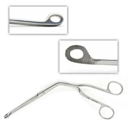
- Magill forceps (spelled McGill in the Core Curriculum): angled forceps the AP uses to grasp and guide the tube, as when a tube is passed through the nose.
- Laryngeal mask airway (LMA): a tube with a small mask on the end that sits over the larynx from above, placed without a laryngoscope. It does not protect the lungs from aspiration. It suits a patient breathing on their own for a short case, and it can rescue a difficult airway.
- Bite block: keeps the patient from biting down on the tube, most of all while waking. A roll of gauze beside an LMA can serve as one, and some LMAs have one built in.
- Oral airway (oropharyngeal airway, OPA): a curved device placed over the tongue to keep the tongue from falling back and blocking the throat, used in a patient who is unresponsive or only semiconscious, such as during recovery.
- Nasal airway (nasopharyngeal airway): a tube from the nostril to the back of the nose (the nasopharynx), used in a semiconscious patient when an oral airway causes gagging or a mouth injury prevents one. An awake patient tolerates it better.

A patient who does not need help breathing gets oxygen through a simple face mask or a nasal cannula, as in the PACU.
Airway equipment is heavily contaminated after use. Disposable airways, masks and hoses are preferred; reusable items are decontaminated and sterilized, and Standard Precautions apply whenever you handle them. The anesthesia team cleans and checks the airway circuit, and the biomedical department maintains the machine.
The difficult-airway cart. When a patient may be hard to intubate, the difficult-airway (emergency airway) cart, stocked with extra laryngoscopes, scopes, tubes and other airway devices, is brought into or near the room. Learn what is on it and where each item sits on the cart.
Other devices
- Infusion pump (infusion control device): delivers IV fluids and drugs at a programmed rate.
- Fluid warmers: warm IV fluids and blood products as they run in. Blood products are warmed for a transfusion, except platelets and cryoprecipitate, which never go through a warmer, and you may help set the warmer up. Chapter 23 covers the warming equipment.
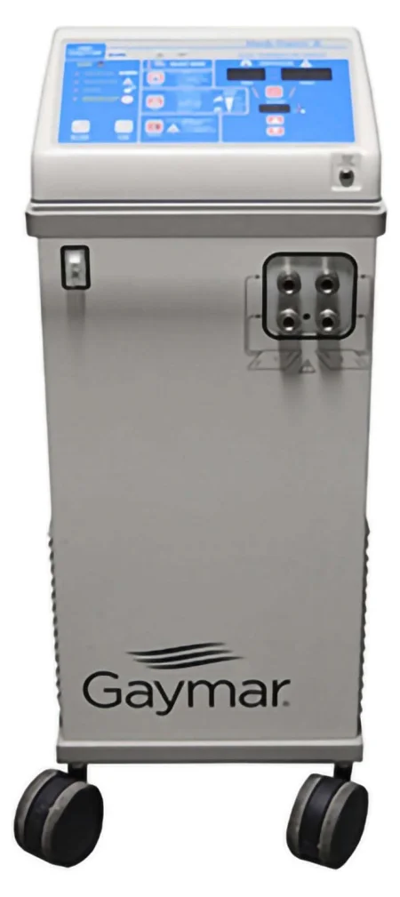
- Hypothermia and hyperthermia unit: pumps warmed or cooled water through a pad under the patient, so it can warm a cold patient or cool a hot one (chapter 23).
- Peripheral nerve stimulator: sends small, painless electrical pulses to a nerve and watches the muscle twitch in response, which shows how deep the muscle relaxation is.
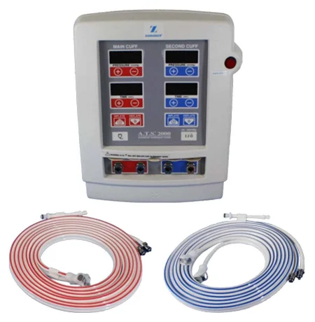
- Double-cuffed pneumatic tourniquet: holds the local anesthetic inside the arm during a Bier block (see Regional anesthesia). The cuff itself is taught in chapter 31.
8Watching the patient
Anesthesia drugs, position changes and the operation itself all change how the body works. The drugs change the blood pressure and heart rate, slow or stop breathing, and take away the protective reflexes, and tilting the table or putting the legs in stirrups can change the body's state fast. So the AP monitors the patient's vital functions continuously, to catch a dangerous change at once. How much monitoring a patient needs depends on the type of anesthesia, the patient's condition and the risks of the operation.
The ASA's basic monitoring standards apply to every general anesthetic, regional anesthetic and MAC: qualified anesthesia personnel stay in the room throughout, the patient's oxygenation, ventilation, circulation and temperature are checked continually, and the blood pressure and heart rate at least every 5 minutes.
The monitors and what they measure
| Monitor | What it shows |
|---|---|
| Blood pressure cuff (automatic) | Blood pressure, usually taken every few minutes by machine |
| Electrocardiogram (ECG) | The heart's rate and rhythm, and signs that the heart muscle is short of oxygen |
| Pulse oximeter | The oxygen saturation of the blood and the pulse rate |
| Capnograph | The carbon dioxide in each exhaled breath, shown as a number and a wave |
| Temperature probes | The core temperature, from a probe in the esophagus, nose and throat, bladder, ear canal or a pulmonary artery catheter |
| Bispectral index (BIS) | How deeply asleep the brain is, from sensors across the forehead |
| Arterial line | Blood pressure beat by beat from a thin catheter in an artery, and arterial blood for testing; for a long case, such as an arterial bypass, it usually goes in the radial artery and is kept open by a pressurized heparin drip to a transducer |
| Central venous pressure (CVP) line | The pressure in the large veins near the heart; it also lets fluids, blood and drugs go in fast |
| Pulmonary artery (Swan-Ganz) catheter | Pressures inside the heart and lungs, cardiac output and the oxygen in mixed venous blood, for selected critically ill patients |
| Transesophageal echocardiogram (TEE) | Ultrasound pictures of the heart from a probe in the esophagus: the heart muscle, the valves, the blood volume and air in the heart |
| Urinary catheter | Urine output, a rough guide to kidney blood flow and fluid volume |
| Nerve stimulator | How deep the muscle relaxation is |
| Precordial Doppler | The sound of air entering the heart, in operations at risk of air embolism, such as in the sitting position |
Pulse oximetry and capnography
The pulse oximeter's sensor goes on a finger, a toe or an earlobe. Anything that weakens the pulse there, such as a low blood volume, a weak heart output, a cold patient or a blood pressure cuff squeezing the same arm, can make the reading fail, and blue, black or green nail polish makes it read falsely low.
The capnograph measures the carbon dioxide in each breath out. The level at the very end of the breath is the end-tidal carbon dioxide (ETCO2). It confirms that the tube is in the trachea, warns of a disconnected circuit, and a rise in it is often the first sign of malignant hyperthermia.
Depth of anesthesia and awareness
Intraoperative awareness is rare: the patient under general anesthesia is aware, and may feel pain, but cannot move or tell anyone. It happens more often in emergencies, such as trauma, obstetric and heart surgery, and it can leave lasting trauma. The bispectral index system (BIS) reads the brain's signals through sensors across the forehead and gives a number from 0 to 100; about 40 to 60 is the usual target for general anesthesia. It helps, but it does not replace a watchful anesthesia provider.
Fluids, blood and the kidneys
The AP keeps the volume in the blood vessels up with IV fluids, given by an infusion pump, and with blood products when needed. Blood loss is measured by taking the fluid in the suction canisters and subtracting the irrigation used, and by weighing used sponges (1 gram counts as 1 mL of blood) or looking at them. That is why the scrub reports the irrigation used (chapter 29). A urinary catheter lets the team measure urine output, in mL an hour, through a long case.
9Vital signs and normal values
Vital signs are the body's most basic measures: temperature, pulse, respiration and blood pressure, with oxygen saturation often taken alongside them. Under general anesthesia or deep sedation, machines measure them almost continuously. Before surgery, the first set taken is the baseline, and every later reading is compared with it; a patient's baseline may sit outside the textbook range.
Chapter 31 teaches taking a baseline set step by step, and your part in it: counting the pulse and the breaths for a full minute, and measuring the blood pressure with a cuff and a stethoscope. This section teaches what the numbers mean.
Temperature
The hypothalamus holds the body's core temperature steady. Normal is about 36 to 37.5°C (97 to 99.5°F); an oral temperature at rest is normally about 37°C (98.6°F). Temperatures are recorded in degrees Celsius. To convert, °C = 5/9 × (°F − 32), and °F = (°C × 9/5) + 32.
The site changes the reading, so the route is recorded and the same route is used each time. Oral (under the tongue) and tympanic (ear canal) readings track the core well; the eardrum shares its blood supply with the carotid artery. A temporal artery (forehead) reading runs about 0.4°C higher than oral, a rectal reading about 0.4 to 0.5°C higher, and an axillary (armpit) reading about 0.3 to 0.6°C lower. Thermometers can carry germs, so gloves are worn and a clean probe cover is used for every patient.
Pulse
The pulse is the artery expanding with each heartbeat. A normal adult pulse at rest is 60 to 100 beats a minute; a child's is faster. It is felt by palpating, pressing gently with the pads of the fingers, never the thumb, which has a pulse of its own.
- The radial artery, at the thumb side of the wrist, is used most. Other pulse points are the temporal (between the ear and the outer corner of the eye), carotid (beside the trachea, in the side of the neck), brachial (the inner upper arm and the bend of the elbow, the artery used for blood pressure), femoral (the groin crease), popliteal (behind the knee), dorsalis pedis (the top of the foot, between the first and second toes) and posterior tibial (behind the inner ankle bone).
- The strength of the pulse is graded: 3+ bounding, 2+ normal, 1+ weak (thready) and 0 absent.
- A baseline pulse is counted for a full minute, and so is any irregular pulse.
Respiration
A normal adult breathes about 12 to 20 times a minute; one reference starts the range at 10. Children breathe faster. Exertion, stress, strong emotion and drugs all change the rate.
Blood pressure
Blood pressure is the push of the blood on the walls of the vessels.
- Systolic pressure: the highest pressure, as the left ventricle pumps.
- Diastolic pressure: the lowest, as the heart relaxes between beats.
- Mean arterial pressure (MAP): the average pressure across the whole heartbeat, the force that pushes blood into the tissues, normally about 70 to 110 mm Hg. It is roughly the diastolic pressure plus a third of the pulse pressure: for 120/80, about 93.
- Pulse pressure: the difference between the systolic and diastolic pressures; 120/80 gives a pulse pressure of 40.
- Orthostatic (postural) blood pressure: the pressure and pulse measured with the patient lying down and again sitting or standing.
The American Heart Association calls a pressure below 120 systolic and below 80 diastolic normal. Blood pressure changes with age, weight and sex (an adult woman's usually runs lower than a man's), with exertion, stress and strong emotion, and with the heart's output and the vessels' resistance.
It is measured with a sphygmomanometer, a cuff (an inflatable bag), a pump and a gauge, either electronic or manual with a stethoscope. For a manual reading, the cuff is first pumped up while the brachial pulse is felt, to 25 to 30 mm Hg past the point where the pulse disappears, then let down, with a wait of 10 to 15 seconds so the blood returns to the artery before the reading itself. The sounds heard through the stethoscope as the cuff is let down slowly are the Korotkoff sounds: the first sound is the systolic pressure, and the point where they disappear is the diastolic. A cuff that is too narrow or too loose, or an arm below heart level, reads falsely high.
An automatic cuff gives the pulse, systolic, diastolic and mean pressures, but it does not pick up changes in the sounds, and it can read wrong if the machine is poor or badly maintained.
Oxygen, carbon dioxide and the ECG
- Oxygen saturation (SpO2): normally 95 to 100 percent. An older patient may run in the low 90s and a patient with severe lung disease lower still, which is why the baseline matters. A saturation above 90 percent corresponds to an arterial oxygen pressure of about 60 mm Hg or more.
- End-tidal CO2: normally 35 to 45 mm Hg, a few points below the arterial carbon dioxide.
- The electrocardiogram (ECG): in electrocardiography, electrodes on the chest and limbs record the heart's electrical activity as a waveform. A three-lead ECG gives a simple picture, and a 12-lead ECG a complete one. A normal heart beats in a regular rhythm at 60 to 100 a minute; faster than 100 in an adult is tachycardia, slower than 60 is bradycardia. ECG monitoring is routine for everyone under general anesthesia or sedation and after surgery. Chapter 5 explains the P, QRS and T waves.
- Arterial blood gas (ABG): a sample of arterial blood that shows the blood's acid balance and its oxygen and carbon dioxide. It can be analyzed in minutes.
| Measure | Normal adult value |
|---|---|
| Temperature | About 36 to 37.5°C (97 to 99.5°F); oral about 37°C (98.6°F) |
| Pulse | 60 to 100 beats a minute |
| Respiration | About 12 to 20 breaths a minute |
| Blood pressure | Below 120/80 mm Hg |
| Mean arterial pressure | About 70 to 110 mm Hg |
| Oxygen saturation (SpO2) | 95 to 100 percent |
| End-tidal CO2 | 35 to 45 mm Hg |
| ABG: pH | 7.35 to 7.45 |
| ABG: PaO2 (arterial oxygen) | 80 to 100 mm Hg |
| ABG: PaCO2 (arterial carbon dioxide) | 35 to 45 mm Hg |
| ABG: bicarbonate (HCO3) | 22 to 26 mEq/L |
Normal ranges differ a little between labs and references, and a patient's own baseline matters more than any table.
Every reading is compared with the patient's baseline. A pressure of 110/70 is normal for most people, but for a patient whose baseline is 160/95 it is a drop of more than 20 percent, which counts as low blood pressure after surgery.
10General anesthesia
General anesthesia is a reversible loss of consciousness with no pain, no awareness, no memory, relaxed muscles and dulled reflexes. The patient cannot be woken even by pain, and often cannot keep their own airway open or breathe enough alone. It is a continuum, from barely responsive to completely unresponsive, rather than one fixed state.
General anesthesia is given by vein, by inhalation, or, most often, both: an IV drug puts the patient to sleep quickly, and an inhaled agent keeps them asleep. In total IV anesthesia (TIVA), short-acting IV drugs, such as propofol with remifentanil, put the patient to sleep, and an infusion keeps them asleep.
The phases and the stages
The phases of general anesthesia are the steps of the anesthetic in time. The stages describe how deep the patient is. The four stages were described by the anesthesiologist Arthur Guedel. Today's fast IV drugs carry the patient from stage 1 to stage 3 in moments, so the stages are seldom distinct, but you still need to know them, because the excitement stage can show itself at induction and again as the patient wakes.
| Phase | When | What happens | Your part |
|---|---|---|---|
| Preinduction | From the premedication until induction starts | The patient comes in and moves to the table, lying supine; monitors go on; the IV is started if it is not in; the AP may give a sedative and has the patient breathe 100% oxygen. Suction must be ready | Have suction working; stay beside the patient, who is never left alone |
| Induction | From the first anesthetic drug until the patient is ready for positioning, prep and incision | The patient loses consciousness; the airway is secured, usually with an endotracheal tube or LMA | Quiet room; stand by the patient; cricoid pressure or other help when the AP asks |
| Maintenance | From the secured airway until near the end of the operation | The operation itself; the AP adjusts the anesthetic, muscle relaxant, fluids and drugs | Report blood loss, irrigation and anything wrong |
| Emergence | From stopping the anesthetic until the patient is ready to leave the OR | Reversal drugs as needed; the patient wakes and the protective reflexes return; the tube comes out (extubation) | Quiet room again; stay sterile until the patient leaves |
| Recovery | From the end of the anesthetic, through the PACU, until the drugs have cleared | Care in the PACU, then on the unit or at home | Hand-off details from the field |
Some references count preinduction as a fifth phase.
| Stage | Name | From and to | What you may see |
|---|---|---|---|
| Stage 1 | Amnesia (induction) | From the first anesthetic drug to the loss of consciousness, usually moments | The patient loses consciousness; hearing is the last sense to go |
| Stage 2 | Excitement or delirium | From the loss of consciousness until breathing becomes regular and the eyelid reflex is gone | Exaggerated reflexes, movements the patient does not control and dilated pupils; vomiting, laryngospasm, a racing heart and high blood pressure can happen. The patient still controls the airway but is unstable |
| Stage 3 | Surgical anesthesia | From regular breathing and the loss of the eyelid reflex until breathing stops | Relaxed; the gag, blink and swallow reflexes are gone; the patient does not respond to pain, cannot keep the airway open, and breathing fails. Surgery is done here |
| Stage 4 | Overdose | Breathing has stopped and the circulation is failing | Dilated pupils that do not react and a falling blood pressure, ending in death if it is not reversed at once. It is never meant to happen |
When the anesthetic is withdrawn, the patient goes back through the stages in reverse, so the excitement stage can return during emergence.
Hearing is the last sense to go. At induction and emergence, stop setting up, stop talking and make no noise. A patient in the excitement stage can be set off into laryngospasm or vomiting, and a patient going to sleep can hear and misunderstand what you say.
Before induction
General anesthesia starts only when monitoring is in place and the team is present. The AP gathers the drugs and equipment, reassures the patient and may have them breathe 100% oxygen through a mask for a few minutes. This builds a reserve of oxygen in the blood, which carries the patient through a short block of the airway, as while the tube goes in. Suction is connected and working, and it stays working as long as the patient is in the room. The patient lies supine for induction; if the operation needs another position, the patient is moved into it after induction and intubation.
The universal protocol, with the team's check of the patient, procedure and site, is done with the whole team; chapter 28 teaches it.
Induction and intubation
The AP gives a fast IV drug, such as propofol, and the patient is unconscious within seconds; putting a patient to sleep by mask with an inhaled agent takes longer. Once breathing stops and the eyelash reflex is gone, the AP checks that a mask can move air into the lungs, and the eyes are protected. If an endotracheal tube is planned, a muscle relaxant is given and, when the patient is paralyzed, the tube is placed: this is intubation.
The AP places the head in a "sniffing" position, looks at the vocal cords with the laryngoscope (direct or video), passes the tube between them and inflates the cuff. The tube's place in the trachea is confirmed by carbon dioxide on the capnograph, equal breath sounds on both sides of the chest and none over the stomach, the chest rising evenly, and mist in the tube. Then the tube is connected to the breathing circuit.
During intubation, the circulator or an anesthesia assistant stands at the head and helps. The AP may ask an assistant to move the larynx to bring the cords into view, with the BURP maneuver (backward, upward and rightward pressure) or optimal external laryngeal manipulation. These maneuvers take training; done blindly, they cost precious seconds and can injure the airway.
Cricoid pressure (the Sellick maneuver)
A patient with a full stomach can vomit and aspirate the moment the protective reflexes go. That includes an emergency patient who has eaten, a patient whose fasting is unknown, and patients with a bowel blockage, a hiatal hernia, pregnancy, diabetes or bad reflux. For these patients, the AP may use a rapid sequence induction: 100% oxygen, then the induction drug, an analgesic and a fast-acting muscle relaxant, and a cuffed tube placed as quickly as possible, with cricoid pressure to keep stomach contents out of the airway.
The Sellick maneuver works because the cricoid cartilage is the only complete ring in the airway; pressed straight back, it squeezes the esophagus shut against the sixth cervical vertebra behind it. When the AP asks:
- Find the cricoid cartilage, the firm ring just below the bulge of the thyroid cartilage (the Adam's apple).
- Form a V with your thumb and index finger and press the cricoid firmly straight back toward the spine.
- Keep pressing, without letting up, until the tube is in, its place is confirmed and its cuff is inflated.
- Let go only when the AP tells you to.
let go of cricoid pressure because the tube looks like it is in, or because your hand is tired. Release it only on the anesthesia provider's word, once the cuff is inflated and the tube's place is confirmed.
Research has questioned how well cricoid pressure works and how often it is done correctly, but many anesthesia providers still use it for rapid sequence induction.
One reference has cricoid pressure applied before induction begins, rather than when the anesthesia provider asks.
Maintenance
Maintenance begins once the airway is secure and runs through the operation itself. The AP balances the anesthetic agents and oxygen, adds analgesics and muscle relaxant as needed, keeps the fluids and blood pressure steady and records every drug and every reading in the anesthesia record. Surgery can change the patient's condition fast, so this is the busiest time for the AP.
Muscle relaxation is needed so the surgeon can retract the body wall. Most anesthetic agents do not relax the muscles enough on their own, so a neuromuscular blocking agent is given. It paralyzes the muscles of breathing too, so the patient must be breathed for by the ventilator or by hand the whole time. The nerve stimulator shows how deep the block is, and more relaxant is given if the surgeon needs more, as for deep retraction.
Emergence
As the operation ends, the AP stops the anesthetic agents and reverses what needs reversing, such as the muscle relaxant. The patient breathes 80 to 100 percent oxygen for several minutes while the inhaled agents wash out, and the AP suctions the mouth and throat to lower the risk of aspiration and laryngospasm after the tube comes out. When the patient is awake enough that the protective reflexes are back and they are breathing well alone, the tube is removed. An oral or nasal airway may go in then.
Emergence usually goes smoothly, but the patient may tremble or stiffen late in emergence, vomiting and aspiration are a risk, and some patients, children most of all, wake in a state of confused, agitated emergence delirium. Laryngospasm is a risk at this time, as it is at induction. An older patient may stay confused for days; that is a complication, not the usual course.
This is why the scrub stays sterile, with a basic set of instruments ready, until the patient has left the room (chapter 30).
Recovery
When the patient is stable, they are moved to the stretcher and taken to the PACU, breathing oxygen from a portable tank if needed. A member of the anesthesia team goes with the patient, and a patient on a cardiac monitor is also accompanied by a licensed nurse or physician.
11The drugs of general anesthesia
The Core Curriculum lists the drugs below. Chapter 12 explains how drugs act in the body and how they are handled; here you learn what each one does in anesthesia and what to watch for.
Inhalation agents
Inhaled agents travel from the lungs into the blood and on to the brain. All of them except nitrous oxide are volatile liquids, turned into vapor in the vaporizer. They are used mainly to keep the patient asleep after an IV induction, though children are often put to sleep by mask. Every volatile agent can trigger malignant hyperthermia; nitrous oxide does not.
- Sevoflurane: pleasant smelling and gentle on the airway, so it is the agent for putting patients to sleep by mask, children above all. Induction and waking are fast.
- Desflurane: the fastest of all to take effect and to wear off, which suits outpatient surgery. It is pungent and irritates the airway, so it is not used to put patients to sleep; in children it brings on laryngospasm and bronchospasm. One reference still allows it for induction in adults. It needs a heated, pressurized vaporizer.
- Isoflurane: inexpensive and widely used; induction and waking are a little slower than with sevoflurane and desflurane.
- Nitrous oxide: a true gas, colorless, with strong pain relief but too weak to be the only anesthetic, so it is mixed with other agents. It does not burn but supports burning (chapter 20), and it spreads into closed air spaces such as the middle ear and the bowel, so it is avoided in some operations.
- Oxygen: essential for life and given with every anesthetic, at 21 to 100 percent. During laser and other surgery near the airway it is kept as low as is safe, because it feeds fire (chapter 20).
Halothane and enflurane are older agents now rarely used.
Intravenous agents
- Propofol: the most common drug for putting patients to sleep, and also used to keep them asleep and for sedation. It works in seconds, and patients wake quickly and clear headed. It is a milky emulsion of fat that can grow germs, so it is drawn up and given with strict aseptic technique. It stings in the vein and lowers the blood pressure. Only trained anesthesia providers should give it.
- Etomidate: puts patients to sleep with very little effect on the heart and blood pressure, so it is chosen for an unstable patient. It can sting, cause muscle jerks, and bring more nausea.
- Ketamine: produces dissociative anesthesia (see Sedation, below).
- Methohexital: an ultrashort-acting barbiturate, for brief anesthesia; it can be given rectally to a child.
- Thiopental: a barbiturate that was once the usual induction drug. It is no longer available in the United States, though some references still list it.
- Benzodiazepines: midazolam, diazepam and lorazepam ease anxiety and cause amnesia, and midazolam is often given at induction. They do not relieve pain.
- Dexmedetomidine: a sedative that leaves the patient's own breathing drive intact; it can slow the heart.
- Droperidol: a neuroleptic that sedates and prevents nausea.
Opioids
Opioids relieve pain by acting on receptors in the brain and spinal cord, and they let the AP use less of the other agents. All of them can slow or stop breathing, lower the blood pressure and slow the heart, and all can be abused, so they are controlled substances. Morphine is the natural opioid; the others are synthetic.
- Morphine: for severe pain, before, during or after surgery, by vein, into a muscle or into the spinal fluid.
- Fentanyl: the opioid used most in anesthesia; about 100 times stronger than morphine, fast and short acting.
- Sufentanil: like fentanyl but stronger still.
- Alfentanil: short acting, though its slowing of breathing can outlast its pain relief.
- Remifentanil: ultrashort acting, broken down within minutes in the blood, so it is given as a continuous infusion and wears off almost as soon as it is stopped.
- Meperidine: like morphine; it is also the drug of choice for shivering after anesthesia.
Non-opioid analgesics, such as acetaminophen, ibuprofen, diclofenac and ketorolac, treat mild to moderate pain, as after minor operations under local anesthesia. Combined with opioids and regional blocks in multimodal pain control, they cut the amount of opioid a patient needs.
Neuromuscular blocking agents
Neuromuscular blocking agents (muscle relaxants) stop the signal from nerve to skeletal muscle at the neuromuscular junction (chapter 2), so the muscles go limp for intubation and surgery. They have little effect on the heart, but they paralyze the breathing muscles, so the patient must be ventilated. They do nothing for pain or awareness: a paralyzed patient who is not anesthetized can feel pain and cannot move to show it.
- Depolarizing: succinylcholine is the only one. It acts within 30 to 90 seconds and lasts only 5 to 10 minutes, which makes it the drug for a rapid sequence induction. First it makes the muscles twitch all over (fasciculations), which can leave sore muscles. It can raise the blood potassium and the pressures inside the eye, skull and stomach, it can cause dysrhythmias, and it is a trigger for malignant hyperthermia. No drug reverses it. A rare patient lacks the enzyme that breaks it down (pseudocholinesterase) and stays paralyzed for hours, needing support until it wears off. It is stored in the refrigerator.
- Nondepolarizing: atracurium, cisatracurium, rocuronium and vecuronium are intermediate acting, and pancuronium is long acting. They block the receptors without twitching, and they can be reversed. They are safe for patients at risk of malignant hyperthermia. Hypothermia makes them last longer.
Reversal agents
| Drug | Reverses | What to know |
|---|---|---|
| Neostigmine | Nondepolarizing muscle relaxants | The main reversal drug; it lets acetylcholine build up again at the junction. Given with atropine or glycopyrrolate to keep the heart from slowing |
| Edrophonium | Nondepolarizing muscle relaxants | The older reversal drug, largely replaced by neostigmine |
| Sugammadex | Rocuronium, vecuronium and pancuronium | Binds the relaxant itself and reverses even a deep block within minutes; it can slow the heart, and it weakens hormonal birth control for about a week |
| Naloxone | Opioids | Given slowly by vein to reverse slowed breathing. It wears off sooner than many opioids, so the breathing can slow again; reversing fast can bring sudden pain, a racing heart and high blood pressure, or withdrawal |
| Nalmefene | Opioids | A long-acting opioid antagonist |
| Naltrexone | Opioids | An opioid antagonist, usually taken by mouth, used mainly to treat opioid and alcohol dependence |
| Flumazenil | Benzodiazepines | Reverses the sedation but may not reverse the amnesia; it can cause seizures, and the sedation can come back |
12Sedation and monitored anesthesia care
Procedural sedation is a general term for drugs that depress the central nervous system, relieve pain and ease anxiety for a procedure, without a full general anesthetic. Modern sedatives and opioids such as propofol and remifentanil wear off fast, need no anesthesia machine, and let the patient recover more quickly than after general anesthesia.
The levels of sedation
Sedation runs on a continuum, and a patient can slide deeper than intended, sometimes fast. The ASA describes the levels this way:
| Level | Response | Airway | Breathing | Heart and circulation |
|---|---|---|---|---|
| Minimal sedation (anxiolysis) | Responds normally to the voice | Unaffected | Unaffected | Unaffected |
| Moderate sedation ("conscious sedation") | Responds on purpose to the voice or a light touch | Needs no help | Adequate | Usually maintained |
| Deep sedation | Responds on purpose only to repeated or painful stimulation | May need help | May be inadequate | Usually maintained |
| General anesthesia | Cannot be roused, even by pain | Often needs help | Often inadequate | May be impaired |
Pulling away from a painful stimulus is a reflex, not a purposeful response.
Moderate sedation
Under moderate sedation, the patient stays able to keep their own airway open, breathes adequately and answers questions or follows commands, while feeling less pain and remembering little. It is used for short procedures, and benzodiazepines such as midazolam and opioids such as fentanyl are the usual drugs. An anesthesia provider may not be present: a registered nurse with extra training and proven competence may give the drugs and monitor the patient, as AORN's guideline and the state's nursing rules allow. That nurse has no other duties, so the patient is never left unwatched, and a second nurse circulates. The nurse watches the level of consciousness, the oxygen saturation, the blood pressure, the breathing, the heart's rhythm and rate, and the skin's color, and records the readings every 5 to 15 minutes. Oxygen and suction are in the room, and an emergency cart with a defibrillator is kept close at hand.
Monitored anesthesia care (MAC)
In monitored anesthesia care, an anesthesia provider gives sedation and pain drugs by vein, often while the surgeon numbs the site with local anesthetic. The AP watches the patient's vital functions throughout and must be able to "rescue" a patient who drifts too deep, converting to general anesthesia if the operation calls for it. MAC is used for healthy patients having fairly minor operations, and for very sick patients who would tolerate general anesthesia poorly. Strictly, MAC is the name of the anesthesia service, not a depth of sedation.
Dissociative anesthesia
Ketamine produces dissociative anesthesia: it isolates the sensory parts of the brain, blocking sensory signals and the pathways that connect them, so the patient may keep their eyes open and look awake while being unaware of what is happening. It gives deep pain relief and amnesia, works quickly, keeps the patient breathing and the airway reflexes intact, and can be given by vein or into a muscle. It is used for short procedures, for children, for burn patients and in trauma. Its drawbacks are a faster heart and higher blood pressure, a raised pressure in the skull, more saliva, increased muscle tone, and vivid dreams or hallucinations as the patient wakes.
Treat any sedated patient as one who can stop breathing. Sedation slides along a scale, and a patient under moderate sedation can drift into deep sedation and lose the airway reflexes within minutes.
13Regional and local anesthesia
Regional anesthesia gives a reversible loss of feeling in one part of the body while the patient stays conscious. A local anesthetic, injected along the path of a nerve or into a space around the spinal cord, stops the nerve from carrying signals, so the tissue it serves feels no pain. It can numb a small patch of skin or a whole region, as in spinal anesthesia, and the patient is always monitored during it.
Regional anesthesia is used most for:
- operations on the limbs, where a nerve can be blocked completely
- procedures in which the patient should be awake, such as many obstetric procedures
- minor operations on the surface of the body
- patients for whom general anesthesia is a serious risk
It also gives excellent pain control after surgery, so patients need fewer opioids and have less nausea, and it is part of many enhanced recovery (ERAS) plans. Resuscitation equipment and drugs must be at hand whenever regional or local anesthesia is given.
How much drug is safe
The safe dose of a local anesthetic depends on the patient's age, weight, heart disease, other drugs and blood flow, and on how fast the body takes the drug up and breaks it down. The more drug injected, the higher the risk. Doses are given in mg per kilogram of body weight, with a total that is never exceeded, and a lower dose is used for a patient who is frail, acutely ill, very young, very old or has liver or artery disease.
Topical anesthesia
Topical anesthesia numbs the nerves at the surface of the skin, the mucous membranes or the eye. It is sprayed or applied in the throat before laryngoscopy, intubation or bronchoscopy to stop gagging, spread as a gel on cystoscopy instruments, and dropped into the eye. A cream of lidocaine and prilocaine (EMLA) on the skin takes the sting out of starting an IV in a child. Mucous membranes absorb the drug quickly, so even a small amount needs watching for toxicity.
Local infiltration
Local infiltration is an injection into the tissue at the operative site, numbing a small area, as for removing a skin lesion. The surgeon injects after the patient is prepped and draped, at the start of the operation and again during it if needed. Epinephrine is sometimes added.
Your role in local infiltration. The scrub handles the local anesthetic on the field, and the steps are strict:
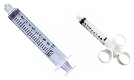
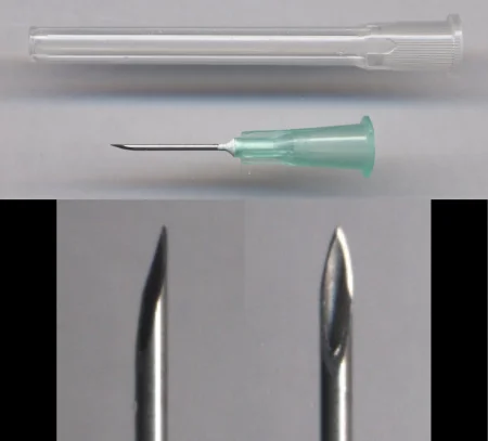
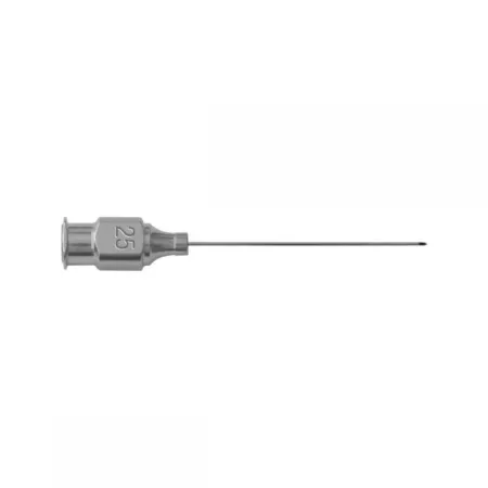
- Prepare the supplies: at least two small needles (25, 26 or 30 gauge, 1 to 1½ inches long), two 10 or 20 mL Luer-lock syringes, radiopaque sponges, a medicine cup, labels and a sterile waterproof marker.
- Receive the drug from the circulator, confirming the name, strength and amount (chapter 12), and label the medicine cup at once.
- Draw the anesthetic up and fill each syringe completely; a syringe filled partway makes it hard to know how much was used. Label every syringe right away.
- Keep the local anesthetic syringes and needles apart from everything else on the table. A small needle starts the injection, and a larger one finishes it.
- When the surgeon asks, pass the syringe and state the drug's name and its strength in percent, such as "lidocaine 1 percent." With epinephrine, know the exact ratio, such as 1:200,000, and label it on the cup and the syringes.
- Keep a running total of every mL used, and report the total to the circulator for the record.

pass an unlabeled syringe or lose track of the local anesthetic used. Deaths from lidocaine with epinephrine have followed a missing or wrong label, a wrong total reported and the wrong drug passed to the surgeon. Epinephrine is a high-alert drug.
Peripheral nerve blocks
A peripheral nerve block numbs the area supplied by one major nerve or a group of nerves (a plexus). The anesthetic is injected into the tissue around the nerve, never into the nerve itself, after the skin is prepped. The AP finds the nerve with a nerve stimulator, often with ultrasound guidance, and the block may take a few minutes to an hour to work fully. Many blocks are placed in the holding area so they have time to take effect before the patient reaches the OR.
- Nerve plexus blocks: a whole limb or region is numbed through a group of nerves, such as the brachial plexus in the armpit (an axillary block) for arm and hand surgery, or the cervical or lumbar plexus.
- The retrobulbar block: an injection behind the eye, into the cone of eye muscles, that numbs the eye and stops it moving for eye surgery. It is given by the ophthalmologist or the AP, sometimes after a sedative by vein, often with hyaluronidase added to spread the drug. It is rarely used for cataract surgery now.
A blocked limb has lost its warning sense of pain. Position and pad it with care, and remind the patient after surgery that a numb limb can be burned without their feeling it and raises the risk of a fall.
The scrub assists when a block is done as part of the operation, the same way as for infiltration.
The IV regional block (Bier block)
In a Bier block, the local anesthetic is injected into a vein of a limb that has been emptied of blood and closed off with a tourniquet. It is used most for the arm.
- A small IV catheter goes into a vein of the hand or arm, as far down the limb below the operative site as it can go, and a pneumatic tourniquet, single or double cuffed, goes around the limb above the site.
- The limb is raised and wrapped tightly from the fingers upward with an Esmarch bandage, which squeezes the blood out of it (exsanguination).
- The tourniquet is inflated, to the pressure the AP sets, and the Esmarch is removed. The tourniquet time starts.
- The local anesthetic, usually lidocaine, is injected through the IV catheter, and the arm goes numb quickly.
- With a double cuff, the upper cuff is inflated first. When the cuff starts to hurt, usually after 35 to 40 minutes, the lower cuff, which sits over numb skin, is inflated before the upper one is let down, so the drug never escapes.
The great danger is the cuff failing or being let down too soon, which sends a toxic dose of local anesthetic straight into the circulation; the cuff is kept up long enough for the drug to bind in the tissues. Cuff pain usually limits a Bier block to about an hour.
The scrub has the supplies ready and helps drape the arm, sometimes with an occlusive drape over the hand; the block is an IV procedure done with aseptic technique. Your facility may require you to change gown and gloves after the block and before the operation. Chapter 31 teaches applying the tourniquet cuff and its safe pressures.

Spinal anesthesia
In spinal anesthesia (a subarachnoid block), the local anesthetic is injected into the cerebrospinal fluid in the subarachnoid space, also called the intrathecal space, low in the back. The nerve roots leaving the cord at that level stop carrying signals, and the lower body goes numb and cannot move. It takes effect almost at once and can last up to about 2 hours, and it is used most for gynecologic, obstetric, orthopedic and urologic surgery. Fentanyl or preservative-free morphine is often added for pain relief after surgery.
The anesthetic is usually mixed with dextrose, which makes it heavier than the spinal fluid (hyperbaric), so it sinks to the lowest part of the spine. Tilting the table moves the block up, down or to one side, and after about 10 to 15 minutes it "sets" and stops spreading. The common drugs are bupivacaine, tetracaine, ropivacaine and lidocaine, and anything injected into the spinal fluid or the epidural space must be preservative-free, as its label states.
Positioning for the spinal. The needle must pass between two vertebrae, so the patient curves the back to open the spaces between them:
- Lying on the side with the knees drawn up; the circulator steadies the shoulders with one hand and supports the back of the knees with the other.
- Sitting on the edge of the table, bent forward with a rounded back, while the circulator supports the patient.
Cover the patient with a blanket so only the injection site shows, for warmth and modesty.
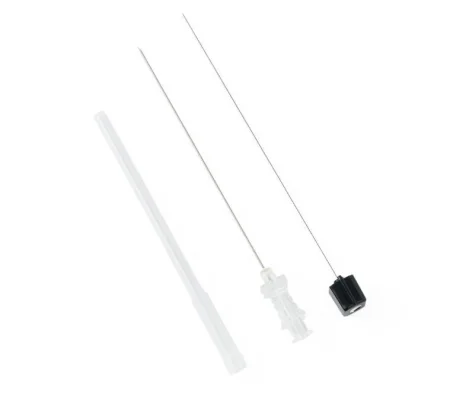
The AP preps the skin, places a small sterile drape, numbs the skin with local anesthetic and passes the spinal needle into a space low in the lumbar spine. Afterward the patient lies supine, and the AP may tilt the table to set the block's level.
Your role. As circulator or assistant, you prepare the spinal tray, the right size of spinal needle, the prep solution and drapes; help the patient hold the position, which matters because even a cold prep solution on the back can make a patient flinch and move out of it; and confirm the type and strength of the anesthetic.
Risks of spinal anesthesia.
- Low blood pressure: the block also stops the sympathetic nerves that keep the blood vessels toned, so blood pools in the legs. IV fluid and a drug to raise the pressure, such as ephedrine, treat it.
- High or total spinal: the anesthetic spreads too high and paralyzes the breathing muscles, so the patient needs intubation and ventilation at once. Any breathing trouble soon after a spinal is a warning.
- Positioning injury: the numb body cannot feel pressure or a strained joint, so it is positioned and padded with care.
- Post-dural puncture headache (spinal headache): spinal fluid leaks through the hole in the dura, and the headache comes on when the patient sits or stands. It is most common in patients under 40 and new mothers, and less common with thin, pencil-point needles. Rest, fluids and caffeine help; an epidural blood patch, the patient's own blood injected into the epidural space at the puncture, seals the leak.
Epidural and caudal anesthesia
In epidural anesthesia, the local anesthetic goes into the epidural space, just outside the dura, which holds fat, many veins and the nerve roots. The needle usually enters between the lumbar vertebrae, though thoracic and neck injections are possible. The drug acts on the nerve roots there, some of it soaks slowly through the dura into the spinal fluid, and it spreads up and down from where it was injected. It needs a much larger dose than a spinal, it takes longer to work (about 10 to 20 minutes), and the patient's position affects its spread much less.
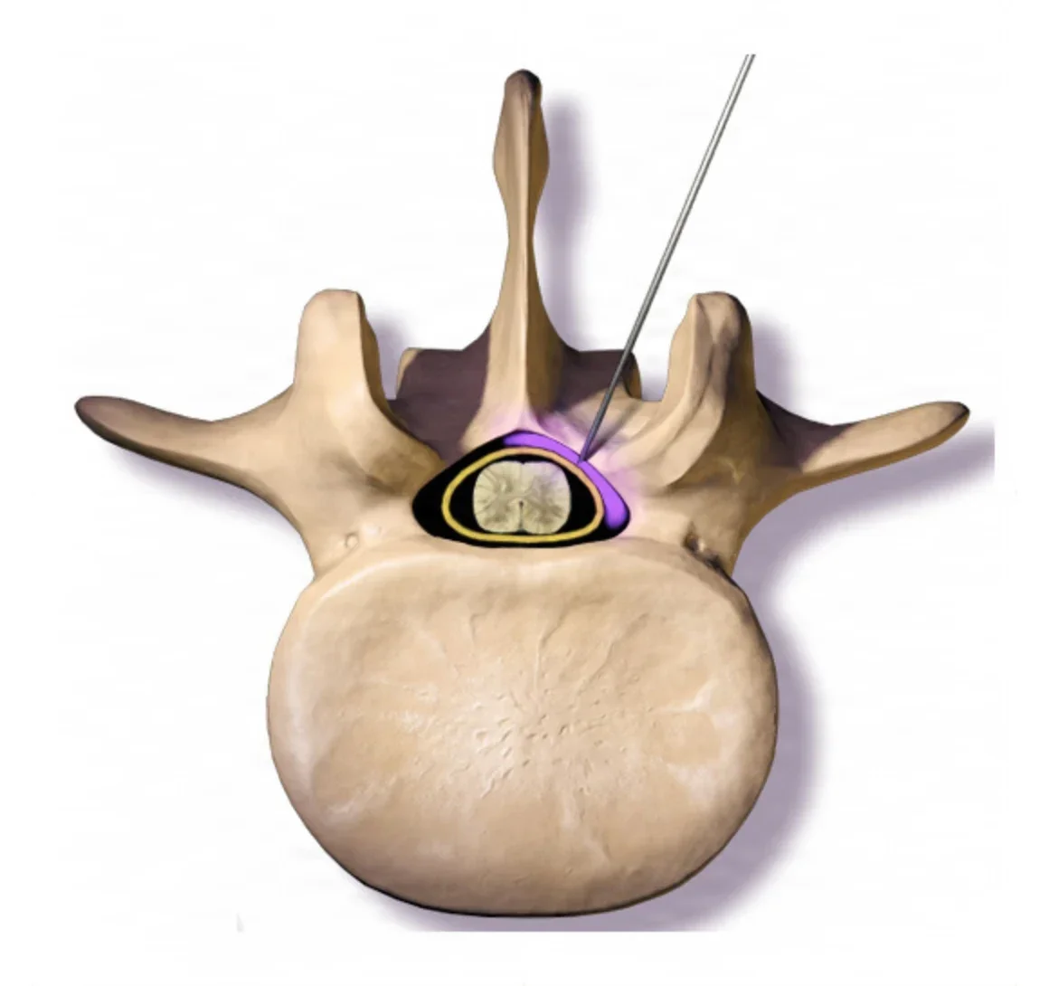
A thin catheter can be left in the epidural space, so more drug can be given through it for as long as needed. That makes the epidural a common choice for obstetric, gynecologic, urologic and rectal surgery, and for pain control for days after major surgery.
A caudal block reaches the same epidural space from below, through an opening in the sacrum. It numbs the perineum and groin and is often given to children, after they are asleep, for pain relief after lower body surgery.
The AP gives a small test dose first, then the rest of the drug in small doses.
Risks of epidural anesthesia.
- A wet tap: the epidural needle punctures the dura by mistake, which can cause a spinal headache.
- Total spinal: a large epidural dose that goes into the spinal fluid paralyzes the breathing muscles, needing intubation and ventilation.
- Injection into a vein: the space is full of veins, and an accidental injection there can cause local anesthetic toxicity.
- Low blood pressure: possible, but it comes on more slowly than with a spinal and is easier to correct.
The circulator's role is the same as for a spinal.
The local anesthetics
Local anesthetics block the sodium channels in nerve cell membranes, so the nerve cannot carry its signal. They come in two chemical families, amides and esters. Allergic reactions are more common with the esters.
| Drug | Family | Notes |
|---|---|---|
| Lidocaine (Xylocaine) | Amide | The local anesthetic used most, usually 0.5 to 2 percent; fast to work, and also a drug for heart rhythm problems |
| Bupivacaine (Marcaine, Sensorcaine) | Amide | About four times stronger than lidocaine, slower to start and much longer lasting; used for nerve blocks, spinals and epidurals, and 0.25 percent for pain relief after surgery. Injected into a vein, it can stop the heart |
| Mepivacaine (Carbocaine) | Amide | Like lidocaine, but longer lasting |
| Ropivacaine (Naropin) | Amide | Long acting like bupivacaine, a little weaker, and less toxic to the heart |
| Tetracaine (Pontocaine) | Ester | For topical use on the throat and the eye, and for spinal anesthesia; not for local injection or nerve blocks. Cetacaine is a topical preparation that contains it |
| Cocaine | Ester | Only as a topical, 4 to 10 percent, on the mucous membranes of the nose and throat, where it also shrinks the vessels; a controlled substance |
Epinephrine with a local anesthetic
Adding epinephrine narrows the blood vessels at the site. That means less bleeding, a slower uptake of the anesthetic into the blood, an anesthetic that lasts longer, and a lower risk of toxicity, so a larger dose can be given safely. Its strength is written as a ratio, and the larger the second number, the weaker the solution: 1:100,000 is twice as strong as 1:200,000. Premixed local anesthetics with epinephrine usually carry red on the label.
Epinephrine is used with caution in patients with high blood pressure, heart disease or diabetes, and with extreme caution, and as little as possible, where the blood supply is limited: the fingers, toes, tip of the nose and penis.
Maximum doses
These are common adult maximums. References differ a little, and the surgeon and the AP set the dose for each patient; chapter 12 teaches how to work out milligrams from a percentage.
| Drug | Maximum adult dose |
|---|---|
| Lidocaine, plain | 4.5 mg/kg, no more than 300 mg in all |
| Lidocaine with epinephrine | 7 mg/kg; references give different totals, from 300 to 500 mg |
| Bupivacaine, plain | 2.5 mg/kg (one reference gives 2 mg/kg), no more than 175 mg in all |
| Mepivacaine | 4 to 7 mg/kg (references differ), no more than 400 mg in all |
| Tetracaine, topical | No more than 20 mg |
Local anesthetic systemic toxicity (LAST)
Local anesthetic systemic toxicity happens when too much local anesthetic reaches the blood: from too large a dose, an injection too fast, a slow clearance, or an injection straight into a vessel. It happens most often with nerve blocks and epidurals, where large doses lie near many vessels. It can come on slowly or all at once, and it can appear up to 30 minutes after the injection.
- Brain signs come first: lightheadedness, restlessness and confusion, numbness or tingling around the mouth, a metallic taste, ringing in the ears, blurred vision, slurred speech, twitching and a sense of doom; the patient may become talkative. Then come seizures, then drowsiness, slowed breathing and apnea.
- Heart signs: first a fast heart and high blood pressure, then low blood pressure, a slow heart, dysrhythmias and cardiac arrest.
The anesthesia provider treats it in order: the airway and oxygen first, then a benzodiazepine to stop seizures, then an infusion of 20% lipid emulsion, which draws the drug out of the tissues.
A true allergic reaction to a local anesthetic is a different thing from toxicity. It can be mild, such as itching or a rash, or it can be anaphylaxis. Talking with an awake patient helps you notice either early.
Under local or regional anesthesia, the patient's words are a monitor. Ringing ears, a metallic taste or tingling lips can be the first sign of toxicity; tell the surgeon or AP at once.
14Your role with the anesthesia team
The Core Curriculum gives the surgical team five jobs while anesthesia is given: assisting the anesthesia team, documenting, handling medications and solutions, following facility policy, and monitoring both the drug doses and the patient.
At induction of general anesthesia
In the assistant circulator role, the surgical technologist may help the AP during induction and intubation. One reference lists this help among the RN circulator's duties, so your facility's policy decides what you may do.
- Before: have the suction on and working at the head of the table, and know where the difficult-airway cart and the crash cart are.
- Stay with the patient: a patient is never left alone in the OR. Stand at the patient's side, ready to help.
- Keep it quiet: stop setting up and talking until the patient is fully asleep.
- Hand what is asked for: the laryngoscope, the tube with its syringe attached for the cuff, the stylet, suction.
- Apply cricoid pressure when the AP asks, and release it only when told.
- In a difficult airway: bring the difficult-airway cart, make sure 100% oxygen and suction are ready, watch the monitors, record the oxygen saturation and end-tidal CO2, and tell the AP about any important change.
- Never move an anesthetized patient, or reposition a limb, without coordinating with the AP.
In the first scrub role, you may already be gowned with the field set up. Keep the room quiet, guard your field, and be ready if an emergency airway is needed.
During local, regional, spinal and epidural anesthesia
- Local: prepare, label, pass and total the local anesthetic as described above.
- Regional and Bier block: have the supplies ready, help drape, and be ready to change gown and gloves if policy requires.
- Spinal and epidural: prepare the tray and needles, help position and steady the patient, and confirm the drug and its strength.
- Watch the awake patient: anxiety, restlessness, a change in breathing, sweating, nausea or ringing ears can be the first sign of trouble.
Documenting
Everything given to the patient is recorded. The AP keeps the anesthesia record; you report to the circulator, for the patient's record, every drug and solution used on the field, its strength and its total.
Handling medications and solutions
Every drug on the field is received, confirmed, labeled and tracked by the rules in chapter 12. Two anesthesia points matter most: label every syringe of local anesthetic and every cup of epinephrine at once, and keep a running total, because the total is what decides whether the dose is safe.
Monitoring the dose and the patient
You watch two things. The dose: know the local anesthetic's strength and its running total, so the total can be checked against the safe limit. The patient: the color of the blood in the field (dark blood can be a sign of poor oxygen or malignant hyperthermia), the amount of bleeding, the patient's position and padding, and, in an awake patient, what they say and how they look. Chapter 29 teaches watching and reporting from the field.
15Keeping the patient warm
AST defines normothermia as a core temperature of at least 36°C and hypothermia as a core temperature below 36°C. Unplanned hypothermia is very common: it is estimated to happen to more than half of surgical patients in the United States, even in short cases.
| Hypothermia | Core temperature |
|---|---|
| Mild | 34 to 36°C |
| Moderate | 32 to 34°C |
| Severe | Below 32°C |
Why patients get cold
The body loses heat in four ways:
- Radiation: heat given off to a cooler room, the biggest loss, about 60 percent of it.
- Conduction: heat lost to anything cold the body touches, such as the OR table's mattress, and to cold IV fluids, blood and irrigation.
- Convection: heat carried off by moving air, as in the OR's air flow.
- Evaporation: heat lost as fluid evaporates from wet skin, prep solution, or open body cavities.
The OR is kept cool for the gowned team under hot lights, but the main cause of unplanned hypothermia is the anesthesia itself. General anesthesia switches off the body's temperature control and widens the vessels, so warm blood moves from the core out to the skin, where the heat is lost. In the first 30 to 40 minutes the core can fall below 35°C. Spinal and epidural anesthesia cause it too: the numb part of the body cannot shiver or narrow its vessels, and the patient may even feel warm while getting colder.
Muscle relaxants stop shivering. The very young and the very old, thin patients, burn and trauma patients, long and open operations, and patients with heart, gland or liver disease or diabetes are at highest risk.
Why it matters
Even mild hypothermia harms the patient:
- the heart beats irregularly and can be starved of oxygen
- the risk of surgical site infection rises, because narrowed vessels bring less oxygen to the wound
- the blood clots poorly, so blood loss and transfusions increase
- drugs, including the muscle relaxants, last longer, and waking is delayed
- shivering afterward can raise the body's oxygen demand by up to 400 percent, and increases pain
- the stay in the PACU and the hospital grows longer
How the team keeps the patient warm
- Before surgery: keep the patient covered on the stretcher, with warm blankets, and ask whether they are warm enough. A patient having planned surgery should be at 36°C or above before going into the OR, and active warming starts at least 30 minutes before induction, unless it would delay emergency surgery. Warming the patient before induction cuts the heat lost when the vessels open.
- Measure: the patient's temperature is taken before induction, recorded through the case (studies support every 15 minutes under general anesthesia), and on arrival in the PACU. Use the same site and method throughout. The most accurate core sites are the distal esophagus, the nasopharynx, the pulmonary artery and the eardrum with a contact probe; the bladder, rectum, armpit, mouth and forehead read near the core. Know how to test and use your facility's temperature device as its maker directs.
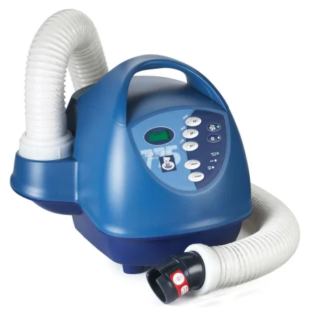
- Warm actively: forced-air warming, circulating-water pads and conductive mattresses all work, and several methods are often combined with passive covers such as blankets and drapes. Use a forced-air hose only with its own blanket, never alone or tucked under the drapes: the air at the hose can be hot enough to burn. Never warm a patient with warm IV bags or hot-water bottles, which can burn.
- Prewarm the table: a warm mattress and linens prevent loss by conduction.
- Warm fluids: IV fluids are warmed when large volumes will be given, more than about 2 liters an hour in an adult, and blood products are warmed before a transfusion, except platelets and cryoprecipitate (chapter 12).
- Prep and drape promptly: expose only the skin that must be prepped, use a warm prep solution only if its maker allows (some are flammable), and drape as soon as the prep is done.
- Warm the room: the room may be warmed, along with other methods, most of all for children. AST's guideline cites a minimum of 21°C (about 70°F) while the patient is uncovered, as during the skin prep; once active warming is under way, the room can be cooled again for the gowned team.
- Warm irrigation: as the scrub, you provide warm irrigation fluid. Irrigation fluid is kept at body temperature, 37°C, and no warmer, because fluid that is too hot can burn the patient.
- Shivering: meperidine is the drug of choice for shivering after anesthesia.
AST's 2019 normothermia guideline, going by the temperatures used in studies, warms irrigation fluid to 33 to 40°C as the fluid's maker allows; one reference's care plan sets the limit at less than 40.5°C.
The goal for a patient under general or neuraxial anesthesia for an hour or more is a core temperature of at least 36°C at the end of the anesthetic, and a normal temperature, 36 to 38°C, by discharge from the PACU.
Deliberate hypothermia
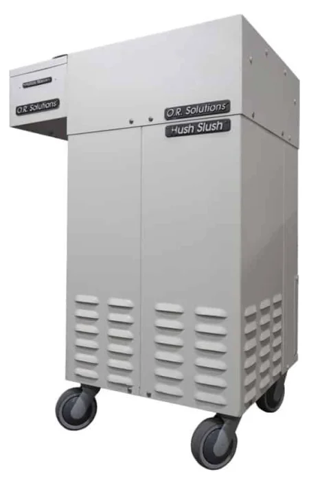
Sometimes the team cools a patient on purpose. Cooling lowers the body's need for oxygen, so it is used in some heart and brain operations, and it is part of treating malignant hyperthermia. Blood can be cooled through the heart-lung machine, and cold IV fluids, cold irrigation of a body cavity and saline slush packed around the heart also cool. How far it goes depends on the operation: light cooling holds the core between 37 and 32°C, moderate between 32 and 26°C, deep between 26 and 20°C, and profound below 20°C. Most cooling stays at 26°C or above; some aortic arch operations, done with the circulation stopped, go colder (chapter 41). Cooling can cause dysrhythmias up to cardiac arrest, so the patient is rewarmed slowly.
16The child under anesthesia
A child's body keeps its balance differently from an adult's, and some of those differences raise the risks of anesthesia.
Temperature
Infants and children lose heat faster than they make it. They have a large skin surface for their weight, blood vessels close to the skin, little insulating fat, and temperatures that swing widely. A cold table, a cold prep solution, wet linens and the anesthetic itself all chill them, and most of their heat leaves by radiation. Infants cannot shiver; they burn a special brown fat for heat, which uses up oxygen and glucose, so a cold baby becomes short of oxygen and blood sugar, and the heart can stop.
Children can also overheat, from a warming device used wrongly, too many covers or waterproof drapes. Malignant hyperthermia is more of a risk in children than in adults. Under forced-air warming, babies and children warm quickly and are watched closely.
To keep a child warm:
- Have plenty of blankets for transport; a newborn may arrive in a heated isolette, an enclosed infant transport unit with its own controlled environment and monitors.
- Keep a cap on an infant's head at all times, and prewarm the holding area and the OR before the child arrives; AST suggests above 26°C for a child.
- Use warm-air blankets and mattresses, water blankets and, for infants, overhead radiant warmers at the distance the maker sets (watch for drying out).
- Warm the IV fluids and the irrigation to body temperature, and wet sponges with warm saline.
- Never use warm IV bags or hot-water bottles to warm a child.
- Monitor the temperature with an esophageal or rectal probe, or an armpit probe for a short case.
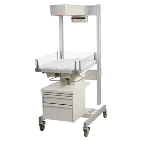
Fluids and blood
Children dehydrate fast, and their large surface for their size raises the risk of acid-base upsets. The IV runs through microdrip tubing with a measuring chamber and a pump, so every mL is counted, and the AP may give a bolus, one large dose given all at once, of fluid or a drug. Children also have little blood to spare and cannot tolerate steady bleeding. Report every mL of irrigation used, keep used sponges for weighing, and have clamps, sutures and hemostatic agents ready before the operation begins.
The airway
A child's airway is small and easily blocked:
- The tongue is large for the mouth.
- Babies under about 8 weeks breathe only through the nose, so a blocked nose can cause serious breathing trouble.
- The trachea is shorter and narrower, and the airway is softer and less rigid than an adult's.
- The narrowest point is just below the vocal cords, at the cricoid, while an adult's is at the cords. Newer tubes with very small cuffs let cuffed tubes be used at any age.
- The chest wall is soft, and the lungs hold a smaller reserve of air, so a child runs out of oxygen much faster than an adult if the airway is lost.
Drugs
Children react strongly to small changes in a dose. Doses are ordered by weight in kilograms, and the surgeon, the circulator and the scrub each verify them. Keep a running total of local anesthetics and vasoconstrictors such as epinephrine through the case, to prevent an overdose.
Anesthesia for a child
Children usually need general anesthesia, even for operations an adult could have under a block, because they cannot hold still and cooperate, and it lets the AP control the airway fully.
- Preparing: the child and family are prepared ahead of time, with the anesthesia devices, attire and steps explained in ways suited to the child's age. Chapter 18 teaches a child's fears and how to talk with a child.
- Premedication: optional, and practice varies. It may be midazolam by mouth, nose or rectum, ketamine into a muscle, methohexital by rectum, or EMLA cream on the skin before the IV. Sedation can delay waking and raise the risk of falls and slowed breathing.
- Parents at induction: many facilities let one or both parents stay with the child through induction, which can reduce the need for premedication. Children older than about 4 do best with it, and an infant may be put to sleep in a parent's or nurse's arms. It is debated: it eases parents and pleases families, but it may not change the child's behavior, and the team must be ready to care for the parent too.
- Induction: often by mask, with sevoflurane, so the IV can wait until the child is asleep. A cooperative child tries the mask on; a sleeping child can be put to sleep by holding the mask near the face ("blow-by"). A child who already has an IV may get propofol. Other routes are into a muscle, by rectum or by mouth.
- Maintenance: a precordial stethoscope, a pulse oximeter and ECG electrodes go on, the IV goes in once the child is asleep, and anesthesia continues by tube, or by mask for a short case.
- Emergence and recovery: the tube comes out once the airway reflexes are back, unless the child goes to the ICU on a ventilator. Parents usually join the child in the PACU. Nausea and vomiting are the most common problems, in 40 to 50 percent of children; emergence delirium is less common, though it is seen most often in children.
Chapter 18 covers the care of children as people, and chapter 31 their transport and positioning.
17When anesthesia goes wrong
An anesthesia emergency comes from the drugs themselves or from something that happens to the body while the patient is anesthetized. Some need the whole team. Your role in every emergency is the same at its core: protect the surgical field, and help as you are directed.
Every surgical technologist must hold a current Basic Life Support (BLS) card, with CPR and the automated external defibrillator (AED), and AST holds CSTs to keeping it current. In the OR, the patient is already in the best place to be rescued, because monitors, drugs and skilled people are all at hand.
Problems at the airway
Aspiration is breathing fluid or solid matter into the lungs, such as stomach contents after vomiting or regurgitation. Anesthetics, sedatives and opioids weaken the gag reflex, and the risk is highest at induction and emergence. Stomach acid in the lungs causes a chemical pneumonitis, which can turn into a bacterial aspiration pneumonia. Aspiration is rare; emergency surgery is the factor most often linked to it. Fasting, drugs that cut stomach acid, cricoid pressure, a cuffed tube and working suction all protect against it, and an LMA does not.
Laryngospasm is a spasm of the muscles of the larynx that closes the vocal cords partly or fully. Secretions, intubation, extubation, suctioning or stimulation of a lightly anesthetized patient set it off, most often at induction or emergence. A partly closed larynx makes a high, crowing sound (stridor); a fully closed one makes no breath sounds at all, while the chest rocks as the patient tries to breathe and the oxygen falls. The AP removes the trigger, suctions, extends the neck, gives oxygen and positive-pressure breaths by mask. If that fails within about a minute, succinylcholine relaxes the larynx, and the patient is breathed for until it wears off.
Bronchospasm is a spasm of the bronchial tubes that narrows or closes the lower airway. The patient wheezes, breathes fast and struggles for air. Aspiration, suctioning, an allergic reaction and some drugs trigger it, and a patient with asthma or a recent cold is at higher risk. It is treated with inhaled bronchodilators first, then IV drugs, and epinephrine if it threatens life.
The difficult airway. A difficult airway is one in which placing a tube or even breathing for the patient by mask is very hard. In an airway emergency, the unconscious patient cannot be intubated at all, and brain damage can follow within minutes. The team brings the emergency airway cart, tries again with the tube or an LMA, and if all fails, the surgeon opens an emergency airway through the neck: a cricothyrotomy, through the membrane between the thyroid and cricoid cartilages, or a tracheotomy. Be ready to open the tray and assist.

Dysrhythmias and cardiac arrest
A dysrhythmia is an abnormal heart rate or rhythm, seen on the ECG. Low oxygen, high carbon dioxide, a low potassium or an acid-base upset, too much fluid, anemia, withdrawal from a substance, an unsettled circulation and the anesthetic drugs themselves, alone or combined, can all cause them, so the AP treats the cause. Atropine speeds a slow heart, and sometimes a pacemaker is needed. The dangerous rhythms, ventricular tachycardia and ventricular fibrillation, are treated with defibrillation and a drug such as amiodarone or lidocaine.
Cardiac arrest is the sudden loss of the heart's pumping, so blood stops reaching the brain and organs. In surgery, its causes include a heart attack, dysrhythmias, anaphylaxis, malignant hyperthermia, respiratory arrest and massive blood loss. A code blue is the hospital's call for a team to come at once and treat a cardiac arrest.
Survival depends on the chain of survival: recognizing the arrest early, calling for help early, starting CPR early, defibrillating early when the rhythm is shockable, and starting advanced life support early. Every minute without CPR and defibrillation lowers the chance of survival by about 7 to 10 percent.
AST's code blue guideline follows the American Heart Association's order: C-A-B, compressions, then airway, then breathing. Push hard and fast, 100 to 120 compressions a minute and at least 2 inches deep but no more than 2.4 inches, let the chest recoil fully, and keep pauses short. In the OR a fourth step is added, D, definitive treatment: the drugs, defibrillation and care after resuscitation that the physicians direct. A patient under general anesthesia usually already has a secured airway, and the AP can quickly intubate a patient under local, regional or MAC.
Some references still list the old order, airway, breathing, then circulation (A-B-C).
The scrub's role in a code blue, from AST's guideline:
- Keep the back table, the Mayo stand and the surgical site sterile, and protect the field while the rest of the team works on the patient.
- Pack the wound with moist sponges and cover it with a sterile drape or sterile towels.
- Keep track of every instrument, sponge and needle on the field, and have ready what the surgeon will need next.
- Be ready to help when asked: giving chest compressions, or breathing for the patient with a bag ("bagging").
- If the surgeon must shock the heart directly or massage it with the chest open, assist with whatever is needed.
When a life is at stake, saving it comes first; sterile technique becomes secondary to the steps that keep the patient alive.
In the assistant circulator role, know where the code cart (crash cart) and the defibrillator are kept, bring them into the room, check that the defibrillator is ready, and know how to run it so the surgeon or team can apply the pads. The crash cart carries the defibrillator and the emergency drugs.
The emergency drugs. The anesthesia provider or the RN circulator gives the code drugs from the crash cart. Know them so you can hand them over fast; in some codes the surgical technologist keeps the record, writing each drug, its dose and the time on the cart's resuscitation form.
- Epinephrine: the first drug for cardiac arrest. It narrows the blood vessels, raises the blood pressure and makes the heart beat harder, and it wears off fast, so it is often given again.
- Vasopressin: another drug that narrows the vessels; in an adult it may take the place of the first or second dose of epinephrine, and it is given only once.
- Amiodarone: for ventricular fibrillation or ventricular tachycardia that goes on after defibrillation; lidocaine is used if amiodarone does not work.
- Atropine: speeds a slow heart, and epinephrine or dopamine can too.
The American Heart Association's 2025 guidelines find that vasopressin, alone or with epinephrine, gives an adult in cardiac arrest no advantage over epinephrine alone.
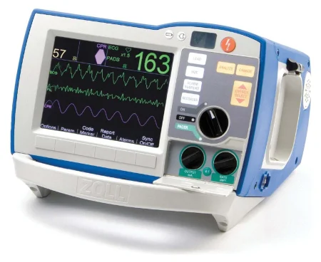
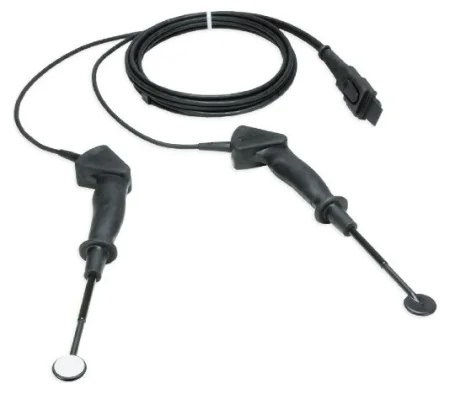
Shock and severe bleeding
Shock is a failing circulation that cannot feed the cells; chapter 11 teaches its types. The Core Curriculum names two you meet under anesthesia: cardiogenic shock, when the heart cannot pump enough blood, as after a heart attack, and hemorrhagic shock, from heavy blood loss. Treatment restores the circulation, the oxygen and the electrolytes, with fluids, blood and drugs to raise the blood pressure, aimed at the cause.
Severe hemorrhage during surgery may come from an injured large vessel, trauma, or a bleeding disorder such as DIC, end-stage kidney disease or liver disease. The first response is to find the bleeding and stop it; then the lost volume is replaced. Have the clamps, sutures and hemostatic materials and agents the procedure needs ready, and report the blood loss you see. Blood products, the blood given in an emergency before the patient's type is known, cell salvage and the checks before a transfusion are in chapter 12, and hemostasis on the field in chapter 29.
Malignant hyperthermia
Malignant hyperthermia (MH) is an inherited disorder of skeletal muscle that turns into a life-threatening crisis when a susceptible person gets a triggering drug. Calcium floods the muscle cells and they cannot relax: the muscles stiffen, burn energy out of control and pour out heat, carbon dioxide, acid and potassium. In 50 to 70 percent of the families who carry it, the faulty gene is the one for the muscle's calcium-release channel (the ryanodine receptor). Untreated, about 80 percent of patients die; with fast treatment with dantrolene, fewer than 5 percent do.
Triggers: all the volatile inhaled anesthetics (sevoflurane, desflurane, isoflurane and the older agents) and succinylcholine. Nitrous oxide does not trigger it. A patient known to be susceptible can be anesthetized safely with drugs that do not trigger it, such as IV opioids and sedatives, benzodiazepines, nondepolarizing relaxants and ester local anesthetics, and dantrolene beforehand is not needed.
Who is at risk: MH is often silent until it is triggered, and about half of patients who have a crisis had a triggering anesthetic before with no problem. A family history of trouble with anesthesia is a warning; all close blood relatives of a person with MH are treated as susceptible. Children and young adults, males, and people with certain muscle diseases (some muscular dystrophies and myopathies) are affected most, and patients under 19 account for about half of cases. It is seen more in orthopedic surgery, cleft palate repair, tonsillectomy and eye muscle surgery. A muscle biopsy test confirms susceptibility.
The signs.
- Early: a rise in end-tidal CO2 that is not explained by the breathing settings is most often the first sign. Then a fast heart, fast breathing, stiffness of the jaw muscles (masseter spasm, or trismus) after succinylcholine, rigid muscles, acidosis, and low oxygen.
- What the team may notice: the carbon dioxide absorber canister changes color fast and feels hot; blood in the field turns dark; the skin is sweaty, mottled or blue; the blood pressure is unstable; the heart rhythm is irregular.
- Late: the temperature rises fast, as much as 1 to 2°C every 5 minutes; the potassium climbs; muscle breaks down (rhabdomyolysis) and its pigment, myoglobin, turns the urine brown or tea colored; the kidneys and circulation fail, DIC can follow, and the heart can stop.
A high temperature is a late sign of malignant hyperthermia. The early warning is a rising end-tidal CO2 with a racing heart. AST counts on you as one more pair of eyes.
The treatment, which follows the MHAUS protocol. Several things happen at once:
- Stop the triggering agents at once. If the operation can end, the surgeon finishes and closes as fast as possible; if it cannot, anesthesia continues with IV drugs that do not trigger MH.
- Call for help, call for the MH cart, and call the MHAUS hotline (1-800-644-9737, 1-800-MH-HYPER), keeping it on speaker.
- The AP breathes for the patient with 100% oxygen at a high flow, to wash out the agent and the carbon dioxide, removes the vaporizers, and changes the soda lime and tubing or adds charcoal filters to the circuit.
- Dantrolene is given by vein as fast as possible, 2.5 mg/kg through a large IV, and repeated as needed, up to 10 mg/kg, until the signs ease. If the patient needs more than 10 mg/kg, another diagnosis is considered.
- The patient is cooled if the core temperature is above 39°C, with ice packs on the groin, armpits and neck, cold IV saline and cold irrigation of an open body cavity, the stomach or the rectum (never the bladder, so the urine can still be measured). Cooling stops when the temperature falls below 38°C, so the patient does not become hypothermic.
- The acid and the high potassium are treated with sodium bicarbonate, calcium, and insulin with glucose; calcium channel blockers are avoided.
- A urinary catheter measures the urine and its color, and diuretics keep the urine flowing to protect the kidneys from myoglobin. Blood gases, potassium, creatine kinase and urine myoglobin are checked, and the core temperature is watched with a probe in the esophagus, nose and throat, bladder or pulmonary artery.
- Once stable, the patient goes to the ICU, and dantrolene continues in smaller doses for at least a day. The episode is reported to the MHAUS registry, and the team reviews how it went.
One reference gives a loading dose of 2.5 mg/kg of dantrolene followed by 1 mg/kg every 5 minutes, up to 10 to 30 mg/kg.
Dantrolene is a muscle relaxant made for MH and the only drug that treats it; it stops the release of calcium inside the muscle cells. It comes freeze-dried. Each 20 mg vial of Dantrium or Revonto is mixed with 60 mL of sterile water (without a bacteriostatic agent) and shaken hard until clear, which takes many vials and several people; AST asks for 2 or 3 people assigned just to mix it. Ryanodex comes in 250 mg vials, each mixed with 5 mL of sterile water and shaken into an orange suspension in about 15 seconds. Warmed sterile water helps it dissolve. It irritates veins, so it goes through a large IV. Afterward it can leave the muscles weak, including the breathing muscles.
The scrub's role in MH (AST):
- If the operation goes on, help the surgeon finish as quickly and efficiently as you can, and guard the back table and Mayo stand closely, because many nonsterile people will be moving around the field.
- Stay scrubbed for the whole case; do not hand over to another scrub, which would break the continuity of care and the counts.
- Help cool the patient: receive and give cold irrigation, ice and cold fluids onto the field as the surgeon directs.
- After the dressings are on and the drapes are off, keep the back table and Mayo stand set up and sterile until the patient has left the room.
The assistant circulator's role in MH (AST):
- Know where the MH cart is, bring it at once, and know what is in it.
- Get the ice, cold IV fluids and cold irrigation; help mix dantrolene with sterile water; hand the AP the drugs they ask for.
- Insert a urinary catheter if one is not in.
- Help cool the patient: a cooling blanket, ice packs on the armpits, groin and head, and a colder room.
- Help the AP with the anesthesia machine, the charcoal filters, an intubation (the laryngoscope, the tube with its syringe, cricoid pressure) and the core temperature probe.
The MH cart. Every surgery department, and every ambulatory surgery center, keeps an MH cart. Its contents include plenty of dantrolene and sterile water to mix it; drugs for acid and potassium (sodium bicarbonate, calcium, insulin, 50% dextrose), diuretics (mannitol, furosemide) and drugs for heart rhythm; refrigerated IV saline for cooling, a bucket and plastic bags for ice, and a cold-irrigation tray with 60 mL syringes; a urinary catheter tray and urine meter, nasogastric tubes, IV catheters, central and arterial line kits, blood-gas kits and blood tubes; temperature probes; and an adhesive drape to cover the wound during treatment. A written treatment plan is posted where everyone can see it.
MH can also begin after the operation, in the PACU, so PACU staff are trained for it too.
Drug reactions
The Core Curriculum names three kinds of adverse reaction.
Anaphylactic. Anaphylaxis is a severe, whole-body allergic reaction (type I hypersensitivity, chapter 10) that comes on fast and can kill. Chemicals released from mast cells widen the vessels and make them leak, so the blood pressure falls fast, while the airway swells and the bronchi clamp down.
- Causes: in anesthesia, the leading cause is the neuromuscular blocking drugs, with succinylcholine the greatest risk; then latex; then antibiotics, mostly penicillins and cephalosporins. Chlorhexidine, dyes such as methylene blue, hyaluronidase, bone cement, heparin, bacitracin irrigation, colloids, induction drugs and ester local anesthetics are rarer causes.
- Timing: most reactions come within minutes of the IV drugs at induction. A latex reaction, through the skin or mucous membranes, may take 15 to 60 minutes to show.
- Signs under anesthesia: low blood pressure, a fast heart and dysrhythmias, and bronchospasm usually come first. Hives and swelling are hard to see under the drapes, and the patient cannot say what they feel.
The five immediate steps (AST):
- Stop the agent that caused it.
- Give epinephrine to the AP or the RN circulator for an immediate injection into a muscle. It raises the blood pressure and opens the airways.
- Open the IV fully to fill the circulation.
- Keep the airway open and give 100% oxygen; a patient without a tube is intubated, perhaps with cricoid pressure.
- Call for more help, including another AP, because many things must happen at once.
Other drugs follow as the AP orders: antihistamines (diphenhydramine and an H2 blocker such as famotidine), a steroid, an inhaled bronchodilator, IV fluids and vasopressin, which reverses shock from widened vessels.
As the scrub, protect the back table and Mayo stand, and be ready to pack the wound or help close quickly. On a late-night case with no one else to help, you may be asked to break scrub to help. As the assistant circulator, bring the emergency cart and the code cart with its defibrillator, have the drugs ready to hand over, epinephrine first, and fetch the TEE probe if the AP asks for it.
Latex allergy. Latex is one of the leading causes of anaphylaxis in surgery, and latex anaphylaxis is treated with the same steps as any other. Chapter 10 teaches latex allergy and how the room is made latex-safe; under anesthesia, the assistant circulator helps the AP gather latex-free supplies and check that the anesthesia machine's parts are latex-free.
Hemolytic. A hemolytic reaction destroys red blood cells, most often when a patient receives blood of the wrong ABO type. An awake patient feels back pain, chills and shortness of breath, and the blood pressure falls; under anesthesia, the signs may be only bleeding everywhere, a fever, a falling blood pressure, dark (blood-colored) urine and little or no urine. The transfusion is stopped at once, and the IV is kept open with saline. The reaction is reported to the surgeon and the blood bank, the unused blood and its tubing go back to the blood bank with a sample of the patient's blood, and fluids and diuretics protect the kidneys.
Idiosyncratic. An idiosyncratic reaction is a rare, unpredictable, unusual response to a drug whose cause is not known, one that does not fit what the drug normally does.
Injury under anesthesia
An anesthetized patient cannot blink, swallow, move away from pressure or complain, so the team protects them.
- The eyes: an eye that stays open can dry out, and pressure on the eye or a scratch can injure the cornea (a corneal abrasion). After induction, the eyes are protected with lubricant and the lids are taped shut; in the prone position, goggles or a padded face rest keep pressure off the eyes. The pulse oximeter is kept off the index finger, because a waking patient may rub an eye with that finger and scratch the cornea. Pressure on the eye can even cause loss of vision after surgery.
- The mouth and teeth: loose or cracked teeth can come out or break, and dental implants and bridgework can be damaged, during intubation; the AP checks the teeth beforehand. Hoarseness and a sore throat are common after anesthesia. The bite block keeps the patient from biting the tube as they wake.
- Clots: a still patient's blood can pool and clot in the deep veins of the legs (deep vein thrombosis), and a clot can travel to the lungs (pulmonary embolism). Antiembolism stockings, sequential compression devices, drugs and slow, careful moves of the limbs help prevent it (chapters 11 and 31).
Nerve and pressure injuries from positioning are taught in chapter 31.
18The PACU
After surgery, most patients who had general anesthesia, a major regional block or MAC go to the postanesthesia care unit (PACU), once called the recovery room. It is a critical care area where specially trained nurses watch patients as they come out of anesthesia, because a patient can become unstable fast in this time. The PACU is the first phase of recovery (phase I); a phase II area, for patients who need less acute care, prepares many of them to go home. Surgical technologists do not work in the PACU, but you need to know what happens there, because it continues the care you began in the OR.
The unit
The PACU sits next to the surgical suite, so patients reach it quickly after surgery. It is usually one large room with patient bays around the walls, all in view of a central nurses' station, with no walls between bays, so a portable x-ray machine, a 12-lead ECG or a crash cart can reach any bed fast. Each bay has suction, oxygen, power and bright lighting, and each patient's monitor shows at the central station. An isolation area separates patients with an active infection, and there may be an area for children.
Admission and the hand-off
The patient goes to the PACU only when the AP judges it safe. A member of the anesthesia team takes them, and the circulator goes too. On arrival, the PACU nurse checks the airway, breathing and circulation at once, starts oxygen and suction, and connects the heart monitor and pulse oximeter. Then the AP and the circulator give the hand-off (handover), a report spoken and written, and the AP stays with the patient until the hand-off is complete.
The hand-off covers:
- The patient's history, age, allergies, medications and other diseases.
- The exact operation, with its side and site.
- The anesthetic, how long it lasted, and every drug and fluid given, with the amounts.
- The estimated blood loss, and the fluids and blood products given.
- The wound, the dressings, drains, catheters and other devices, and what is draining.
- The ASA class.
- Any complications during surgery.
- Where the family is waiting and how to reach them.
This is why the totals you report from the field (drugs, irrigation, specimens and blood loss) matter after the operation too.
Care in the PACU
The Core Curriculum lists what PACU care covers:
- The airway: kept open, with breathing and oxygen watched closely.
- Comfort: pain is assessed and treated, and the patient is positioned for comfort and a clear airway.
- Dressings and fluid collection devices: the wound and drains are checked for the amount, color and kind of drainage.
- The IV line: kept running and intact.
- Nausea and vomiting: prevented and treated.
- Privacy: the patient is covered and their dignity kept, with emotional support, reassurance and explanations.
- The skin: its color and condition are checked.
- Urinary catheters: urine output is measured in mL an hour.
- Vital signs and monitors: the breathing rate, oxygen saturation and sometimes end-tidal CO2, the blood pressure, the heart rate and rhythm, and the temperature, compared with the patient's baseline.
The nurse may check the whole body from head to toe or focus on the main systems: the breathing (breath sounds, rate and rhythm), the circulation (pulse oximetry, ECG, blood pressure, pulses; blue lips are a late sign of low oxygen), the temperature, the abdomen (swelling or silent bowel sounds), the fluids and electrolytes, the level of consciousness, the kidneys, the wound, the tubes and drains, and the patient's emotional state.
Level of consciousness. The Glasgow Coma Scale scores three responses: eye opening (1 to 4), the best verbal response (1 to 5) and the best motor response (1 to 6). The total runs from 3, the worst, to 15, fully alert.
Pain is what the patient says it is, and each patient responds to it differently, shaped by past experience, anxiety, the drugs given and their culture. The nurse uses a pain scale, or a behavior scale such as the FLACC scale (face, legs, activity, cry, consolability) for a baby or a child who cannot talk. Pain is managed with more than one kind of drug, because opioids alone bring more sedation, and sedation comes before the slowed breathing that can kill.
Blood sugar is checked with a glucometer when needed. The CDC's guideline for preventing surgical site infection calls for keeping the blood sugar below 200 mg/dL around surgery, in patients with and without diabetes.
A 2022 update from three US infection prevention societies (SHEA, IDSA and APIC) sets a lower target: keep every patient's blood sugar between 110 and 150 mg/dL in the first day or two after surgery, with or without diabetes.
Complications in the PACU
The patient is still unstable, still has drugs in the body, and can react to the operation or the drugs, so complications can come fast.
- Airway occlusion: breathing problems are the most common life-threatening complication after surgery. The tongue falling back in a relaxed, drowsy patient is the most common cause; the patient snores or moves little air, and the oxygen falls. The nurse stimulates the patient to breathe deeply, turns them on the side, gives oxygen, lifts the chin or thrusts the jaw forward, and may insert an oral airway (for an unresponsive patient) or a nasal airway (for a waking one). Laryngospasm, bronchospasm, aspiration, and bleeding into the neck after neck surgery can also block the airway; rarely a new tube or a tracheostomy is needed.
- Breathing problems: drugs that dull the urge to breathe, and pain that stops deep breaths after chest and abdominal incisions, let carbon dioxide build up. Atelectasis, collapse of part of the lung, is prevented with deep breathing and coughing. A pulmonary embolism is a sudden danger, most of all after a deep vein clot.
- Cardiac arrest and the circulation: the blood pressure may fall (a drop of 20 percent from the baseline), most often from too little volume, so fluids are given first; it may rise from pain, anxiety, a cold patient, low oxygen, high carbon dioxide or a distended organ; and the heart may race or slow. Cardiac arrest is rare in the PACU, and a crash cart stands ready for it.
- Hemorrhage: watch for pallor, a falling blood pressure, a rising pulse, sweating, cool skin, restlessness and pain, and a dressing that soaks through. It can come from a slipped tie, poor hemostasis, a leaking join of two vessels or a clotting disorder, and the patient may go back to the OR.
- Hypothermia: a temperature below 36°C slows recovery, raises the risk of infection and heart problems and slows the breakdown of drugs. The patient is rewarmed with forced air and warm blankets, and shivering is treated.
- Malignant hyperthermia: it can begin after the operation, in the PACU.
- Nausea and vomiting (PONV): it is uncomfortable and a risk, because vomiting in a drowsy patient can lead to aspiration; drugs given before surgery help prevent it, and more are given in the PACU to treat it.
- Changes in consciousness and neurologic deficits: confusion, agitation and delirium are most common in children and older adults. Low oxygen is ruled out first, because it is the most common cause of agitation after surgery. A lasting change from the preoperative baseline, such as new weakness or numbness, needs the physician's assessment; a patient who had a spinal or epidural should regain feeling and movement down to the toes.
- Patient injury: a confused patient can fall or pull at tubes and lines, so side rails are padded and the patient is watched.
- Wound disruption: a wound can open (dehiscence; chapter 11).
The surgical technologist's part
The Core Curriculum gives you two jobs in PACU emergencies: respond and observe and communicate.
- Respond: if the patient bleeds, stops breathing or has an arrest, the team may need to reopen the wound or open an emergency airway, in the PACU or back in the OR. This is the reason you stay sterile with a basic set ready until the patient leaves the room, and why the crash cart, the MH cart, a tracheotomy tray and sterile supplies are kept in the PACU. Be ready to set up quickly and help.
- Observe and communicate: report anything abnormal you see as the case ends, such as bleeding at the incision, a swelling that may be a hematoma, or a rash (chapter 30). Report the totals of drugs, irrigation and blood loss accurately, because the PACU relies on them.
Equipment and supplies in the PACU
The Core Curriculum's list:
- For monitoring and care: airways, dressings, an ECG, a glucometer, IV setups, oxygen, a pulse oximeter, a sphygmomanometer, a stethoscope and urinary catheters.
- For the patient's comfort: a bedpan, an emesis basin and side rail pads.
- For emergencies: the crash cart, the MH cart, suction, suture, a tracheotomy tray or cart, and sterile supplies (drapes, gloves, gowns and a minor instrument tray).
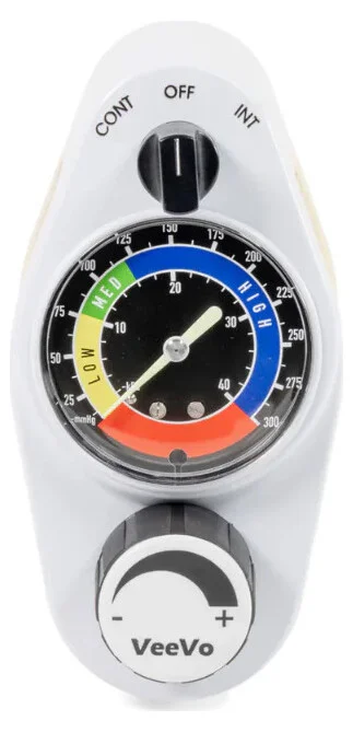
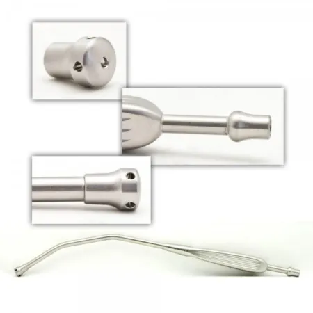
Discharge from the PACU
Patients leave the PACU only when they meet the discharge criteria the facility sets, which are mainly measures of the body's stability. The AP may write the discharge order, or the nurse may use a scoring system the anesthesia department approves.
The Aldrete score rates five things, each from 0 to 2: activity, breathing, circulation, consciousness and oxygen saturation. The total runs from 0 to 10, and a score of 8 to 10 is generally enough to leave the PACU, unless a physician orders otherwise. The score does not measure pain, which is checked too. Outpatients going home may be scored on a modified system that rates the vital signs (within 20 percent of the preoperative values), walking, nausea and vomiting, and bleeding.
Before a patient goes home, they should have:
- stable vital signs near the patient's baseline, with normal skin color
- nausea and vomiting under control, and the ability to drink fluids
- the ability to walk, alone or with help, without dizziness or weakness
- the ability to urinate, which shows the bladder is not holding urine back
- a dry incision, with drainage absent or as expected
- orientation to time, place and person, and pain at a level the patient accepts
- written discharge orders, a responsible adult to escort them and drive them home (never public transportation), and help at home as needed
Postoperative instructions are given before surgery and again at discharge, spoken and written, in plain words, to the patient and the family, because a recovering patient may not remember. For the next 24 hours, the patient does not drive, operate machinery, drink alcohol or sign important papers, and an adult stays with them. The instructions cover the medications, activity, diet, the wound, follow-up appointments, and when to call the surgeon: a fever, worsening pain, unexpected bleeding, redness or swelling or pus at the wound, persistent vomiting, or being unable to urinate. Discharge planning starts at admission. Its purpose is to make sure the patient will be safe after leaving: the discharge criteria, safe transport with an escort, care at home, teaching for the patient and family, follow-up appointments and the paperwork. A patient with no help at home is connected with social services or home nursing.
When the plan changes: unanticipated PACU outcomes.
- A patient who does not meet the discharge criteria, such as one who stays cold, bled, or has vital signs that will not settle, stays longer, goes to the ICU, or is admitted overnight.
- A patient may choose to leave against medical advice (AMA). Patients have the right to leave as long as they are not a danger to themselves or others. The facility explains the risks and tries to have the patient sign a waiver stating they were advised not to leave and accept the consequences.
- Rarely, a patient dies in the PACU. The family is told, the surgeon meets with them, and a staff member stays with them for support (chapter 18).
The PACU continues the care that starts on your field. Your counts of drugs, irrigation and blood loss, and what you saw at the wound, become the PACU nurse's starting point.
Lo que te llevas de este capítulo
- Anesthesia takes away sensation so the patient can bear surgery while the body stays in balance. General anesthesia gives sleep, pain relief, amnesia, relaxed muscles and dulled reflexes, built from several drugs at once.
- The patient, the AP and the surgeon choose the anesthetic from the patient's health (the ASA class 1 to 6, with E for an emergency), the operation and everyone's preference: general, regional, local, MAC or moderate sedation.
- The AP monitors oxygenation, ventilation, circulation and temperature continually. Know the normal values: pulse 60 to 100, breathing 12 to 20, blood pressure below 120/80, SpO2 95 to 100 percent, end-tidal CO2 35 to 45 mm Hg, core temperature about 36 to 37.5°C, always compared with the patient's baseline.
- General anesthesia runs through induction, maintenance, emergence and recovery, and through stages 1 to 4. Keep the room silent at induction and emergence, and hold cricoid pressure until the AP says to let go.
- Neuromuscular blockers paralyze but do not relieve pain or awareness. Succinylcholine and every volatile agent can trigger malignant hyperthermia; nitrous oxide does not.
- A spinal goes into the subarachnoid space, an epidural into the epidural space outside the dura. Label, total and report every local anesthetic, know its maximum dose, and treat ringing ears, a metallic taste or tingling lips as toxicity until proven otherwise.
- In any emergency, protect the field and help as directed. In a code blue, keep the field sterile, pack and cover the wound and be ready to give compressions. In MH, the first sign is a rising end-tidal CO2; dantrolene 2.5 mg/kg is the treatment, the patient is cooled to below 38°C, and you bring the MH cart and help mix the drug. In anaphylaxis, epinephrine comes first.
- Keep every patient warm, at a core temperature of at least 36°C, children above all. In the PACU, the airway comes first, and the patient goes home only after meeting the discharge criteria, such as an Aldrete score of 8 to 10.
Ponte a prueba5 preguntas rápidas
Gratis para todos. Elige una respuesta para cada una y luego revísalas todas juntas.
En el quirófanoDónde vas a usar esto
Todo el banco de preguntas del capítulo en series de 10, con la respuesta explicada después de cada una.
Parte del programa para educadoresLas palabras del capítulo en tarjetas de estudio: las que fallas vuelven antes.
Parte del programa para educadoresLo asigna tu docente. Cada grupo saca sus propias 25 preguntas del banco del capítulo.
Parte del programa para educadores
Sugerir una corrección
¿Algo está mal, confuso o desactualizado en este capítulo? Avísanos y lo corregimos para todos. Tu correo solo se usa para responderte.