Breathing: the respiratory system
The airway from the nose to the air sacs, the lungs and the pleura, how breathing works and what controls it, and why the anesthesia provider and the surgeon guard it.
1Why it matters
Every cell in the body burns oxygen to make energy and gives off carbon dioxide as waste. The respiratory system brings oxygen in from the air, hands it to the blood, and sends the carbon dioxide back out. By controlling how much carbon dioxide leaves, it also helps hold the blood's acid-base balance (its pH) steady.
It has other jobs too. The nose smells, the larynx makes the voice, and a held breath helps the body push, as in coughing or giving birth.
You meet this system in every case.
- Anesthesia takes it over. Under general anesthesia, the patient loses the reflexes that protect the airway, cannot keep the airway open, and stops breathing well alone. The anesthesia provider places an airway, such as an endotracheal tube, and breathes for the patient.
- Some operations are on the airway itself: the nose and sinuses, the tonsils, the larynx and the trachea.
- Chest surgery opens the space the lungs need to work. Once the chest wall is opened, the lung on that side collapses. The anesthesia provider inflates the lungs with a ventilator during the operation, and afterward a drain restores the pressure around the lung so it can expand.
- Positions and incisions change breathing. Most positions push the abdominal organs up against the diaphragm, and after surgery, pain in the chest or upper abdomen keeps patients from breathing deeply.
This chapter follows a breath from the nose to the air sacs, then covers the lungs and their coverings, how air moves, what controls breathing, the lung volumes and the words for breathing problems. It ends with what all of this means to anesthesia. Other chapters hold the rest: the cells and the lining of the airway in chapter 2; the phrenic and vagus nerves as nerves in chapter 4; the heart and the pulmonary circulation in chapter 5; the pharynx as part of swallowing in chapter 7; respiratory infections and airborne spread in chapter 10; airway management, intubation, oxygen monitoring and anesthesia complications in chapter 13; positioning in chapter 31; and the operations of the nose, throat, airway and chest in chapters 36 and 41.
2The path of a breath
Two zones
The airway has two zones, divided by what they do.
- The conducting zone moves air in and out: the nose, pharynx, larynx, trachea, bronchi and bronchioles, down to the smallest terminal bronchioles. It cleans, warms and moistens the air, but no oxygen enters the blood there.
- The respiratory zone is where gas exchange happens: the respiratory bronchioles, the alveolar ducts and the alveoli, the tiny air sacs.
Surgical texts also split the tract into an upper and a lower part. The upper respiratory tract is the nose and nasal cavity, the pharynx and the larynx. The lower respiratory tract is the trachea, the bronchi and bronchioles, and the lungs. Some anatomy books put the larynx in the lower tract instead.
One breath, stop by stop
Follow one breath in. These are the stops the Core Curriculum names, in the order air meets them.
- The nares, or nostrils, the two openings of the nose.
- The nasal cavity, where the nasal conchae (turbinates) stir the air against a warm, wet lining.
- The choanae, the two openings at the back of the nasal cavity into the throat.
- The nasopharynx, then the oropharynx, then the laryngopharynx: the three parts of the throat.
- The larynx, past the epiglottis and between the vocal cords.
- The trachea, down to the carina, where it splits in two.
- The primary bronchi, one to each lung; then the secondary bronchi, one to each lobe; then the tertiary bronchi.
- The bronchioles, smaller and smaller, to the terminal bronchioles and the respiratory bronchioles.
- The alveolar ducts and the alveoli, wrapped in pulmonary capillaries, where oxygen passes into the blood.
Nose, pharynx, larynx, trachea, bronchi, bronchioles, alveoli. Only the last stretch, from the respiratory bronchioles to the alveoli, exchanges gas; everything above it is a pipe that cleans and warms the air.
How the airway guards the lungs
Most of the conducting zone is lined with a mucous membrane whose cells carry cilia, with goblet cells that make mucus (chapter 2). The mucus traps dust and germs, and the cilia beat it up and out toward the throat, where it is swallowed.
Smoke slows and destroys the cilia, so mucus and debris build up in a smoker's airways.
3The nose and the sinuses
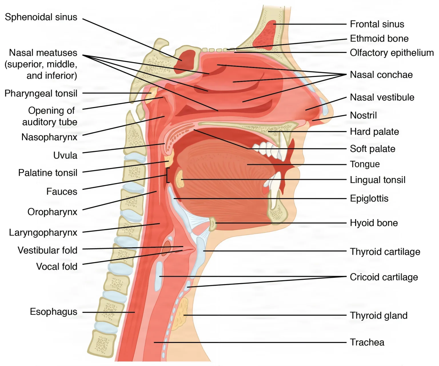
The outside of the nose
The upper part of the nose is bone, the paired nasal bones. The lower, movable part is cartilage, which is why the nose is missing on a skull. The nares (one is a naris) open the nose to the air.
Hairs just inside the nares strain out larger particles before they go deeper.
The nasal cavity
The nasal septum is the wall down the middle of the nasal cavity, splitting it into a left and a right side. Its front part is flexible cartilage, the part you can feel with your fingers; its back part is bone, from the ethmoid bone and the vomer.
The floor of the nasal cavity is the palate: the hard palate of bone in front and the soft palate of muscle behind. The same palate is the roof of the mouth.
The roof is the cribriform plate of the ethmoid bone, a thin plate that separates the nose from the cranial cavity. Infection in the nose can spread through it to the brain. High in the cavity, the olfactory epithelium holds the nerve endings for smell.
Each side wall carries three curled shelves of bone, the nasal conchae, also called turbinates: superior, middle and inferior. The superior and middle conchae are part of the ethmoid bone; the inferior conchae are separate bones. Below each concha lies a groove called a meatus, named for the concha above it.
The conchae make the air tumble against the lining, which cleans and warms it, and they trap moisture as you breathe out so the lining stays wet. The lining is full of blood vessels, and the warm blood heats the incoming air.
At the back, the nasal cavity opens into the nasopharynx through the two choanae, also called the internal nares. A baby born with one or both choanae closed, a condition called choanal atresia, needs emergency surgery to open the airway (chapter 36).
The nasolacrimal duct, which drains tears from the eye, empties into the inferior meatus.
When the nose is blocked
A septum bent off the midline, from an injury (including one at birth), an infection or a tumor, can block airflow, disturb sleep and cause headaches. In a septoplasty, the surgeon moves the septum back to its correct position. The same approach is also used to reach the sphenoid sinus to remove a pituitary tumor.
A chronically swollen inferior turbinate can also block the nose. The surgeon may shrink it with a bipolar electrode or radiofrequency energy, remove part of it through the lining, or trim it with a microdebrider in an inferior turbinoplasty.
The paranasal sinuses
Air-filled spaces in the bones around the nose open into the nasal cavity. These are the paranasal sinuses, and each pair is named for its bone: frontal, maxillary, ethmoid and sphenoid. They are lined with mucous membrane, make mucus, warm and moisten the air, and lighten the skull.
| Sinus | Where it is | What to know |
|---|---|---|
| Frontal | Behind the lower forehead | Drains into the middle meatus |
| Maxillary | A large pair in the upper jaw bones (the maxillae), below the eye sockets | The roots of the upper teeth reach its floor; drains into the middle meatus |
| Ethmoid | Many small air cells in the side wall of the nasal cavity | The front cells drain into the middle meatus, the back cells into the superior meatus (one reference says the sphenoethmoidal recess) |
| Sphenoid | Deep, at the back and top of the nasal cavity | Its side walls hold the optic nerves and the carotid arteries, and the pituitary gland sits above and behind it, so surgeons can reach the pituitary through it (the transsphenoidal approach); drains into the sphenoethmoidal recess |
Each sinus drains into the nose through a small opening. A polyp, an extra fold of lining, can block that drainage and the airflow through the nose. Most endoscopic surgery of the nose treats inflammation or infection of the sinuses.
Functional endoscopic sinus surgery (FESS) works through the nostrils with an endoscope and a video monitor. Its aim is to open the sinuses' drainage and let air into them, removing the diseased tissue while keeping normal structures and their function. Because the sinuses border the eye socket and the brain, it is technically demanding surgery.
Before endoscopic surgery, sinus disease was treated with open operations, such as the Caldwell-Luc operation on the maxillary sinus, an external ethmoidectomy or obliteration of the frontal sinus. Endoscopic sinus surgery has largely replaced them, but the Caldwell-Luc, which opens the maxillary sinus through the upper jaw, is still taught and is also used to reach the maxillary artery in a severe nosebleed.
4The pharynx
The pharynx, or throat, is a tube of skeletal muscle lined with mucous membrane. It runs from behind the nose down to where it divides into the larynx in front and the esophagus behind, and it has three parts.
- Nasopharynx: behind the nasal cavity, from the choanae down to the soft palate. It carries air only. The adenoids (pharyngeal tonsils) sit high on its back wall, and the auditory (Eustachian) tubes from the middle ears open on its sides, which is how a cold can spread to the ear. When you swallow, the soft palate and the uvula swing up and close it off, so food cannot go up into the nose.
- Oropharynx: behind the mouth, from the soft palate down to the level of the hyoid bone. It carries both air and food. The palatine tonsils, the ones people mean by "the tonsils," sit in its side walls, and the lingual tonsil is at the base of the tongue.
- Laryngopharynx, also called the hypopharynx: from the hyoid bone down, behind the larynx. It too carries air and food. At its lower end, air goes forward into the larynx and food goes back into the esophagus.
The adenoids are large in children and shrink with age. Tonsils that are infected again and again or that block breathing during sleep are removed, often with the adenoids; chapter 5 covers them as lymph tissue.
The nasopharynx carries air only. The oropharynx and laryngopharynx carry both air and food, so breathing and swallowing share them.
5The larynx
The larynx, or voice box, sits in the front of the neck, in front of the fourth to sixth cervical vertebrae, between the root of the tongue and the trachea. It joins the laryngopharynx to the trachea.
It does three jobs: it is an open passage for air, a valve that keeps food and drink out of the lungs, and the source of the voice.
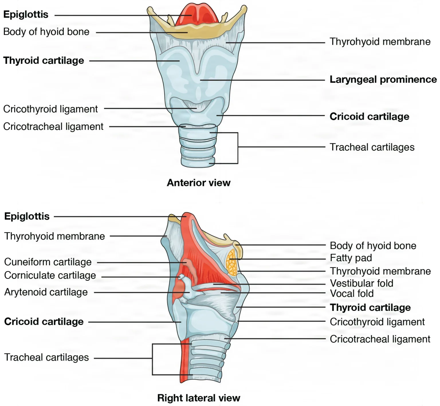
The cartilages
Nine pieces of cartilage, joined by ligaments and membranes, build the larynx: three single ones and three pairs.
| Cartilage | What it is and does |
|---|---|
| Thyroid cartilage | The largest, a shield that forms the front wall. Its ridge is the laryngeal prominence, the Adam's apple, usually bigger in males |
| Cricoid cartilage | A ring just below the thyroid cartilage, narrow in front and wide at the back, like a signet ring. It is the only complete ring of cartilage in the upper airway (the tracheal rings below it are C-shaped), and it holds the airway open |
| Epiglottis | A leaf-shaped flap of flexible elastic cartilage at the top of the larynx, attached to the thyroid cartilage. It folds down over the opening of the larynx when you swallow |
| Arytenoid cartilages (a pair) | Small cartilages that sit on the top of the back of the cricoid. The back ends of the true vocal cords attach to them, and they move the cords to make sound |
| Corniculate and cuneiform cartilages (two pairs) | Small cartilages attached to the arytenoids; the cuneiforms link the arytenoids to the epiglottis |
Between the thyroid and cricoid cartilages, in front, is a membrane, the cricothyroid membrane (ligament). In an emergency, when no other airway can be placed, the surgeon can open it and insert a tube, a cricothyrotomy. One reference notes that it can be faster than a standard tracheotomy, with fewer complications.
The vocal cords and the glottis
Inside the larynx, two pairs of folds of mucous membrane run from front to back.
- The upper pair are the vestibular folds, or false vocal cords.
- The lower pair are the true vocal cords, or vocal folds: white folds attached in front to the thyroid cartilage and behind to the arytenoid cartilages. Their inner edges are free, and they vibrate as air passes to make the voice.
The glottis is at the level of the true vocal cords. Surgical texts use the word for the opening between the true cords, the entrance to the trachea; one anatomy text counts both pairs of folds as part of the glottis too. The part of the larynx above the cords, with the epiglottis and false cords, is the supraglottis; below the cords, down to the bottom of the cricoid, is the subglottis.
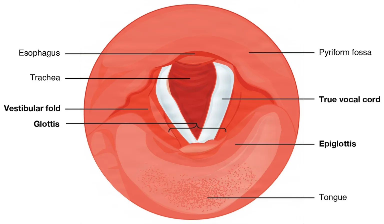
When you swallow, the larynx rises, the epiglottis swings down over its opening and the glottis closes, so food slides past into the esophagus instead of the trachea.
The small muscles inside the larynx that move the vocal cords are worked by the recurrent laryngeal nerve, a branch of the vagus. Surgeons protect it during thyroid surgery, because an injured nerve leaves a vocal cord paralyzed (chapter 4).
In an adult, the opening at the vocal cords is the narrowest part of the airway. A child's airway is narrowest lower down, at the cricoid; chapter 13 explains the child's airway.
In a laryngoscopy, the surgeon looks directly at the larynx through a rigid laryngoscope passed through the mouth, often with the operating microscope, to examine it, take a biopsy or remove small growths. While the surgeon looks through the microscope, the scrub guides the long, fine instruments into the laryngoscope, because the surgeon does not look away to take them.
6The trachea and the carina
The trachea
The trachea, or windpipe, runs from the bottom of the cricoid cartilage, at about the level of the sixth cervical vertebra, down into the chest. It is about 9 to 15 cm long.
It is built of about 16 to 20 C-shaped rings of hyaline cartilage, stacked and joined by connective tissue. The rings keep the trachea from collapsing. They are open at the back, where smooth muscle (the trachealis) and elastic tissue close the tube and let it stretch a little with each breath. The esophagus lies right behind that soft back wall.
Its lining has cilia and goblet cells, like the nose, and keeps sweeping mucus up toward the larynx to be swallowed.
In the neck, the isthmus of the thyroid gland crosses the front of the trachea.
A tracheotomy or tracheostomy makes an opening into the front of the trachea for an airway tube. It is done for a patient who needs a ventilator for a long time, or whose upper airway is blocked by a tumor, an infection, secretions, a foreign body or extra tissue in the throat that causes sleep apnea. The surgeon cuts over the front of the trachea, may divide the thyroid isthmus to see it, lifts the trachea with a tracheal hook placed in the cricoid cartilage, and opens the trachea, usually between the third and fourth rings.
The carina
In the chest, at about the level of the sternal angle, the trachea divides into the right and left primary bronchi. The ridge on the inside of that split is the carina.
The carina is packed with sensitive nerve tissue, so anything that lands on it, such as a bit of food, sets off violent coughing. It is a landmark the surgeon uses during bronchoscopy, and the tip of an endotracheal tube is placed a few centimeters above it.
The right primary bronchus is wider and runs more in line with the trachea than the left one, which bends off at a sharper angle. So an inhaled object, or a tube pushed in too far, usually ends up on the right side.
In a bronchoscopy, the surgeon passes a scope through the mouth or nose, through the larynx and trachea, past the carina and into the bronchi, to look, take biopsies or remove a foreign body. A flexible bronchoscope reaches farther into the smaller branches, and the patient breathes around it. A rigid bronchoscope is a wider tube, used under general anesthesia when large instruments are needed, and the patient can be ventilated through it. When cancer is suspected, the healthy lung is examined first, so the scope does not carry cancer cells into it.
7The bronchial tree
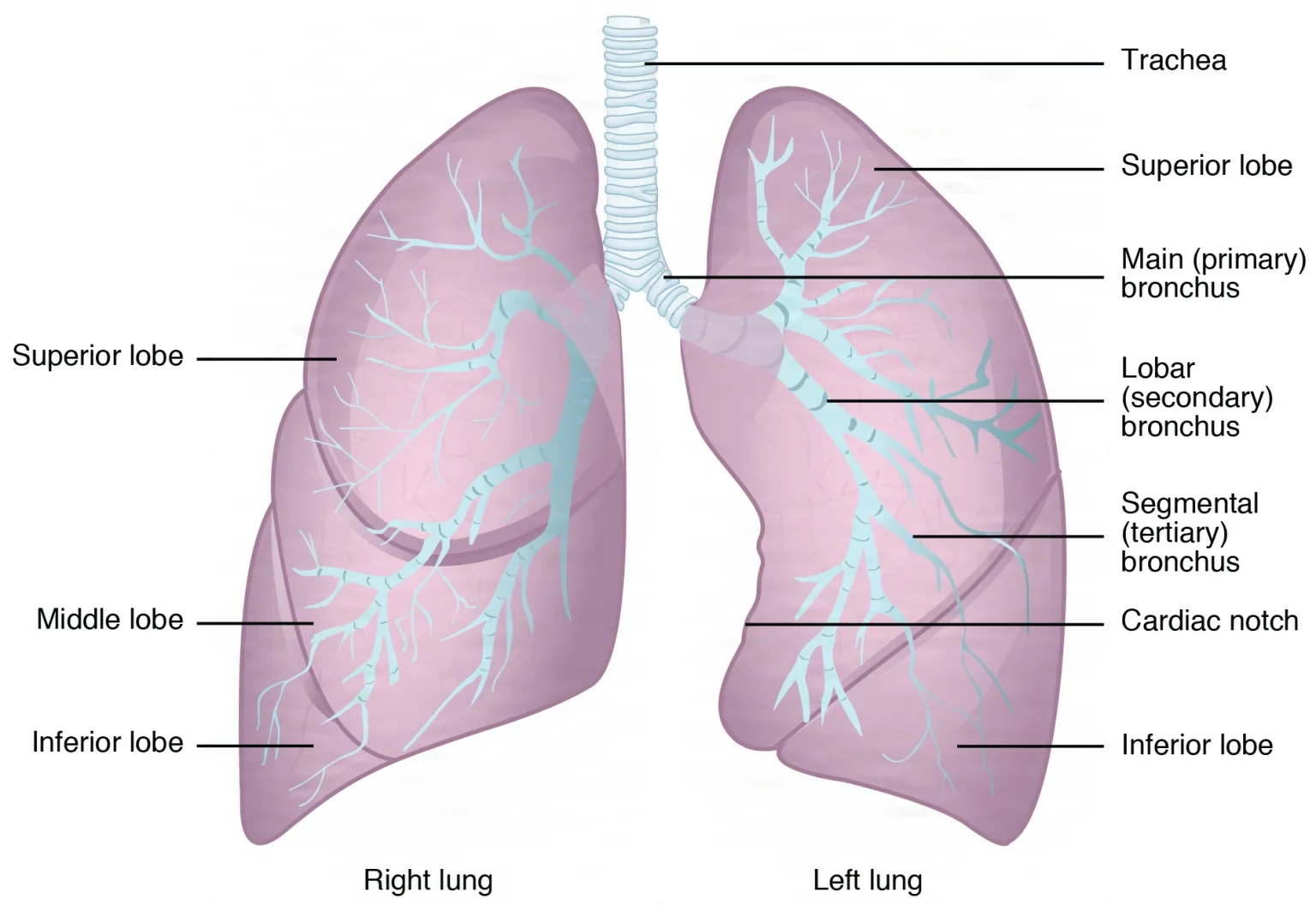
The primary bronchi enter the lungs and branch again and again, like an upside-down tree. All of these branches together are the bronchial tree.
- Each primary (main) bronchus serves one lung.
- It divides into secondary (lobar) bronchi, one for each lobe: three on the right and two on the left.
- These divide into tertiary (segmental) bronchi, one for each bronchopulmonary segment.
- The tertiary bronchi branch into bronchioles, about 1 mm across, which keep branching down to the terminal bronchioles. Each lung has more than 1000 of these.
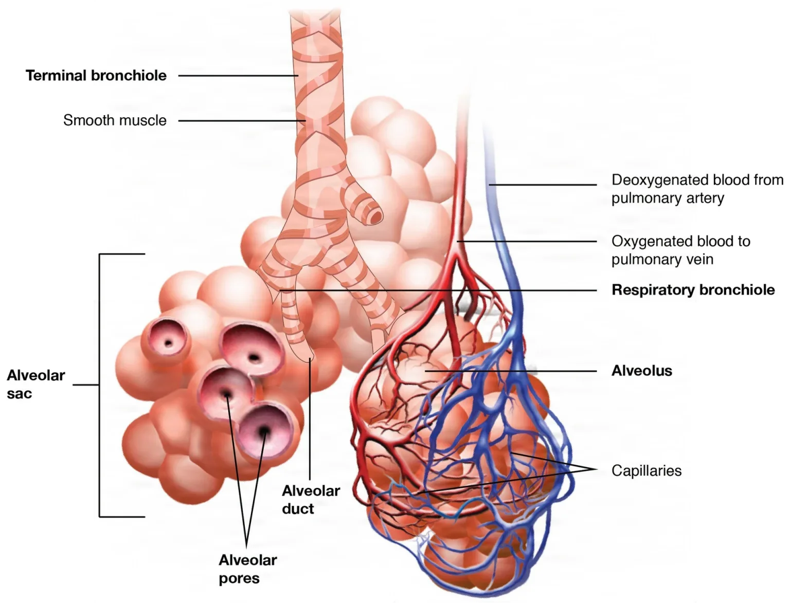
- Each terminal bronchiole leads into respiratory bronchioles, the smallest kind, which open into alveolar ducts and clusters of alveoli.
Rings of cartilage hold the primary bronchi open. As the branches get smaller, the rings become plates, and the bronchioles have no cartilage at all, only smooth muscle that can narrow or widen the tube.
That muscle answers to the autonomic nerves: the parasympathetic nerves narrow the airways and the sympathetic nerves widen them. In asthma, the bronchioles go into spasm (bronchospasm), the lining swells and extra mucus clogs them, so air has trouble getting in and out.
8The alveoli and gas exchange
The air sacs
An alveolus is a tiny, grape-like air sac about 0.2 mm (200 micrometers) across, with stretchy walls. Clusters of them, the alveolar sacs, open off the alveolar ducts, and small pores between neighbors keep the air pressure even.
Three kinds of cells make up the alveolar wall.
- Type I cells: very thin, flat cells that cover most of the surface, up to 97 percent. Gases pass through them easily.
- Type II cells: scattered among the type I cells. They make surfactant, a mix of fats and proteins that lowers the surface tension of the thin film of water lining each alveolus. Without surfactant, the alveoli would collapse every time you breathe out.
- Alveolar macrophages: roaming immune cells that eat the dust and germs that reach the alveoli.
When alveoli collapse, the lung or part of it goes airless. This is atelectasis. Mucus plugs, too little surfactant, pressure on the lung, and shallow breathing all cause it, and shallow breathing because of pain after chest or upper abdominal surgery makes it a known complication of those operations.
How the gases cross
The thin alveolar wall and the thin wall of the capillary pressed against it form the respiratory membrane, only about 0.5 micrometer thick. Spread out, the lungs' exchange surface covers about 70 square meters.
Gases cross it by simple diffusion, with no energy spent: each gas moves from where its pressure is higher to where it is lower.
- Oxygen is at about 104 mm Hg in the alveoli but only about 40 mm Hg in the blood coming back from the body, so it moves into the blood.
- Carbon dioxide is at about 45 mm Hg in that blood and about 40 mm Hg in the alveoli, so it moves out into the air to be breathed out. That gap is small, but carbon dioxide dissolves about 20 times more easily than oxygen, so about the same amount of each gas crosses.
Most of the oxygen rides on hemoglobin in the red blood cells, which turns bright red when it is loaded (chapter 5). Most of the carbon dioxide, about 70 percent, travels in the blood as bicarbonate.
This exchange in the lungs is external respiration. The reverse exchange out in the tissues, where oxygen leaves the blood and carbon dioxide enters it, is internal respiration.
For the exchange to work, two things have to match. Ventilation means air moving in and out of the lungs. Perfusion means blood flowing through the capillaries around the alveoli. An area that gets air but little blood, or blood but little air, exchanges gas poorly, so the body narrows the vessels to poorly aired alveoli and sends the blood where the air is.
9The lungs
The lungs are two pyramid-shaped organs in the chest, one on each side of the mediastinum, the middle space that holds the heart, the great vessels, the trachea, the esophagus and the thymus (chapter 2).
- The apex, the narrow top of each lung, rises into the base of the neck, just above the first rib and the collarbone.
- The base rests on the diaphragm.
- The surface that faces the ribs is the costal surface, and the surface that faces the middle is the mediastinal surface.
Right and left sides, and the lobes
Deep grooves called fissures divide each lung into lobes. The two lungs are not mirror images.
| Right lung | Left lung | |
|---|---|---|
| Lobes | Three: superior (upper), middle and inferior (lower) | Two: superior (upper) and inferior (lower) |
| Fissures | Two, the oblique and the horizontal | One, the oblique |
| Shape | Shorter and wider | Smaller, with the cardiac notch, a dent that makes room for the heart |
Bronchopulmonary segments
Each lobe is made of smaller units, the bronchopulmonary segments. Each segment gets its air from its own tertiary bronchus and its blood from its own branch of the pulmonary artery. Each lung has about 10. On the left, some segments share a bronchus and are counted together, so you may see fewer listed.
Segments barely connect to each other, so they are the lung's surgical units: a diseased segment can be removed with little effect on its neighbors.
The hilum
On the mediastinal surface of each lung is a dent, the hilum. The primary bronchus, the pulmonary artery and veins, the bronchial vessels, the nerves and the lymph vessels all enter or leave the lung there, and together they form the root of the lung. The nerves gather there in a network called the pulmonary plexus.
At the hilum of the right lung, the phrenic nerve runs down in front of the root, and the vagus nerve passes behind it with the esophagus. The surgeon keeps both out of harm when retracting the lung.
Removing lung tissue
Lung operations are named for how much they remove.
- Wedge resection: a small wedge of lung taken with a stapler, as in a lung biopsy.
- Segmentectomy (segmental resection): one bronchopulmonary segment, taken at its own artery and bronchus.
- Lobectomy: one or more lobes, most often for lung cancer. The surgeon divides the lobe's branches of the pulmonary artery and vein at the hilum, then clamps and divides its bronchus. While the bronchus is open, suction keeps blood and fluid from running into the other lung.
- Pneumonectomy: a whole lung.
Most lung operations, 80 to 90 percent, are now done by video-assisted thoracoscopic surgery (VATS), through small ports instead of a painful muscle-splitting incision. The instruments for an open chest are still kept close by, so the team can convert to an open operation quickly.
10The blood supply of the lungs
The lungs have two blood supplies.
- The pulmonary circulation brings blood to the alveoli for gas exchange. The right ventricle pumps oxygen-poor blood into the pulmonary trunk, which splits into the right and left pulmonary arteries. Each artery branches alongside the bronchi until it becomes the capillary network around the alveoli. The oxygen-rich blood leaves through the pulmonary veins, two from each lung, which exit at the hilum and empty into the left atrium.
- The bronchial arteries, small branches of the aorta, carry oxygen-rich blood to feed the lung tissue itself: the bronchi, the bronchioles and the visceral pleura. Their number varies from person to person.
So in the lungs, as chapter 5 explains, the arteries carry oxygen-poor blood and the veins carry oxygen-rich blood, the reverse of the rest of the body.
The pulmonary vessels are fragile and are handled gently at surgery. A blood clot that lodges in a pulmonary artery, a pulmonary embolism, can be fatal; the clot usually comes from a deep vein, most often in a leg.
11The pleura
Two layers and a space
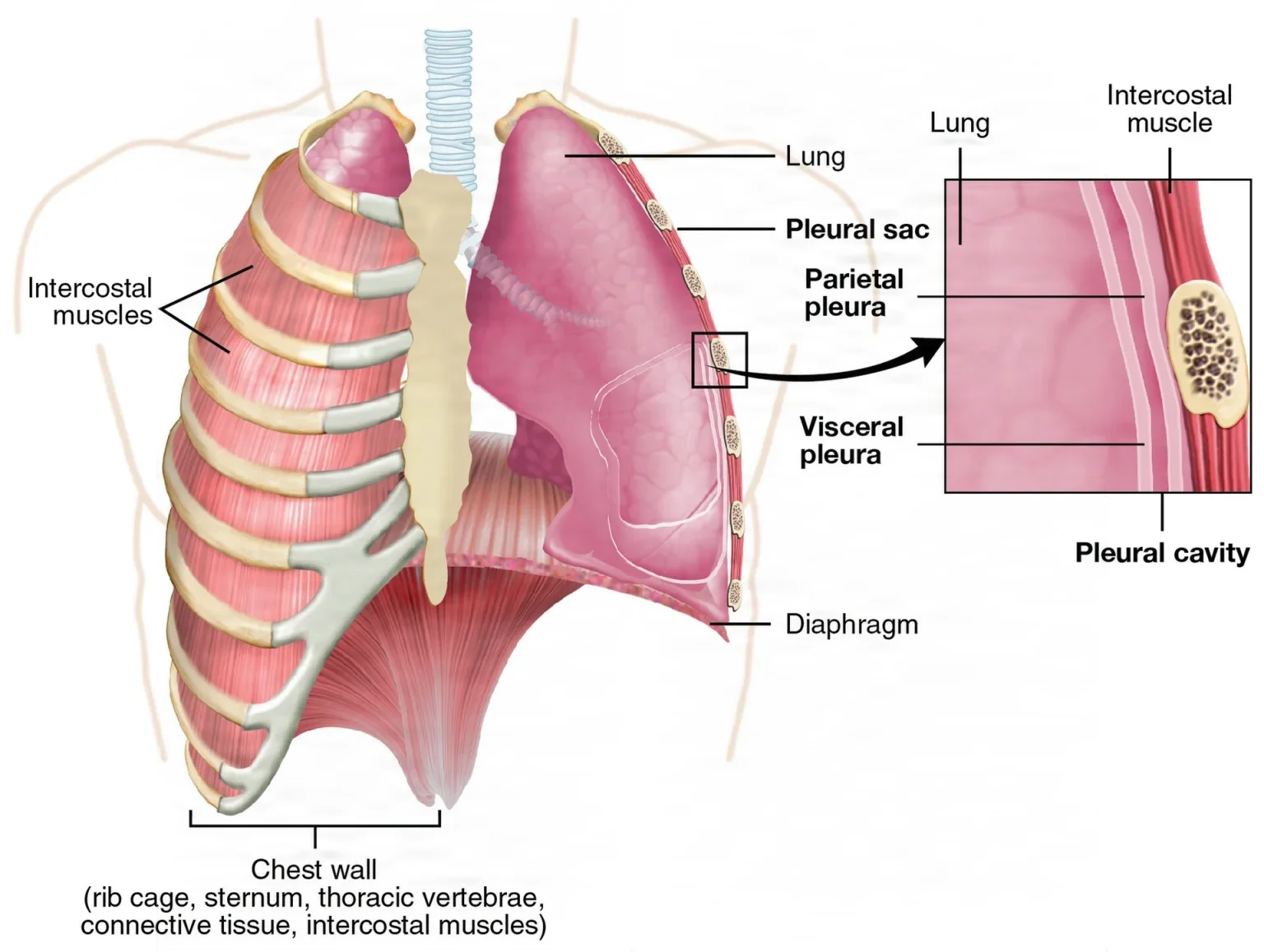
Each lung lies in its own sac of serous membrane, the pleura (chapter 2).
- The visceral pleura covers the lung itself and dips into its fissures.
- The parietal pleura lines the inside of the chest wall, the side of the mediastinum and the top of the diaphragm.
- The two layers are one sheet: at the hilum, the membrane folds back over the root of the lung.
Between the layers is the pleural space, or pleural cavity. It is a potential space: the two layers touch, with only a thin film of pleural fluid between them, made by both layers. The fluid lets the layers slide without friction as you breathe, and its surface tension holds the lung against the chest wall, so the lung follows the chest wall out. The pleura also walls each lung off from the other organs, which keeps their movements apart and helps stop infection from spreading.
Inflammation of the pleura is pleuritis.
The pleural recesses
The lungs do not fill the pleural sacs all the way to their edges. Where two parts of the parietal pleura lie against each other with no lung between them, they form pockets, the pleural recesses.
- The costodiaphragmatic recess is the lowest edge of each sac, where the pleura on the ribs meets the pleura on the diaphragm. Fluid in the chest can collect there.
- The costomediastinal recesses lie behind the sternum, where the pleura on the ribs meets the pleura on the mediastinum. The left one is larger, because of the cardiac notch.
The diaphragm is a dome that rises as high as the fourth intercostal space, and the pleural sac reaches down around its edges, to about the eighth rib in front and the tenth rib at the side. So an incision made too low can miss the chest altogether and pass through the diaphragm into the abdomen.
The pressure that holds the lung open
The pressure inside the pleural space stays a little lower than the pressure in the lungs, about 4 mm Hg below it. The lungs' elastic tissue keeps pulling inward, away from the chest wall, while the chest wall pulls outward; the slight vacuum between them keeps the lungs stretched open.
When air, blood or fluid gets in
Anything that fills the pleural space takes away the negative pressure. The lung can no longer follow the chest wall, so it cannot expand fully, and it may collapse.
| Problem | What is in the pleural space |
|---|---|
| Pneumothorax | Air, for example from a wound through the chest wall; it keeps the lung from expanding and can push the structures in the middle of the chest aside |
| Hemothorax | Blood, usually from an injury |
| Pleural effusion | Extra fluid, such as serous fluid, pus or blood |
| Empyema | Pus, from an infection |
Long-lasting inflammation, infection or a tumor can lay down a stiff fibrous layer, called a peel, over the pleura, which traps the lung. Decortication strips it away so the lung can expand again.
Opening the chest
When the surgeon opens the chest wall in a thoracotomy, air rushes into the pleural space and the lung on that side collapses. During the operation, the anesthesia provider ventilates the lungs with a machine. For many chest operations, a double-lumen endotracheal tube lets the operated lung stay collapsed, out of the surgeon's way, while the other lung gets the oxygen and anesthetic.
At the end, chest tubes are left in the pleural space and connected to a closed drainage system. It removes air and fluid and restores the negative pressure, so the lung can expand again. A chest X-ray afterward checks that the lung has not collapsed.
12How air moves
Pressure and volume
Air moves for one reason: it flows from higher pressure to lower pressure.
In a closed space at a steady temperature, pressure and volume work in opposite directions. Make the space bigger and the pressure inside drops; make it smaller and the pressure rises. This is Boyle's law, and it runs every breath.
Three pressures matter.
- Atmospheric pressure: the pressure of the air around the body, 760 mm Hg at sea level.
- Intrapulmonary pressure, also called intra-alveolar pressure: the pressure of the air inside the alveoli. It dips and rises during each breath and evens out with atmospheric pressure between breaths.
- Intrapleural pressure: the pressure in the pleural space, which stays a little below the pressure in the lungs.
The lungs have no muscle to move air themselves. They are passive: the pleural fluid ties them to the chest wall, so they grow and shrink as the chest does.
Breathing in
Inspiration takes muscle work.
- The diaphragm contracts. Its dome flattens and moves down toward the abdomen, making the chest taller.
- The external intercostal muscles, between the ribs, contract and lift the ribs up and out, making the chest deeper from front to back and wider.
- The lungs stretch with the chest, so the pressure in the alveoli falls a little below atmospheric pressure.
- Air flows in through the airway until the two pressures are equal again.
Breathing out
Quiet expiration is passive. The diaphragm and the intercostal muscles relax, the stretchy lung tissue recoils, and the chest gets smaller. The pressure in the alveoli rises above atmospheric pressure, and air flows out.
Breathing hard
Quiet breathing at rest is called eupnea. When the body needs more air, as in exercise, more muscles join in.
- To breathe in harder, accessory muscles in the neck, the sternocleidomastoid and the scalenes, lift the top of the chest.
- To breathe out hard, the internal intercostal muscles pull the ribs down and together, and the abdominal muscles squeeze the abdominal organs up against the diaphragm.
Diaphragm down and ribs up: the chest gets bigger, the pressure in the lungs drops, and air flows in. The muscles relax: the lungs recoil, the pressure rises, and air flows out.
Two things make breathing harder work. Narrow airways resist airflow, and the size of the airway matters more than anything else. A stiff chest wall resists stretching: thick scar from deep burns over the chest, for example, can keep the chest from expanding.
13What controls breathing
You breathe without thinking about it, yet you can hold your breath, talk or sing at will. Breathing is under both nervous and chemical control.
Nervous control
- The medullary respiratory center: groups of neurons in the medulla oblongata, the lowest part of the brain stem (chapter 4), set the basic rhythm. They fire to start each breath in; when they go quiet, the muscles relax and the breath goes out.
- The pons: respiratory centers in the pons, just above the medulla, adjust the rate and the depth of breathing.
- The phrenic nerves carry the signal from the spinal cord in the neck to the diaphragm. They come from C3 to C5, so a patient whose spinal cord is cut above C3 cannot breathe without help (chapter 4).
- The intercostal nerves, the thoracic spinal nerves that run between the ribs, carry the signal to the intercostal muscles.
- The vagus nerve carries signals from stretch receptors in the lungs back to the brain. As the lungs fill, these signals stop the breath in before the lungs overinflate; this is the inflation, or Hering-Breuer, reflex. The vagus also carries the parasympathetic fibers that narrow the airways.
- The cerebral cortex lets you take over for a while, to hold a breath or to speak.
Breathing depends on all of this working. Drugs that depress the brain, such as barbiturates and morphine, can slow breathing or stop it altogether.
Chemical control
Sensors called chemoreceptors watch the chemistry of the blood and adjust breathing to match.
- Carbon dioxide is the main driver. As it builds up, it makes the body's fluids more acid. Central chemoreceptors in the brain stem sense this, and the respiratory center makes breathing faster and deeper, which blows off the extra carbon dioxide. When carbon dioxide is low, breathing slows and becomes shallow.
- Peripheral chemoreceptors, in the carotid bodies where each common carotid artery divides and in the aortic arch, sense carbon dioxide, acid and oxygen in the arterial blood.
- Oxygen is a weaker trigger. Low oxygen speeds breathing only when it falls a long way, to about 60 mm Hg or less, because hemoglobin keeps releasing oxygen as the dissolved oxygen drops.
Body temperature, pain and emotion change breathing too; a fever, for example, speeds it up.
The drugs given during surgery blunt the response to carbon dioxide, so a patient who is sedated or recovering from anesthesia may not breathe faster even as carbon dioxide rises.
14Lung volumes
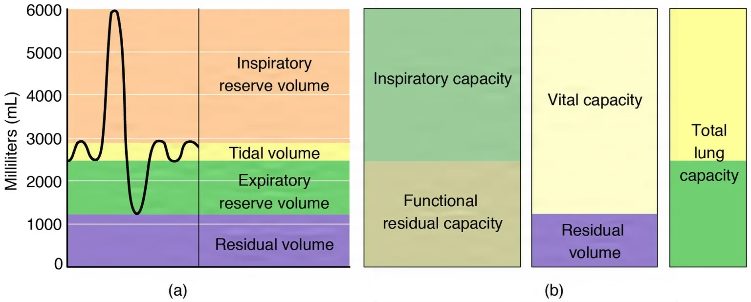
A spirometer measures how much air a person moves, as part of the pulmonary function tests done before lung surgery. The four basic volumes are separate slices of the air in the lungs, and the capacities add two or more volumes together. A person's size, age, sex and lung disease all change the vital capacity.
| Term | What it is |
|---|---|
| Tidal volume (TV) | The air moved in or out with one normal, quiet breath, about 500 mL |
| Inspiratory reserve volume (IRV) | The extra air you can pull in with effort after a normal breath in |
| Expiratory reserve volume (ERV) | The extra air you can force out after a normal breath out, up to about 1200 mL in males |
| Residual volume (RV) | The air still in the lungs after you breathe out as hard as you can; it can never be breathed out, and it keeps the alveoli from collapsing |
| Vital capacity (VC) | The most air you can breathe out after the deepest breath in: TV + IRV + ERV, about 4000 to 5000 mL |
| Total lung capacity (TLC) | All the air the lungs hold after the deepest breath in: TV + IRV + ERV + RV, about 6000 mL in males and 4200 mL in females |
Two other capacities appear in reports. The inspiratory capacity is the tidal volume plus the inspiratory reserve volume. The functional residual capacity is the air left in the lungs after a normal breath out, the expiratory reserve volume plus the residual volume.
The minute volume is the air breathed out in one minute. Not all of each breath reaches the alveoli: the air that fills the conducting airways, the anatomical dead space, never takes part in gas exchange.
Before lung surgery, these tests help predict how well the patient will breathe afterward. A patient who can force out less than 1 liter in the first second of a hard breath out is at extremely high risk of lung complications after surgery.
15Words for breathing problems
These are the Core Curriculum's terms. You will hear them in report and read them in the chart.
| Term | What it means |
|---|---|
| Apnea | A stop in breathing. It happens at the start of some types of general anesthesia, one reason the patient is ventilated |
| Dyspnea | Difficult, labored or uncomfortable breathing; shortness of breath |
| Cyanosis | A blue, gray or dark purple color of the skin or mucous membranes, such as the lips, from blood that is low in oxygen |
| Cheyne-Stokes respiration | A repeating pattern: breaths grow deeper and faster, then fade into a pause with no breathing that lasts 10 to 60 seconds, then start again. It is seen with heart failure, with brain damage or raised pressure inside the skull, and near the end of life |
| Hypercapnia | Too much carbon dioxide in the blood, usually above 45 to 50 mm Hg; also called hypercarbia. It can bring dizziness, drowsiness and confusion |
| Hyperventilation | Breathing faster or deeper than the body needs, so the blood's carbon dioxide falls too low. The Core Curriculum lists tachypnea and hyperpnea under it |
| Tachypnea | Fast breathing: in an adult, more than 20 breaths a minute |
| Hyperpnea | Deeper, and often faster, breathing. A degree of it is normal after exercise; pain, fever and some lung and heart diseases also cause it |
| Hypoxia | Too little oxygen reaching the tissues; hypoxemia is too little oxygen in the blood |
Hypoxia has four kinds, named for the cause.
- Hypoxic hypoxia: too little oxygen gets into the arterial blood, as at high altitude, with a blocked airway or with fluid in the lungs.
- Anemic hypoxia: too little working hemoglobin to carry the oxygen, as after a hemorrhage, in anemia or in carbon monoxide poisoning.
- Ischemic hypoxia: the blood flow to a tissue is too low to deliver enough oxygen.
- Histotoxic hypoxia: the blood brings enough oxygen, but the cells cannot use it, as in cyanide poisoning.
Two more words: bradypnea is breathing that is slower than normal, and atelectasis, from earlier in this chapter, is collapsed, airless lung.
Counting breaths
An adult at rest breathes about 12 to 20 times a minute; one reference starts the normal range at 10. Children breathe faster: a baby under 1 year, about 30 to 60 times a minute.
To count breaths, watch the chest rise and fall without making a point of it, and count for one full minute. Note how deep and how regular the breaths are, as well as how many. Chapter 13 teaches the vital signs and the monitors in the OR.
16Why anesthesia cares
The airway under general anesthesia
As general anesthesia deepens, the patient loses the protective reflexes, such as gagging and swallowing, can no longer keep the airway open, and the breathing response fails. Breathing may stop at induction, so the anesthesia provider places an airway and breathes for the patient. The drugs also blunt the carbon dioxide drive, and that effect can last into recovery.
The path of the endotracheal tube
An endotracheal tube runs from the mouth into the trachea, through the anatomy of this chapter.
- The anesthesia provider uses a laryngoscope, a lighted instrument, to look past the tongue and the epiglottis to the vocal cords.
- The tube passes through the glottis, between the vocal cords, into the trachea.
- A balloon cuff near its tip is inflated to seal it against the wall of the trachea.
- In an adult, its tip should sit about 2 to 6 cm above the carina. Too high, it can damage the vocal cords; too deep, it slides into the right primary bronchus, and only the right lung gets air.
A laryngeal mask airway (LMA) is a tube with a small mask on its end. It is placed without a laryngoscope, and the mask fits snugly over the larynx from above.
Guarding the lungs
- Aspiration: stomach contents can come up and be breathed into the lungs, most of all at induction and as the patient wakes, causing an inflammation of the lungs called chemical pneumonitis. For a patient at risk, such as one with a full stomach, the anesthesia provider may ask for cricoid pressure, the Sellick maneuver. The cricoid cartilage is a complete ring, unlike the C-shaped tracheal rings, so pressing it back toward the spine squeezes the esophagus shut behind it. The pressure is held until the tube is in place and the anesthesia provider says to let go.
- Laryngospasm: a spasm of the muscles of the larynx that closes the glottis and can block the airway.
- Bronchospasm: the bronchioles clamp down, as in an asthma attack, and air has trouble moving.
Chapter 13 teaches how the anesthesia provider prevents and treats these problems.
The lungs during and after surgery
- Most positions push the abdominal organs up against the diaphragm, which cuts its pull to about two-thirds and lowers the tidal volume (chapter 31). A patient who is short of breath while awake, such as during transport or under local or regional anesthesia, breathes easier with the head raised.
- In chest surgery, the anesthesia provider can let one lung collapse on purpose, through a double-lumen tube, so the surgeon can work.
- After surgery, pain in a chest or upper abdominal incision keeps patients from breathing deeply. Carbon dioxide builds up, the oxygen falls, and atelectasis can follow.
If the blood on the field turns darker, or the patient's lips or skin look blue, tell the anesthesia provider and the surgeon at once. Both can mean the blood is short of oxygen.
Lo que te llevas de este capítulo
- The conducting zone, from the nose to the terminal bronchioles, cleans, warms and moistens the air. Gas exchange happens only in the respiratory zone: the respiratory bronchioles, alveolar ducts and alveoli, across a membrane about 0.5 micrometer thick.
- A breath runs nares, nasal cavity, choanae, nasopharynx, oropharynx, laryngopharynx, larynx, trachea, carina, primary and secondary bronchi, bronchioles, alveolar ducts and alveoli. The nasopharynx carries air only; the lower two parts of the pharynx carry air and food.
- The nasal septum divides the nose, three conchae (turbinates) line each side wall with a meatus under each, and the frontal, maxillary, ethmoid and sphenoid sinuses drain into the nose. FESS reopens that drainage.
- The larynx has nine cartilages. The thyroid cartilage is the largest, the cricoid is the only complete ring in the upper airway, the epiglottis covers the larynx when you swallow, and the true vocal cords run from the thyroid cartilage in front to the arytenoids behind.
- The trachea's C-shaped rings are open at the back. It splits at the carina, and the right primary bronchus is wider and straighter, so inhaled objects and tubes pushed too far go right.
- The right lung has three lobes and the left two, with the cardiac notch; each lung has about 10 bronchopulmonary segments, and everything enters at the hilum. Pulmonary arteries bring oxygen-poor blood for exchange, and bronchial arteries from the aorta feed the lung tissue.
- The visceral and parietal pleura hold a film of fluid and a slight negative pressure that keeps the lungs open. Air, blood, fluid or pus in the pleural space keeps the lung from expanding and can collapse it, and chest tubes on a closed drainage system restore the pressure.
- The diaphragm and external intercostals make inspiration; quiet expiration is passive recoil. The medulla sets the rhythm through the phrenic and intercostal nerves, the vagus reports stretch, and carbon dioxide is the main chemical drive. Know the volumes and the words for breathing problems.
Ponte a prueba5 preguntas rápidas
Gratis para todos. Elige una respuesta para cada una y luego revísalas todas juntas.
En el quirófanoDónde vas a usar esto
Todo el banco de preguntas del capítulo en series de 10, con la respuesta explicada después de cada una.
Parte del programa para educadoresLas palabras del capítulo en tarjetas de estudio: las que fallas vuelven antes.
Parte del programa para educadoresLo asigna tu docente. Cada grupo saca sus propias 25 preguntas del banco del capítulo.
Parte del programa para educadores
Sugerir una corrección
¿Algo está mal, confuso o desactualizado en este capítulo? Avísanos y lo corregimos para todos. Tu correo solo se usa para responderte.