Breathing: the respiratory system
The airway from the nose to the air sacs, the lungs and the pleura, how breathing works and what controls it, and why the anesthesia provider and the surgeon guard it.
1Why it matters
Every cell in the body burns oxygen to make energy and gives off carbon dioxideKAR-bun dy-OK-sideA gas made by the body's cells as waste, carried in the blood to the lungs and breathed out.Full entry as waste. The respiratory system brings oxygen in from the air, hands it to the blood, and sends the carbon dioxide back out. By controlling how much carbon dioxide leaves, it also helps hold the blood's acid-base balanceAS-id BAYS BAL-ensThe body's control of how acidic or alkaline the blood is, keeping its pH in a narrow normal range.Full entry (its pH) steady.
It has other jobs too. The nose smells, the larynxLAIR-inksThe voice box: the part of the airway in the neck, above the windpipe, that holds the vocal cords.laryng/o larynxFull entry makes the voice, and a held breath helps the body push, as in coughing or giving birth.
You meet this system in every case.
- anesthesiaan-us-THEE-zhuhLoss of feeling or sensation, often brought on by drugs so that surgery or a procedure can be done without pain.an- withoutesthesi/o feeling, sensation-ia conditionFull entry takes it over. Under general anesthesiaJEN-er-ul an-es-THEE-zhuhDrug-induced loss of consciousness and feeling over the whole body, so a person feels nothing and is not aware during surgery.an- withoutesthesi/o feeling, sensation-ia conditionFull entry , the patient loses the reflexREE-fleksAn automatic body response to something that stimulates it, which happens without thought, such as the knee jerk.Full entry that protect the airway, cannot keep the airway open, and stops breathing well alone. The anesthesia provideran-us-THEE-zhuh pruh-VY-derThe licensed professional who gives anesthesia and watches over the client's breathing and vital functions during a procedure.Full entry places an airway, such as an endotracheal tubeen-doh-TRAY-kee-ul toobA flexible plastic tube passed through the mouth or nose into the windpipe to keep the airway open and allow breathing support.endo- withintrache/o windpipe-al relating toFull entry , and breathes for the patient.
- Some operations are on the airway itself: the nose and sinusSY-nusA hollow space in the body, most often one of the air-filled spaces in the skull bones around the nose.Full entry , the tonsilTON-sulOne of the masses of lymph tissue at the back of the throat that help fight germs entering the mouth and nose.Full entry , the larynx and the tracheaTRAY-kee-uhThe windpipe: the tube that carries air from the throat to the lungs.Full entry .
- Chest surgery opens the space the lungs need to work. Once the chest wall is opened, the lung on that side collapses. The anesthesia provider inflates the lungs with a mechanical ventilationmeh-KAN-ih-kul ven-tuh-LAY-shunBreathing support from a machine, a ventilator, that pushes air into the lungs through a tube or a mask.Full entry during the operation, and afterward a drain restores the pressure around the lung so it can expand.
- Positions and incisions change breathing. Most positions push the abdominal organs up against the diaphragmDY-uh-framThe dome-shaped sheet of muscle under the lungs that is the main muscle of breathing; also the flat side of a stethoscope.Full entry , and after surgery, pain in the chest or upper abdomen keeps patients from breathing deeply.
This chapter follows a breath from the nose to the air sacs, then covers the lungs and their coverings, how air moves, what controls breathing, the lung volumes and the words for breathing problems. It ends with what all of this means to anesthesia. Other chapters hold the rest: the cells and the lining of the airway in chapter 2; the phrenic and vagus nerveVAY-gus nervThe 10th cranial nerve, which runs from the brain stem to the chest and belly and slows the heart and helps digestion.Full entry as nerves in chapter 4; the heart and the pulmonary circulationPUL-muh-nair-ee ser-kyuh-LAY-shunThe path blood takes from the heart to the lungs, where it picks up oxygen, and back to the heart.pulmon/o lung-ary relating toFull entry in chapter 5; the pharynxFAIR-inksThe throat: the passage behind the nose and mouth that carries air to the voice box and food to the esophagus.pharyng/o throatFull entry as part of swallowing in chapter 7; respiratory infections and airborne transmissionAIR-born trans-MISH-unThe spread of germs in tiny particles that stay floating in the air and can travel across a room or farther.Full entry in chapter 10; airway management, endotracheal intubationen-doh-TRAY-kee-ul in-too-BAY-shunPutting a breathing tube through the mouth or nose into the windpipe (trachea).endo- withintrache/o windpipe-al relating toFull entry , oxygen monitoring and anesthesia complications in chapter 13; positioning in chapter 31; and the operations of the nose, throat, airway and chest in chapters 36 and 41.
2The path of a breath
Two zones
The airway has two zones, divided by what they do.
- The conducting zone moves air in and out: the nose, pharynxFAIR-inksThe throat: the passage behind the nose and mouth that carries air to the voice box and food to the esophagus.pharyng/o throatFull entry , larynxLAIR-inksThe voice box: the part of the airway in the neck, above the windpipe, that holds the vocal cords.laryng/o larynxFull entry , tracheaTRAY-kee-uhThe windpipe: the tube that carries air from the throat to the lungs.Full entry , bronchusBRONG-kusOne of the large air tubes that branch from the windpipe into each lung.bronch/o airwayFull entry and bronchioleBRONG-kee-ohlOne of the smallest airways in the lungs, branching from the bronchi and leading to the air sacs.bronch/o airway (bronchus)-iole smallFull entry , down to the smallest terminal bronchioles. It cleans, warms and moistens the air, but no oxygen enters the blood there.
- The respiratory zone is where gas exchangeGAS eks-CHAYNJThe movement of oxygen from the air sacs of the lungs into the blood and of carbon dioxide from the blood into the air sacs.Full entry happens: the respiratory bronchioles, the alveolusal-VEE-uh-lus (plural alveoli, al-VEE-uh-ly)One of the tiny air sacs at the ends of the smallest airways in the lungs.Full entry ducts and the alveoli, the tiny air sacs.
Surgical texts also split the tract into an upper and a lower part. The upper respiratory tract is the nose and nasal cavity, the pharynx and the larynx. The lower respiratory tract is the trachea, the bronchi and bronchioles, and the lungs. Some anatomy books put the larynx in the lower tract instead.
One breath, stop by stop
Follow one breath in. These are the stops the Core Curriculum names, in the order air meets them.
- The narisNAIR-isA nostril, one of the two openings of the nose; the plural is nares.nas/o noseFull entry , or nostrils, the two openings of the nose.
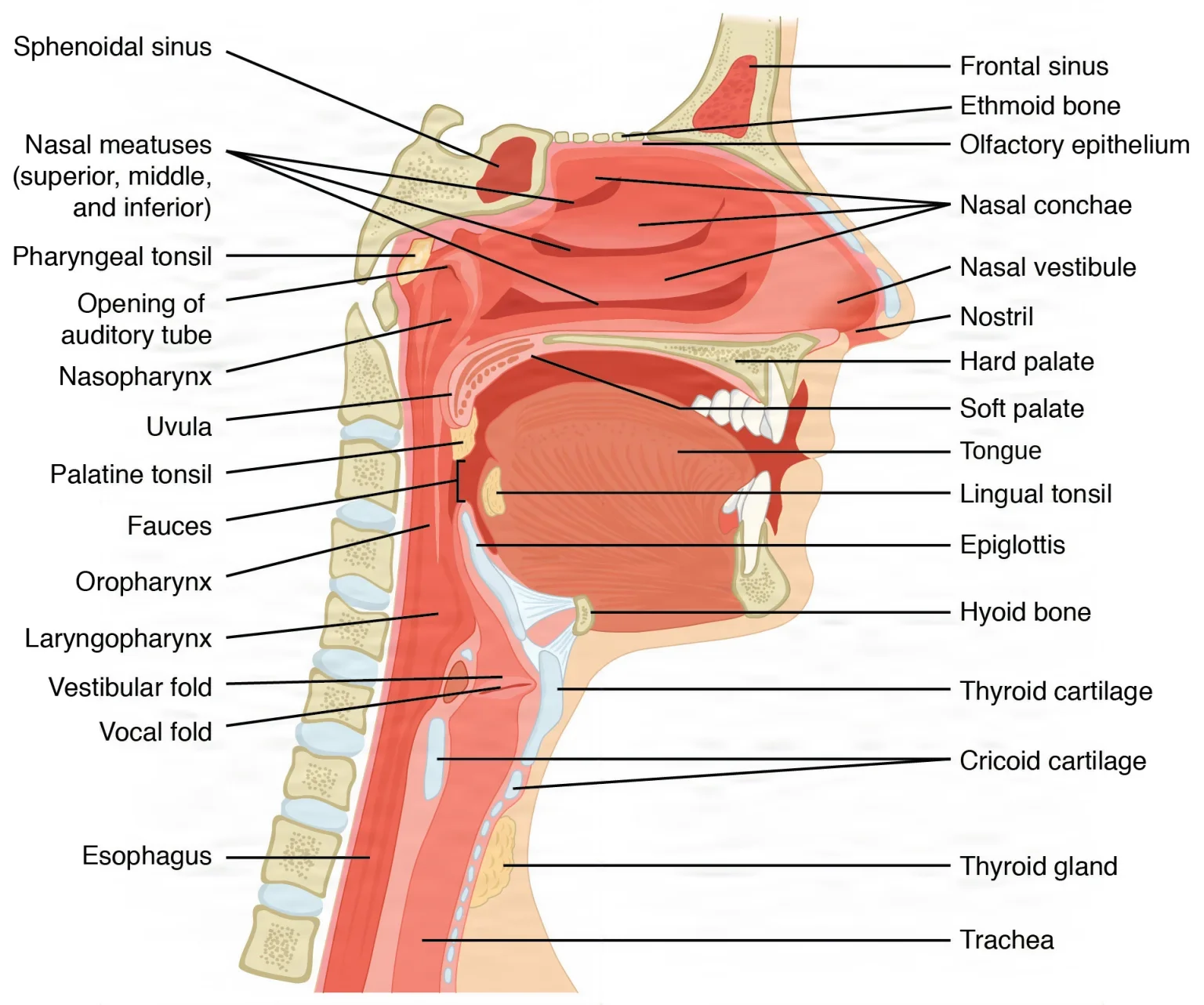
- The nasal cavity, where the nasal conchae (turbinates) stir the air against a warm, wet lining.
- The choanae, the two openings at the back of the nasal cavity into the throat.
- The nasopharynxnay-zoh-FAIR-inksThe top part of the throat, behind the nose and above the soft palate.nas/o nosepharyng/o throatFull entry , then the oropharynxor-oh-FAIR-inksThe middle part of the throat, behind the mouth, from the soft palate down to the top of the voice box.or/o mouthpharyng/o throatFull entry , then the laryngopharynxluh-ring-goh-FAIR-inksThe lowest part of the throat, below the back of the mouth, where the airway and the food passage separate.laryng/o larynx, voice boxpharyng/o pharynx, throatFull entry : the three parts of the throat.
- The larynx, past the epiglottisep-ih-GLOT-isA leaf-shaped flap of cartilage at the back of the throat that covers the windpipe during swallowing.epi- upon, overglott/o the opening of the larynxFull entry and between the vocal cords.
- The trachea, down to the carina, where it splits in two.
- The primary bronchi, one to each lung; then the secondary bronchi, one to each lobe; then the tertiary bronchi.
- The bronchioles, smaller and smaller, to the terminal bronchioles and the respiratory bronchioles.
- The alveolar ducts and the alveoli, wrapped in pulmonary capillaryKAP-ih-lair-eeOne of the tiniest blood vessels, joining the smallest arteries to the smallest veins.Full entry , where oxygen passes into the blood.
Nose, pharynx, larynx, trachea, bronchi, bronchioles, alveoli. Only the last stretch, from the respiratory bronchioles to the alveoli, exchanges gas; everything above it is a pipe that cleans and warms the air.
How the airway guards the lungs
Most of the conducting zone is lined with a mucous membrane whose cells carry ciliaSIL-ee-uhTiny hair-like structures on the surface of some cells that move in waves.Full entry , with goblet cells that make mucus (chapter 2). The mucus traps dust and germs, and the cilia beat it up and out toward the throat, where it is swallowed.
Smoke slows and destroys the cilia, so mucus and debris build up in a smoker's airways.
3The nose and the sinuses
The outside of the nose
The upper part of the nose is bone, the paired nasal bones. The lower, movable part is cartilageKAR-tih-lijFirm, smooth, rubbery tissue that cushions joints and gives shape to parts such as the ear, nose and windpipe.chondr/o cartilageFull entry , which is why the nose is missing on a skull. The narisNAIR-isA nostril, one of the two openings of the nose; the plural is nares.nas/o noseFull entry (one is a naris) open the nose to the air.
Hairs just inside the nares strain out larger particles before they go deeper.
The nasal cavity
The nasal septumNAY-zul SEP-tumThe wall of cartilage and bone that divides the inside of the nose into right and left sides.nas/o nosesept/o dividing wallFull entry is the wall down the middle of the nasal cavity, splitting it into a left and a right side. Its front part is flexible cartilage, the part you can feel with your fingers; its back part is bone, from the ethmoid bone and the vomer.
The floor of the nasal cavity is the palate: the hard palateHARD PAL-itThe bony front part of the roof of the mouth.Full entry of bone in front and the soft palateSOFT PAL-itThe soft, muscular back part of the roof of the mouth, with no bone in it.Full entry of muscle behind. The same palate is the roof of the mouth.
The roof is the cribriform plate of the ethmoid bone, a thin plate that separates the nose from the cranialKRAY-nee-ulRelating to the skull, or toward the head end of the body.crani/o skull-al relating toFull entry cavity. Infection in the nose can spread through it to the brain. High in the cavity, the olfactoryol-FAK-tuh-reeRelating to the sense of smell.olfact smell-ory relating toFull entry epitheliumep-ih-THEE-lee-umThe tissue that covers the body's surfaces and lines its organs, vessels and cavities.Full entry holds the nerve endings for smell.
Each side wall carries three curled shelves of bone, the nasal conchae, also called turbinates: superior, middle and inferior. The superior and middle conchae are part of the ethmoid bone; the inferior conchae are separate bones. Below each concha lies a groove called a meatusmee-AY-tusAn opening or passage in the body, such as the urinary opening or the ear canal.Full entry , named for the concha above it.
The conchae make the air tumble against the lining, which cleans and warms it, and they trap moisture as you breathe out so the lining stays wet. The lining is full of blood vessels, and the warm blood heats the incoming air.
At the back, the nasal cavity opens into the nasopharynxnay-zoh-FAIR-inksThe top part of the throat, behind the nose and above the soft palate.nas/o nosepharyng/o throatFull entry through the two choanae, also called the internal nares. A baby born with one or both choanae closed, a condition called choanal atresia, needs emergency surgery to open the airway (chapter 36).
The nasolacrimal duct, which drains tears from the eye, empties into the inferior meatus.
When the nose is blocked
A septumSEP-tumA wall of tissue that divides two spaces or cavities, such as the two sides of the heart or nose.sept/o wall, partitionFull entry bent off the midlineMID-lineAn imaginary line down the center of the body; also, a type of IV catheter placed in a vein of the upper arm.Full entry , from an injury (including one at birth), an infection or a tumor, can block airflow, disturb sleep and cause headaches. In a septoplasty, the surgeon moves the septum back to its correct position. The same approach is also used to reach the sphenoid sinusSY-nusA hollow space in the body, most often one of the air-filled spaces in the skull bones around the nose.Full entry to remove a pituitary tumor.
A chronically swollen inferior turbinate can also block the nose. The surgeon may shrink it with a bipolarby-POH-lerHaving two poles or two opposite ends; most often used as a short form of bipolar disorder.bi- twopolar having polesFull entry electrode or radiofrequency energy, remove part of it through the lining, or trim it with a microdebrider in an inferior turbinoplasty.
The paranasal sinuses
Air-filled spaces in the bones around the nose open into the nasal cavity. These are the paranasal sinuses, and each pair is named for its bone: frontalFRUN-tulRelating to the front of the body, or to the forehead and the bone or brain area behind it.Full entry , maxillary, ethmoid and sphenoid. They are lined with mucous membrane, make mucus, warm and moisten the air, and lighten the skull.
| Sinus | Where it is | What to know |
|---|---|---|
| Frontal | Behind the lower forehead | Drains into the middle meatus |
| Maxillary | A large pair in the upper jaw bones (the maxillae), below the eye sockets | The roots of the upper teeth reach its floor; drains into the middle meatus |
| Ethmoid | Many small air cells in the side wall of the nasal cavity | The front cells drain into the middle meatus, the back cells into the superior meatus (one reference says the sphenoethmoidal recess) |
| Sphenoid | Deep, at the back and top of the nasal cavity | Its side walls hold the optic nerveOP-tik nervThe nerve that carries visual signals from the back of the eye to the brain; the second cranial nerve.Full entry and the carotidkuh-ROT-idHaving to do with the carotid arteries, the two large arteries on each side of the neck that carry blood to the head and brain.Full entry arteries, and the pituitary glandpih-TOO-ih-tair-ee glandA gland the size of a pea, hanging below the brain, whose hormones control many other glands.Full entry sits above and behind it, so surgeons can reach the pituitary through it (the transsphenoidal approach); drains into the sphenoethmoidal recess |
Each sinus drains into the nose through a small opening. A polypPOL-ipA growth that sticks out from the lining of a hollow organ, such as the colon, nose or uterus.Full entry , an extra fold of lining, can block that drainage and the airflow through the nose. Most endoscopicen-doh-SKOP-ikDone with an endoscope, a thin tube with a light and camera passed into the body.endo- within-scopic viewingFull entry surgery of the nose treats inflammation or infection of the sinuses.
Functional endoscopic sinus surgery (FESS) works through the nostrils with an endoscopeEN-duh-skohpA thin tube with a light and a camera that is passed into the body to look inside an organ or cavity.endo- within-scope instrument for viewingFull entry and a video monitor. Its aim is to open the sinuses' drainage and let air into them, removing the diseased tissue while keeping normal structures and their function. Because the sinuses border the eye socket and the brain, it is technically demanding surgery.
Before endoscopic surgery, sinus disease was treated with open operations, such as the Caldwell-Luc operation on the maxillary sinus, an external ethmoidectomy or obliteration of the frontal sinus. Endoscopic sinus surgery has largely replaced them, but the Caldwell-Luc, which opens the maxillary sinus through the upper jaw, is still taught and is also used to reach the maxillary artery in a severe nosebleed.
4The pharynx
The pharynxFAIR-inksThe throat: the passage behind the nose and mouth that carries air to the voice box and food to the esophagus.pharyng/o throatFull entry , or throat, is a tube of skeletal muscleSKEL-uh-tul MUS-ulMuscle attached to bones that a person moves by choice, such as the muscles of the arms and legs.Full entry lined with mucous membrane. It runs from behind the nose down to where it divides into the larynxLAIR-inksThe voice box: the part of the airway in the neck, above the windpipe, that holds the vocal cords.laryng/o larynxFull entry in front and the esophagusih-SOF-uh-gusThe muscular tube that carries food and liquid from the throat to the stomach.esophag/o esophagusFull entry behind, and it has three parts.
- nasopharynxnay-zoh-FAIR-inksThe top part of the throat, behind the nose and above the soft palate.nas/o nosepharyng/o throatFull entry : behind the nasal cavity, from the choanae down to the soft palateSOFT PAL-itThe soft, muscular back part of the roof of the mouth, with no bone in it.Full entry . It carries air only. The adenoids (pharyngeal tonsilTON-sulOne of the masses of lymph tissue at the back of the throat that help fight germs entering the mouth and nose.Full entry ) sit high on its back wall, and the auditory (Eustachian) tubes from the middle ears open on its sides, which is how a cold can spread to the ear. When you swallow, the soft palate and the uvula swing up and close it off, so food cannot go up into the nose.
- oropharynxor-oh-FAIR-inksThe middle part of the throat, behind the mouth, from the soft palate down to the top of the voice box.or/o mouthpharyng/o throatFull entry : behind the mouth, from the soft palate down to the level of the hyoid bone. It carries both air and food. The palatine tonsils, the ones people mean by "the tonsils," sit in its side walls, and the lingual tonsil is at the base of the tongue.
- laryngopharynxluh-ring-goh-FAIR-inksThe lowest part of the throat, below the back of the mouth, where the airway and the food passage separate.laryng/o larynx, voice boxpharyng/o pharynx, throatFull entry , also called the hypopharynx: from the hyoid bone down, behind the larynx. It too carries air and food. At its lower end, air goes forward into the larynx and food goes back into the esophagus.
The adenoids are large in children and shrink with age. Tonsils that are infected again and again or that block breathing during sleep are removed, often with the adenoids; chapter 5 covers them as lymphLIMFThe clear fluid that flows through the lymphatic vessels and carries white blood cells.Full entry tissue.
The nasopharynx carries air only. The oropharynx and laryngopharynx carry both air and food, so breathing and swallowing share them.
5The larynx
The larynxLAIR-inksThe voice box: the part of the airway in the neck, above the windpipe, that holds the vocal cords.laryng/o larynxFull entry , or voice box, sits in the front of the neck, in front of the fourth to sixth cervicalSER-vih-kulRelating to the neck, or to the cervix, the lower end of the uterus.cervic/o neck, cervix-al relating toFull entry vertebrae, between the root of the tongue and the tracheaTRAY-kee-uhThe windpipe: the tube that carries air from the throat to the lungs.Full entry . It joins the laryngopharynxluh-ring-goh-FAIR-inksThe lowest part of the throat, below the back of the mouth, where the airway and the food passage separate.laryng/o larynx, voice boxpharyng/o pharynx, throatFull entry to the trachea.
It does three jobs: it is an open passage for air, a valve that keeps food and drink out of the lungs, and the source of the voice.
The cartilages
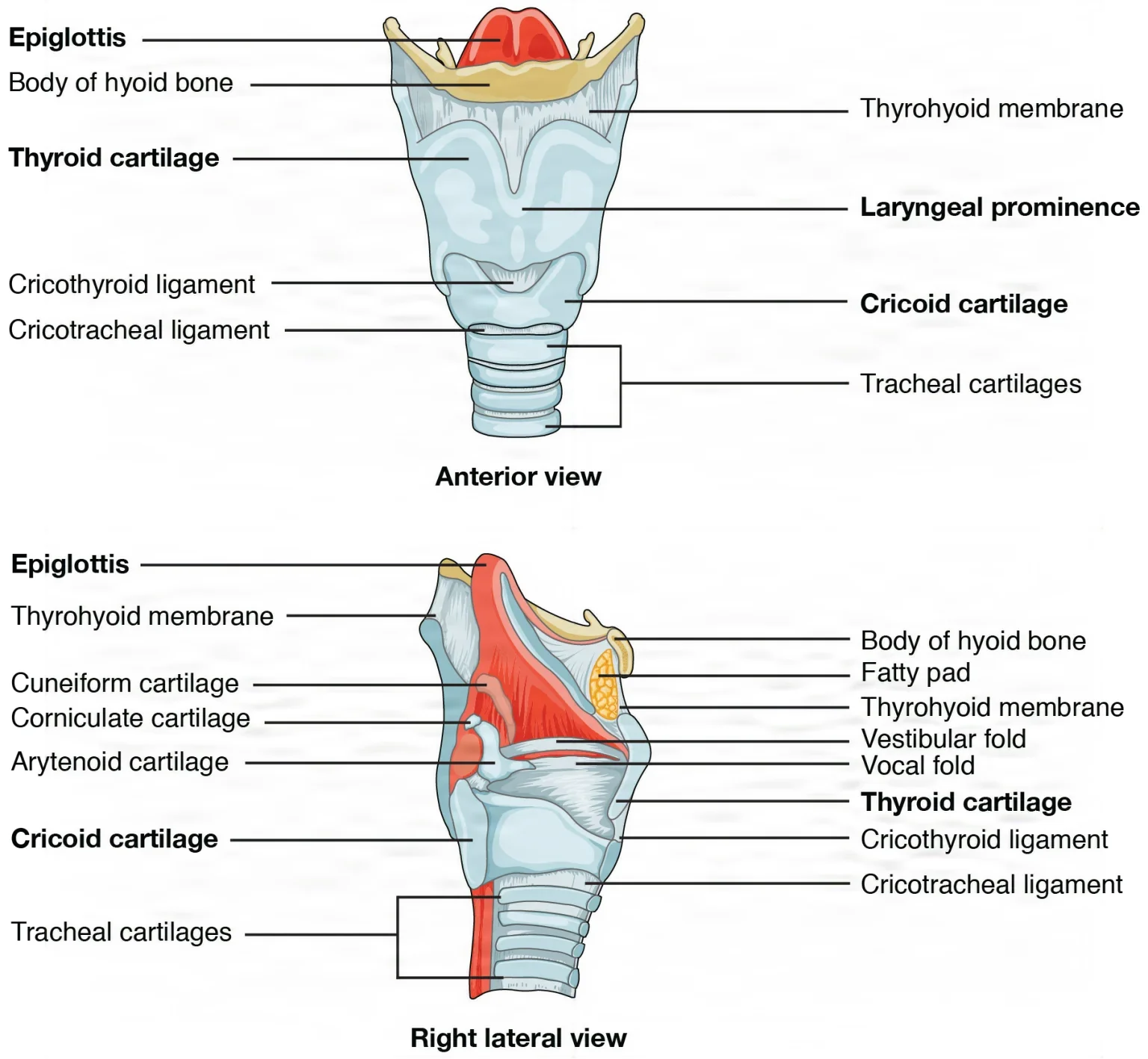
Nine pieces of cartilageKAR-tih-lijFirm, smooth, rubbery tissue that cushions joints and gives shape to parts such as the ear, nose and windpipe.chondr/o cartilageFull entry , joined by ligaments and membranes, build the larynx: three single ones and three pairs.
| Cartilage | What it is and does |
|---|---|
| Thyroid cartilage | The largest, a shield that forms the front wall. Its ridge is the laryngeal prominence, the Adam's apple, usually bigger in males |
| Cricoid cartilage | A ring just below the thyroid cartilage, narrow in front and wide at the back, like a signet ring. It is the only complete ring of cartilage in the upper airway (the tracheal rings below it are C-shaped), and it holds the airway open |
| epiglottisep-ih-GLOT-isA leaf-shaped flap of cartilage at the back of the throat that covers the windpipe during swallowing.epi- upon, overglott/o the opening of the larynxFull entry | A leaf-shaped flap of flexible elastic cartilage at the top of the larynx, attached to the thyroid cartilage. It folds down over the opening of the larynx when you swallow |
| Arytenoid cartilages (a pair) | Small cartilages that sit on the top of the back of the cricoid. The back ends of the true vocal cords attach to them, and they move the cords to make sound |
| Corniculate and cuneiform cartilages (two pairs) | Small cartilages attached to the arytenoids; the cuneiforms link the arytenoids to the epiglottis |
Between the thyroid and cricoid cartilages, in front, is a membrane, the cricothyroid membrane (ligament). In an emergency, when no other airway can be placed, the surgeon can open it and insert a tube, a cricothyrotomy. One reference notes that it can be faster than a standard tracheotomytray-kee-OT-uh-meeThe surgical cut made into the windpipe through the front of the neck.trache/o windpipe-tomy cutting, incisionFull entry , with fewer complications.
The vocal cords and the glottis
Inside the larynx, two pairs of folds of mucous membrane run from front to back.
- The upper pair are the vestibular folds, or false vocal cords.
- The lower pair are the true vocal cords, or vocal folds: white folds attached in front to the thyroid cartilage and behind to the arytenoid cartilages. Their inner edges are free, and they vibrate as air passes to make the voice.
The glottis is at the level of the true vocal cords. Surgical texts use the word for the opening between the true cords, the entrance to the trachea; one anatomy text counts both pairs of folds as part of the glottis too. The part of the larynx above the cords, with the epiglottis and false cords, is the supraglottis; below the cords, down to the bottom of the cricoid, is the subglottis.
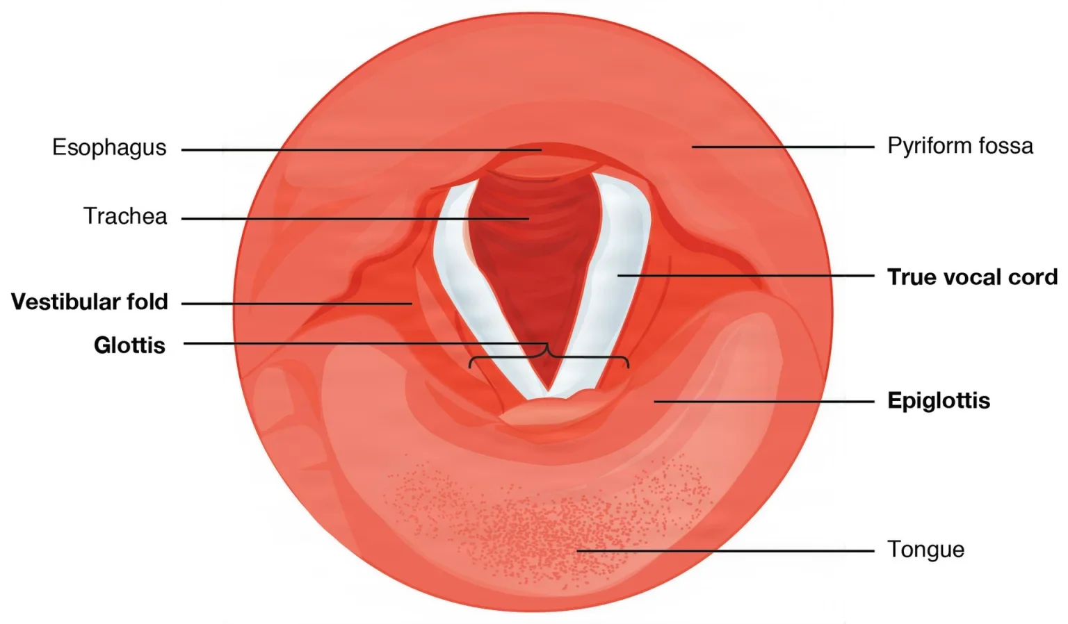
When you swallow, the larynx rises, the epiglottis swings down over its opening and the glottis closes, so food slides past into the esophagusih-SOF-uh-gusThe muscular tube that carries food and liquid from the throat to the stomach.esophag/o esophagusFull entry instead of the trachea.
The small muscles inside the larynx that move the vocal cords are worked by the recurrent laryngeal nerve, a branch of the vagus. Surgeons protect it during thyroid surgery, because an injured nerve leaves a vocal cord paralysispuh-RAL-uh-sisLoss of the ability to move a part of the body, and sometimes loss of feeling in it.para- beside, abnormal-lysis looseningFull entry (chapter 4).
In an adult, the opening at the vocal cords is the narrowest part of the airway. A child's airway is narrowest lower down, at the cricoid; chapter 13 explains the child's airway.
In a laryngoscopy, the surgeon looks directly at the larynx through a rigid laryngoscope passed through the mouth, often with the operating microscope, to examine it, take a biopsy or remove small growths. While the surgeon looks through the microscope, the scrub guides the long, fine instruments into the laryngoscope, because the surgeon does not look away to take them.
6The trachea and the carina
The trachea
The tracheaTRAY-kee-uhThe windpipe: the tube that carries air from the throat to the lungs.Full entry , or windpipe, runs from the bottom of the cricoid cartilageKAR-tih-lijFirm, smooth, rubbery tissue that cushions joints and gives shape to parts such as the ear, nose and windpipe.chondr/o cartilageFull entry , at about the level of the sixth cervicalSER-vih-kulRelating to the neck, or to the cervix, the lower end of the uterus.cervic/o neck, cervix-al relating toFull entry vertebra, down into the chest. It is about 9 to 15 cm long.
It is built of about 16 to 20 C-shaped rings of hyaline cartilage, stacked and joined by connective tissuekuh-NEK-tiv TISH-ooBody tissue that supports, binds together and protects other tissues and organs.Full entry . The rings keep the trachea from collapsing. They are open at the back, where smooth musclesmooth MUS-ulMuscle that works without conscious control and lines hollow organs such as the stomach, intestines, bladder, airways and blood vessels.Full entry (the trachealis) and elastic tissue close the tube and let it stretch a little with each breath. The esophagusih-SOF-uh-gusThe muscular tube that carries food and liquid from the throat to the stomach.esophag/o esophagusFull entry lies right behind that soft back wall.
Its lining has ciliaSIL-ee-uhTiny hair-like structures on the surface of some cells that move in waves.Full entry and goblet cells, like the nose, and keeps sweeping mucus up toward the larynxLAIR-inksThe voice box: the part of the airway in the neck, above the windpipe, that holds the vocal cords.laryng/o larynxFull entry to be swallowed.
In the neck, the isthmus of the thyroid gland crosses the front of the trachea.
A tracheotomytray-kee-OT-uh-meeThe surgical cut made into the windpipe through the front of the neck.trache/o windpipe-tomy cutting, incisionFull entry or tracheostomytray-kee-OS-tuh-meeA surgical opening through the front of the neck into the windpipe, usually holding a tube that the person breathes through.trache/o windpipe-stomy surgical openingFull entry makes an opening into the front of the trachea for an airway tube. It is done for a patient who needs a mechanical ventilationmeh-KAN-ih-kul ven-tuh-LAY-shunBreathing support from a machine, a ventilator, that pushes air into the lungs through a tube or a mask.Full entry for a long time, or whose upper airway is blocked by a tumor, an infection, secretionsih-KREE-shunA substance a gland or cell makes and releases, such as mucus, saliva, sweat or a hormone.Full entry , a foreign body or extra tissue in the throat that causes sleep apneasleep AP-nee-uhA sleep disorder in which breathing stops and starts again and again during sleep.Full entry . The surgeon cuts over the front of the trachea, may divide the thyroid isthmus to see it, lifts the trachea with a tracheal hook placed in the cricoid cartilage, and opens the trachea, usually between the third and fourth rings.
The carina
In the chest, at about the level of the sternal angle, the trachea divides into the right and left primary bronchusBRONG-kusOne of the large air tubes that branch from the windpipe into each lung.bronch/o airwayFull entry . The ridge on the inside of that split is the carina.
The carina is packed with sensitive nerve tissue, so anything that lands on it, such as a bit of food, sets off violent coughing. It is a landmark the surgeon uses during bronchoscopybrong-KOS-kuh-peeA procedure in which a thin, lighted tube with a camera is passed through the nose or mouth to look inside the airways.bronch/o airway (bronchus)-scopy looking with an instrumentFull entry , and the tip of an endotracheal tubeen-doh-TRAY-kee-ul toobA flexible plastic tube passed through the mouth or nose into the windpipe to keep the airway open and allow breathing support.endo- withintrache/o windpipe-al relating toFull entry is placed a few centimeters above it.
The right primary bronchus is wider and runs more in line with the trachea than the left one, which bends off at a sharper angle. So an inhaled object, or a tube pushed in too far, usually ends up on the right side.
In a bronchoscopy, the surgeon passes a scope through the mouth or nose, through the larynx and trachea, past the carina and into the bronchi, to look, take biopsies or remove a foreign body. A flexible bronchoscope reaches farther into the smaller branches, and the patient breathes around it. A rigid bronchoscope is a wider tube, used under general anesthesiaJEN-er-ul an-es-THEE-zhuhDrug-induced loss of consciousness and feeling over the whole body, so a person feels nothing and is not aware during surgery.an- withoutesthesi/o feeling, sensation-ia conditionFull entry when large instruments are needed, and the patient can be ventilated through it. When cancer is suspected, the healthy lung is examined first, so the scope does not carry cancer cells into it.
7The bronchial tree
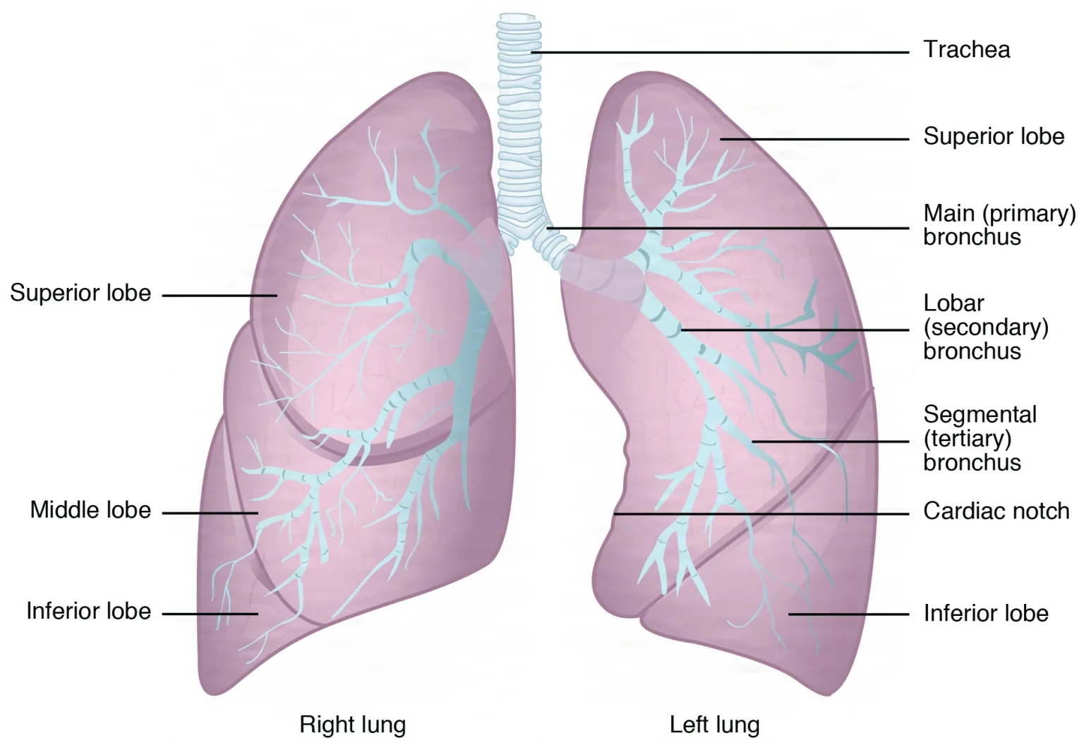
The primary bronchusBRONG-kusOne of the large air tubes that branch from the windpipe into each lung.bronch/o airwayFull entry enter the lungs and branch again and again, like an upside-down tree. All of these branches together are the bronchial tree.
- Each primary (main) bronchus serves one lung.
- It divides into secondary (lobar) bronchi, one for each lobe: three on the right and two on the left.
- These divide into tertiary (segmental) bronchi, one for each bronchopulmonary segment.
- The tertiary bronchi branch into bronchioleBRONG-kee-ohlOne of the smallest airways in the lungs, branching from the bronchi and leading to the air sacs.bronch/o airway (bronchus)-iole smallFull entry , about 1 mm across, which keep branching down to the terminal bronchioles. Each lung has more than 1000 of these.
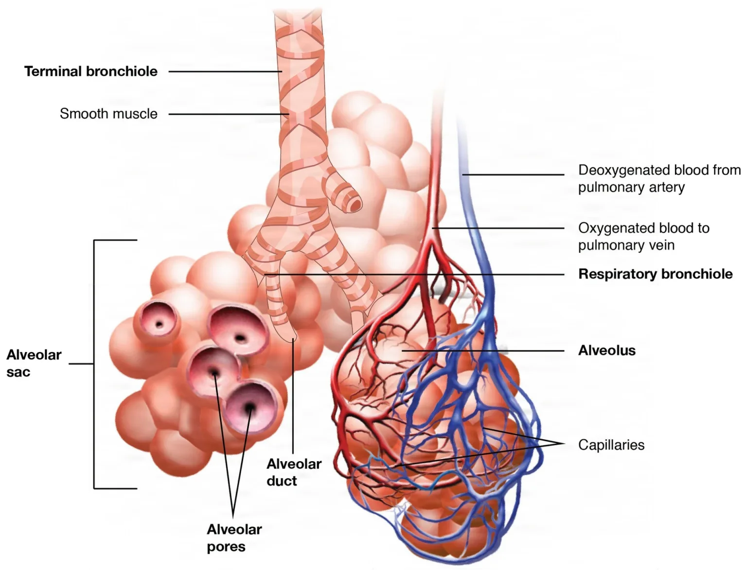
- Each terminal bronchiole leads into respiratory bronchioles, the smallest kind, which open into alveolusal-VEE-uh-lus (plural alveoli, al-VEE-uh-ly)One of the tiny air sacs at the ends of the smallest airways in the lungs.Full entry ducts and clusters of alveoli.
Rings of cartilageKAR-tih-lijFirm, smooth, rubbery tissue that cushions joints and gives shape to parts such as the ear, nose and windpipe.chondr/o cartilageFull entry hold the primary bronchi open. As the branches get smaller, the rings become plates, and the bronchioles have no cartilage at all, only smooth musclesmooth MUS-ulMuscle that works without conscious control and lines hollow organs such as the stomach, intestines, bladder, airways and blood vessels.Full entry that can narrow or widen the tube.
That muscle answers to the autonomicaw-tuh-NOM-ikWorking on its own, without conscious control; used for the nerves that run the organs.auto- selfnom/o rule, law-ic relating toFull entry nerves: the parasympatheticpair-uh-sim-puh-THET-ikRelating to the part of the autonomic nervous system that calms the body and supports "rest and digest" functions.para- besidesympathetic the other autonomic divisionFull entry nerves narrow the airways and the sympathetic nerves widen them. In asthma, the bronchioles go into spasmSPAZ-umA sudden, involuntary tightening of a muscle or of the muscle in a hollow organ or tube.Full entry (bronchospasmBRONG-koh-spaz-umA sudden tightening of the muscles around the airways that narrows them.bronch/o bronchus-spasm sudden involuntary contractionFull entry ), the lining swells and extra mucus clogs them, so air has trouble getting in and out.
8The alveoli and gas exchange
The air sacs
An alveolusal-VEE-uh-lus (plural alveoli, al-VEE-uh-ly)One of the tiny air sacs at the ends of the smallest airways in the lungs.Full entry is a tiny, grape-like air sac about 0.2 mm (200 micrometers) across, with stretchy walls. Clusters of them, the alveolar sacs, open off the alveolar ducts, and small pores between neighbors keep the air pressure even.
Three kinds of cells make up the alveolar wall.
- Type I cells: very thin, flat cells that cover most of the surface, up to 97 percent. Gases pass through them easily.
- Type II cells: scattered among the type I cells. They make surfactantser-FAK-tuntA fatty substance coating the inside of the lungs' air sacs that lowers surface tension so they stay open.Full entry , a mix of fats and proteins that lowers the surface tension of the thin film of water lining each alveolus. Without surfactant, the alveoli would collapse every time you breathe out.
- Alveolar macrophageMAK-roh-fayjA large white blood cell in the tissues that swallows and digests germs, dead cells and debris.macro- large-phage eaterFull entry : roaming immune cells that eat the dust and germs that reach the alveoli.
When alveoli collapse, the lung or part of it goes airless. This is atelectasisat-uh-LEK-tuh-sisThe collapse of part or all of a lung, in which the tiny air sacs close and hold no air.atel- incomplete-ectasis expansionFull entry . Mucus plugs, too little surfactant, pressure on the lung, and shallow breathing all cause it, and shallow breathing because of pain after chest or upper abdominal surgery makes it a known complication of those operations.
How the gases cross
The thin alveolar wall and the thin wall of the capillaryKAP-ih-lair-eeOne of the tiniest blood vessels, joining the smallest arteries to the smallest veins.Full entry pressed against it form the respiratory membrane, only about 0.5 micrometer thick. Spread out, the lungs' exchange surface covers about 70 square meters.
Gases cross it by simple diffusiondih-FYOO-zhunThe movement of particles from an area where they are more concentrated to one where they are less concentrated.dif- apartfus pour-ion processFull entry , with no energy spent: each gas moves from where its pressure is higher to where it is lower.
- Oxygen is at about 104 mm Hg in the alveoli but only about 40 mm Hg in the blood coming back from the body, so it moves into the blood.
- carbon dioxideKAR-bun dy-OK-sideA gas made by the body's cells as waste, carried in the blood to the lungs and breathed out.Full entry is at about 45 mm Hg in that blood and about 40 mm Hg in the alveoli, so it moves out into the air to be breathed out. That gap is small, but carbon dioxide dissolves about 20 times more easily than oxygen, so about the same amount of each gas crosses.
Most of the oxygen rides on hemoglobin in the red blood cells, which turns bright red when it is loaded (chapter 5). Most of the carbon dioxide, about 70 percent, travels in the blood as bicarbonateby-KAR-buh-naytA base in the blood that balances acid; the kidneys control it, and its level is measured on blood gas and chemistry tests.Full entry .
This exchange in the lungs is external respiration. The reverse exchange out in the tissues, where oxygen leaves the blood and carbon dioxide enters it, is internal respiration.
For the exchange to work, two things have to match. ventilationven-tuh-LAY-shunAir moving in and out of the lungs as a person breathes.Full entry means air moving in and out of the lungs. perfusionper-FYOO-zhunThe flow of blood through the body's tissues, bringing them oxygen and nutrients.per- throughfus pour-ion processFull entry means blood flowing through the capillaries around the alveoli. An area that gets air but little blood, or blood but little air, exchanges gas poorly, so the body narrows the vessels to poorly aired alveoli and sends the blood where the air is.
9The lungs
The lungs are two pyramid-shaped organs in the chest, one on each side of the mediastinum, the middle space that holds the heart, the great vessels, the tracheaTRAY-kee-uhThe windpipe: the tube that carries air from the throat to the lungs.Full entry , the esophagusih-SOF-uh-gusThe muscular tube that carries food and liquid from the throat to the stomach.esophag/o esophagusFull entry and the thymusTHY-musA small gland in the upper chest, behind the breastbone, where T cells (a kind of white blood cell) mature.Full entry (chapter 2).
- The apex, the narrow top of each lung, rises into the base of the neck, just above the first rib and the collarbone.
- The base rests on the diaphragmDY-uh-framThe dome-shaped sheet of muscle under the lungs that is the main muscle of breathing; also the flat side of a stethoscope.Full entry .
- The surface that faces the ribs is the costal surface, and the surface that faces the middle is the mediastinal surface.
Right and left sides, and the lobes
Deep grooves called fissureFISH-erA narrow crack or split in the skin, a lining or an organ.Full entry divide each lung into lobes. The two lungs are not mirror images.
| Right lung | Left lung | |
|---|---|---|
| Lobes | Three: superior (upper), middle and inferior (lower) | Two: superior (upper) and inferior (lower) |
| Fissures | Two, the oblique and the horizontal | One, the oblique |
| Shape | Shorter and wider | Smaller, with the cardiac notch, a dent that makes room for the heart |
Bronchopulmonary segments
Each lobe is made of smaller units, the bronchopulmonary segments. Each segment gets its air from its own tertiary bronchusBRONG-kusOne of the large air tubes that branch from the windpipe into each lung.bronch/o airwayFull entry and its blood from its own branch of the pulmonary arteryPUL-muh-nair-ee AR-ter-eeThe large blood vessel that carries blood low in oxygen from the right side of the heart to the lungs.Full entry . Each lung has about 10. On the left, some segments share a bronchus and are counted together, so you may see fewer listed.
Segments barely connect to each other, so they are the lung's surgical units: a diseased segment can be removed with little effect on its neighbors.
The hilum
On the mediastinal surface of each lung is a dent, the hilum. The primary bronchus, the pulmonary artery and veins, the bronchial vessels, the nerves and the lymphLIMFThe clear fluid that flows through the lymphatic vessels and carries white blood cells.Full entry vessels all enter or leave the lung there, and together they form the root of the lung. The nerves gather there in a network called the pulmonary plexusPLEK-susA network of nerves or blood vessels that branch and join together.Full entry .
At the hilum of the right lung, the phrenic nerve runs down in front of the root, and the vagus nerveVAY-gus nervThe 10th cranial nerve, which runs from the brain stem to the chest and belly and slows the heart and helps digestion.Full entry passes behind it with the esophagus. The surgeon keeps both out of harm when retracting the lung.
Removing lung tissue
Lung operations are named for how much they remove.
- Wedge resectionrih-SEK-shunSurgery to remove part of an organ or tissue.re- back, againsect to cut-ion processFull entry : a small wedge of lung taken with a stapler, as in a lung biopsy.
- Segmentectomy (segmental resection): one bronchopulmonary segment, taken at its own artery and bronchus.
- Lobectomy: one or more lobes, most often for lung cancer. The surgeon divides the lobe's branches of the pulmonary artery and vein at the hilum, then clamps and divides its bronchus. While the bronchus is open, suction keeps blood and fluid from running into the other lung.
- Pneumonectomy: a whole lung.
Most lung operations, 80 to 90 percent, are now done by video-assisted thoracoscopic surgery (VATS), through small ports instead of a painful muscle-splitting incision. The instruments for an open chest are still kept close by, so the team can convert to an open operation quickly.
10The blood supply of the lungs
The lungs have two blood supplies.
- The pulmonary circulationPUL-muh-nair-ee ser-kyuh-LAY-shunThe path blood takes from the heart to the lungs, where it picks up oxygen, and back to the heart.pulmon/o lung-ary relating toFull entry brings blood to the alveolusal-VEE-uh-lus (plural alveoli, al-VEE-uh-ly)One of the tiny air sacs at the ends of the smallest airways in the lungs.Full entry for gas exchangeGAS eks-CHAYNJThe movement of oxygen from the air sacs of the lungs into the blood and of carbon dioxide from the blood into the air sacs.Full entry . The right ventriclerite VEN-trih-kulThe lower right chamber of the heart, which pumps blood to the lungs.Full entry pumps oxygen-poor blood into the pulmonary trunk, which splits into the right and left pulmonary arteryPUL-muh-nair-ee AR-ter-eeThe large blood vessel that carries blood low in oxygen from the right side of the heart to the lungs.Full entry . Each artery branches alongside the bronchusBRONG-kusOne of the large air tubes that branch from the windpipe into each lung.bronch/o airwayFull entry until it becomes the capillaryKAP-ih-lair-eeOne of the tiniest blood vessels, joining the smallest arteries to the smallest veins.Full entry network around the alveoli. The oxygen-rich blood leaves through the pulmonary veinPUL-muh-nair-ee VAYNOne of the four veins that carry oxygen-rich blood from the lungs to the left atrium of the heart.Full entry , two from each lung, which exit at the hilum and empty into the left atriumAY-tree-umOne of the two upper chambers of the heart, which receive blood coming back to the heart.atri/o atrium-um noun endingFull entry .
- The bronchial arteries, small branches of the aorta, carry oxygen-rich blood to feed the lung tissue itself: the bronchi, the bronchioleBRONG-kee-ohlOne of the smallest airways in the lungs, branching from the bronchi and leading to the air sacs.bronch/o airway (bronchus)-iole smallFull entry and the visceralVIS-er-ulHaving to do with the internal organs, especially those in the chest and belly.viscer/o internal organs-al relating toFull entry pleuralPLOOR-ulHaving to do with the pleura, the thin two-layer membrane that covers the lungs and lines the chest wall.pleur/o pleura-al relating toFull entry . Their number varies from person to person.
So in the lungs, as chapter 5 explains, the arteries carry oxygen-poor blood and the veins carry oxygen-rich blood, the reverse of the rest of the body.
The pulmonary vessels are fragile and are handled gently at surgery. A blood clot that lodges in a pulmonary artery, a pulmonary embolismPUL-muh-nair-ee EM-buh-liz-umA blockage of an artery in the lungs, usually by a blood clot that has traveled there from elsewhere in the body.pulmon/o lung-ary relating toembol/o plug-ism conditionFull entry , can be fatal; the clot usually comes from a deep vein, most often in a leg.
11The pleura
Two layers and a space
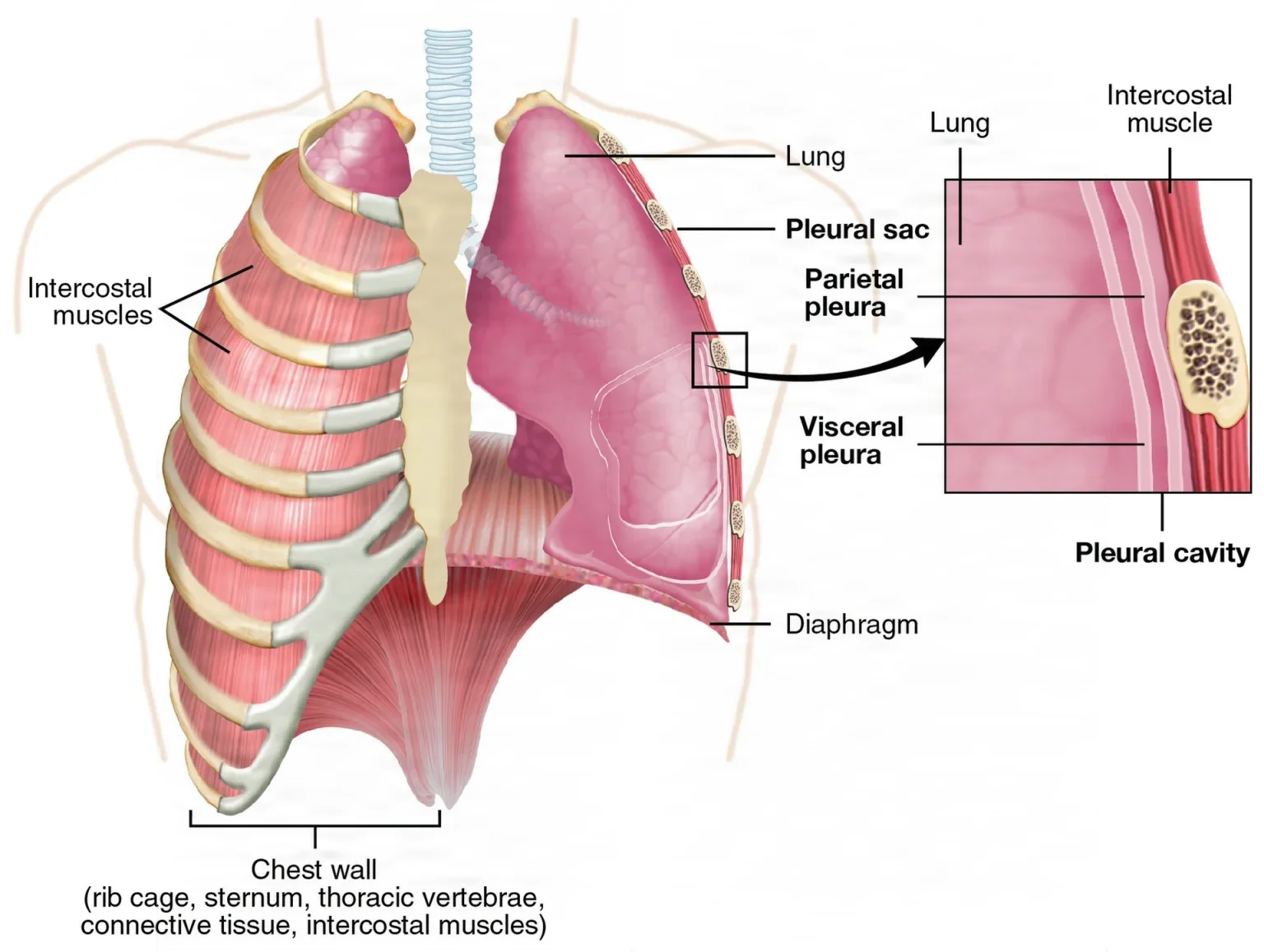
Each lung lies in its own sac of serousSEER-usThin, watery and clear to pale yellow, like the fluid part of blood.Full entry membrane, the pleuralPLOOR-ulHaving to do with the pleura, the thin two-layer membrane that covers the lungs and lines the chest wall.pleur/o pleura-al relating toFull entry (chapter 2).
- The visceralVIS-er-ulHaving to do with the internal organs, especially those in the chest and belly.viscer/o internal organs-al relating toFull entry pleura covers the lung itself and dips into its fissureFISH-erA narrow crack or split in the skin, a lining or an organ.Full entry .
- The parietalpuh-RY-uh-tulRelating to the wall of a body cavity; also names a lobe of the brain and two bones of the skull.pariet/o wall-al relating toFull entry pleura lines the inside of the chest wall, the side of the mediastinum and the top of the diaphragmDY-uh-framThe dome-shaped sheet of muscle under the lungs that is the main muscle of breathing; also the flat side of a stethoscope.Full entry .
- The two layers are one sheet: at the hilum, the membrane folds back over the root of the lung.
Between the layers is the pleural spacePLOOR-ul SPAYSThe thin space between the two layers of the pleura, the membrane that covers the lungs and lines the chest wall.Full entry , or pleural cavity. It is a potential space: the two layers touch, with only a thin film of pleural fluid between them, made by both layers. The fluid lets the layers slide without frictionFRIK-shunThe rubbing of one surface against another, such as skin against bed sheets.Full entry as you breathe, and its surface tension holds the lung against the chest wall, so the lung follows the chest wall out. The pleura also walls each lung off from the other organs, which keeps their movements apart and helps stop infection from spreading.
Inflammation of the pleura is pleuritis.
The pleural recesses
The lungs do not fill the pleural sacs all the way to their edges. Where two parts of the parietal pleura lie against each other with no lung between them, they form pockets, the pleural recesses.
- The costodiaphragmatic recess is the lowest edge of each sac, where the pleura on the ribs meets the pleura on the diaphragm. Fluid in the chest can collect there.
- The costomediastinal recesses lie behind the sternum, where the pleura on the ribs meets the pleura on the mediastinum. The left one is larger, because of the cardiac notch.
The diaphragm is a dome that rises as high as the fourth intercostalin-ter-KOS-tulBetween the ribs.inter- betweencost/o rib-al relating toFull entry space, and the pleural sac reaches down around its edges, to about the eighth rib in front and the tenth rib at the side. So an incision made too low can miss the chest altogether and pass through the diaphragm into the abdomen.
The pressure that holds the lung open
The pressure inside the pleural space stays a little lower than the pressure in the lungs, about 4 mm Hg below it. The lungs' elastic tissue keeps pulling inward, away from the chest wall, while the chest wall pulls outward; the slight vacuum between them keeps the lungs stretched open.
When air, blood or fluid gets in
Anything that fills the pleural space takes away the negative pressureNEG-uh-tiv PRESH-erPressure that is lower than the pressure around it, so air or fluid is drawn in.Full entry . The lung can no longer follow the chest wall, so it cannot expand fully, and it may collapse.
| Problem | What is in the pleural space |
|---|---|
| pneumothoraxnoo-moh-THOR-aksAir in the space between a lung and the chest wall, which lets part or all of that lung collapse.pneum/o air, lungthorac/o chestFull entry | Air, for example from a wound through the chest wall; it keeps the lung from expanding and can push the structures in the middle of the chest aside |
| Hemothorax | Blood, usually from an injury |
| pleural effusionPLOOR-ul eh-FYOO-zhunA buildup of extra fluid in the pleural space, between the lining of the lung and the chest wall.Full entry | Extra fluid, such as serous fluid, pus or blood |
| Empyema | Pus, from an infection |
Long-lasting inflammation, infection or a tumor can lay down a stiff fibrousFY-brusMade of or containing tough threads (fibers), especially of connective tissue.fibr/o fiber-ous full of, relating toFull entry layer, called a peel, over the pleura, which traps the lung. Decortication strips it away so the lung can expand again.
Opening the chest
When the surgeon opens the chest wall in a thoracotomythor-uh-KOT-uh-meeA surgical cut through the chest wall to reach the lungs, heart or other organs in the chest.thorac/o chest-tomy cutting intoFull entry , air rushes into the pleural space and the lung on that side collapses. During the operation, the anesthesia provideran-us-THEE-zhuh pruh-VY-derThe licensed professional who gives anesthesia and watches over the client's breathing and vital functions during a procedure.Full entry ventilates the lungs with a machine. For many chest operations, a double-lumenLOO-menThe open space inside a tube, such as a blood vessel, the intestine or a catheter.Full entry endotracheal tubeen-doh-TRAY-kee-ul toobA flexible plastic tube passed through the mouth or nose into the windpipe to keep the airway open and allow breathing support.endo- withintrache/o windpipe-al relating toFull entry lets the operated lung stay collapsed, out of the surgeon's way, while the other lung gets the oxygen and anesthetican-us-THET-ikA drug that causes loss of feeling, with or without loss of consciousness.an- withoutesthesi/o feeling, sensation-ic relating toFull entry .
At the end, chest tubechest toobA tube placed through the chest wall into the space around the lung to drain air, blood or fluid.Full entry are left in the pleural space and connected to a closed drainage system. It removes air and fluid and restores the negative pressure, so the lung can expand again. A chest X-ray afterward checks that the lung has not collapsed.
12How air moves
Pressure and volume
Air moves for one reason: it flows from higher pressure to lower pressure.
In a closed space at a steady temperature, pressure and volume work in opposite directions. Make the space bigger and the pressure inside drops; make it smaller and the pressure rises. This is Boyle's law, and it runs every breath.
Three pressures matter.
- Atmospheric pressure: the pressure of the air around the body, 760 mm Hg at sea level.
- Intrapulmonary pressure, also called intra-alveolusal-VEE-uh-lus (plural alveoli, al-VEE-uh-ly)One of the tiny air sacs at the ends of the smallest airways in the lungs.Full entry pressure: the pressure of the air inside the alveoli. It dips and rises during each breath and evens out with atmospheric pressure between breaths.
- Intrapleural pressure: the pressure in the pleural spacePLOOR-ul SPAYSThe thin space between the two layers of the pleura, the membrane that covers the lungs and lines the chest wall.Full entry , which stays a little below the pressure in the lungs.
The lungs have no muscle to move air themselves. They are passive: the pleuralPLOOR-ulHaving to do with the pleura, the thin two-layer membrane that covers the lungs and lines the chest wall.pleur/o pleura-al relating toFull entry fluid ties them to the chest wall, so they grow and shrink as the chest does.
Breathing in
Inspiration takes muscle work.
- The diaphragmDY-uh-framThe dome-shaped sheet of muscle under the lungs that is the main muscle of breathing; also the flat side of a stethoscope.Full entry contracts. Its dome flattens and moves down toward the abdomen, making the chest taller.
- The external intercostalin-ter-KOS-tulBetween the ribs.inter- betweencost/o rib-al relating toFull entry muscles, between the ribs, contract and lift the ribs up and out, making the chest deeper from front to back and wider.
- The lungs stretch with the chest, so the pressure in the alveoli falls a little below atmospheric pressure.
- Air flows in through the airway until the two pressures are equal again.
Breathing out
Quiet expirationek-spuh-RAY-shunBreathing out; letting air flow out of the lungs. Also the date after which a product should not be used.ex- outspir/o breathe-ation processFull entry is passive. The diaphragm and the intercostal muscles relax, the stretchy lung tissue recoils, and the chest gets smaller. The pressure in the alveoli rises above atmospheric pressure, and air flows out.
Breathing hard
Quiet breathing at rest is called eupnea. When the body needs more air, as in exercise, more muscles join in.
- To breathe in harder, accessory muscles in the neck, the sternocleidomastoid and the scalenes, lift the top of the chest.
- To breathe out hard, the internal intercostal muscles pull the ribs down and together, and the abdominal muscles squeeze the abdominal organs up against the diaphragm.
Diaphragm down and ribs up: the chest gets bigger, the pressure in the lungs drops, and air flows in. The muscles relax: the lungs recoil, the pressure rises, and air flows out.
Two things make breathing harder work. Narrow airways resist airflow, and the size of the airway matters more than anything else. A stiff chest wall resists stretching: thick scar from deep burns over the chest, for example, can keep the chest from expanding.
13What controls breathing
You breathe without thinking about it, yet you can hold your breath, talk or sing at will. Breathing is under both nervous and chemical control.
Nervous control
- The medullamuh-DUL-uhThe inner part of an organ; most often the medulla oblongata, the lowest part of the brainstem.Full entry respiratory center: groups of neuronNOOR-onA nerve cell: a cell that carries electrical and chemical signals through the nervous system.neur/o nerveFull entry in the medulla oblongata, the lowest part of the brain stemBRAYN STEMThe lowest part of the brain, which connects it to the spinal cord and controls breathing, heart rate and other automatic functions.Full entry (chapter 4), set the basic rhythm. They fire to start each breath in; when they go quiet, the muscles relax and the breath goes out.
- The ponsPONZThe part of the brain stem between the midbrain and the medulla.Full entry : respiratory centers in the pons, just above the medulla, adjust the rate and the depth of breathing.
- The phrenic nerves carry the signal from the spinal cord in the neck to the diaphragmDY-uh-framThe dome-shaped sheet of muscle under the lungs that is the main muscle of breathing; also the flat side of a stethoscope.Full entry . They come from C3 to C5, so a patient whose spinal cord is cut above C3 cannot breathe without help (chapter 4).
- The intercostalin-ter-KOS-tulBetween the ribs.inter- betweencost/o rib-al relating toFull entry nerves, the thoracicthuh-RAS-ikRelating to the chest, or to the middle part of the spine.thorac/o chest-ic relating toFull entry spinal nerveSPY-nul nervOne of the 31 pairs of nerves that branch from the spinal cord to carry signals to and from the body.Full entry that run between the ribs, carry the signal to the intercostal muscles.
- The vagus nerveVAY-gus nervThe 10th cranial nerve, which runs from the brain stem to the chest and belly and slows the heart and helps digestion.Full entry carries signals from stretch receptorrih-SEP-terA structure on or in a cell that a specific chemical, such as a hormone or drug, attaches to in order to act.Full entry in the lungs back to the brain. As the lungs fill, these signals stop the breath in before the lungs overinflate; this is the inflation, or Hering-Breuer, reflexREE-fleksAn automatic body response to something that stimulates it, which happens without thought, such as the knee jerk.Full entry . The vagus also carries the parasympatheticpair-uh-sim-puh-THET-ikRelating to the part of the autonomic nervous system that calms the body and supports "rest and digest" functions.para- besidesympathetic the other autonomic divisionFull entry fibers that narrow the airways.
- The cerebral cortexsuh-REE-brul KOR-teksThe thin, folded outer layer of the cerebrum, where thinking, language, memory, voluntary movement and sensation are handled.Full entry lets you take over for a while, to hold a breath or to speak.
Breathing depends on all of this working. Drugs that depress the brain, such as barbituratebar-BICH-er-itA type of sedative drug that slows the brain and nervous system, used for some seizures and in anesthesia.Full entry and morphineMOR-feenA strong opioid drug used to relieve moderate to severe pain.Full entry , can slow breathing or stop it altogether.
Chemical control
Sensors called chemoreceptors watch the chemistry of the blood and adjust breathing to match.
- carbon dioxideKAR-bun dy-OK-sideA gas made by the body's cells as waste, carried in the blood to the lungs and breathed out.Full entry is the main driver. As it builds up, it makes the body's fluids more acid. Central chemoreceptors in the brain stem sense this, and the respiratory center makes breathing faster and deeper, which blows off the extra carbon dioxide. When carbon dioxide is low, breathing slows and becomes shallow.
- Peripheral chemoreceptors, in the carotidkuh-ROT-idHaving to do with the carotid arteries, the two large arteries on each side of the neck that carry blood to the head and brain.Full entry bodies where each common carotid artery divides and in the aortic arch, sense carbon dioxide, acid and oxygen in the arterial blood.
- Oxygen is a weaker trigger. Low oxygen speeds breathing only when it falls a long way, to about 60 mm Hg or less, because hemoglobin keeps releasing oxygen as the dissolved oxygen drops.
Body temperature, pain and emotion change breathing too; a fever, for example, speeds it up.
The drugs given during surgery blunt the response to carbon dioxide, so a patient who is sedationsih-DAY-shunA calm, relaxed or sleepy state brought on by drugs, or the use of drugs to cause it.sed calm, settle-ation processFull entry or recovering from anesthesiaan-us-THEE-zhuhLoss of feeling or sensation, often brought on by drugs so that surgery or a procedure can be done without pain.an- withoutesthesi/o feeling, sensation-ia conditionFull entry may not breathe faster even as carbon dioxide rises.
14Lung volumes
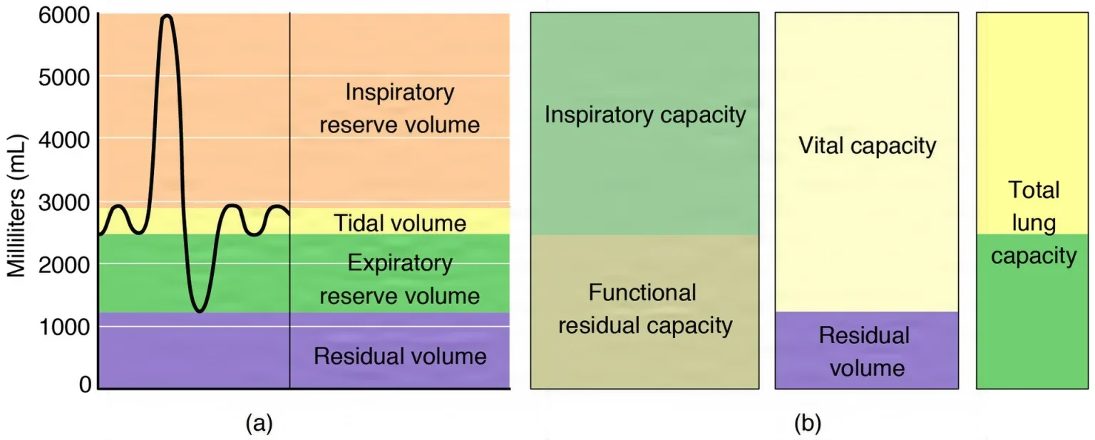
A spirometerspy-ROM-eh-terA device that measures how much air a person breathes in or out, and how fast.spir/o breathing-meter instrument for measuringFull entry measures how much air a person moves, as part of the pulmonary function testsPUL-muh-nair-ee FUNK-shun testsBreathing tests that measure how much air the lungs hold and how fast air moves in and out.pulmon/o lung-ary relating toFull entry done before lung surgery. The four basic volumes are separate slices of the air in the lungs, and the capacities add two or more volumes together. A person's size, age, sex and lung disease all change the vital capacity.
| Term | What it is |
|---|---|
| tidal volumeTY-dul VOL-yoomThe amount of air moved in or out of the lungs with one normal breath.Full entry (TV) | The air moved in or out with one normal, quiet breath, about 500 mL |
| Inspiratory reserve volume (IRV) | The extra air you can pull in with effort after a normal breath in |
| expirationek-spuh-RAY-shunBreathing out; letting air flow out of the lungs. Also the date after which a product should not be used.ex- outspir/o breathe-ation processFull entry reserve volume (ERV) | The extra air you can force out after a normal breath out, up to about 1200 mL in males |
| Residual volume (RV) | The air still in the lungs after you breathe out as hard as you can; it can never be breathed out, and it keeps the alveolusal-VEE-uh-lus (plural alveoli, al-VEE-uh-ly)One of the tiny air sacs at the ends of the smallest airways in the lungs.Full entry from collapsing |
| Vital capacity (VC) | The most air you can breathe out after the deepest breath in: TV + IRV + ERV, about 4000 to 5000 mL |
| Total lung capacity (TLC) | All the air the lungs hold after the deepest breath in: TV + IRV + ERV + RV, about 6000 mL in males and 4200 mL in females |
Two other capacities appear in reports. The inspiratory capacity is the tidal volume plus the inspiratory reserve volume. The functional residual capacity is the air left in the lungs after a normal breath out, the expiratory reserve volume plus the residual volume.
The minute volume is the air breathed out in one minute. Not all of each breath reaches the alveoli: the air that fills the conducting airways, the anatomical dead space, never takes part in gas exchangeGAS eks-CHAYNJThe movement of oxygen from the air sacs of the lungs into the blood and of carbon dioxide from the blood into the air sacs.Full entry .
Before lung surgery, these tests help predict how well the patient will breathe afterward. A patient who can force out less than 1 liter in the first second of a hard breath out is at extremely high risk of lung complications after surgery.
15Words for breathing problems
These are the Core Curriculum's terms. You will hear them in report and read them in the chart.
| Term | What it means |
|---|---|
| apneaAP-nee-uhA pause in breathing, or breathing that has stopped.a- without-pnea breathingFull entry | A stop in breathing. It happens at the start of some types of general anesthesiaJEN-er-ul an-es-THEE-zhuhDrug-induced loss of consciousness and feeling over the whole body, so a person feels nothing and is not aware during surgery.an- withoutesthesi/o feeling, sensation-ia conditionFull entry , one reason the patient is mechanical ventilationmeh-KAN-ih-kul ven-tuh-LAY-shunBreathing support from a machine, a ventilator, that pushes air into the lungs through a tube or a mask.Full entry |
| dyspneaDISP-nee-uhDifficult or labored breathing, or the feeling of not getting enough air.dys- difficult-pnea breathingFull entry | Difficult, labored or uncomfortable breathing; shortness of breath |
| cyanosissy-uh-NOH-sisA blue or gray color of the skin, lips or nail beds.cyan/o blue-osis abnormal conditionFull entry | A blue, gray or dark purple color of the skin or mucous membranes, such as the lips, from blood that is low in oxygen |
| Cheyne-Stokes respirationsCHAYN STOHKS res-puh-RAY-shunzA breathing pattern in which breaths grow deeper and then shallower in a repeating cycle, with pauses in breathing between cycles.Full entry | A repeating pattern: breaths grow deeper and faster, then fade into a pause with no breathing that lasts 10 to 60 seconds, then start again. It is seen with heart failurehart FAYL-yerA long-term condition in which the heart cannot pump enough blood to meet the body's needs.Full entry , with brain damage or raised pressure inside the skull, and near the end-of-life careEND-uv-LYF KAIRThe care given to a person and their family in the last days, weeks or months of life.Full entry |
| hypercapniahy-per-KAP-nee-uhToo much carbon dioxide in the blood, a PaCO2 above 45 mm Hg.hyper- abovecapn/o carbon dioxide-ia conditionFull entry | Too much carbon dioxideKAR-bun dy-OK-sideA gas made by the body's cells as waste, carried in the blood to the lungs and breathed out.Full entry in the blood, usually above 45 to 50 mm Hg; also called hypercarbia. It can bring dizziness, drowsiness and confusion |
| hyperventilationhy-per-ven-tih-LAY-shunBreathing faster or deeper than the body needs, so too much carbon dioxide is breathed out.hyper- above, excessiveventilation moving air in and out of the lungsFull entry | Breathing faster or deeper than the body needs, so the blood's carbon dioxide falls too low. The Core Curriculum lists tachypneatak-ip-NEE-uhA breathing rate faster than normal, above 20 breaths per minute in an adult.tachy- fast-pnea breathingFull entry and hyperpnea under it |
| Tachypnea | Fast breathing: in an adult, more than 20 breaths a minute |
| Hyperpnea | Deeper, and often faster, breathing. A degree of it is normal after exercise; pain, fever and some lung and heart diseases also cause it |
| hypoxiahy-POK-see-uhA lower than normal level of oxygen in the body's tissues.hypo- belowox/o oxygen-ia conditionFull entry | Too little oxygen reaching the tissues; hypoxemiahy-pok-SEE-mee-uhA level of oxygen in the blood below normal.hypo- belowox oxygen-emia in the bloodFull entry is too little oxygen in the blood |
Hypoxia has four kinds, named for the cause.
- Hypoxic hypoxia: too little oxygen gets into the arterial blood, as at high altitude, with a blocked airway or with fluid in the lungs.
- anemiauh-NEE-mee-uhA lower than normal number of red blood cells or amount of hemoglobin in the blood.a- without, lacking-emia blood conditionFull entry hypoxia: too little working hemoglobin to carry the oxygen, as after a hemorrhageHEM-uh-rijA large loss of blood from a blood vessel, inside the body or out of it.hem/o blood-rrhage bursting forthFull entry , in anemia or in carbon monoxide poisoning.
- ischemiais-KEE-mee-uhToo little blood flow to a part of the body, so the tissue does not get enough oxygen.isch- hold back-emia blood conditionFull entry hypoxia: the blood flow to a tissue is too low to deliver enough oxygen.
- Histotoxic hypoxia: the blood brings enough oxygen, but the cells cannot use it, as in cyanide poisoning.
Two more words: bradypneabrad-ip-NEE-uhBreathing that is slower than normal for the person's age.brady- slow-pnea breathingFull entry is breathing that is slower than normal, and atelectasisat-uh-LEK-tuh-sisThe collapse of part or all of a lung, in which the tiny air sacs close and hold no air.atel- incomplete-ectasis expansionFull entry , from earlier in this chapter, is collapsed, airless lung.
Counting breaths
An adult at rest breathes about 12 to 20 times a minute; one reference starts the normal range at 10. Children breathe faster: a baby under 1 year, about 30 to 60 times a minute.
To count breaths, watch the chest rise and fall without making a point of it, and count for one full minute. Note how deep and how regular the breaths are, as well as how many. Chapter 13 teaches the vital signs and the monitors in the OR.
16Why anesthesia cares
The airway under general anesthesia
As general anesthesiaJEN-er-ul an-es-THEE-zhuhDrug-induced loss of consciousness and feeling over the whole body, so a person feels nothing and is not aware during surgery.an- withoutesthesi/o feeling, sensation-ia conditionFull entry deepens, the patient loses the protective reflexREE-fleksAn automatic body response to something that stimulates it, which happens without thought, such as the knee jerk.Full entry , such as gagging and swallowing, can no longer keep the airway open, and the breathing response fails. Breathing may stop at inductionin-DUK-shunThe starting of a process on purpose, such as bringing on labor or putting a client under anesthesia.Full entry , so the anesthesia provideran-us-THEE-zhuh pruh-VY-derThe licensed professional who gives anesthesia and watches over the client's breathing and vital functions during a procedure.Full entry places an airway and breathes for the patient. The drugs also blunt the carbon dioxideKAR-bun dy-OK-sideA gas made by the body's cells as waste, carried in the blood to the lungs and breathed out.Full entry drive, and that effect can last into recovery.
The path of the endotracheal tube
An endotracheal tubeen-doh-TRAY-kee-ul toobA flexible plastic tube passed through the mouth or nose into the windpipe to keep the airway open and allow breathing support.endo- withintrache/o windpipe-al relating toFull entry runs from the mouth into the tracheaTRAY-kee-uhThe windpipe: the tube that carries air from the throat to the lungs.Full entry , through the anatomy of this chapter.
- The anesthesia provider uses a laryngoscope, a lighted instrument, to look past the tongue and the epiglottisep-ih-GLOT-isA leaf-shaped flap of cartilage at the back of the throat that covers the windpipe during swallowing.epi- upon, overglott/o the opening of the larynxFull entry to the vocal cords.
- The tube passes through the glottis, between the vocal cords, into the trachea.
- A balloon cuff near its tip is inflated to seal it against the wall of the trachea.
- In an adult, its tip should sit about 2 to 6 cm above the carina. Too high, it can damage the vocal cords; too deep, it slides into the right primary bronchusBRONG-kusOne of the large air tubes that branch from the windpipe into each lung.bronch/o airwayFull entry , and only the right lung gets air.
A larynxLAIR-inksThe voice box: the part of the airway in the neck, above the windpipe, that holds the vocal cords.laryng/o larynxFull entry mask airway (LMA) is a tube with a small mask on its end. It is placed without a laryngoscope, and the mask fits snugly over the larynx from above.
Guarding the lungs
- aspirationas-puh-RAY-shunBreathing food, liquid, saliva or stomach contents into the airway and lungs; also, drawing fluid out with suction or a needle.Full entry : stomach contents can come up and be breathed into the lungs, most of all at induction and as the patient wakes, causing an inflammation of the lungs called chemical pneumonitis. For a patient at risk, such as one with a full stomach, the anesthesia provider may ask for cricoid pressure, the Sellick maneuver. The cricoid cartilageKAR-tih-lijFirm, smooth, rubbery tissue that cushions joints and gives shape to parts such as the ear, nose and windpipe.chondr/o cartilageFull entry is a complete ring, unlike the C-shaped tracheal rings, so pressing it back toward the spine squeezes the esophagusih-SOF-uh-gusThe muscular tube that carries food and liquid from the throat to the stomach.esophag/o esophagusFull entry shut behind it. The pressure is held until the tube is in place and the anesthesia provider says to let go.
- laryngospasmluh-RING-goh-spaz-umA sudden, involuntary closing of the vocal cords that partly or fully blocks air from entering the lungs.laryng/o larynx (voice box)-spasm sudden involuntary tighteningFull entry : a spasmSPAZ-umA sudden, involuntary tightening of a muscle or of the muscle in a hollow organ or tube.Full entry of the muscles of the larynx that closes the glottis and can block the airway.
- bronchospasmBRONG-koh-spaz-umA sudden tightening of the muscles around the airways that narrows them.bronch/o bronchus-spasm sudden involuntary contractionFull entry : the bronchioleBRONG-kee-ohlOne of the smallest airways in the lungs, branching from the bronchi and leading to the air sacs.bronch/o airway (bronchus)-iole smallFull entry clamp down, as in an asthma attackAZ-muh uh-TAKA sudden worsening of asthma in which the airways tighten, swell and fill with mucus.Full entry , and air has trouble moving.
Chapter 13 teaches how the anesthesia provider prevents and treats these problems.
The lungs during and after surgery
- Most positions push the abdominal organs up against the diaphragmDY-uh-framThe dome-shaped sheet of muscle under the lungs that is the main muscle of breathing; also the flat side of a stethoscope.Full entry , which cuts its pull to about two-thirds and lowers the tidal volumeTY-dul VOL-yoomThe amount of air moved in or out of the lungs with one normal breath.Full entry (chapter 31). A patient who is short of breath while awake, such as during transport or under local or regional anesthesiaan-us-THEE-zhuhLoss of feeling or sensation, often brought on by drugs so that surgery or a procedure can be done without pain.an- withoutesthesi/o feeling, sensation-ia conditionFull entry , breathes easier with the head raised.
- In chest surgery, the anesthesia provider can let one lung collapse on purpose, through a double-lumenLOO-menThe open space inside a tube, such as a blood vessel, the intestine or a catheter.Full entry tube, so the surgeon can work.
- After surgery, pain in a chest or upper abdominal incision keeps patients from breathing deeply. Carbon dioxide builds up, the oxygen falls, and atelectasisat-uh-LEK-tuh-sisThe collapse of part or all of a lung, in which the tiny air sacs close and hold no air.atel- incomplete-ectasis expansionFull entry can follow.
If the blood on the field turns darker, or the patient's lips or skin look blue, tell the anesthesia provider and the surgeon at once. Both can mean the blood is short of oxygen.
What to take from this chapter
- The conducting zone, from the nose to the terminal bronchioles, cleans, warms and moistens the air. Gas exchange happens only in the respiratory zone: the respiratory bronchioles, alveolar ducts and alveoli, across a membrane about 0.5 micrometer thick.
- A breath runs nares, nasal cavity, choanae, nasopharynx, oropharynx, laryngopharynx, larynx, trachea, carina, primary and secondary bronchi, bronchioles, alveolar ducts and alveoli. The nasopharynx carries air only; the lower two parts of the pharynx carry air and food.
- The nasal septum divides the nose, three conchae (turbinates) line each side wall with a meatus under each, and the frontal, maxillary, ethmoid and sphenoid sinuses drain into the nose. FESS reopens that drainage.
- The larynx has nine cartilages. The thyroid cartilage is the largest, the cricoid is the only complete ring in the upper airway, the epiglottis covers the larynx when you swallow, and the true vocal cords run from the thyroid cartilage in front to the arytenoids behind.
- The trachea's C-shaped rings are open at the back. It splits at the carina, and the right primary bronchus is wider and straighter, so inhaled objects and tubes pushed too far go right.
- The right lung has three lobes and the left two, with the cardiac notch; each lung has about 10 bronchopulmonary segments, and everything enters at the hilum. Pulmonary arteries bring oxygen-poor blood for exchange, and bronchial arteries from the aorta feed the lung tissue.
- The visceral and parietal pleura hold a film of fluid and a slight negative pressure that keeps the lungs open. Air, blood, fluid or pus in the pleural space keeps the lung from expanding and can collapse it, and chest tubes on a closed drainage system restore the pressure.
- The diaphragm and external intercostals make inspiration; quiet expiration is passive recoil. The medulla sets the rhythm through the phrenic and intercostal nerves, the vagus reports stretch, and carbon dioxide is the main chemical drive. Know the volumes and the words for breathing problems.
Check yourself5 quick questions
Free for everyone. Pick an answer for each, then check them all at once.
In the ORWhere you'll use this
The chapter's whole question pool in sets of 10, with the answer explained after each one.
Part of the educators programThe chapter's words on spaced cards: the ones you miss come back sooner.
Part of the educators programGiven by your class's teacher. Each class draws its own 25 from the chapter's pool.
Part of the educators program
Suggest a fix
Something wrong, unclear or out of date in this chapter? Tell us and we will fix it for everyone. Your email is only used to reply.