The heart, blood and vessels
The blood, the heart that pumps it, the vessels that carry it and the lymph system that brings fluid home, with what each one means at the surgical field.
1Why it matters
Every cell in the body needs a steady supply of oxygen and food, and a way to get rid of its wastes. The circulatory system does that work. The heart pumps blood through a loop of vessels, out to the tissues in arteries and back in veins, and the blood carries what the cells need and takes away what they do not.
A second set of vessels, the lymphatic systemlim-FAT-ik SIS-tumThe network of lymph vessels, lymph nodes and organs such as the spleen and thymus that drains tissue fluid and helps fight infection.Full entry , runs alongside. It collects the fluid that leaks out of the blood vessels into the tissues, filters it through lymph nodelimf nohdA small, bean-shaped organ of the immune system that filters lymph fluid and holds white blood cells.Full entry and returns it to the blood.
You meet this system in every case.
- Bleeding in the surgical wound is expected. Stopping bleeding, knowing bright red arterial blood from dark venous blood, and helping the team keep track of blood loss start with the anatomy in this chapter.
- Some operations are on the vessels themselves. Surgeons open blocked arteries, bypass them with graftGRAFTTissue or a man-made material moved into place in surgery to replace or repair a damaged part.Full entry , repair bulging ones and build new paths for dialysisdy-AL-ih-sisA treatment that removes wastes and extra fluid from the blood when the kidneys can no longer do it.dia- through, apart-lysis separation, looseningFull entry .
- Heart surgery replaces valves, bypasses blocked coronary arteryKOR-uh-nair-ee AR-ter-eeOne of the blood vessels on the surface of the heart that bring oxygen-rich blood to the heart muscle.Full entry with grafts taken from the chest wall and the leg, and places pacemakerPAYS-may-kerA small device that sends electrical pulses to the heart to keep it beating at a set rate; also the heart's own natural rhythm center.Full entry .
- The lymphLIMFThe clear fluid that flows through the lymphatic vessels and carries white blood cells.Full entry system guides cancer surgery. The first lymph node a tumor drains to, the sentinel node, tells the surgeon whether the cancer has begun to spread.
This chapter starts with the blood, then the heart, then the vessels, and ends with the lymph system. Other chapters teach what it leaves out: cells and tissues in chapter 2; the blood vessels of the brain as part of the nervous system in chapter 4; the lungs and gas exchangeGAS eks-CHAYNJThe movement of oxygen from the air sacs of the lungs into the blood and of carbon dioxide from the blood into the air sacs.Full entry in chapter 6; immunityih-MYOO-nih-teeThe body's ability to resist or fight off a particular infection or disease.immun/o protection-ity stateFull entry and how infection spreads in chapter 10; shockshokA life-threatening state in which blood flow is too low to bring enough oxygen to the body's tissues.Full entry , hemorrhageHEM-uh-rijA large loss of blood from a blood vessel, inside the body or out of it.hem/o blood-rrhage bursting forthFull entry and wound healingwoond HEE-lingThe body's process of repairing damaged skin and tissue.Full entry in chapter 11; anticoagulantan-tee-koh-AG-yoo-luntA drug that slows the blood's ability to form clots.anti- againstcoagul/o clotting-ant an agent that doesFull entry , heparinHEP-uh-rinAn anticoagulant drug, given by injection or IV, that keeps blood clots from forming or growing.Full entry and blood productBLUD PROD-uktAny part of donated blood prepared for transfusion, such as red cells, plasma or platelets.Full entry as drugs in chapter 12; monitoring blood pressure, the electrocardiogramih-lek-troh-KAR-dee-oh-gramA test that records the heart's electrical activity as waves on paper or a screen.electr/o electricitycardi/o heart-gram recordFull entry and blood loss under anesthesiaan-us-THEE-zhuhLoss of feeling or sensation, often brought on by drugs so that surgery or a procedure can be done without pain.an- withoutesthesi/o feeling, sensation-ia conditionFull entry in chapter 13; hemostasishee-moh-STAY-sisThe stopping of bleeding, either by the body's own clotting process or by medical or surgical methods.hem/o blood-stasis stopping, standing stillFull entry at the field in chapter 29; and the heart and vascular operations in chapters 40 and 41.
2Blood
What blood is made of
Blood is a connective tissuekuh-NEK-tiv TISH-ooBody tissue that supports, binds together and protects other tissues and organs.Full entry with a liquid matrix. It has two parts: the formed elements, which are the red blood cells, the white blood cells and the plateletPLAYT-litA tiny cell fragment in the blood that sticks together at a wound to help form a clot.Full entry , and plasmaPLAZ-muhThe pale yellow liquid part of the blood, which carries the blood cells, proteins, nutrients and wastes.Full entry , the straw-colored fluid they float in.
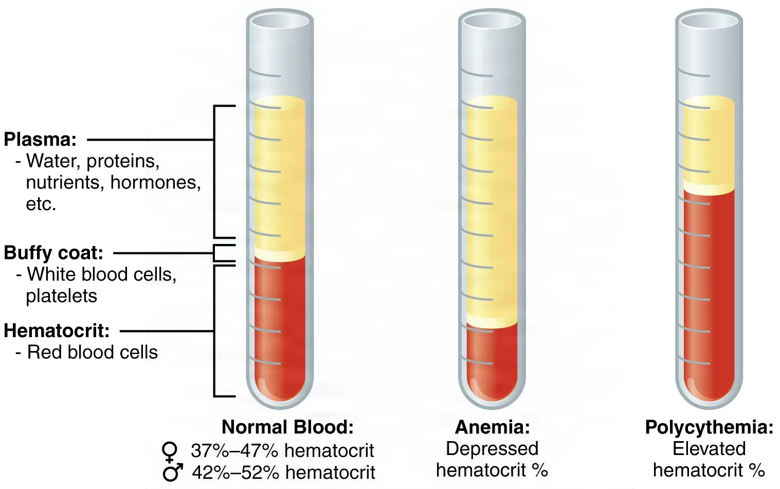
When a tube of blood is spun in a centrifuge, the heavy red cells sink to the bottom. The white cells and platelets form a thin pale band above them, called the buffy coat, which is less than 1 percent of the sample. The plasma sits on top. In normal blood, red cells make up about 45 percent of the volume.
Plasma is about 92 percent water. Most of what is dissolved in it is protein.
- albuminal-BYOO-minThe main protein in blood plasma, made by the liver, which holds fluid inside the blood vessels and carries drugs and hormones.Full entry , made by the liver, is the most plentiful plasma protein. It carries fats and some hormones, and it holds water inside the blood vessels, which helps keep up blood volume and blood pressure.
- Globulins carry iron, fats and vitamins. One group, the gamma globulins, are the antibodyAN-tih-bod-eeA Y-shaped protein made by the immune system that sticks to one specific germ or foreign substance and marks it for destruction.anti- againstbody Full entry the immune system makes.
- fibrinogen levelfy-BRIN-uh-jen LEV-ulA blood test that measures fibrinogen, a protein made by the liver that the body turns into fibrin to form clots.Full entry , also made by the liver, is the protein that turns into the threads of a blood clot.
- Plasma also carries electrolytes such as sodium, potassium and calcium, dissolved gases, nutrients such as glucose, and wastes.
serumSEER-umThe clear, yellow liquid left from blood after it clots and the cells are removed.Full entry is plasma with its clotting factorKLOT-ing FAK-terAny of the proteins in blood plasma that work together in a chain to form a blood clot.Full entry removed, the fluid left behind when blood clots. Many lab tests are run on serum.
An adult male has about 5 to 6 liters of blood and an adult female about 4 to 5 liters, roughly 8 percent of body weight. Blood is about five times thicker than water, and its normal pH, a measure of how acid or alkaline it is, stays between 7.35 and 7.45.
What blood does
The Core Curriculum names four jobs.
- Transport: blood carries oxygen from the lungs and nutrients from the gut to every cell, carries hormones to their targets, and takes carbon dioxideKAR-bun dy-OK-sideA gas made by the body's cells as waste, carried in the blood to the lungs and breathed out.Full entry to the lungs and other wastes to the kidneys and liver.
- Acid-base (pH) buffers: proteins and other chemicals in the blood act as buffers. They soak up extra acid or base and keep the pH of the body's fluids in its narrow normal range, working with the lungs and the kidneys.
- clottingKLOT-ingThe process by which blood thickens into a gel to plug a damaged vessel and stop bleeding; also called coagulation.Full entry : platelets and clotting proteins seal a torn vessel so the body does not bleed out.
- Protection: white blood cells attack germs that get in through a wound and destroy infected or abnormal cells.
Blood also spreads heat. It carries heat from the core out to the skin to cool the body, and pulls away from the skin to hold heat in when the body is cold.
Red blood cells
Red blood cells, or erythrocytes, are by far the most common formed element. A microliter of blood (a tiny drop) holds about 5 million of them. Their job is to carry oxygen from the lungs to the tissues, and to carry some carbon dioxide back.
A red blood cell is a flexible disk, thick at the rim and thin in the middle. As it matures in the bone marrowbone MAIR-ohThe soft, spongy tissue inside bones that makes blood cells.Full entry it pushes out its nucleus and most of its other parts, which leaves room for more hemoglobin. Its flexible shape lets it fold to squeeze through the narrowest capillaryKAP-ih-lair-eeOne of the tiniest blood vessels, joining the smallest arteries to the smallest veins.Full entry .
Hemoglobin is the protein that carries oxygen. Each hemoglobin molecule has four protein chains, and each chain holds a heme group with an iron atom at its center. Each iron atom binds one oxygen molecule. Hemoglobin full of oxygen is bright red, and hemoglobin that has given up oxygen in the tissues is a darker red. That is why arterial blood is brighter than venous blood.
Red cells are made in the red bone marrow, at a rate of more than 2 million a second, and the kidneys control the pace with a hormone called erythropoietinih-rith-roh-POY-uh-tinA hormone made mainly by the kidneys that tells the bone marrow to make red blood cells.erythr/o red-poietin substance that makesFull entry (EPO). They need iron, B vitamins and other raw materials. A red cell lives about 120 days. Then macrophageMAK-roh-fayjA large white blood cell in the tissues that swallows and digests germs, dead cells and debris.macro- large-phage eaterFull entry , large white cells in the spleenspleenAn organ in the upper left abdomen that filters the blood and helps fight infection.splen/o spleenFull entry , liver and bone marrow, break it down: the iron is saved and reused, and the rest of the heme becomes bilirubinbil-ih-ROO-binA yellow pigment made when old red blood cells break down, which the liver clears into bile.Full entry , a yellow waste the liver puts into bilebileA yellow-green fluid made by the liver and stored in the gallbladder that helps digest fats.chol/e bileFull entry .
Too few red cells or too little hemoglobin is anemiauh-NEE-mee-uhA lower than normal number of red blood cells or amount of hemoglobin in the blood.a- without, lacking-emia blood conditionFull entry , and the tissues may not get enough oxygen. Too many red cells is polycythemiapol-ee-sy-THEE-mee-uhHaving too many red blood cells in the blood.poly- manycyt/o cell-emia blood conditionFull entry , which makes the blood thick and harder for the heart to pump.
White blood cells
White blood cells, or leukocytes, defend the body. They are far fewer than red cells. Unlike red cells, they leave the blood vessels and squeeze into the tissues to fight infection where it is.
They come in two groups. The granular leukocytes have many visible granules in their cytoplasm and a nucleus with two to five lobes, which is why they are also called polymorphonuclear ("many-shaped nucleus"). The agranular leukocytes have smaller, less visible granules and a simple nucleus without distinct lobes, so they are also called mononuclear.
| Type | Share of white cells | What it does |
|---|---|---|
| neutrophilNOO-truh-filThe most common type of white blood cell, which is first to reach and destroy bacteria at a site of infection.Full entry (granular) | 55 to 70 percent, the most common | Rushes first to an infection and eats bacteria; a high count suggests a bacterial infection or inflammation |
| eosinophilee-oh-SIN-uh-filA type of white blood cell that fights parasites and takes part in allergic reactions.eosin a pink dye-phil attracted toFull entry (granular) | 1 to 3 percent | Fights parasitic worms and calms allergic reactions; a high count is seen in allergies |
| basophilBAY-soh-filThe least common type of white blood cell, which releases histamine during allergic and inflammatory reactions.bas/o base-phil attracted toFull entry (granular) | 1 percent or less, the least common | Releases histamineHIS-tuh-meenA chemical released by certain immune cells that widens blood vessels, makes them leaky and tightens airway muscles.hist/o tissue-amine a type of nitrogen-containing chemicalFull entry , which drives inflammation, and heparinHEP-uh-rinAn anticoagulant drug, given by injection or IV, that keeps blood clots from forming or growing.Full entry , which slows clotting |
| lymphocyteLIM-foh-syteA type of white blood cell that targets specific germs and abnormal cells, including T cells, B cells and natural killer cells.Full entry (agranular) | 20 to 35 percent | B cells make antibodies, T cells attack infected or abnormal cells, and natural killer cells destroy virus-infected and cancer cells |
| Monocyte (agranular) | 4 to 8 percent, the largest | Leaves the blood and becomes a macrophage, which eats germs, debris and worn-out cells |
All white cells start in the red bone marrow. Lymphocytes then do much of their growing and multiplying in lymphLIMFThe clear fluid that flows through the lymphatic vessels and carries white blood cells.Full entry tissue, such as the lymph nodelimf nohdA small, bean-shaped organ of the immune system that filters lymph fluid and holds white blood cells.Full entry . T lymphocytes mature in the thymusTHY-musA small gland in the upper chest, behind the breastbone, where T cells (a kind of white blood cell) mature.Full entry , and B lymphocytes in the bone marrow.
Platelets
Platelets, also called thrombocytes, are not whole cells. They are small fragments broken off a giant bone marrow cell called a megakaryocyte, each wrapped in a bit of membrane. A microliter of blood normally holds about 150,000 to 400,000 of them. A platelet lives about 10 days, and about one third of the body's platelets are stored in the spleen, ready for a bleed.
Platelets plug holes in damaged vessels and start clotting. They also release growth factors that help tissue repair. Too few platelets is thrombocytopeniathrom-boh-sy-toh-PEE-nee-uhA blood platelet count below normal, under 150,000/mm3.thromb/o clotcyt/o cell-penia lack, too fewFull entry , and the blood may not clot well.
How blood clots
Stopping bleeding is called hemostasishee-moh-STAY-sisThe stopping of bleeding, either by the body's own clotting process or by medical or surgical methods.hem/o blood-stasis stopping, standing stillFull entry . The body does it in three steps, one after the other.
- Vascular spasmSPAZ-umA sudden, involuntary tightening of a muscle or of the muscle in a hollow organ or tube.Full entry : the smooth musclesmooth MUS-ulMuscle that works without conscious control and lines hollow organs such as the stomach, intestines, bladder, airways and blood vessels.Full entry in the wall of the cut vessel clamps down hard, which slows the flow. The spasm usually lasts up to 30 minutes, sometimes for hours.
- Platelet plug: platelets stick to the torn edge where collagenKOL-uh-jenThe main structural protein of the body, which gives strength to skin, bones, tendons, ligaments and scar tissue.coll/a glue-gen producingFull entry is exposed, turn spiky and sticky, and call in more platelets. The pile seals a small hole for a while.
- coagulationkoh-ag-yuh-LAY-shunThe process by which blood changes from a liquid into a gel, forming a clot.coagul clot-ation processFull entry : a chain of reactions, the clotting cascade, builds a tough mesh of fibrin threads through the plug. Red cells and platelets are trapped in the mesh, and that is the clot.
The cascade is a row of clotting factors, mostly proteins made by the liver, each switching on the next. Two pathways start it: the extrinsic pathway, set off within seconds when damaged tissue releases tissue factor, and the slower intrinsicin-TRIN-zikComing from inside, or belonging naturally to something itself.Full entry pathway, set off inside the blood. Both lead to the common pathway: the enzymeEN-zimeA protein that speeds up a chemical reaction in the body without being used up.Full entry thrombinTHROM-binAn enzyme in the blood that turns fibrinogen into fibrin, the threads that hold a blood clot together.thromb/o clot-in substanceFull entry is made from prothrombin, and thrombin turns the dissolved protein fibrinogen into solid strands of fibrin. Calcium is needed at every stage, and the liver needs vitamin K to make several of the factors.
Once the clot forms, the platelets inside it contract and pull its edges together, squeezing out serum. As the vessel heals, the clot is slowly dissolved, a process called fibrinolysisfy-brin-OL-ih-sisThe body's process of breaking down a blood clot once it is no longer needed.fibrin/o fibrin, the protein threads of a clot-lysis breaking downFull entry , in which an enzyme called plasmin breaks down the fibrin. The body also makes natural anticoagulantan-tee-koh-AG-yoo-luntA drug that slows the blood's ability to form clots.anti- againstcoagul/o clotting-ant an agent that doesFull entry , substances that keep clotting from spreading past the injury. Heparin is one, and a drug form of heparin is given to surgical patients at risk of clots.
Clotting can fail. In hemophiliahee-muh-FIL-ee-uhAn inherited disorder in which the blood lacks a clotting factor, so it does not clot normally.hem/o blood-philia attraction, tendencyFull entry , an inherited disorder, the body does not make enough of one clotting factor, most often factor VIII, and the patient bleeds even from small wounds. Clotting can also happen where it should not. A thrombosisthrom-BOH-sisThe forming of a blood clot (a thrombus) inside a blood vessel or the heart.thromb/o clot-osis conditionFull entry is a clot inside an intact vessel; if a piece breaks off and travels, it is an embolismEM-buh-liz-umThe blocking of a blood vessel by a mass carried there in the blood (an embolus), such as a clot, fat or an air bubble.embol/o plug-ism conditionFull entry , and where it lodges it can block a vessel to the heart, brain or lungs.
disseminated intravascular coagulationdih-SEM-ih-nay-tid in-truh-VAS-kyuh-ler koh-ag-yuh-LAY-shunA disorder in which clotting is switched on throughout the blood vessels until the clotting factors are used up.intra- withinvascul/o vesselcoagul/o clottingFull entry (DIC) is a dangerous mix of both. Some serious condition, such as sepsisSEP-sisA severe whole-body response to an infection that damages the body's own organs.Full entry , major trauma or a massive blood transfusionBLUD trans-FYOO-zhunGiving blood, or a part of blood, from a donor into a person's vein.trans- acrossfus/o pourFull entry , sets off clotting all over the body at once. Tiny clots form throughout the small vessels, and they use up the platelets and clotting factors so fast that the patient then bleeds: from the surgical wound, from the gums, mouth and nose, and sometimes from the IV sites.
At the field, these steps explain what you see.
- A cut artery may pull back into the tissue as its wall goes into spasm, which can make it hard for the surgeon to find, clamp and tie.
- A cold patient bleeds more. hypothermiahy-poh-THER-mee-uhA body temperature that has fallen below the normal range.hypo- belowtherm/o heat-ia conditionFull entry makes platelets and fibrinolysis work poorly, so AST guidance calls for warm irrigationeer-ih-GAY-shunWashing out a wound, body cavity, tube or eye with a steady flow of liquid.Full entry fluid for a patient who is bleeding heavily.
- When the team saves the patient's own shed blood with a cell salvage machine, a heparin and saline dripDRIPA continuous IV infusion of fluid or a drug, given slowly over time by gravity or a pump.Full entry at the suction tip keeps the blood from clotting in the tubing, and topical hemostatichee-moh-STAT-ikStopping bleeding; also, a product or agent used to stop bleeding.hem/o blood-stasis stopping, standing still-ic relating toFull entry must never be suctioningSUK-shun-ingRemoving mucus, saliva or other secretions from the mouth, nose or airway with a tube connected to a suction source.Full entry into it, because they could set off clotting inside the patient when the blood goes back in.
- In a large bleed, you keep careful track of the irrigation you use, so the surgeon and anesthesia provideran-us-THEE-zhuh pruh-VY-derThe licensed professional who gives anesthesia and watches over the client's breathing and vital functions during a procedure.Full entry can estimate the blood loss.
Hemostasis is spasm, plug, clot. The vessel squeezes, platelets plug the hole, and fibrin threads lock the plug into a clot.
The lab values the team reads
Before surgery, the patient usually has a complete blood count (CBC), a blood test that counts and measures the formed elements, along with tests of clotting and a blood typeBLUD typeThe group a person's blood belongs to, based on markers on the red blood cells: A, B, AB or O, and Rh positive or negative.Full entry . Normal ranges can differ a little from one lab to another.
| Test | What it measures | About normal for adults |
|---|---|---|
| hematocrithih-MAT-uh-kritA blood test that shows what share of the blood's volume is made up of red blood cells, given as a percentage.hemat/o blood-crit to separateFull entry (Hct) | The percent of the blood's volume that is red cells | 36 to 47 percent in females, 38 to 51 percent in males |
| Hemoglobin (Hgb) | The oxygen-carrying protein, in grams per deciliter | 12 to 16 g/dL in females, 14 to 17 g/dL in males |
| red blood cell countRED BLUD sel KOWNTA blood test that counts the red blood cells in a set amount of blood.Full entry | Red cells in a microliter of blood | 4.5 to 5.5 million in females, 4.5 to 6.0 million in males |
| white blood cell countwite blud sel kowntA blood test that counts the white blood cells, the cells that fight infection, in a sample of blood.Full entry | White cells in a microliter; a "differential" counts each type | 5,000 to 10,000 |
| Platelet count | Platelets in a microliter | 150,000 to 400,000 |
| Serologic studies | Any test done on serum, such as the screen for blood type antibodies before a transfusion, or a test for antibodies to an infection | Positive or negative for each antibody |
Some references list a normal platelet count up to 500,000.
Low hemoglobin and hematocrit point to anemia or blood loss, a high white count to infection, and a low platelet count to a risk of bleeding.
Blood types and the Rh factor
Red cells carry markerMAR-kerSomething that can be measured, such as a substance in the blood, that shows a disease or body process is present.Full entry molecules, called antigenAN-tih-jenA substance, often a protein on a germ or cell, that the immune system recognizes and responds to.anti- against (short for antibody)-gen something that producesFull entry , on their surface. An antigen is a substance the immune system can recognize as "not self." The immune system answers a foreign antigen with antibodies, Y-shaped proteins made by plasma cells, a kind of B lymphocyte. More than 50 antigens have been found on red cells, but two groups matter most: the ABO group and the Rh group.
The ABO group is based on two antigens, A and B. Each person's type is inherited. People are exposed to A and B look-alikes in food and germs early in life, so they make antibodies against whichever antigen they do not have, without ever being transfused.
| Blood type | Antigens on the red cells | Antibodies in the plasma |
|---|---|---|
| A | A | Anti-B |
| B | B | Anti-A |
| AB | A and B | Neither |
| O | Neither | Anti-A and anti-B |
The Rh factorar-AYCH FAK-terA protein that may be present on the surface of red blood cells; people who have it are Rh-positive and those without it are Rh-negative.Full entry is a separate antigen, the D antigen. A person who has it is Rh positive, and one who lacks it is Rh negative; about 85 percent of Americans are Rh positive. The two systems are written together, so A positive means type A with the Rh antigen. Unlike the ABO antibodies, anti-Rh antibodies are made only after an Rh-negative person is exposed to Rh-positive blood.
In pregnancy, this matters when an Rh-negative person carries an Rh-positive baby. During the birth, or bleeding in pregnancy, some of the baby's blood can enter the parent's circulation, and the parent's immune system starts making anti-Rh antibodies. In a later pregnancy with another Rh-positive baby, those antibodies can cross the placentapluh-SEN-tuhThe organ that grows in the uterus during pregnancy and passes oxygen and food from the parent's blood to the fetus.Full entry and destroy the baby's red cells, called hemolysishee-MOL-ih-sisThe breaking apart of red blood cells, which releases hemoglobin into the blood.hem/o blood-lysis breaking downFull entry disease of the newborn. An injection of Rho(D) immune globulinROH-dee ih-MYOON GLOB-yoo-linAn injection of antibodies given to an Rh-negative person to stop their body from making antibodies against Rh-positive blood.Full entry (RhoGAM), given around the 28th week of pregnancy and again after an Rh-positive baby is born, keeps the parent from making the antibodies.
Before a transfusion, blood is tested in three steps. The type finds the ABO and Rh type. The screen looks for unexpected antibodies. The type and crossmatchTYPE and KROS-machBlood tests done before a transfusion to find a person's blood type and check that donor blood will not react with it.Full entry mixes the patient's serum with the donor's red cells to prove they are compatible. The blood bank needs a correctly labeled sample from the right patient, because a mislabeled sample can kill.
If incompatible blood is given, the patient's antibodies clump the donor red cells together (agglutination) and burst them (hemolysis). The clumps block small vessels, and the freed hemoglobin can shut down the kidneys. An awake patient may have back pain, chills and trouble breathing. Under anesthesiaan-us-THEE-zhuhLoss of feeling or sensation, often brought on by drugs so that surgery or a procedure can be done without pain.an- withoutesthesi/o feeling, sensation-ia conditionFull entry , those signs are hidden: the only signs may be little or no urine and oozing of blood all over the field. If you see that after blood is hung, tell the team at once.
In an emergency, when there is no time to crossmatch, the patient can be given type O red cells, the universal donor, because they carry neither the A nor the B antigen. Most trauma centers keep type O-negative blood on hand for a patient who needs blood before a type and crossmatch can be done. Some regions run short of O-negative blood, so O-positive blood can be used for male patients and for adult female patients who are not of childbearing age. Once the patient's type is known, blood of the patient's own type can be given while the crossmatch is finished. A type AB positive patient, who has no anti-A or anti-B antibodies, is called the universal recipient.
AST's 2018 guideline says that when the patient's blood type is not known and the transfusion must start before it is confirmed, type O Rh-negative blood should be given, as packed red cells.
A patient may also get their own blood back. They can donate it ahead of time for planned surgery, or the team can save and wash their shed blood during surgery with a cell saver, which lowers the need for donor blood and its risks.
If the field starts oozing everywhere after a transfusion starts, or the patient's urine stops, say so out loud right away. Under anesthesia, those may be the only signs of a transfusion reactiontrans-FYOO-zhun ree-AK-shunA harmful response of the body to a blood transfusion.Full entry .
3The heart
Where the heart sits
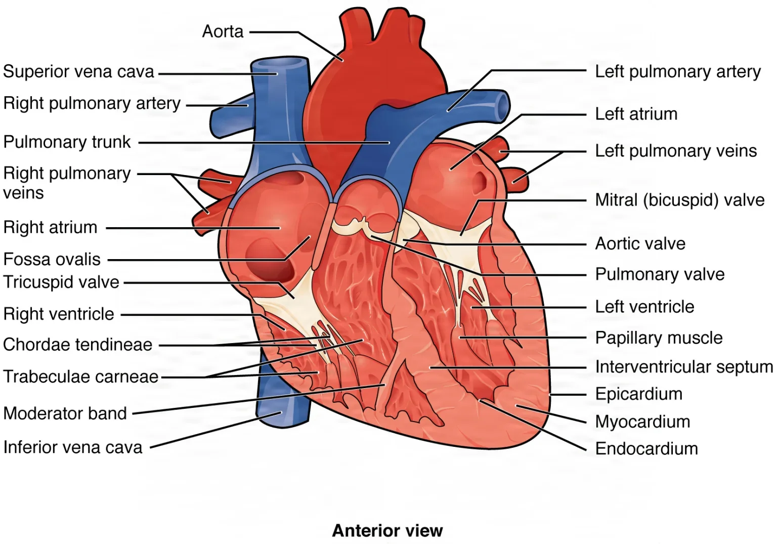
The heart is a muscular pump about the size of a fist. It lies in the chest, in the mediastinum, the space between the two lungs. The sternum is in front of it, the spine, esophagusih-SOF-uh-gusThe muscular tube that carries food and liquid from the throat to the stomach.esophag/o esophagusFull entry and descending aorta are behind it, and the diaphragmDY-uh-framThe dome-shaped sheet of muscle under the lungs that is the main muscle of breathing; also the flat side of a stethoscope.Full entry is below it. Most of the heart lies left of the midlineMID-lineAn imaginary line down the center of the body; also, a type of IV catheter placed in a vein of the upper arm.Full entry .
The heart is shaped a little like a pinecone, broad at the top and tapering to a point. Its broad top, the base, is where the great vessels attach, at about the level of the third rib cartilageKAR-tih-lijFirm, smooth, rubbery tissue that cushions joints and gives shape to parts such as the ear, nose and windpipe.chondr/o cartilageFull entry . Its pointed bottom, the apex, points down and to the left and sits just left of the sternum, near where the fourth and fifth ribs meet their cartilages.
The pericardiumpair-ih-KAR-dee-umThe thin, two-layered sac that surrounds the heart.peri- aroundcardi/o heart-um a structureFull entry is the sac around the heart. It has two parts.
- The fibrousFY-brusMade of or containing tough threads (fibers), especially of connective tissue.fibr/o fiber-ous full of, relating toFull entry pericardium, the tough outer bag of dense connective tissuekuh-NEK-tiv TISH-ooBody tissue that supports, binds together and protects other tissues and organs.Full entry , protects the heart and holds it in place.
- The serousSEER-usThin, watery and clear to pale yellow, like the fluid part of blood.Full entry pericardium has two thin layers: the parietalpuh-RY-uh-tulRelating to the wall of a body cavity; also names a lobe of the brain and two bones of the skull.pariet/o wall-al relating toFull entry layer lines the inside of the fibrous bag, and the visceralVIS-er-ulHaving to do with the internal organs, especially those in the chest and belly.viscer/o internal organs-al relating toFull entry layer covers the heart itself. A thin film of slippery fluid between them lets the heart beat without rubbing.
If blood or fluid fills the pericardial sac, it presses on the heart so it cannot relax and fill fully between beats. This is cardiac tamponade, and after an injury as little as 100 mL collecting quickly can cause it. Cardiac surgery is one cause, and it is one reason drainage tubes are left in the chest after heart surgery and must not clot or come out too early.
The fluid can be drawn off with a needle, or drained through an opening the surgeon cuts in the sac, called a pericardial window.
The heart wall
The wall of the heart has three layers, from outside to inside.
- Epicardium: the thin outer layer. It is the same tissue as the visceral layer of the serous pericardium, so the pericardium and the heart wall share it. The coronary vessels run on this surface, in grooves filled with fat.
- myocardiummy-oh-KAR-dee-umThe thick middle layer of the heart wall, made of heart muscle that contracts to pump blood.my/o musclecardi/o heart-um structureFull entry : the thick middle layer of cardiac muscleKAR-dee-ak MUS-ulThe muscle tissue that makes up the walls of the heart.cardi/o heart-ac relating toFull entry . Its contraction pumps the blood. Its fibers swirl around the chambers, a pattern that helps the heart pump blood well.
- Endocardium: the smooth inner lining of the chambers. It also covers the valves, and it is the same endothelium that lines every blood vessel.
Chambers and septa
The heart has four chambers. The two upper chambers, the atriumAY-tree-umOne of the two upper chambers of the heart, which receive blood coming back to the heart.atri/o atrium-um noun endingFull entry (one is an atrium), receive blood coming back to the heart. The two lower chambers, the ventricleVEN-trih-kulOne of the two lower chambers of the heart, or one of the fluid-filled spaces inside the brain.Full entry , are the main pumps. Each atrium has a small, ear-shaped pouch on its surface, the auricleOR-ih-kulThe outer part of the ear that you can see, on the side of the head.Full entry .
The heart is really two pumps side by side.
- The right side receives oxygen-poor blood from the body and pumps it through the lungs. This loop is the pulmonary circuit.
- The left side receives oxygen-rich blood from the lungs and pumps it out to the whole body. This loop is the systemicsis-TEM-ikAffecting the whole body rather than one part.Full entry circuit.
Both ventricles pump the same amount of blood with each beat, but the wall of the left ventricleLEFT VEN-trih-kulThe heart's lower left chamber, which pumps oxygen-rich blood into the aorta and out to the body.Full entry is much thicker. It has to push blood through the long, high-resistance systemic circuit, while the right ventriclerite VEN-trih-kulThe lower right chamber of the heart, which pumps blood to the lungs.Full entry only pushes blood next door to the lungs.
Walls called septumSEP-tumA wall of tissue that divides two spaces or cavities, such as the two sides of the heart or nose.sept/o wall, partitionFull entry divide the chambers. The interatrial septum separates the atria. It carries a small oval dent, the fossaFOS-uhA shallow hollow or dip in a bone or on the body's surface.Full entry ovalis, left from an opening the heart had before birth. The thick interventricular septum separates the ventricles. A ring of dense connective tissue, the skeleton of the heart, surrounds the four valve openings and gives the valves something firm to attach to.
The valves
Four valves keep blood moving one way through the heart. Each one opens when the pressure behind it is higher and snaps shut when blood tries to flow backward.
| Valve | Where it is | Built of |
|---|---|---|
| Tricuspid (right atrioventricular) | Between the right atrium and the right ventricle | Three flaps, tied by chordae tendineae to papillary muscles |
| Pulmonary (pulmonic, a semilunar valve) | Between the right ventricle and the pulmonary trunk | Three pocket-shaped cusps, with no chordae |
| mitral valveMY-trul valvThe heart valve between the left atrium and the left ventricle.Full entry (bicuspid, left atrioventricular) | Between the left atrium and the left ventricle | Two flaps, tied by chordae tendineae to papillary muscles |
| Aortic (a semilunar valve) | Between the left ventricle and the aorta | Usually three pocket-shaped cusps, with no chordae |
The two valves between the atria and ventricles are the atrioventricular (AV) valves. Their flaps are tied to the ventricle wall by strong cords, the chordae tendineae, which run to small papillary muscles. When the ventricle squeezes, the papillary muscles tighten the cords so the flaps cannot flip back into the atrium. The two semilunar valves, named for their half-moon cusps, have no cords: blood falling back fills their pockets and presses them shut.
A valve can fail in two ways. In stenosisstuh-NOH-sisNarrowing of a passage or opening in the body, such as a blood vessel, heart valve or the spinal canal.sten/o narrow-osis conditionFull entry , the valve becomes stiff, may calcify over time, and does not open fully, so the heart must work harder to push blood through. In regurgitationrih-gur-jih-TAY-shunA flowing backward: blood leaking back through a heart valve, or stomach contents coming back up into the mouth.Full entry , also called insufficiencyin-suh-FISH-un-seeA state in which an organ or part does not work well enough to meet the body's needs.in- notsufficient enoughFull entry , the valve does not close fully and blood leaks backward. Either one disturbs the flow and makes an extra sound called a murmurMER-merAn extra whooshing or swishing sound heard with a stethoscope over the heart, made by turbulent blood flow.Full entry . The mitral and aortic valveay-OR-tik valvThe heart valve between the left ventricle and the aorta, which opens to let blood out to the body.Full entry are the ones most often diseased, and the higher pressure on the left side of the heart makes the disease worse.
Surgeons can replace a diseased aortic valve with a mechanical valve or one made from biological tissue. When they can, many surgeons repair a leaking mitral valve rather than replace it, and may sew in a ring that makes a stretched valve opening smaller, because a replacement valve can mean anticoagulantan-tee-koh-AG-yoo-luntA drug that slows the blood's ability to form clots.anti- againstcoagul/o clotting-ant an agent that doesFull entry for life.
The great vessels
The great vessels carry blood into and out of the heart. The two vena cavaVEE-nuh KAY-vuhOne of the two large veins that return oxygen-poor blood from the body to the right atrium of the heart.Full entry , the pulmonary trunk and the aorta attach at its base.
- superior vena cavasuh-PEER-ee-er VEE-nuh KAY-vuhThe large vein that carries blood from the head, neck, arms and upper chest back to the right atrium of the heart.superior uppervena veincava hollowFull entry : brings oxygen-poor blood from everything above the diaphragm (the head, neck, arms and chest) into the right atrium.
- inferior vena cavain-FEER-ee-er VEE-nuh KAY-vuhThe large vein that carries blood from the lower body back to the right atrium of the heart.inferior lowervena veincava hollowFull entry : brings oxygen-poor blood from everything below the diaphragm (the abdomen, pelvis and legs) into the right atrium.
- Pulmonary trunk and pulmonary arteryPUL-muh-nair-ee AR-ter-eeThe large blood vessel that carries blood low in oxygen from the right side of the heart to the lungs.Full entry : the pulmonary trunk leaves the right ventricle and splits into the right and left pulmonary arteries, one to each lung. They are the only arteries after birth that carry oxygen-poor blood.
- pulmonary veinPUL-muh-nair-ee VAYNOne of the four veins that carry oxygen-rich blood from the lungs to the left atrium of the heart.Full entry : four of them, two from each lung, bring oxygen-rich blood back to the left atrium.
- Aorta: the largest artery in the body. It leaves the left ventricle and carries oxygen-rich blood to the whole body.
The rule to keep straight is simple: arteries carry blood away from the heart and veins carry it toward the heart, whether or not the blood is rich in oxygen.
The path of blood through the heart
Trace one trip through the heart, in order.
- Oxygen-poor blood from the body enters the right atrium through the superior and inferior venae cavae, and blood from the heart muscle itself enters through the coronary sinusSY-nusA hollow space in the body, most often one of the air-filled spaces in the skull bones around the nose.Full entry .
- It passes through the tricuspid valvetry-KUS-pid VALVThe heart valve between the right atrium and the right ventricle.tri- threecusp point, flapFull entry into the right ventricle.
- The right ventricle pumps it through the pulmonary valve into the pulmonary trunk and the right and left pulmonary arteries.
- In the capillaryKAP-ih-lair-eeOne of the tiniest blood vessels, joining the smallest arteries to the smallest veins.Full entry of the lungs, the blood gives off carbon dioxideKAR-bun dy-OK-sideA gas made by the body's cells as waste, carried in the blood to the lungs and breathed out.Full entry and picks up oxygen.
- Oxygen-rich blood returns through the four pulmonary veins to the left atrium.
- It passes through the mitral valve into the left ventricle.
- The left ventricle pumps it through the aortic valve into the aorta and out to the body.
- In the body's capillaries the blood delivers oxygen and picks up wastes, and it returns through the veins to the right atrium to start again.
Right side to the lungs, left side to the body. Blood passes the tricuspid, pulmonary, mitral and aortic valves, in that order.
The coronary vessels
The heart muscle works without rest, and it needs its own blood supply, the coronary circulation. The two coronary arteryKOR-uh-nair-ee AR-ter-eeOne of the blood vessels on the surface of the heart that bring oxygen-rich blood to the heart muscle.Full entry are the first branches of the aorta. They start from small pouches in the aortic wall just above the aortic valve. Blood flows into them mostly while the heart muscle is relaxed, between beats.
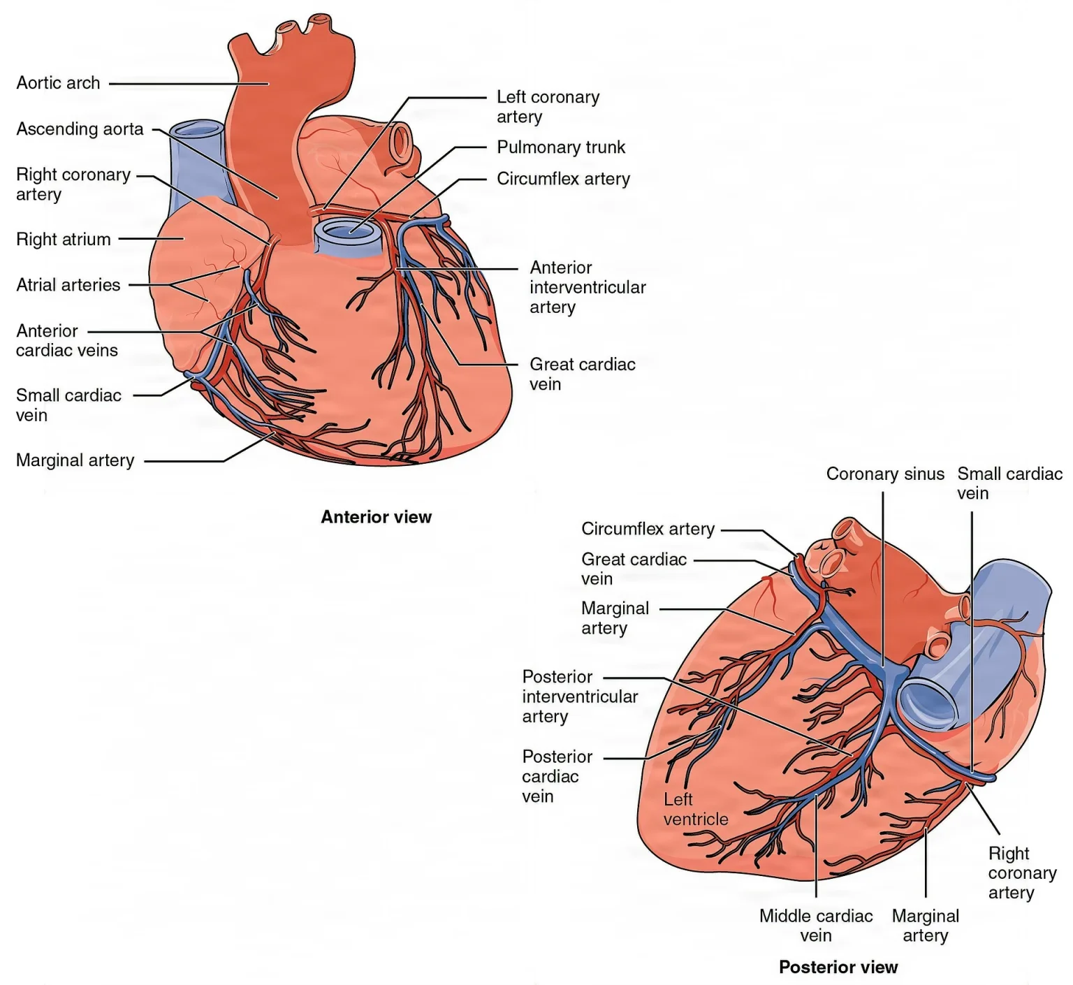
| Vessel | Course and job |
|---|---|
| Left main coronary artery | The first part of the left coronary artery, before it divides into the LAD and the circumflex; the left system feeds the left atrium, the left ventricle and the septum |
| Left anterior descending (LAD) artery | Also called the anterior interventricular artery; runs down the front of the heart in the groove between the two ventricles |
| Left circumflex artery | Curves to the left around the heart in the groove between the atria and ventricles |
| Right coronary artery | Runs in the groove on the right; feeds the right atrium, parts of both ventricles and the conductionkun-DUK-shunThe passing of something, such as an electrical signal or heat, from one place to another through a material.Full entry system, and gives off marginal branches and the posterior descending (posterior interventricular) artery |
| Great cardiac vein | Runs up beside the LAD, then around the left side to the back of the heart, where it joins the coronary sinus |
| Oblique vein of the left atrium | A small vein on the back of the left atrium; where it meets the great cardiac vein, the coronary sinus begins |
| Coronary sinus | A wide, thin-walled vein on the back of the heart that collects most of the cardiac veins and empties into the right atrium |
| Anterior cardiac veins | Small veins on the front of the right ventricle that skip the coronary sinus and drain straight into the right atrium |
The middle and small cardiac veins also drain into the coronary sinus.
The small coronary branches link up with each other only a little. So when plaquePLAKA deposit that builds up on a surface in the body, such as a sticky film of bacteria on teeth or a fatty buildup in an artery wall.Full entry blocks a coronary artery, the muscle it feeds is starved of oxygen and often dies. That death of tissue from a lost blood supply is an infarctionin-FARK-shunDeath of an area of tissue (an infarct) because its blood supply has been cut off.Full entry , and in the heart it is a myocardial infarctionmy-oh-KAR-dee-ul in-FARK-shunDeath of part of the heart muscle because its blood supply is blocked; a heart attack.my/o musclecardi/o heart-al having to do withinfarct area of dead tissueFull entry , a heart attack.
Grafts for the coronary arteries
In coronary artery bypass graftGRAFTTissue or a man-made material moved into place in surgery to replace or repair a damaged part.Full entry (CABG), the surgeon routes blood around a blocked coronary artery using a length of the patient's own vessel, an autograft. Two grafts come from structures in this chapter.
- The internal mammary artery (also called the internal thoracicthuh-RAS-ikRelating to the chest, or to the middle part of the spine.thorac/o chest-ic relating toFull entry artery) runs down the inside of the front chest wall. With the sternum split open, a retractorrih-TRAK-terA surgical instrument that holds back tissue or organs so the surgeon can see and reach the operative site.Full entry lifts one edge of it so the surgeon can reach the artery. The surgeon frees it from the chest wall but leaves its upper end attached to the subclavian artery, so it keeps its own blood supply, then sews its cut lower end to the coronary artery past the block. Most often, the surgeon joins the left internal mammary artery (LIMA) to the LAD. Arterial grafts like this stay open for many years.
- The great saphenous vein is taken from the inside of the leg. One end is sewn to the coronary artery and the other to the aorta. Veins have one-way valves, so the vein graft is turned end for end (reversed), or its valves are cut, so they cannot block the flow. The vein can be taken through one long cut or, more often now, through small incisions with an endoscopeEN-duh-skohpA thin tube with a light and a camera that is passed into the body to look inside an organ or cavity.endo- within-scope instrument for viewingFull entry , which means less pain and fewer wound problems.
While the vein is out of the body, the scrub keeps it moist in a basin of solution and handles it gently; it must be protected at all times.
Many heart operations use the heart-lung machine, or cardiopulmonarykar-dee-oh-PUL-muh-nair-eeRelating to the heart and lungs together.cardi/o heartpulmon/o lung-ary relating toFull entry bypass. The patient is given heparinHEP-uh-rinAn anticoagulant drug, given by injection or IV, that keeps blood clots from forming or growing.Full entry so the blood will not clot in the tubing. Blood is drained from the right atrium, the machine adds oxygen to it, and it is pumped back into the ascending aorta, or sometimes the femoral arteryFEM-er-ul AR-ter-eeThe large artery that carries blood to the leg, running through the groin and down the thigh.Full entry . The machine does the work of the heart and lungs, so the heart can be stopped while the surgeon works. Some bypass operations are done on the beating heart, without the machine.
The cardiac cycle and the heart sounds
One full heartbeat, from one contraction to the next, is the cardiac cycleKAR-dee-ak SY-kulThe series of events in one heartbeat, from the start of one beat to the start of the next.cardi/o heart-ac relating toFull entry . Contraction of a chamber is systoleSIS-tuh-leeThe part of each heartbeat when the heart muscle squeezes and pumps blood out.Full entry , and relaxation, when the chamber fills, is diastoledy-AS-tuh-leeThe part of each heartbeat when the heart muscle relaxes and the chambers fill with blood.Full entry .
The Core Curriculum follows the flow of blood in three steps.
- Relaxation: at the start, all four chambers are relaxed. Blood flows into the atria from the veins and on through the open tricuspid and mitral valves into the ventricles. This passive flow does most of the filling, about 70 to 80 percent.
- atrialAY-tree-ulRelating to an atrium, one of the two upper chambers of the heart.atri/o atrium-al relating toFull entry contraction: the atria squeeze and top up the ventricles with the last 20 to 30 percent, sometimes called the atrial kick.
- Ventricular contraction: the ventricles squeeze. The pressure inside them snaps the tricuspid and mitral valves shut, then forces the pulmonary and aortic valves open, and blood rushes into the pulmonary trunk and aorta. Then the ventricles relax, the semilunar valves close, and the cycle starts again.
The sounds you hear through a stethoscope are the valves closing.
- First heart sound (S1), the "lub": the tricuspid and mitral valves closing as the ventricles start to contract.
- Second heart sound (S2), the "dub": the aortic and pulmonary valves closing as the ventricles relax.
A third heart sound is rarely heard in healthy people, though some young people, athletes and pregnant people have one; heard later in life, it may point to heart failurehart FAYL-yerA long-term condition in which the heart cannot pump enough blood to meet the body's needs.Full entry . A fourth heart sound comes from the atria pushing blood into a stiff ventricle and points to a failing left ventricle. A murmur is an extra whooshing sound from blood flowing roughly through a narrowed or leaky valve or a hole in a septum.
Heart terms you will hear
| Term | Meaning |
|---|---|
| Blood pressure | The force the blood puts on the walls of the vessels and the heart chambers; usually the pressure in the large arteries |
| Heart rate, pulse rate | How many times the heart beats in a minute; the pulse felt at an artery tells you the heart rate |
| normal sinus rhythmNOR-mul SY-nus RITH-umThe heart's normal rhythm, started by the sinoatrial (SA) node, regular and at 60 to 100 beats a minute in adults.Full entry | The normal pattern, set by the sinoatrial (SA) node; at rest an adult's heart rate is 60 to 100 beats a minute |
| bradycardiabray-dee-KAR-dee-uhA heart rate slower than normal; in an adult, fewer than 60 beats per minute.brady- slowcardi/o heart-ia conditionFull entry | A resting heart rate below 60 beats a minute |
| tachycardiatak-ih-KAR-dee-uhA heart rate faster than normal; in an adult, more than 100 beats per minute.tachy- fastcardi/o heart-ia conditionFull entry | A resting heart rate above 100 beats a minute |
| stroke volumeSTROHK VOL-yoomThe amount of blood the left ventricle pumps out with each heartbeat.Full entry | The blood one ventricle pumps with each beat, about 70 mL at rest |
| cardiac outputKAR-dee-ak OWT-putThe amount of blood the heart pumps out in one minute.Full entry | The blood one ventricle pumps in a minute: heart rate times stroke volume, about 5 liters a minute at rest |
| Ectopic beat | An early beat started by some spot in the heart other than the SA nodeES-AY NOHDSinoatrial node: a small cluster of cells in the right atrium that starts each heartbeat.sin/o sinusatri/o atriumFull entry |
| fibrillationfib-rih-LAY-shunFast, uncoordinated twitching of muscle fibers, most often in the heart, so the muscle quivers instead of contracting together.fibr/o fiber-ation processFull entry | Fast, uncoordinated quivering of the heart muscle that cannot pump well; in the ventricles it is an emergency that needs defibrillationdee-fib-ruh-LAY-shunAn electric shock given to the heart to stop a chaotic, life-threatening rhythm so a normal rhythm can return.Full entry |
| Heart block | A delay or break in the electrical pathway, most often at the AV nodeay-VEE nohdAtrioventricular node: a small group of cells between the upper and lower chambers of the heart that passes on the heartbeat signal.atri/o atriumventricul/o ventricle-ar relating toFull entry or in the AV bundle just below it |
| Infarction | Death of tissue that has lost its blood supply; in the heart, a heart attack |
A heart rate that is too slow pumps too little blood. One that is too fast leaves too little time for the ventricles to fill, so either one can lower cardiac output.
The conduction pathway
Heart muscle sets its own beat. Special muscle cells in the heart make electrical signals on their own and carry them along a set path, the conduction system, so the chambers squeeze in the right order.
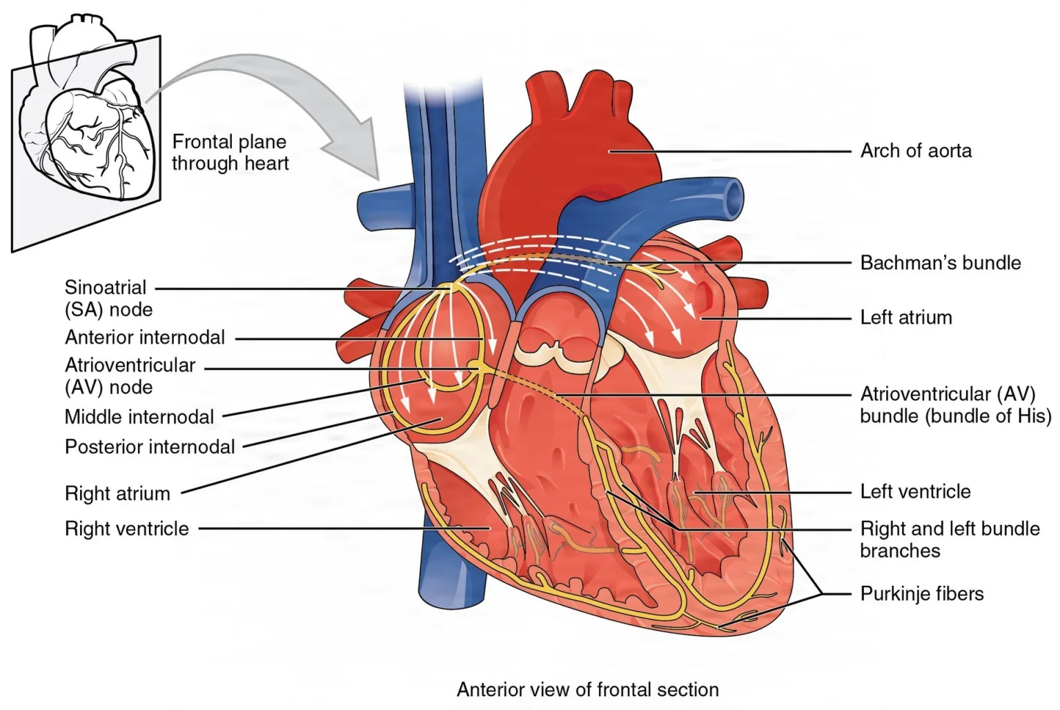
- Sinoatrial (SA) node: a small cluster of cells in the upper back wall of the right atrium, near where the superior vena cava enters. It fires faster than any other part of the heart, so it sets the pace; it is the heart's natural pacemakerPAYS-may-kerA small device that sends electrical pulses to the heart to keep it beating at a set rate; also the heart's own natural rhythm center.Full entry .
- Atrial myocardium: the signal spreads across the muscle of both atria, and they contract.
- Atrioventricular (AV) node: in the lower part of the right atrium. The connective tissue between the atria and ventricles blocks the signal everywhere except here. The AV node holds the signal for about a tenth of a second, which gives the atria time to empty into the ventricles.
- AV bundle (bundle of HisBUN-dul uv HISSA band of special heart fibers that carries the electrical signal from the AV node down to the ventricles.Full entry ) and bundle branches: the signal runs from the AV node down the atrioventricular bundle in the interventricular septum, which splits into right and left bundle branches.
- Purkinje fiberspur-KIN-jee FY-berzSpecial fibers in the walls of the heart's ventricles that spread the electrical signal so the ventricles contract.Full entry : from the apex, these fibers spread the signal fast through the walls of both ventricles.
- Ventricular myocardium: the ventricles contract from the apex upward, squeezing blood up and out, the way you squeeze toothpaste from the bottom of the tube.
If the SA node fails, the next part down takes over, but more slowly: the AV node beats about 40 to 60 times a minute, and the Purkinje fibers only 15 to 20.
The electrocardiogramih-lek-troh-KAR-dee-oh-gramA test that records the heart's electrical activity as waves on paper or a screen.electr/o electricitycardi/o heart-gram recordFull entry (ECG) records this electrical activity. The P wave is the signal spreading across the atria, the QRS complexKYOO-ar-es KOM-pleksThe tall, spiky wave on an ECG that shows the electrical signal spreading through the ventricles.Full entry is the signal spreading through the ventricles, and the T waveTEE waveThe wave on an electrocardiogram that shows the heart's lower chambers recovering electrically after each beat.Full entry is the ventricles resetting. Chapter 13 teaches the monitor.
When the conduction system fails, a surgeon or cardiologist can implant an artificial pacemaker. A lead is passed through a vein, such as the cephalic or subclavian vein, under X-ray guidance into the right atrium or ventricle, and attached to a pulse generator placed in a pocket under the skin.
What sets the heart rate
The SA node alone would fire about 100 times a minute. The autonomic nervous systemaw-tuh-NOM-ik NUR-vus SIS-tumThe part of the nervous system that controls body functions without conscious thought, such as heart rate, breathing and digestion.auto- selfnom/o law, control-ic relating toFull entry speeds it up or slows it down, from two cardiac centers in the medullamuh-DUL-uhThe inner part of an organ; most often the medulla oblongata, the lowest part of the brainstem.Full entry of the brain stemBRAYN STEMThe lowest part of the brain, which connects it to the spinal cord and controls breathing, heart rate and other automatic functions.Full entry .
- Sympathetic nerves speed the heart. Their nerve endings release norepinephrinenor-ep-ih-NEF-rinA hormone and nerve messenger that narrows blood vessels and raises blood pressure, also given as an IV drug.Full entry .
- The vagus nerveVAY-gus nervThe 10th cranial nerve, which runs from the brain stem to the chest and belly and slows the heart and helps digestion.Full entry , which is parasympatheticpair-uh-sim-puh-THET-ikRelating to the part of the autonomic nervous system that calms the body and supports "rest and digest" functions.para- besidesympathetic the other autonomic divisionFull entry , slows the heart. Its endings release acetylcholineuh-see-tul-KOH-leenA chemical messenger that nerves release to signal muscles, glands and other nerves.Full entry . At rest, the vagus normally holds the rate down, which is why a resting heart beats slower than 100.
Sensors feed the cardiac centers. Stretch sensors called baroreceptors, in the aortic sinus and the carotidkuh-ROT-idHaving to do with the carotid arteries, the two large arteries on each side of the neck that carry blood to the head and brain.Full entry sinus of the neck, report blood pressure. When pressure rises, the centers slow the heart; when it falls, they speed it up. Hormones such as epinephrineep-ih-NEF-rinA hormone and drug, also called adrenaline, that raises heart rate and blood pressure and opens the airways.Full entry , blood levels of calcium and potassium, and body temperature also change the rate.
You will see this reflexREE-fleksAn automatic body response to something that stimulates it, which happens without thought, such as the knee jerk.Full entry in surgery. When a surgeon handles the carotid sinus during a carotid endarterectomy, the heart rate and blood pressure can drop, and the surgeon may inject a little lidocaineLY-doh-kaynA local anesthetic that numbs an area, also given by IV to treat some fast, abnormal heart rhythms.Full entry , a local anestheticLOH-kul an-es-THET-ikA drug that blocks feeling in one part of the body without making the person unconscious.Full entry , around the sinus to prevent it.
4Blood vessels
Arteries, capillaries and veins
Blood leaves the heart in arteries, passes through capillaryKAP-ih-lair-eeOne of the tiniest blood vessels, joining the smallest arteries to the smallest veins.Full entry in the tissues, and comes back in veins. In a healthy vessel it moves in straight lines parallel to each other, which is called laminar flow. The Core Curriculum names six kinds of vessel.
- Artery: a vessel that carries blood away from the heart. Arteries have thick walls to stand up to the high pressure of each heartbeat. The big arteries near the heart are stretchy: they swell with each beat and spring back, which keeps blood moving between beats. Farther out, arteries have more muscle and less elastic tissue.
- arteriolear-TEER-ee-ohlA very small artery that carries blood from an artery into the capillaries.arteri/o artery-ole smallFull entry : the smallest kind of artery, leading into the capillaries. Arterioles have a thin layer of smooth musclesmooth MUS-ulMuscle that works without conscious control and lines hollow organs such as the stomach, intestines, bladder, airways and blood vessels.Full entry , and by narrowing or widening they control how much blood each tissue gets. They are where most of the resistance to blood flow is, so they are the main control of blood pressure.
- Capillary: a microscopic vessel whose wall is a single layer of cells; the smallest are so narrow that a red cell can only just squeeze through. Capillaries are where oxygen, nutrients and wastes pass between the blood and the tissues.
- venuleVEN-yoolA very small vein that collects blood from the capillaries.ven vein-ule smallFull entry : a very small vein that collects blood from the capillaries. White blood cells slip out of the blood mainly through capillaries and venules.
- Vein: a vessel that carries blood back toward the heart. Veins have thin walls and wide openings and work at low pressure. Because they stretch easily, they hold about 64 percent of the body's blood at any moment.
- Venous sinusSY-nusA hollow space in the body, most often one of the air-filled spaces in the skull bones around the nose.Full entry : a wide, irregular channel for venous blood, lined by endothelium and without valves. The dural sinuses inside the skull and the coronary sinus of the heart are examples, and the spleenspleenAn organ in the upper left abdomen that filters the blood and helps fight infection.splen/o spleenFull entry has small ones.
The capillaries of the liver, spleen and bone marrowbone MAIR-ohThe soft, spongy tissue inside bones that makes blood cells.Full entry are an extra-leaky kind called sinusoids, with large gaps that let big proteins and even whole cells pass. That is how new blood cells leave the marrow. Tissues with such rich capillary networks, like the liver and spleen, bleed easily and heavily when cut, and the surgeon controls that capillary bleeding with topical thrombinTHROM-binAn enzyme in the blood that turns fibrinogen into fibrin, the threads that hold a blood clot together.thromb/o clot-in substanceFull entry or a tissue sealant (chapter 29).
Low pressure is a problem for veins, especially in the legs, where blood must climb against gravity. Three things help it. One-way valves in the veins keep blood from falling back. When leg muscles contract, they squeeze the veins and push blood up, the skeletal muscleSKEL-uh-tul MUS-ulMuscle attached to bones that a person moves by choice, such as the muscles of the arms and legs.Full entry pump. And breathing in lowers the pressure in the chest, which draws blood up toward the heart, the respiratory pump. The perforating veins connect the superficial veins of the leg to the deep ones.
The three layers of the vessel wall
Arteries and veins have the same three layers, called tunics. Capillaries have only the innermost one.
- Tunica intima (also called the tunica interna): the inner lining. Its surface is endothelium, the same smooth, slippery layer that lines the heart, on a thin layer of connective tissuekuh-NEK-tiv TISH-ooBody tissue that supports, binds together and protects other tissues and organs.Full entry . Damage to the endothelium exposes the collagenKOL-uh-jenThe main structural protein of the body, which gives strength to skin, bones, tendons, ligaments and scar tissue.coll/a glue-gen producingFull entry beneath it, which is one of the main things that starts a clot. In many veins, especially in the legs, folds of the intima form the one-way valves.
- Tunica media: the middle layer of smooth muscle and elastic fibers. It is usually the thickest layer in an artery. When its muscle contracts, the vessel narrows (vasoconstrictionvay-zoh-kun-STRIK-shunThe narrowing of blood vessels when the muscle in their walls tightens.vas/o vesselconstriction narrowingFull entry ); when the muscle relaxes, the vessel widens (vasodilationvay-zoh-dy-LAY-shunThe widening of blood vessels as the muscle in their walls relaxes, which lets more blood flow through.vas/o vesseldilat widen-ion processFull entry ). Nerves of the sympathetic system, hormones and local chemicals control it. Because of this thicker muscle, a cut artery can often contract and narrow enough to stop its own bleeding.
- Tunica adventitia (also called the tunica externa): the tough outer layer of collagen connective tissue. It anchors the vessel to the tissue around it and gives it strength, and in veins it is usually the thickest layer. Large vessels have their own tiny blood vessels, the vasa vasorum, running in their walls.
These layers explain several operations.
- In atherosclerosisath-er-oh-skluh-ROH-sisA buildup of fatty deposits (plaque) inside the walls of the arteries.ather/o fatty pastescler/o hard-osis conditionFull entry , fatty, calcified plaquePLAKA deposit that builds up on a surface in the body, such as a sticky film of bacteria on teeth or a fatty buildup in an artery wall.Full entry builds up in the intima and narrows the artery. Plaque is often thickest where an artery branches. In a carotidkuh-ROT-idHaving to do with the carotid arteries, the two large arteries on each side of the neck that carry blood to the head and brain.Full entry endarterectomy, the surgeon opens the carotid artery in the neck and lifts out the plaque, a thick yellow rubbery layer stuck to the intima. A flexible shuntSHUNTA passage that moves blood or other fluid from one place to another, either made by a surgeon or present in the body.Full entry may carry blood past the clamps to the brain while the surgeon works; the scrub flushes it with heparinized saline before passing it.
- In an aortic dissectiondy-SEK-shunA tear in the inner layer of an artery wall that lets blood split the layers apart; also, the cutting apart of tissue in surgery.Full entry , the intima tears and blood forces its way into the media, splitting the wall.
- In varicose veinVAIR-ih-kohs VAYNA swollen, twisted vein just under the skin, most often in the legs, in which the valves do not work well.varic/o swollen, twisted vein-ose full ofFull entry , the valves fail, blood pools, and the superficial veins of the legs swell and twist. Severe cases may need surgery to remove the damaged veins; because other veins link up and can carry the blood, removing them does not usually harm the circulation.
The major arteries
The aorta and its branches carry oxygen-rich blood to almost every tissue. The table follows the Core Curriculum's list, from the heart outward.
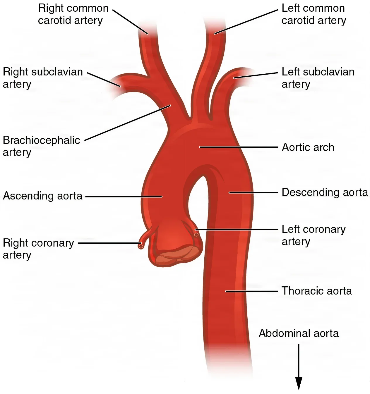
| Artery | Where it comes from | What it feeds, and where you meet it |
|---|---|---|
| Aortic arch | The ascending aorta rises about 5 cm from the left ventricleLEFT VEN-trih-kulThe heart's lower left chamber, which pumps oxygen-rich blood into the aorta and out to the body.Full entry , then curves to the left as the arch | Gives off the three great arteries to the head and arms |
| Brachiocephalic (innominate) trunk | The first branch of the arch, on the right only | Splits into the right subclavian and right common carotid arteries |
| Common carotid | The right one from the brachiocephalic trunk, the left one straight from the arch | Runs up the side of the neck and divides into the external and internal carotids |
| External carotid | The common carotid | The face, lower jaw, neck and larynxLAIR-inksThe voice box: the part of the airway in the neck, above the windpipe, that holds the vocal cords.laryng/o larynxFull entry ; its branches include the facial and superficial temporal arteries |
| Internal carotid | The common carotid; it starts with a small swelling, the carotid sinus | Enters the skull and feeds the front of the brain and the eyes |
| Circle of Willis | Branches of the two internal carotids and the vertebral arteries, joined in a ring | Sits under the base of the brain; because the ring is connected, blood can still reach the brain if one feeding artery is blocked |
| Subclavian | The right one from the brachiocephalic trunk, the left one from the arch | Runs under the collarbone to the arm; also feeds the chest wall, shoulder and brain |
| Vertebral | The subclavian | Climbs through openings in the neck vertebrae into the skull; the two join into the basilar artery, which feeds the back of the brain |
| Internal thoracicthuh-RAS-ikRelating to the chest, or to the middle part of the spine.thorac/o chest-ic relating toFull entry (internal mammary) | The subclavian | Runs down inside the front chest wall and feeds the chest wall, the breast and the thymusTHY-musA small gland in the upper chest, behind the breastbone, where T cells (a kind of white blood cell) mature.Full entry ; used as a bypass graftGRAFTTissue or a man-made material moved into place in surgery to replace or repair a damaged part.Full entry for the heart |
| Axillary | The subclavian, renamed as it enters the armpit | The shoulder and upper arm |
| Brachial | The axillary, renamed in the upper arm | Runs down the inner arm and divides at the elbow into the radial and ulnar arteries; the blood pressure cuff goes over it |
| Radial | The brachial | Runs down the thumb side of the forearm to the hand; the usual site for feeling a pulse and for drawing arterial blood gasar-TEER-ee-ul BLUD GASA blood test from an artery that measures oxygen, carbon dioxide, bicarbonate and the blood's pH.Full entry |
| Ulnar | The brachial | Runs down the little-finger side of the forearm; joins the radial artery in the palmarPAL-merHaving to do with the palm of the hand.Full entry arches of the hand |
| Thoracic aorta | The aorta from the end of the arch down to the diaphragmDY-uh-framThe dome-shaped sheet of muscle under the lungs that is the main muscle of breathing; also the flat side of a stethoscope.Full entry | Feeds the chest wall, esophagusih-SOF-uh-gusThe muscular tube that carries food and liquid from the throat to the stomach.esophag/o esophagusFull entry and the tissue of the lungs |
| intercostalin-ter-KOS-tulBetween the ribs.inter- betweencost/o rib-al relating toFull entry arteries | The thoracic aorta (intercostal means between the ribs) | The muscles of the chest wall and spine |
| Abdominal aorta | The aorta below the diaphragm; it ends at about the fourth lumbarLUM-barHaving to do with the lower back, the part of the spine between the ribs and the pelvis.Full entry vertebra by splitting into the two common iliacIL-ee-akHaving to do with the ilium, the large, wing-shaped upper part of the hip bone.Full entry arteries | Feeds the abdomen, pelvis and legs |
| Celiac trunk (celiac axis) | A single short branch of the abdominal aorta, just above the superior mesenteric artery | Divides into the left gastric, splenic and common hepatichih-PAT-ikRelating to the liver.hepat/o liver-ic relating toFull entry arteries |
| Gastric arteries | The left gastric from the celiac trunk; the right gastric from the common hepatic | The stomach |
| Hepatic artery | The common hepatic, from the celiac trunk | The liver, and through its cystic branch, the gallbladder |
| Splenic | The celiac trunk | The spleen |
| Superior mesenteric | The aorta, just below the celiac trunk | The small intestine, the pancreasPAN-kree-usA long, flat gland behind the stomach that makes digestive enzymes and the hormones insulin and glucagon.pan- allcreas fleshFull entry and most of the large intestine |
| Renal | The aorta, one to each side | The kidneys |
| gonadGOH-nadA sex gland: an ovary or a testis, which makes eggs or sperm and sex hormones.gon seed, generationFull entry | The aorta, below the renal arteries | The ovaries or the testes |
| Inferior mesenteric | The aorta, just above where it divides | The last part of the large intestine and the rectum |
| Common iliac | The aorta divides into right and left | Each divides into an internal and an external iliac artery |
| Internal iliac | The common iliac | The bladder, pelvic walls, external genitals, and the uterus and vagina |
| External iliac | The common iliac | Passes out of the body cavity into the thigh and becomes the femoral arteryFEM-er-ul AR-ter-eeThe large artery that carries blood to the leg, running through the groin and down the thigh.Full entry |
| femoralFEM-er-ulRelating to the thigh or the femur, the thigh bone.Full entry | The external iliac, renamed in the groin | The thigh; the common femoral artery divides into the superficial femoral artery, which continues down the thigh, and the deep femoral artery; a common entry point for catheters and stentstentA small tube, often of metal mesh, placed inside a vessel or duct to hold it open.Full entry |
| Deep femoral (profunda femoris) | The femoral | The deep muscles of the thigh |
| Popliteal | The femoral, renamed behind the knee | Divides into the anterior and posterior tibiaTIB-ee-uhThe large inner bone of the lower leg, between the knee and the ankle; the shinbone.tibi/o tibiaFull entry arteries |
| Anterior tibial | The popliteal | The front of the lower leg; continues onto the foot as the dorsalis pedis |
| Posterior tibial | The popliteal | The back of the lower leg and the sole of the foot |
| peronealper-uh-NEE-ulHaving to do with the outer side of the lower leg, near the fibula bone.Full entry (fibulaFIB-yuh-luhThe thin outer bone of the lower leg, beside the larger tibia.Full entry ) | The posterior tibial | Runs deep in the back of the leg, between the tibia and the fibula |
| Dorsalis pedis | The anterior tibial, renamed at the ankle | The ankle and the top of the foot |
Learn the arteries in the order blood meets them: aorta, arch, then out to the head and arms; then down the aorta, out to the gut and kidneys, and down the legs to the foot.
The major veins
Veins often run beside the artery of the same name, but there are also superficial veins just under the skin with no matching artery. Veins vary more from person to person than arteries do.
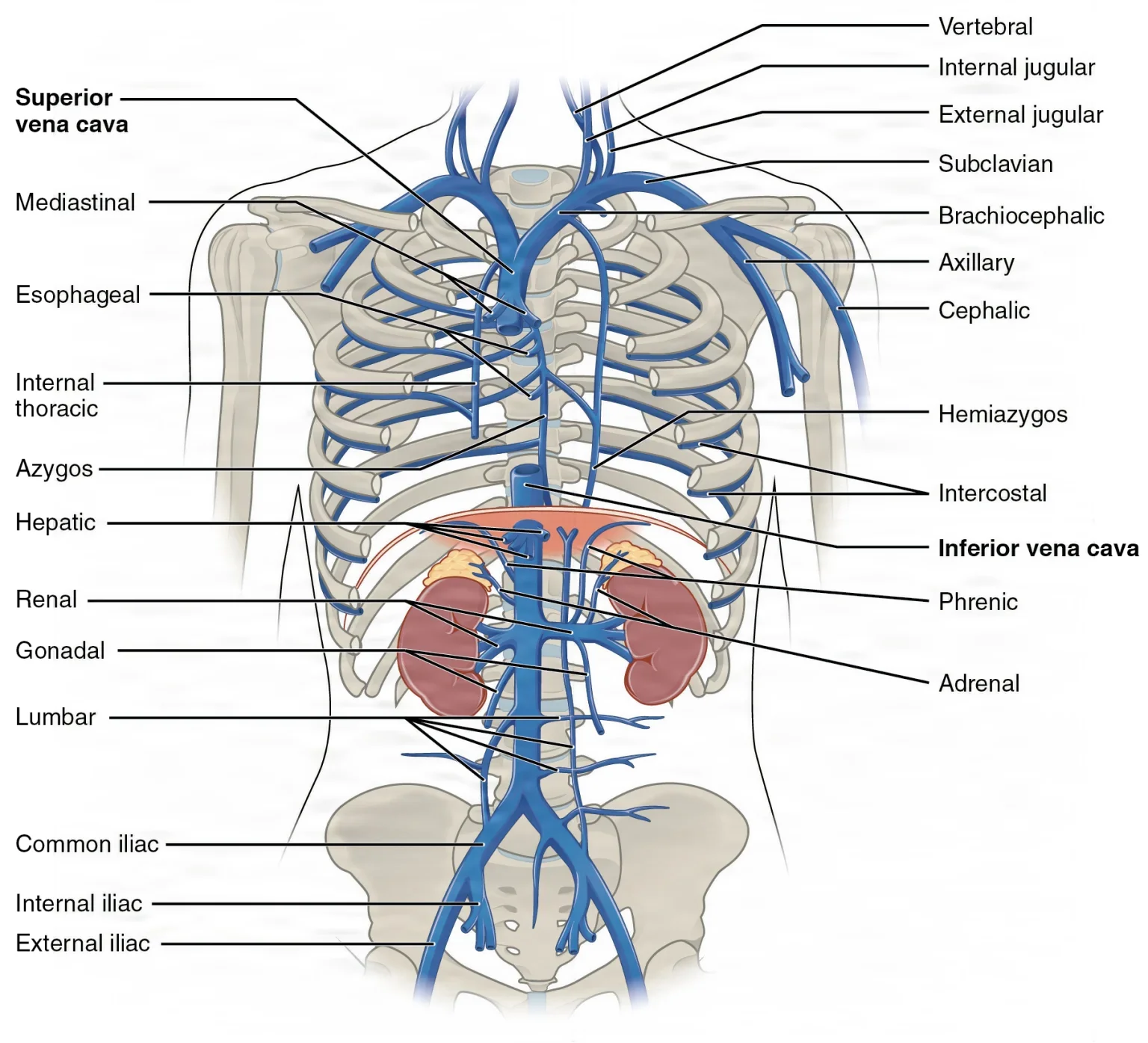
| Vein | Where it runs and drains |
|---|---|
| superior vena cavasuh-PEER-ee-er VEE-nuh KAY-vuhThe large vein that carries blood from the head, neck, arms and upper chest back to the right atrium of the heart.superior uppervena veincava hollowFull entry | Formed where the right and left brachiocephalic veins meet; carries blood from above the diaphragm into the right atriumAY-tree-umOne of the two upper chambers of the heart, which receive blood coming back to the heart.atri/o atrium-um noun endingFull entry |
| inferior vena cavain-FEER-ee-er VEE-nuh KAY-vuhThe large vein that carries blood from the lower body back to the right atrium of the heart.inferior lowervena veincava hollowFull entry | Formed where the two common iliac veins meet, at about the fifth lumbar vertebra; runs up the right side of the abdomen beside the aorta and carries blood from below the diaphragm into the right atrium |
| Internal jugularJUG-yuh-lerRelating to the neck, especially the large veins in the neck.Full entry | Drains the brain; runs down the neck beside the common carotid artery and joins the subclavian vein to form the brachiocephalic vein |
| cranialKRAY-nee-ulRelating to the skull, or toward the head end of the body.crani/o skull-al relating toFull entry venous sinuses | Channels in the duraDOOR-uhThe tough outer layer of the membranes that cover the brain and spinal cord.Full entry mater, the brain's tough outer covering, that collect the blood of the brain and empty into the internal jugular veinin-TUR-nul JUG-yuh-ler vaynA large vein deep on each side of the neck that carries blood from the brain, face and neck back toward the heart.Full entry (chapter 4) |
| Azygos system | The azygos vein on the right and the hemiazygos vein on the left drain the chest wall, esophagus and bronchusBRONG-kusOne of the large air tubes that branch from the windpipe into each lung.bronch/o airwayFull entry ; the azygos empties into the superior vena cava |
| Portal system | The hepatic portal vein, formed by the superior mesenteric and splenic veins, carries blood from the stomach, intestines, spleen and pancreas to the liver |
| Cephalic | A superficial vein up the outer (thumb) side of the arm, from the hand to the axillary vein near the shoulder |
| Basilic | A superficial vein up the inner side of the arm to the axillary vein |
| Median cubital | Crosses the inside of the elbow, linking the cephalic and basilic veins; the most common place to draw blood |
| Great (greater) saphenous | A superficial vein up the inner side of the leg and thigh to the femoral vein near the groin |
| Small (lesser) saphenous | A superficial vein up the outer side of the lower leg to the popliteal vein behind the knee |
The portal system is unusual: it runs from one set of capillaries to another. Blood leaving the capillaries of the stomach and intestines, full of absorptionub-ZORP-shunThe taking in of a substance, such as a drug or a nutrient, into the bloodstream.Full entry nutrients, goes first to the liver, which processes it before it reaches the rest of the body. So the liver gets blood from two sources, the hepatic artery and the portal vein, and all of it leaves through the hepatic veins into the inferior vena cava.
The big veins of the neck and chest are the way in for central venous catheterSEN-trul VEE-nus KATH-uh-terAn IV catheter whose tip sits in a large vein near the heart, used for long-term drugs, fluids, nutrition or blood draws.Full entry and ports. A central venous catheter is passed through the subclavian or internal jugular vein so its tip lies in the superior vena cava near the right atrium. An implanted port is a small chamber placed under the skin, with a catheter running from it through the cephalic, jugular or subclavian vein into the superior vena cava.
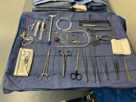
For kidney dialysisdy-AL-ih-sisA treatment that removes wastes and extra fluid from the blood when the kidneys can no longer do it.dia- through, apart-lysis separation, looseningFull entry , a patient needs a vessel that can take large needles again and again. In an arteriovenousar-teer-ee-oh-VEE-nusRelating to both arteries and veins, or joining an artery to a vein.arteri/o arteryven/o vein-ous relating toFull entry (AV) fistulaFIS-choo-luhAn abnormal passage between two organs or vessels, or between an organ and the skin.Full entry , the surgeon joins an artery directly to a vein, often the radial artery to the cephalic vein in the forearm. The arterial blood swells the vein until it is large enough to take the big dialysis needles.
Fetal circulation
Before birth, the fetus's lungs are collapsed and full of fluid, so they cannot take in oxygen. The placentapluh-SEN-tuhThe organ that grows in the uterus during pregnancy and passes oxygen and food from the parent's blood to the fetus.Full entry , the organ of exchange between the pregnant parent and the fetus, does that job, and the fetal circulation has shortcuts that send most of the blood past the lungs and the liver.
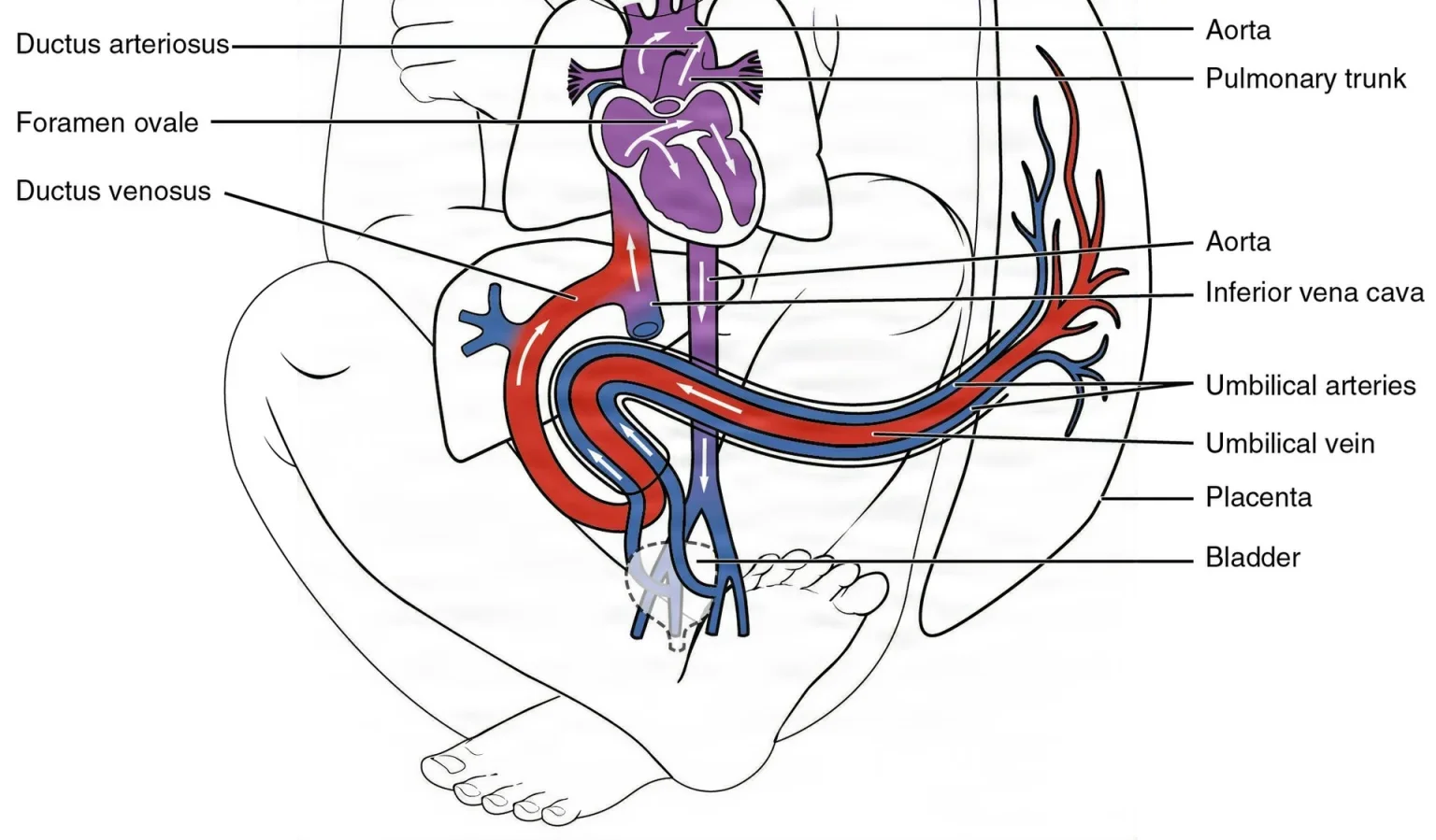
| Structure | What it does before birth | What happens after birth |
|---|---|---|
| Placenta | Exchanges oxygen, nutrients and wastes between the parent's blood and the fetus's blood | The baby's own lungs take over gas exchangeGAS eks-CHAYNJThe movement of oxygen from the air sacs of the lungs into the blood and of carbon dioxide from the blood into the air sacs.Full entry with the first breaths |
| Umbilical vein | One vein in the cord; carries oxygen-rich blood from the placenta toward the fetus's heart | Collapses when the cord is clamped and cut and turns into fibrousFY-brusMade of or containing tough threads (fibers), especially of connective tissue.fibr/o fiber-ous full of, relating toFull entry tissue; in the adult it is the ligamentum teres |
| Umbilical arteries | Two arteries in the cord, from branches of the internal iliac arteries; carry oxygen-poor blood and wastes back to the placenta | Collapse when the cord is clamped and cut; the parts nearest the body stay in use, and the rest turn into fibrous remnants that remain in the adult |
| Ductus venosus | A shortcut from the umbilical vein to the inferior vena cava, so most of the blood bypasses the liver | Closes slowly over the first weeks and becomes a ligament, the ligamentum venosum |
| foramen ovalefuh-RAY-men oh-VAH-leeAn opening between the right and left upper chambers of the fetal heart that lets blood bypass the lungs before birth.foramen openingovale ovalFull entry | An opening in the wall between the atria, so blood passes from the right atrium straight to the left and skips the lungs | Closes as the baby starts breathing and the pressure in the atria rises; the fossaFOS-uhA shallow hollow or dip in a bone or on the body's surface.Full entry ovalis marks the spot |
| ductus arteriosusDUK-tus ar-teer-ee-OH-susA blood vessel in the fetus that connects the pulmonary artery to the aorta, so blood can skip the lungs.Full entry | A short vessel from the pulmonary trunk to the aorta, so most blood pumped toward the lungs goes to the body instead | Its muscle squeezes shut as oxygen rises after the first breaths, and it becomes the ligamentum arteriosum |
Sometimes a shunt stays open. A patent ("open") foramen ovale is common, found in as many as 20 to 25 percent of people, and most never know they have it. A patent ductus arteriosusPAY-tent DUK-tus ar-teer-ee-OH-susA heart defect in which the ductus arteriosus, a blood vessel between the aorta and pulmonary artery, stays open after birth.Full entry (PDA) lets blood flow from the high-pressure aorta back into the pulmonary trunk, which overloads the lungs and the heart. It can be closed with drugs, with a device placed through a catheter, or by surgical ligation, tying it off, through a left thoracotomythor-uh-KOT-uh-meeA surgical cut through the chest wall to reach the lungs, heart or other organs in the chest.thorac/o chest-tomy cutting intoFull entry or by minimally invasive surgery.
Pulse points
A pulse is the swelling and recoil of an artery each time the heart pumps blood into it. You can feel it where an artery runs close to the skin, by pressing it lightly against the bone or muscle beneath it. The rate of the pulse is the heart rate, and its strength tells you how hard the ventricleVEN-trih-kulOne of the two lower chambers of the heart, or one of the fluid-filled spaces inside the brain.Full entry is pushing.
| Pulse point | Where to feel it |
|---|---|
| Superficial temporal | In front of the ear, just above the cheekbone |
| Facial | Where the facial artery crosses the lower edge of the jaw, a few centimeters in front of its angle |
| Carotid | The side of the neck, in the groove between the tracheaTRAY-kee-uhThe windpipe: the tube that carries air from the throat to the lungs.Full entry and the large neck muscle (sternocleidomastoid) |
| Brachial | The inner side of the upper arm, in the groove beside the biceps, and at the inside of the elbow |
| Radial | The thumb side of the wrist; the site used most often |
| Femoral | The crease of the groin; it lies deep, so you may need to press firmly |
| Popliteal | Behind the knee |
| Posterior tibial | Behind the bump of the inner ankle bone |
| Dorsalis pedis | On top of the foot, between the big toe and the second toe |
Feel for a pulse with the pads of your index and middle fingers, never your thumb, which has a pulse of its own. Press just enough to feel the beat without squeezing the artery shut. When a pulse is too weak to feel, a Doppler ultrasoundDOP-ler UL-truh-soundAn ultrasound that uses sound waves to hear or show blood flowing through vessels or the fetal heartbeat.Full entry probe, which picks up moving blood with sound waves, can find it.
Pulse checks after vascular surgery
In surgery on the arteries of the legs, the team checks the pulses of the feet before, during and after the repair.
- Before: the circulator checks the color, warmth and pulses of the legs and feet so there is a baseline to compare with, and marks the pedal pulses, the dorsalis pedis and posterior tibial, on the skin with a marking pen before the prep.
- During: the patient is given heparinHEP-uh-rinAn anticoagulant drug, given by injection or IV, that keeps blood clots from forming or growing.Full entry before the vessels are clamped. When the repair is done and the clamps come off, the surgeon checks the pulses with a sterile Doppler probe, and the circulator may check the feet for warmth and color. Have the sterile Doppler ready on the field.
- After: a pulse that fades or disappears after surgery can mean a new clot is blocking the repair or the graft, and the patient may need to go back to the OR.
Three operations show why the pulses matter.
- In a femoral thromboembolectomy, the surgeon opens the femoral artery and pulls out a clot with a balloon-tipped embolectomy catheter, such as a Fogarty. About 80 percent of the clots that travel out to the body's peripheral arteries lodge in a leg, and about half of all of them where the common femoral artery divides. They may come from the heart, for example from the left atrium in a patient with atrial fibrillationAY-tree-ul fib-rih-LAY-shunAn irregular heart rhythm in which the upper chambers of the heart quiver instead of beating in an organized way.atri/o atrium (upper heart chamber)fibrill/o small fibers, quivering-ation processFull entry .
- In a femoropopliteal bypass, the surgeon places a graft, the patient's own great saphenous vein or a synthetic tube, from the femoral artery to the popliteal artery, around a blocked stretch of the femoral artery.
- In an open abdominal aortic aneurysmab-DOM-ih-nul ay-OR-tik AN-yuh-riz-umA weak, bulging section of the aorta where it passes through the belly.Full entry repair, the surgeon replaces a bulging, weakened part of the abdominal aorta with a graft. Most of these aneurysmAN-yuh-riz-umA weak spot in the wall of an artery that bulges outward like a balloon.aneurysm a widening (Greek)Full entry lie below the renal arteries, which lets the surgeon clamp the aorta above the aneurysm without cutting off the kidneys.
Know where the pedal pulses are before the case starts, and have the sterile Doppler on the field. After the clamps come off, a good pulse in the foot is the sign that blood is getting past the repair.
Blood pressure and what changes it
Blood pressure is the push of blood against the walls of the vessels. When people say "blood pressure," they mean the pressure in the large arteries, usually measured with a cuff over the brachial artery.
- Systolic pressure, the higher number, is the peak pressure while the ventricles contract.
- diastolicdy-uh-STOL-ikRelating to diastole, the part of each heartbeat when the heart muscle relaxes and its chambers fill with blood.diastole the heart's relaxing, filling phase (Greek, to expand)-ic relating toFull entry pressure, the lower number, is the pressure while the ventricles relax.
- A normal adult reading is 120/80 mm Hg (millimeters of mercury) or a little below.
- pulse pressurepuls PRESH-erThe difference between the systolic and the diastolic blood pressure.Full entry is the difference between the two numbers. mean arterial pressureMEEN ar-TEER-ee-ul PRESH-erThe average pressure in the arteries over one heartbeat, used as a measure of how well blood reaches the organs.Full entry (MAP) is the average pressure pushing blood into the tissues; it is normally 70 to 110 mm Hg, and if it stays below 60, the tissues do not get enough blood.
Since 2017, American heart guidelines call a reading below 120/80 normal and a reading of 130/80 or higher hypertension, high blood pressure.
With a cuff and stethoscope, the clinician pumps the cuff up to stop the flow in the brachial artery, then lets the air out slowly. The first tapping sound is the systolic pressure, and the point where the sounds disappear is the diastolic pressure.
The Core Curriculum names five things that change blood pressure.
- cardiac outputKAR-dee-ak OWT-putThe amount of blood the heart pumps out in one minute.Full entry : anything that raises the heart rate or the stroke volumeSTROHK VOL-yoomThe amount of blood the left ventricle pumps out with each heartbeat.Full entry raises the pressure, and anything that lowers them lowers it.
- Blood volume: less blood means lower pressure. Low blood volume, hypovolemiahy-poh-voh-LEE-mee-uhA lower than normal volume of fluid in the blood vessels; also called fluid volume deficit.hypo- belowvol volume-emia in the bloodFull entry , comes from bleeding, dehydration, vomiting or burns. The body hides a loss well, and a patient may show no signs until 10 to 20 percent of the blood volume is gone.
- Blood viscosity: thicker blood flows with more resistance. Viscosity rises with more red cells, as in polycythemiapol-ee-sy-THEE-mee-uhHaving too many red blood cells in the blood.poly- manycyt/o cell-emia blood conditionFull entry , and falls with fewer, as in anemiauh-NEE-mee-uhA lower than normal number of red blood cells or amount of hemoglobin in the blood.a- without, lacking-emia blood conditionFull entry .
- Elasticity of the arteries: healthy arteries stretch with each beat and spring back. Arteries stiffened by arteriosclerosisar-teer-ee-oh-skluh-ROH-sisThickening and stiffening of the walls of the arteries, so they lose their ability to stretch.arteri/o arteryscler/o hard-osis conditionFull entry cannot stretch, so pressure inside them rises and the heart works harder.
- Peripheral vascular resistance: the frictionFRIK-shunThe rubbing of one surface against another, such as skin against bed sheets.Full entry the blood meets in the vessels, set mostly by the width of the arterioles. Vasoconstriction narrows them and raises the pressure; dilationdy-LAY-shunThe widening or opening up of a body part, such as the cervix during labor, the pupils or a blood vessel.dilat widen-ion processFull entry (vasodilation) widens them and lowers it. Width matters enormously: an arteriole squeezed to half its width resists the flow 16 times as much.
The body adjusts these all the time. When pressure drops, the baroreceptors fire less, and the brain speeds the heart and narrows the vessels. In a heavy bleed, the vessels of the limbs, muscles and gut clamp down, which keeps blood going to the heart, brain and kidneys. Chapter 11 teaches shockshokA life-threatening state in which blood flow is too low to bring enough oxygen to the body's tissues.Full entry .
5The lymphatic system
What the lymph system does
As blood runs through the capillaryKAP-ih-lair-eeOne of the tiniest blood vessels, joining the smallest arteries to the smallest veins.Full entry , its pressure pushes fluid out into the spaces between the cells. Each day about 20 liters leaves the capillaries; about 17 liters seeps back in, and the other 3 liters would build up in the tissues if the lymphLIMFThe clear fluid that flows through the lymphatic vessels and carries white blood cells.Full entry system did not collect it. That fluid, once it enters a lymph vessel, is lymph.
The lymph system has three jobs.
- It drains extra fluid from the tissues and returns it to the blood.
- It carries fats absorptionub-ZORP-shunThe taking in of a substance, such as a drug or a nutrient, into the bloodstream.Full entry from the small intestine, through lymph vessels in the gut lining called lacteals. Lymph full of fat looks milky and is called chyle.
- It filters the lymph through lymph nodelimf nohdA small, bean-shaped organ of the immune system that filters lymph fluid and holds white blood cells.Full entry and is home base for many of the cells of the immune system. Chapter 10 teaches immunityih-MYOO-nih-teeThe body's ability to resist or fight off a particular infection or disease.immun/o protection-ity stateFull entry .
Lymph is a clear fluid that starts as blood plasmaPLAZ-muhThe pale yellow liquid part of the blood, which carries the blood cells, proteins, nutrients and wastes.Full entry . It carries water, proteins, white blood cells, chiefly lymphocyteLIM-foh-syteA type of white blood cell that targets specific germs and abnormal cells, including T cells, B cells and natural killer cells.Full entry , and, from the gut, fats.
When lymph cannot drain, because vessels or nodes are blocked by cancer, injured or removed in surgery, protein-rich fluid collects in the tissues and the area swells. This is lymphedemalimf-uh-DEE-muhSwelling of a body part from a buildup of lymph fluid in the tissues.lymph/o lymph, the clear fluid of the lymph systemedema swellingFull entry .
Lymphoid tissue: where it is
| Lymphoid tissue | Where it is | What it does |
|---|---|---|
| tonsilTON-sulOne of the masses of lymph tissue at the back of the throat that help fight germs entering the mouth and nose.Full entry | A ring of lymph tissue in the throat: the pharynxFAIR-inksThe throat: the passage behind the nose and mouth that carries air to the voice box and food to the esophagus.pharyng/o throatFull entry tonsil (called the adenoids when swollen) high at the back of the nose, the palatine tonsils on each side of the throat, and the lingual tonsil at the base of the tongue | Trap germs breathed in or swallowed and help children build immunity to them |
| thymusTHY-musA small gland in the upper chest, behind the breastbone, where T cells (a kind of white blood cell) mature.Full entry | In the upper chest, behind the sternum and in front of the aorta | T lymphocytes mature here; it begins to shrink after puberty |
| spleenspleenAn organ in the upper left abdomen that filters the blood and helps fight infection.splen/o spleenFull entry | The upper left abdomen, beside the stomach | Filters the blood (see below) |
| Peyer's patches | Clusters of lymph tissue in the wall of the small intestine | Watch for germs in swallowed food |
| Regional lymph nodes | Small bean-shaped organs along the lymph vessels, in groups in the neck, armpits, chest, abdomen and groin; there are about 500 to 600 | Filter the lymph from the region they drain |
The palatine tonsils and adenoids are often removed in children who have throat infections again and again, or whose swollen tonsils make breathing or swallowing hard.
What lymph nodes do
Lymph enters a node through several incoming vessels, flows through it and leaves through outgoing vessels. On the way, the node does four jobs.
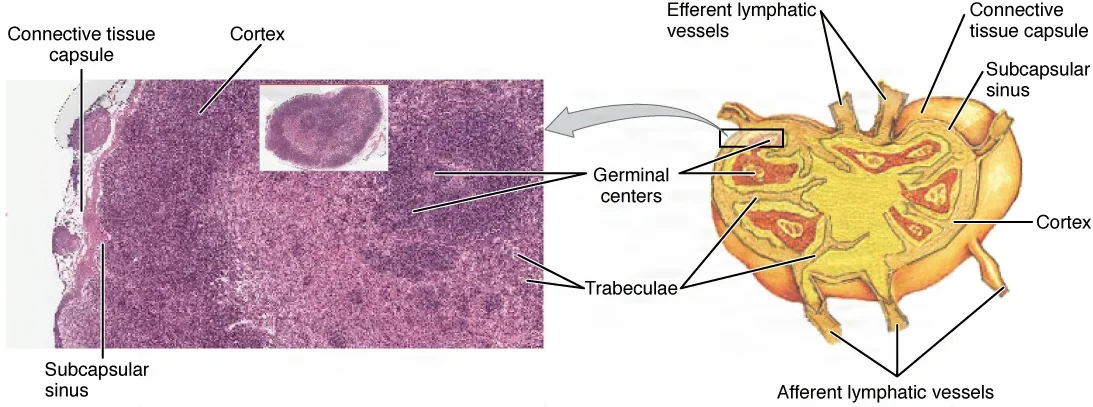
- Filters lymph: macrophageMAK-roh-fayjA large white blood cell in the tissues that swallows and digests germs, dead cells and debris.macro- large-phage eaterFull entry and other cells in the node trap and destroy bacteria, other germs and debris carried in the lymph. That is one reason lymph nodes swell during an infection.
- Makes T and B lymphocytes: lymphocytes form in the node's lymphoid follicleFOL-ih-kulA small sac or cavity in the body, such as the pocket a hair grows from or the sac in the ovary that holds an egg.Full entry , which have areas rich in B cells and areas rich in T cells, and B cells multiply fast in the follicles' germinal centers. Nodes are where the immune system meets germs and mounts its attack.
- Forms antibodyAN-tih-bod-eeA Y-shaped protein made by the immune system that sticks to one specific germ or foreign substance and marks it for destruction.anti- againstbody Full entry : B cells that meet their antigenAN-tih-jenA substance, often a protein on a germ or cell, that the immune system recognizes and responds to.anti- against (short for antibody)-gen something that producesFull entry in the node become plasma cells, which make antibodies.
- Makes monocytes: stem cellSTEM SELAn unspecialized cell that can divide to make more of itself and develop into specialized cells.Full entry that settle in the lymph nodes and spleen can become lymphocytes or monocytes, and the nodes are full of macrophages, the grown form of monocytes.
The spleen's special jobs
The spleen is a dark red organ about 12 cm (5 inches) long in the upper left abdomen, attached to the stomach. It is a major lymphoid organ, and it filters the blood the way lymph nodes filter lymph.
- Its red pulp filters the blood and removes old and damaged red blood cells. Macrophages break them down and save the iron.
- Its white pulp is lymph tissue, where T and B cells fight germs that get into the blood.
- It stores plateletPLAYT-litA tiny cell fragment in the blood that sticks together at a wound to help form a clot.Full entry : about one third of the body's platelets wait there, ready for a bleed.
- Before birth, it helps make blood cells, and it keeps that ability throughout life.
The spleen is fragile and lacks a strong capsule, and it is the organ injured most often in blunt trauma. It is full of blood and can bleed heavily. Because the spleen helps fight infection, surgeons try to stop the bleeding and save it when they can. After a splenectomy, the patient faces a lifelong risk of a sudden, severe infection with certain bacteria, such as pneumococci, especially in young children.
Splenectomy is also done for some blood diseases and cancers, such as idiopathicid-ee-oh-PATH-ikHaving no known cause.idi/o one's own, separatepath/o disease-ic relating toFull entry thrombocytopenic purpuraPUR-pyuh-ruhPurple or red-brown patches on the skin caused by bleeding under the skin, which do not fade when pressed.Full entry (ITP), a disorder with a low platelet count, and lymphomalim-FOH-muhA cancer of lymphocytes, the white blood cells of the lymph system.lymph/o lymph-oma tumorFull entry .
How lymph drains
Lymph flows one way, toward the heart, through a chain of vessels.
- lymphaticlim-FAT-ikRelating to lymph, the clear fluid that drains from body tissues, or to the vessels and nodes that carry it.lymph clear body fluid-atic relating toFull entry capillaries begin as tiny tubes closed at one end, woven among the small blood vessels in the tissues. Their cells overlap like flaps that let fluid in but not out.
- Lymphatic vessels are built like veins, with one-way valves so close together that the vessels look beaded. Lymph passes through lymph nodes along the way.
- Lymphatic trunks form as the vessels merge.
- Two ducts empty the lymph into the veins at the base of the neck, where the internal jugularJUG-yuh-lerRelating to the neck, especially the large veins in the neck.Full entry and subclavian veins meet.
No heart pumps lymph. It is pushed along by the squeeze of skeletal muscleSKEL-uh-tul MUS-ulMuscle attached to bones that a person moves by choice, such as the muscles of the arms and legs.Full entry , by breathing and by body movement, and the valves keep it moving the right way.
The two ducts drain very unequal areas.
- Right lymphatic duct: drains only the upper right part of the body (the right side of the head, neck and chest and the right arm) into the right subclavian vein.
- thoracicthuh-RAS-ikRelating to the chest, or to the middle part of the spine.thorac/o chest-ic relating toFull entry duct: drains everything else, the whole lower body and the left side of the upper body, into the left subclavian vein. It begins just below the diaphragmDY-uh-framThe dome-shaped sheet of muscle under the lungs that is the main muscle of breathing; also the flat side of a stethoscope.Full entry in a pouch called the cisterna chyli, which collects lymph from the legs, pelvis and intestines, and runs up through the chest.
An injured thoracic duct, during neck or chest surgery, can leak milky chyle into the wound or into the chest.
The right lymphatic duct drains only the upper right part of the body. The thoracic duct drains all the rest into the left subclavian vein.
The sentinel node
Cancer cells often spread through the lymph vessels. The sentinel node is the first lymph node, or the first few, that a tumor drains to, so it is the node most likely to hold cancer cells if they have begun to spread.
In a sentinel node biopsy, the tumor area is injected with a radioactive tracer, a blue dye or both. The tracer and dye travel the same lymph path to the sentinel node. The surgeon finds it with a gamma probe in a sterile sleeve, which picks up the tracer, and by the blue color, and removes it for the pathologist. If the sentinel node is free of cancer, the patient may be spared a larger node dissectiondy-SEK-shunA tear in the inner layer of an artery wall that lets blood split the layers apart; also, the cutting apart of tissue in surgery.Full entry and its risk of lymphedema.
In the breast, more than three quarters of the lymph drains to the axillary nodes in the armpit, so that is where the sentinel node usually lies. The blue dye can cause a severe allergic reactionsuh-VEER uh-LUR-jik ree-AK-shunAn allergic reaction strong enough to affect breathing, circulation or several body systems at once, not just the skin.Full entry , anaphylaxisan-uh-fuh-LAK-sisA sudden, severe allergic reaction that affects the whole body.ana- away, against-phylaxis protectionFull entry , in some patients, so the patient is watched closely and a crash cart must be close at hand.
What to take from this chapter
- Blood is plasma plus formed elements. Red cells carry oxygen on hemoglobin, white cells defend the body, and platelets start clotting. Hemostasis is vascular spasm, a platelet plug and a fibrin clot.
- ABO type depends on the A and B antigens on red cells, and each person has antibodies against the antigens they lack. Rh-negative people make anti-Rh antibodies only after exposure, which matters in pregnancy. Type O red cells can go to anyone in an emergency.
- The heart sits in the mediastinum inside the pericardium. Its wall is epicardium, myocardium and endocardium, and its four chambers pump blood from the right side to the lungs and from the left side to the body, through the tricuspid, pulmonary, mitral and aortic valves.
- The coronary arteries leave the aorta just above the aortic valve. The left main divides into the LAD and circumflex; a blocked coronary artery can cause an infarction. CABG bypasses it with the internal mammary artery or a reversed saphenous vein.
- The SA node sets the beat; the signal runs through the atria to the AV node, the AV bundle, the bundle branches and the Purkinje fibers. Sympathetic nerves speed the heart and the vagus nerve slows it.
- Arteries and veins have an intima, a media and an adventitia; capillaries have only the intima. Plaque builds in the intima, the media narrows and widens the vessel, and valves in the veins keep blood moving toward the heart.
- Blood pressure depends on cardiac output, blood volume, viscosity, the elasticity of the arteries and peripheral resistance. Know the nine pulse points; the pedal pulses are checked before and after vascular repair.
- The lymph system returns tissue fluid to the blood through the thoracic duct and right lymphatic duct. Lymph nodes filter lymph and make lymphocytes, the spleen filters blood, and the sentinel node shows whether a cancer has spread.
Check yourself5 quick questions
Free for everyone. Pick an answer for each, then check them all at once.
In the ORWhere you'll use this
The chapter's whole question pool in sets of 10, with the answer explained after each one.
Part of the educators programThe chapter's words on spaced cards: the ones you miss come back sooner.
Part of the educators programGiven by your class's teacher. Each class draws its own 25 from the chapter's pool.
Part of the educators program
Suggest a fix
Something wrong, unclear or out of date in this chapter? Tell us and we will fix it for everyone. Your email is only used to reply.