Splenectomy
The partial or total surgical removal of the spleen, often due to trauma, rupture, or hematologic disorders. The splenic artery and vein are ligated and divided to allow for the safe excision of the organ from the left upper quadrant.
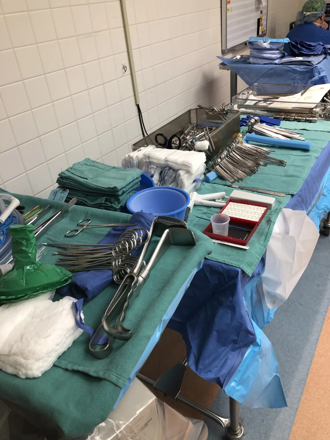
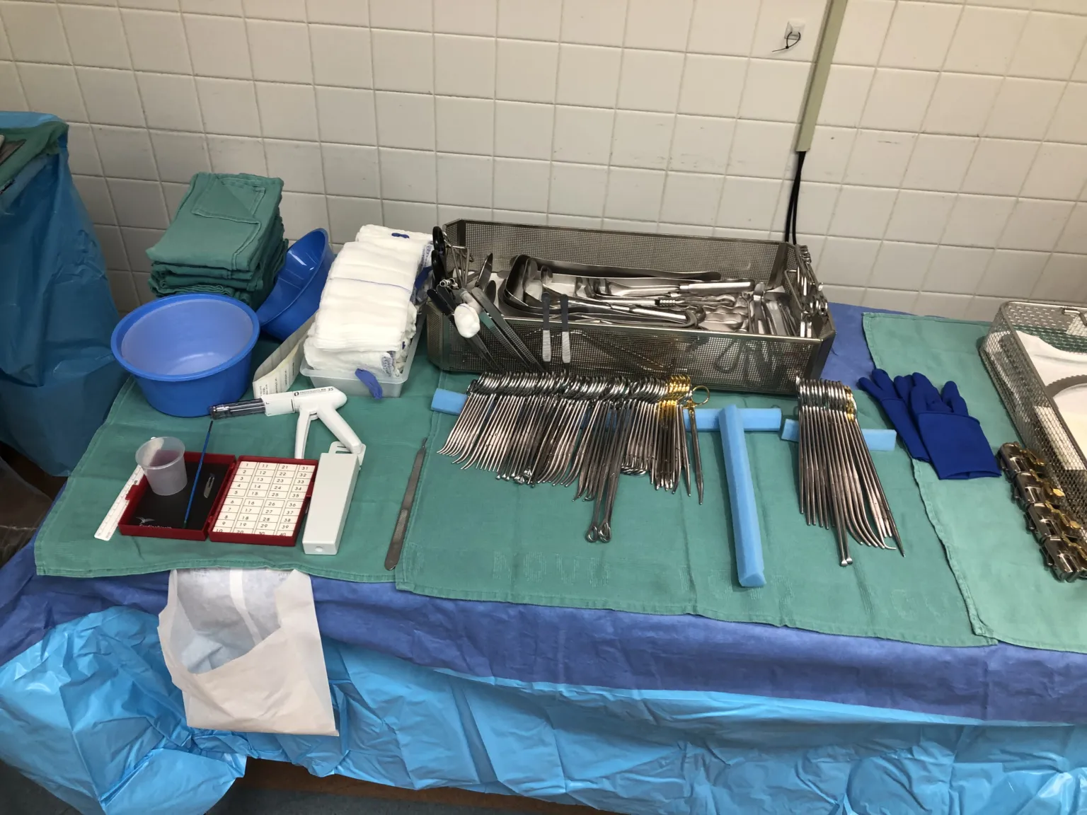
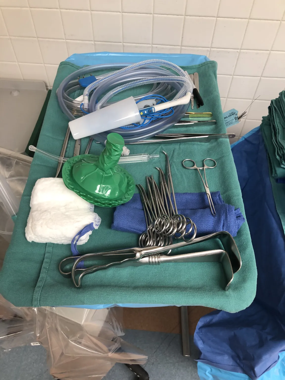
- Position
- Supine
In 30 seconds
Scan this outside the room.
- The Surgery Sparknotes
- Make initial incision with a 15 or 10 blade on a #3 knife handle.
- dissectdy-SEKT, or dih-SEKTTo cut apart or separate tissues carefully during surgery.Full entry through tissue using Adsons, Metz, Bovie, and retractorrih-TRAK-terA surgical instrument that holds back tissue or organs so the surgeon can see and reach the operative site.Full entry .
- Ligate all attachments and blood vessels using Bovie, Metz, or ties.
- Remove spleenspleenAn organ in the upper left abdomen that filters the blood and helps fight infection.splen/o spleenFull entry carefully and pass it off as a specimen.
- Stop bleeding, close incision with staples, and no drains are needed.
The tray
18 instruments, by what they do. Tap a name for its card.
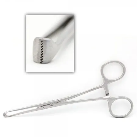
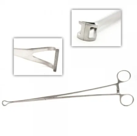
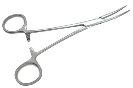
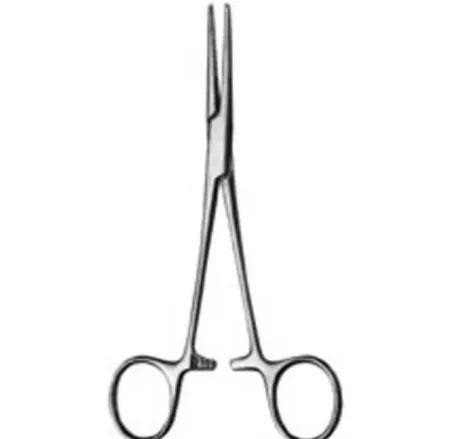
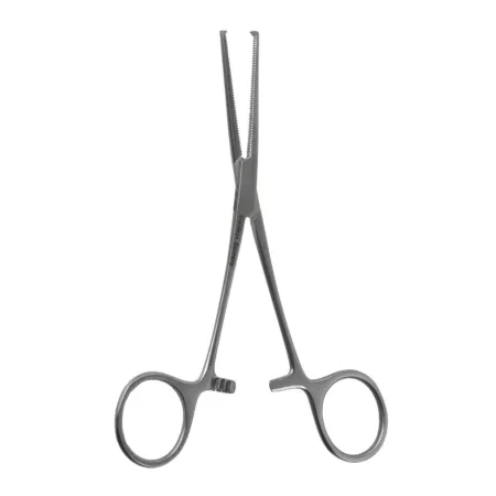
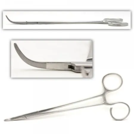
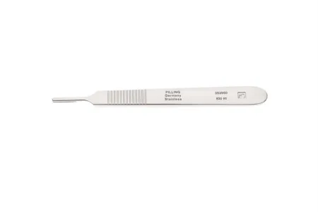
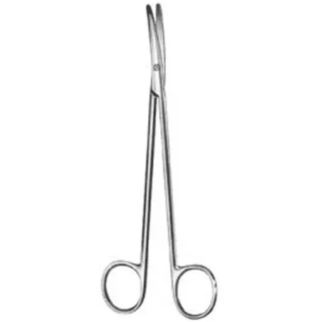
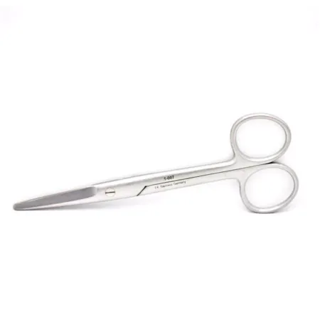
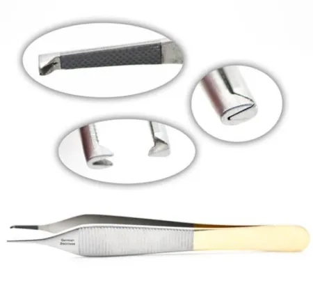
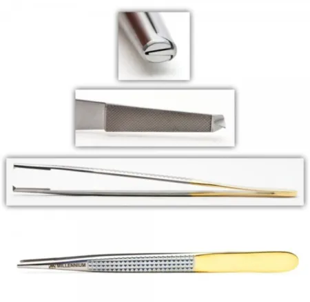
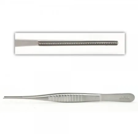
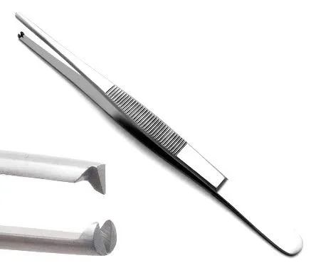
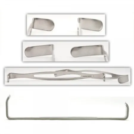
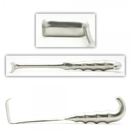
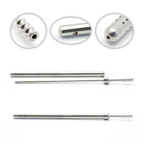
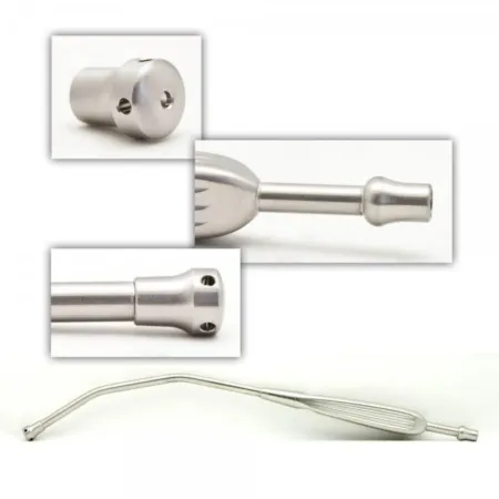
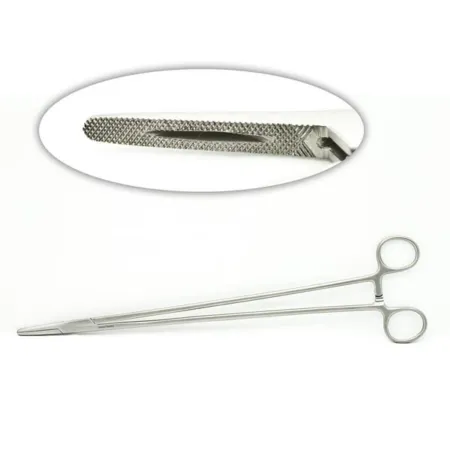
Clamping & Occluding6
Cutting & Dissecting3
Grasping & Holding4
Retracting & Exposing2
Suctioning2
Suturing & Stapling1
Know this tray before you scrub in.
With Pro, for every setup in every specialty. Cancel any time.
Or scrub in free: every General setup and 5 questions a day.
The room
The equipment, the prep and drape, the suture and the medicines.
Folded towels or utility drapes and a laparotomy drape
Step by step
5 steps, one line each.
- 1
Initial Incision
During a Splenectomy, the surgical team will first need to make an incision to gain access to the abdomen. For this, a 15 or 10 blade loaded on a #3 knife handle is typically used. This initial incision will be made with precision to ensure minimal tissue damage and create enough space for the surgical team to work effectively.
- 2
Dissecting Through Tissue
Once the incision has been made, the doctor will start to dissectdy-SEKT, or dih-SEKTTo cut apart or separate tissues carefully during surgery.Full entry through the tissue to enter the abdomen. To do this, they will use a combination of Adsons, Metz, Bovie, and smaller retractorrih-TRAK-terA surgical instrument that holds back tissue or organs so the surgeon can see and reach the operative site.Full entry . These instruments will allow the surgical team to carefully maneuver through the tissue and minimize damage to the surrounding structures.
- 3
Ligation of Attachments and Blood Vessels
Once the spleenspleenAn organ in the upper left abdomen that filters the blood and helps fight infection.splen/o spleenFull entry has been reached, the surgical team will need to ligate all of its attachments and blood vessels to remove it successfully. For this, they will typically use a combination of Bovie, Metz, or ties loaded on tonsilTON-sulOne of the masses of lymph tissue at the back of the throat that help fight germs entering the mouth and nose.Full entry or used freehand. The surgical team should have a lot of 0 and 2-0 ties available, along with vessel loops, to ensure they have enough supplies to complete the procedure effectively.
- 4
Removal of the Spleen
After all of the attachments have been ligated and removed, the spleenspleenAn organ in the upper left abdomen that filters the blood and helps fight infection.splen/o spleenFull entry will be passed off as a specimen. This requires careful handling to avoid damaging the organ or spilling its contents. The surgical team must ensure that the spleen is removed entirely and that there are no remaining attachments or vessels that could cause bleeding or other complications.
- 5
Stop Bleeding and Close Incision
Once the spleenspleenAn organ in the upper left abdomen that filters the blood and helps fight infection.splen/o spleenFull entry has been removed, the surgical team will need to stop any bleeding and close the incision. Drains are usually not used for splenectomies, and staples are typically used to close the skin. The surgical team will need to ensure that the incision is closed correctly to prevent any postoperative complications.
The case
Splenectomy is a surgical procedure that involves the removal of the spleen, which is an organ located in the upper left side of the abdomen. This procedure may be necessary if the spleen is enlarged, damaged, or affected by a medical condition such as cancer, infection, or blood disorders. The surgery is performed under general anesthesia, and the spleen is removed through a small incision in the abdomen. After the surgery, patients may need to take medications and follow certain precautions to prevent infections. Splenectomy can improve or resolve symptoms associated with spleen-related conditions, but it can also increase the risk of certain infections and other health issues.
Built by the techs who scrub it.
Every surgeon runs it a little differently. Send your table, or a fix for this page.
 Diana Lei-alohaReviewer
 LucyReviewer
 Julia BlockPhotos
The rest of this setup is Pro.
Everything unlocked above stays free. Pro opens the rest, on every setup: where new techs slip, what to load next at every step, the circulator’s side, the medicines, the specimen, wound class and trays on standby, what to grab if it goes wrong, and practice for every tray.
Free, for good. No card.
- All 2,882 instrument cards
- Every General setup in full
- 5 exam questions a day
- Your own preference cards
Pass your exam. Know every case.
Everything in Free, plus:
- All 260 setups in every specialty: back table, Mayo and every step
- Practice that never runs out and full timed exams for the CST, TS-C, CRCST, CSPDT and CSFA, with the why behind every answer
- Flashcards on any tray, timed to come back before you forget
- Every instrument card in full: the 3D model, cautions, inspection, how to pass it
Cancel any time from your account. Not worth it? Email us within 30 days of a charge for a refund, one per customer. Refund policy
Pro is also in the CSTSetup app, on the same account. Already have Pro? Log in
Send your table
Your Mayo, your back table, your surgeon’s way, or anything wrong on this page. Your email is only used to credit you. Submissions stay private and are reviewed by our team; accepted ones earn a Contributor credit here.