Sentinel Node/Lymph Node Dissection
The identification and surgical excision of the primary lymph nodes that drain a specific anatomical area to evaluate the potential spread of malignant cells.
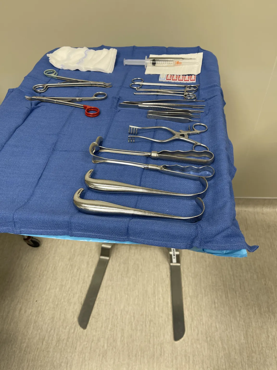
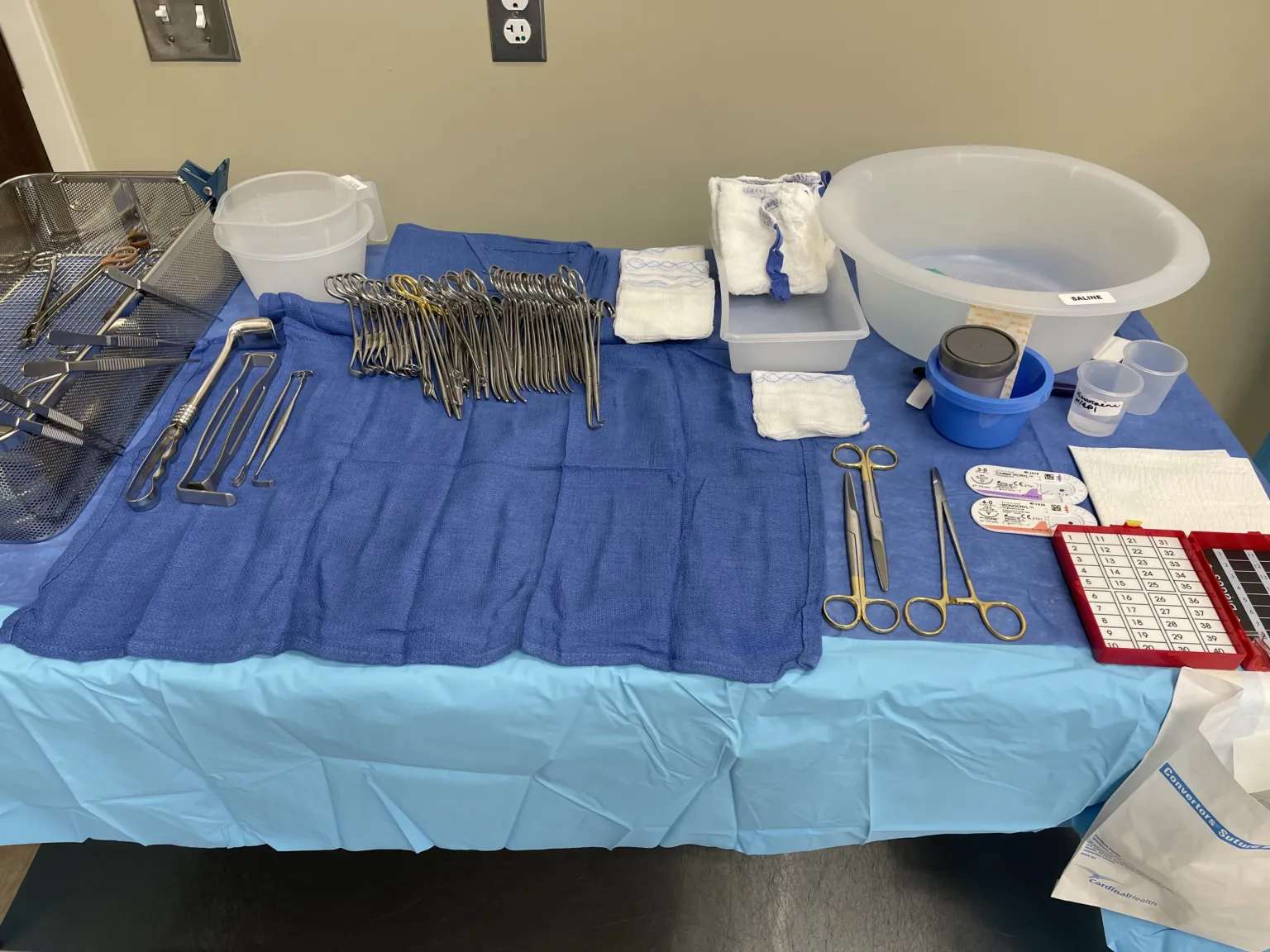
In 30 seconds
Scan this outside the room.
- The Surgery Sparknotes
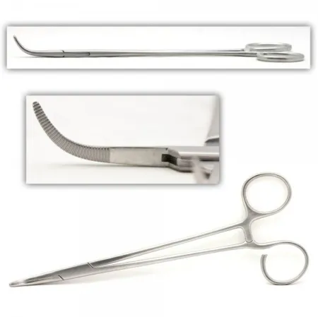
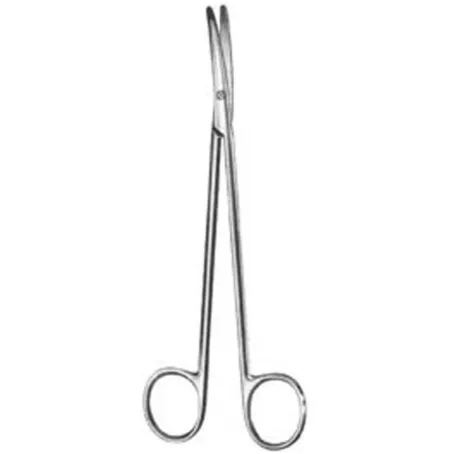
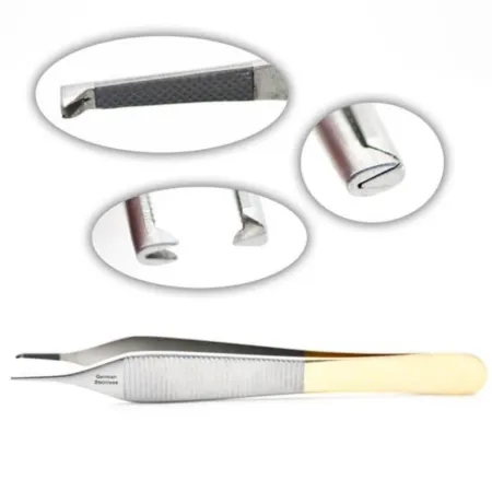
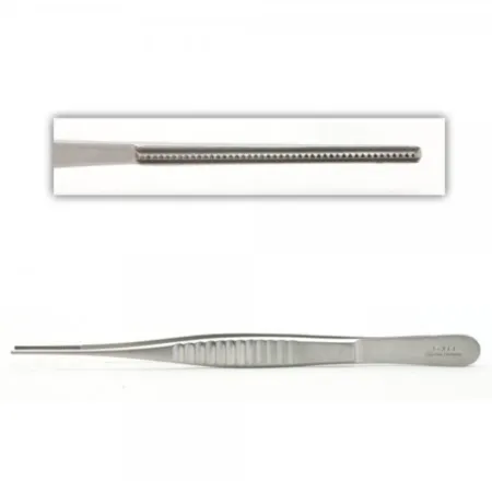
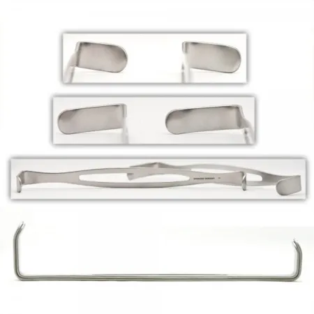
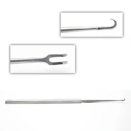
- Make incision and dissectdy-SEKT, or dih-SEKTTo cut apart or separate tissues carefully during surgery.Full entry using 15 blade, metz, debakeys, and tonsilTON-sulOne of the masses of lymph tissue at the back of the throat that help fight germs entering the mouth and nose.Full entry . Use skin hooks or army-navys for retractionrih-TRAK-shunA drawing back or pulling in of tissue; in breathing, the skin sinking in between, below or above the ribs with each breath.Full entry .
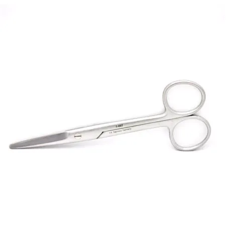
- Collect specimens using forcepsFOR-sepsA hand instrument with two blades or arms used to grasp, hold or pull tissue or objects.Full entry and scissors. Send to lab for analysis.
- Inject methylene blue before surgery to locate nodes. Use sterile or unsterile technique. Use a probe if necessary.
The tray
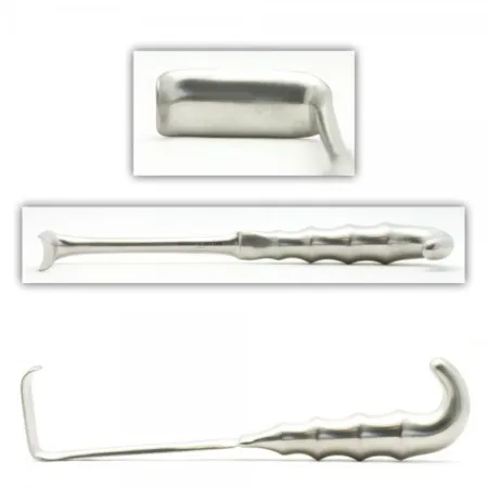
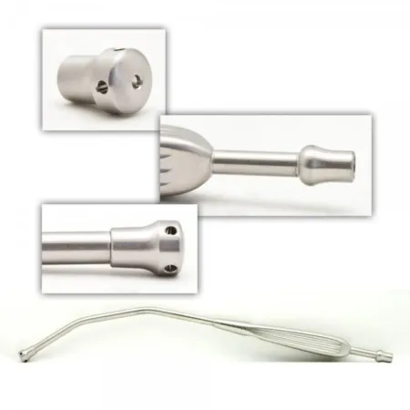
13 instruments, by what they do. Tap a name for its card.
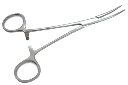
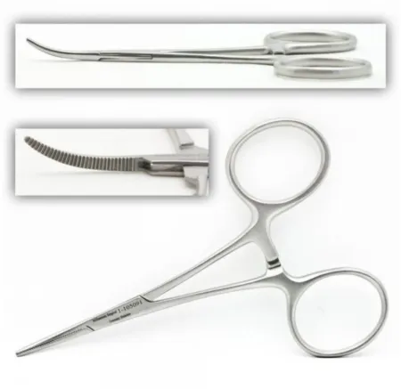
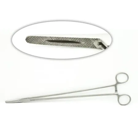
Clamping & Occluding3
Cutting & Dissecting3
Grasping & Holding2
Retracting & Exposing3
Suctioning1
Suturing & Stapling1
Know this tray before you scrub in.
With Pro, for every setup in every specialty. Cancel any time.
Or scrub in free: every General setup and 5 questions a day.
The room
The equipment, the prep and drape, the suture and the medicines.
Folded blue towels or utility drapes and a pediatric laparotomy drape, thyroid drape, or two split U drapes
Step by step
3 steps, one line each.
- 1
Incision and Dissection
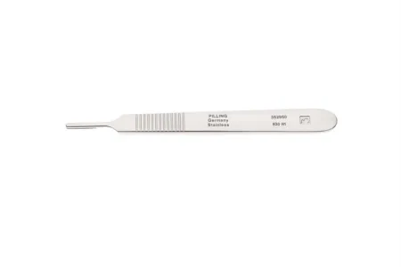
During a Sentinel Node/lymph nodelimf nohdA small, bean-shaped organ of the immune system that filters lymph fluid and holds white blood cells.Full entry dissectiondy-SEK-shunA tear in the inner layer of an artery wall that lets blood split the layers apart; also, the cutting apart of tissue in surgery.Full entry , the surgeon will make an incision with a 15 blade loaded on a #3 knife handle. They will then dissectdy-SEKT, or dih-SEKTTo cut apart or separate tissues carefully during surgery.Full entry down to the nodes using instruments such as metz, debakeys, and a tonsilTON-sulOne of the masses of lymph tissue at the back of the throat that help fight germs entering the mouth and nose.Full entry . The surgeon may also use skin hooks or army-navys for retractionrih-TRAK-shunA drawing back or pulling in of tissue; in breathing, the skin sinking in between, below or above the ribs with each breath.Full entry , depending on the depth of the dissection.
- 2
Specimen Collection
Once the nodes have been located, the surgeon will collect the specimens using a combination of forcepsFOR-sepsA hand instrument with two blades or arms used to grasp, hold or pull tissue or objects.Full entry and scissors. These specimens will then be sent to the lab for analysis to determine if there is any cancer present. Depending on the surgeon's preference, they may choose to close the incision before receiving the lab results.
- 3
Methylene Blue Injection
To make the nodes easier to locate, methylene blue may be injected prior to the start of the surgery. This can be done either unsterile or sterile, depending on the surgeon's preference. A probe may also be draped and used to assist in finding the nodes.
The case
Sentinel Node/Lymph Node Dissection is a surgical procedure used to remove lymph nodes from various areas of the body for the purpose of detecting or treating cancer. The surgeon makes an incision and uses a combination of instruments to locate and remove the nodes, which are then sent to the lab for analysis. Methylene blue may be injected before the surgery to make the nodes easier to locate. Understanding the steps involved and the instruments used can help surgical technologists better assist the surgeon and ensure a successful outcome for the patient.
Built by the techs who scrub it.
Every surgeon runs it a little differently. Send your table, or a fix for this page.
 Diana Lei-alohaReviewer
 Jocelyn MillerPhotos
The rest of this setup is Pro.
Everything unlocked above stays free. Pro opens the rest, on every setup: where new techs slip, what to load next at every step, the circulator’s side, the medicines, the specimen, wound class and trays on standby, what to grab if it goes wrong, and practice for every tray.
Free, for good. No card.
- All 2,882 instrument cards
- Every General setup in full
- 5 exam questions a day
- Your own preference cards
Pass your exam. Know every case.
Everything in Free, plus:
- All 260 setups in every specialty: back table, Mayo and every step
- Practice that never runs out and full timed exams for the CST, TS-C, CRCST, CSPDT and CSFA, with the why behind every answer
- Flashcards on any tray, timed to come back before you forget
- Every instrument card in full: the 3D model, cautions, inspection, how to pass it
Cancel any time from your account. Not worth it? Email us within 30 days of a charge for a refund, one per customer. Refund policy
Pro is also in the CSTSetup app, on the same account. Already have Pro? Log in
Send your table
Your Mayo, your back table, your surgeon’s way, or anything wrong on this page. Your email is only used to credit you. Submissions stay private and are reviewed by our team; accepted ones earn a Contributor credit here.