Digestion
The tube that runs from the mouth to the anus, the liver, gallbladder and pancreas that feed it, and how they turn a meal into fuel: the organs general surgery works on.
1Why it matters
Every cell in the body needs fuel and raw materials, and food carries both. But food arrives as large molecules, such as starches, proteins and fats, that cannot cross into the blood. The digestive system breaks food down into pieces small enough to absorb, takes them into the body, and gets rid of what is left.
You meet this system in a large share of general surgery.
- Surgeons remove and rejoin these organs. A gallbladder or an appendix comes out, and a diseased stretch of stomach or bowel is removed and the ends joined.
- The bowel is full of bacteria. The colon holds the most of them. Once the bowel is opened, its contents can infect the wound, so the team handles the field differently.
- The liver and pancreas reach far beyond digestion. The liver makes clotting factors and breaks down drugs, and the pancreas makes insulin, so disease in either one can change how a patient bleeds and clots, handles anesthetic drugs and controls blood sugar.
- Scopes look inside. Flexible endoscopes pass through the mouth or the anus to look at the lining and to find, mark and sometimes treat disease there, such as polyps that can be removed before they turn into cancer.
This chapter follows a meal from the mouth to the anus, then turns to the liver, gallbladder and pancreas, the nutrients food supplies, the words for digestion and elimination, and what all of it means at the field. Other chapters hold the rest: the abdominal wall, quadrants and regions in chapters 2 and 3; the nerves that drive the gut in chapter 4; the arteries and veins of the gut, the portal vein among them, in chapter 5; the pharynx and larynx as an airway in chapter 6; the kidneys in chapter 8; insulin, glucagon and the endocrine pancreas in chapter 9; the gut's germs and infection in chapter 10; tumors and wound healing in chapter 11; drugs for the stomach and bowel in chapter 12; bowel technique in chapters 29 and 32; the parotid operation in chapter 36; surgery of the teeth and jaws in chapter 37; and hernias and the general surgery operations, with their setups, in chapter 32.
2How digestion works
A tube and its helpers
The digestive system has two groups of organs.
- The alimentary canal, also called the gastrointestinal (GI) tract or the gut, is one long tube. It runs from the mouth through the pharynx and esophagus to the stomach, then through the small intestine and the large intestine, and ends at the anus.
- The accessory organs help from outside the tube: the teeth, tongue and salivary glands in the mouth, and the liver, gallbladder and pancreas, which send their juices into the small intestine through ducts.
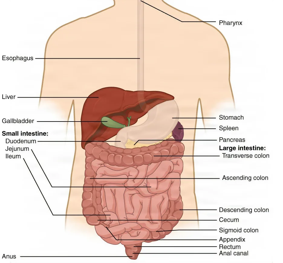
In a living person the tube is about 6 to 7.6 meters long. After death, once its muscle loses its tone, it measures longer, about 10.7 meters.
The mouth and the anus both open to the outside, so food inside the tube is, strictly speaking, still outside the body. Nutrients enter the body only when the lining absorbs them.
Six processes
Digestion is the breaking down of food into molecules small enough to be absorbed. Six processes carry a meal through the system.
- Ingestion: taking food into the mouth.
- Propulsion: moving food along the tube. It starts with swallowing, which you control, and goes on as peristalsis, which you do not: waves in which the smooth muscle of the wall tightens and relaxes in turn, pushing the contents forward. Peristalsis is strong enough to carry a drink to your stomach even if you are upside down.
- Mechanical digestion: breaking food into smaller pieces without changing what it is made of. Chewing (mastication), the churning of the stomach and the squeezing of the small intestine all do it. Smaller pieces give the digestive juices more surface to work on.
- Chemical digestion: enzymes in the digestive juices split large molecules into their building blocks, for example proteins into amino acids. It starts in the mouth and is finished in the small intestine.
- Absorption: the building blocks, with water, vitamins and minerals, pass through the lining into the blood or the lymph. Almost all of it happens in the small intestine.
- Defecation: what cannot be digested or absorbed leaves the body as feces.
Some references count the steps a little differently and list secretion of the digestive juices, or mixing, as steps of their own.
mechanical digestion makes the pieces smaller, and chemical digestion changes the molecules. A meal needs both before anything can be absorbed.
What runs it
The gut has its own nervous system, the enteric nervous system, built into its wall from the esophagus to the anus. One network of nerves, in the muscle layer, controls how the wall moves; another, in the submucosa, controls what it secretes.
The autonomic nervous system adjusts it from outside. Parasympathetic signals, carried by the vagus nerves and the pelvic splanchnic nerves, speed up secretion and movement. Sympathetic signals, the fight-or-flight response, slow them down. Chapter 4 teaches these two divisions.
Hormones also run digestion. Cells in the lining of the stomach and small intestine release them into the blood when food arrives, and they switch the other organs' secretions on or off. The main ones appear with their organs below.
3The abdominal cavity and the peritoneum
Two layers and the space between
Most digestive organs sit in the abdominal cavity, held in place by the peritoneum, the largest serous membrane in the body. Like the pleura around the lungs, it has two layers with a thin film of fluid between them.
- The parietal peritoneum lines the inside of the abdominal wall.
- The visceral peritoneum wraps the organs. On the stomach and intestines it forms their outer layer, the serosa.
The peritoneal cavity is the thin space between the two layers. In health it holds only a few milliliters of watery fluid, which lets the organs slide against each other and the wall without rubbing.
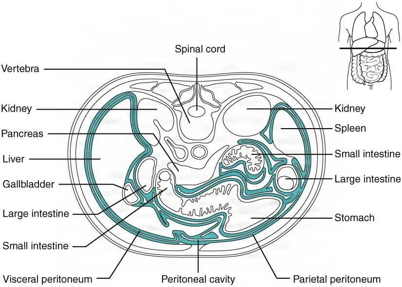
Peritonitis is inflammation of the peritoneum. It follows anything that lets gut contents, blood or germs into the peritoneal cavity: an ulcer that eats through the stomach wall, a burst appendix, a hole in the bowel, or a bleeding liver or spleen. It is life-threatening and often needs emergency surgery.
The retroperitoneal space
Some organs lie behind the peritoneum, against the back wall, in the retroperitoneal space, or retroperitoneum. The peritoneum covers only their front.
- Digestive organs there: most of the duodenum, the pancreas, the ascending and descending colon, and the rectum.
- Others: the kidneys and the ureters, which chapter 8 teaches.
The surgeon reaches these organs either through the peritoneum from the front or from the flank.
The folds that hold the organs
The visceral peritoneum folds into double sheets that tie the organs to the back wall and to each other. Blood vessels, lymph vessels and nerves run between the two layers of each sheet to reach the organs.
| Fold | Where it runs | What it does |
|---|---|---|
| Greater omentum | Hangs from the greater curvature of the stomach like an apron in front of the intestines, then folds back up to the transverse colon | Covers the intestines and stores fat, more of it in people who are overweight |
| Lesser omentum | From the underside of the liver to the lesser curvature of the stomach | Holds the stomach to the liver; the hepatic artery, the portal vein and the common hepatic duct run behind its free edge |
| Mesentery | A fan from the back wall to the jejunum and ileum | Anchors the small intestine below the duodenum and carries its blood vessels, nerves and lymph vessels |
| Mesocolon | From the back wall to the transverse and sigmoid colon | Ties these two loose parts of the colon to the back wall |
| Falciform ligament | From the liver to the front abdominal wall and the diaphragm | Anchors the liver and marks the line between its right and left lobes |
These folds matter at the field. Before part of the stomach is removed, its omentum is separated from it, and when a segment of jejunum or ileum comes out, its mesentery, with the vessels inside, is clamped and divided too.
4The wall of the digestive tube
Along its whole length, the wall of the tube follows one plan of four layers, changed a little in each organ to suit its job. From the inside out:
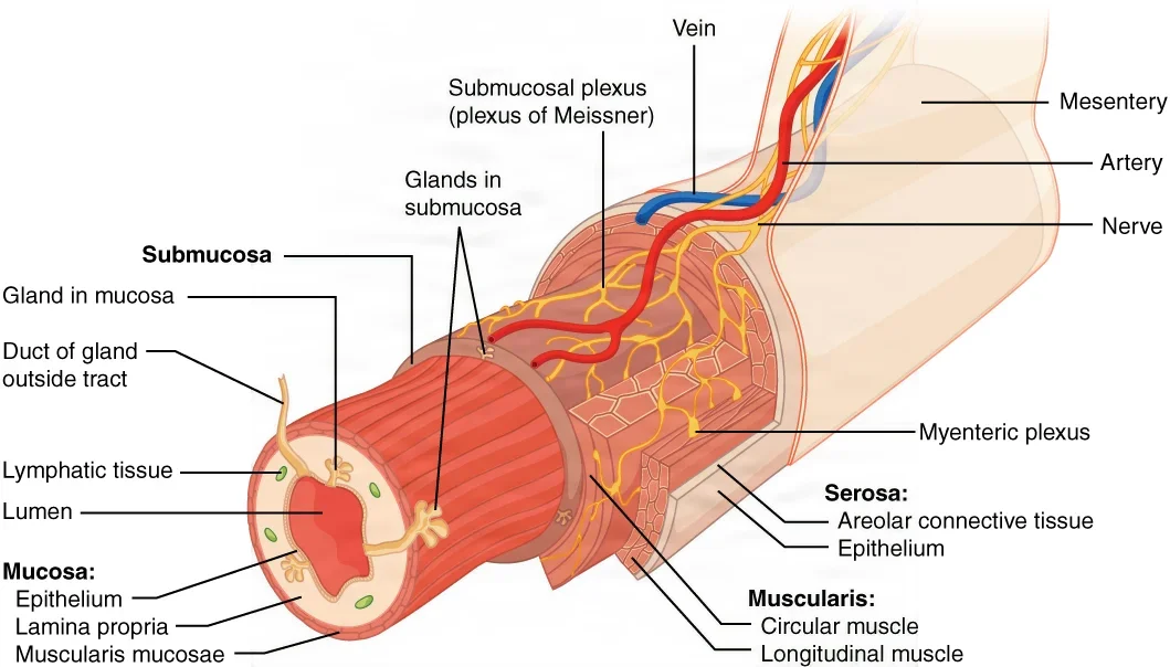
- Mucosa: the lining, a mucous membrane that touches the food. It has three parts: the epithelium on the surface; a thin connective tissue under it, the lamina propria, with blood and lymph vessels and clusters of lymphocytes that guard against swallowed germs; and a thin sheet of smooth muscle, the muscularis mucosae, that ripples the lining into folds. Goblet cells in the epithelium make mucus, and other cells release hormones. The epithelium is stratified squamous where it takes the most wear (the mouth, pharynx, esophagus and anal canal) and a single layer of tall columnar cells in the stomach and intestines. Its cells live only a few days to about a week before new ones replace them.
- Submucosa: dense connective tissue that binds the mucosa to the muscle layer. It carries blood and lymph vessels, some glands, and the nerve network that controls secretion.
- Muscularis, also called the muscularis externa: smooth muscle in two layers, an inner circular layer and an outer longitudinal layer, with the nerve network that controls movement between them. Their contractions mix the food and move it along.
- Serosa: the outer covering, a layer of visceral peritoneum over loose connective tissue. It is found only where the tube lies inside the abdominal cavity, and it continues into the mesentery.
The pattern changes where the job changes.
- The stomach adds a third muscle layer, an oblique one, so it can churn food hard.
- In the large intestine, the outer longitudinal muscle gathers into three narrow bands, the teniae coli.
- At both ends of the tube (the mouth, the pharynx, the top of the esophagus and the external anal sphincter) the muscle is skeletal, so you control swallowing and defecation.
- The mouth, pharynx and esophagus have no serosa. A tough sheath of collagen fibers, the adventitia, holds them in place instead.
from the inside out, the wall is mucosa, submucosa, muscularis and serosa. Read it from the lumen, the space inside the tube, outward to the peritoneal cavity.
5The mouth
Palate, uvula, tongue and teeth
The mouth, or oral cavity, is framed by the cheeks, the tongue and the palate. The lips close its front, and at the back it opens into the throat through an opening called the fauces.
The roof of the mouth is the palate.
- The hard palate, in front, is a shelf of bone. It separates the mouth from the nose and gives the tongue something firm to press food against.
- The soft palate, behind it, is mostly skeletal muscle.
- The uvula is the small fleshy piece that hangs from the middle of the soft palate's back edge. When you swallow, the soft palate and the uvula rise and keep food and drink out of the nose.
The tongue is skeletal muscle covered with mucous membrane, tied to the floor of the mouth by a fold called the lingual frenulum. Its muscles move food into place for chewing, gather it into a bolus and push it back to be swallowed. Its surface carries papillae, some of them holding taste buds, and its glands release lingual lipase, an enzyme for fats that starts working only once it reaches the acid of the stomach.
The teeth tear and grind food. A child has 20 deciduous (baby) teeth, and an adult has 32 permanent teeth: 8 incisors for biting, 4 cuspids (canines) for tearing, 8 premolars for mashing and 12 molars for crushing. Each tooth has a crown above the gum and a root set in the jaw, and enamel, the hardest substance in the body, covers the crown. Under the enamel lies dentin, a bone-like tissue, which in the root is covered by cementum, a layer of modified bone. At the tooth's center the pulp cavity holds nerves and blood vessels, and the part of it running through the root is the root canal. The gums (gingivae) surround each tooth's neck, and a periodontal ligament holds it in its socket.
The salivary glands and their ducts
Saliva comes from many small glands in the lining of the mouth and from three pairs of large glands outside it, which empty into the mouth through ducts.
| Gland | Where it lies | Its duct | Its saliva |
|---|---|---|---|
| Parotid, the largest | In front of the ear, over the jaw and the masseter muscle; its tail reaches below the jaw into the upper neck | The parotid duct, or Stensen duct, which opens inside the cheek opposite the upper second molar | Watery, with salivary amylase |
| Submandibular, the second largest | Curled around the lower border of the jaw | The submandibular duct, or Wharton duct, which opens into the front of the floor of the mouth | Amylase, in saliva thickened with mucus |
| Sublingual, the smallest | In the floor of the mouth, just under the lining | Several small ducts, the ducts of Rivinus | The thickest, mostly mucus, with the least amylase |
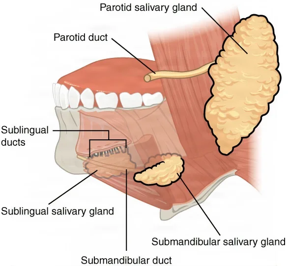
The glands make about 1 to 1.5 liters of saliva a day. It is more than 99 percent water, and it does several jobs.
- It moistens food and binds it into the soft, easily swallowed ball called the bolus.
- It starts chemical digestion: salivary amylase begins breaking down starch, and keeps working in the stomach until the acid stops it.
- It protects the mouth: lysozyme and antibodies (IgA) fight germs, and buffers keep its pH close to neutral.
- It dissolves food so you can taste it, and it rinses the teeth.
The sight, smell, taste or even the thought of food increases the flow. Parasympathetic nerves drive it, and in stress the sympathetic nerves cut it back, which is why fear dries the mouth.
The facial nerve runs through the parotid gland and divides it into a superficial lobe and a deep lobe. Most parotid tumors are benign, and in a parotidectomy the surgeon identifies the facial nerve, sometimes with a nerve stimulator, so it can be kept safe; chapter 36 teaches the operation.
6The pharynx and swallowing
The pharynx
The pharynx, the throat, is a short tube of skeletal muscle lined with mucous membrane. It runs from the back of the nose and mouth down to the esophagus behind and the larynx in front, so it carries both air and food. Chapter 6 teaches its three parts and the airway; here it is the passage for food.
Three groups of constrictor muscles wrap the pharynx. When they squeeze, they push the bolus down into the esophagus.
Swallowing
Swallowing, or deglutition, moves food from the mouth to the stomach. Solid food takes about 4 to 8 seconds, liquids about 1 second, in three phases.
- Voluntary (oral) phase: once chewing is done, the tongue presses up and back against the palate and pushes the bolus into the oropharynx. You choose when this happens.
- Pharyngeal phase, automatic: receptors in the throat signal the swallowing center in the medulla. The soft palate and uvula rise to close off the nose, the larynx rises and the epiglottis folds down over its opening, and breathing stops for a moment. The constrictors squeeze the bolus down, and the upper esophageal sphincter relaxes to let it in.
- Esophageal phase, automatic: peristalsis carries the bolus down the esophagus, and the lower esophageal sphincter relaxes to let it into the stomach.
When food "goes down the wrong way" into the trachea, a cough usually forces it back up. Under general anesthesia the protective reflexes, swallowing and gagging among them, are lost, which is why the anesthesia provider guards the airway; chapter 13 teaches how.
7The esophagus
Where it runs
The esophagus is a muscular tube about 25 cm (10 inches) long that carries food from the pharynx to the stomach. It lies behind the trachea and in front of the spine, runs down through the chest in the mediastinum, and passes through an opening in the diaphragm, the esophageal hiatus, to join the stomach at its cardia. When it is empty, it lies flat.
Its layers
Its wall has the usual layers, with differences in the lining, the muscle and the outer coat.
- Mucosa: stratified squamous epithelium, which stands up to rough food, with glands that make mucus to ease the bolus down.
- Submucosa: connective tissue carrying vessels and glands.
- Muscularis: skeletal muscle in the upper third, a mix of skeletal and smooth muscle in the middle third, and smooth muscle in the lower third.
- Adventitia in place of a serosa, because most of the esophagus lies in the chest, outside the peritoneum.
Two sphincters
A sphincter is a ring of muscle around a tube: it closes the tube when it contracts and opens it when it relaxes.
- The upper esophageal sphincter, made of skeletal muscle, lets the bolus in from the pharynx.
- The lower esophageal sphincter (LES), made of smooth muscle where the esophagus meets the stomach, is also called the gastroesophageal or cardiac sphincter. It relaxes to let food into the stomach, then closes to keep stomach acid out of the esophagus. The diaphragm around it helps hold it shut.
When the LES does not close fully, acid washes back up into the esophagus. This backward flow is reflux. It causes heartburn, and when it keeps happening it is gastroesophageal reflux disease (GERD), which over time can inflame the esophagus, scar and narrow it, and raise the risk of cancer. In a hiatal hernia, a weak hiatus lets the lowest part of the esophagus and the top of the stomach push up into the chest.
When medicine does not control severe reflux, a fundoplication can: the surgeon folds the top of the stomach, the fundus, around the lower esophagus so it acts as a new sphincter. A Nissen fundoplication wraps it all the way around. Chapter 32 teaches the operation.
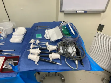
An esophagogastroduodenoscopy (EGD) passes a flexible scope through the mouth to look at the lining of the esophagus, the stomach and the first parts of the duodenum.
8The stomach
The stomach is a widening of the tube, a bag in the upper left abdomen just under the diaphragm, between the esophagus and the duodenum. Empty, it is about the size of a fist; it can stretch to hold about 4 liters and then shrink back.
Regions and curvatures
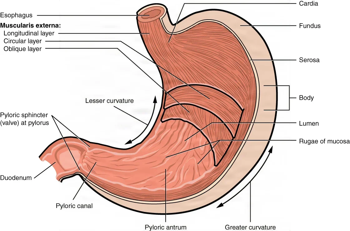
The stomach has four regions.
- Cardia: the area around the opening from the esophagus.
- Fundus: the dome that rises above and to the left of the cardia, under the diaphragm. It holds food for a while, and the gas made during digestion.
- Body: the large middle part.
- Pylorus: the funnel at the end that leads to the duodenum. Its wide part, the pyloric antrum, joins the body; its narrow part, the pyloric canal, ends at the pyloric sphincter, a ring of smooth muscle that controls how fast the stomach empties.
References divide it a little differently. Some name three parts (the fundus, the body and the antrum), and others name the antrum and the pyloric canal as separate regions. The Core Curriculum's list names the antrum and the pylorus separately.
The stomach's short, concave inner border, which faces the liver, is the lesser curvature. Its long, convex outer border is the greater curvature. The lesser omentum ties the lesser curvature to the liver, and the greater omentum hangs from the greater curvature.
The wall and its glands
When the stomach is empty, its mucosa and submucosa lie in folds called rugae, which flatten out as it fills.
Its muscularis has three layers instead of two: an inner oblique layer, a middle circular layer and an outer longitudinal layer. The extra layer lets it churn food hard.
The mucosa is dotted with tiny pits that lead down into the gastric glands, which make gastric juice. Their main cells:
| Cell | What it makes | Why it matters |
|---|---|---|
| Surface mucus cells | A thick coat of alkaline mucus | Shields the lining from the acid and enzymes |
| Parietal cells | Hydrochloric acid (HCl) and intrinsic factor | The acid holds the stomach at a pH of about 1.5 to 3.5, kills many of the bacteria swallowed with food and turns pepsinogen into pepsin; intrinsic factor lets the small intestine absorb vitamin B12 |
| Chief cells | Pepsinogen, and gastric lipase | Pepsinogen becomes pepsin, the enzyme that starts protein digestion; gastric lipase works on fats |
| G cells | The hormone gastrin | Raises the glands' secretion and speeds the stomach's emptying |
What the stomach does
The stomach is first a holding tank. You can eat a meal far faster than the small intestine can digest it, so the stomach keeps it and lets it out a little at a time.
- Mechanical digestion: about every 20 seconds, mixing waves churn the food with gastric juice until it becomes chyme, a thick, soupy liquid.
- Chemical digestion: HCl and pepsin start breaking down proteins. Salivary amylase goes on working on starch for a while in the fundus until the acid stops it, and the lipases begin on fats.
- Absorption: very little, because the stomach wall lets few substances through. It does absorb some water and certain salts (ions), alcohol, some simple sugars, and some drugs such as aspirin. Nearly everything else waits for the small intestine.
- Intrinsic factor: the one stomach job the body cannot do without. Vitamin B12, which the body needs to make mature red blood cells and to keep the nerves working, cannot be absorbed without it, so a patient whose whole stomach is removed needs B12 injections.
The pylorus lets only liquid and very small pieces through, a few milliliters at a time, and pushes the rest back for more mixing. A meal leaves the stomach in about 2 to 4 hours: carbohydrates fastest, then proteins, and fatty meals slowest.
Why does the stomach not digest itself? Its mucosal barrier has three parts: a thick coat of mucus with bicarbonate in it to neutralize acid, tight seals between the lining cells, and fast repair, so the surface lining is completely replaced every 3 to 6 days.
When the barrier breaks down, gastric juice eats into the lining. A shallow break is an erosion. A deeper one is an ulcer, a crater-like sore in a membrane; one in a part of the tract bathed in stomach acid is a peptic ulcer. Most come from Helicobacter pylori infection or from heavy use of NSAIDs such as aspirin. An ulcer that eats through the wall, a perforated ulcer, spills stomach contents into the abdomen and causes peritonitis; it must be repaired surgically.
Weight-loss operations work on the stomach. A sleeve gastrectomy cuts the stomach's size by 70 to 90 percent. A Roux-en-Y gastric bypass makes a small pouch at the top of the stomach and joins it to the jejunum, so food bypasses most of the stomach. Chapter 32 teaches both.
9The small intestine
The small intestine runs from the pyloric sphincter to the ileocecal valve. It is the longest part of the tube, about 3 meters in a living person and about twice that after death, when its muscle tone is gone. Its name comes from its width, about 2.5 cm (1 inch) across, against about 7.6 cm (3 inches) for the large intestine. Most chemical digestion and nearly all absorption happen here.
Three parts
- Duodenum: the first and shortest part, about 20 to 25 cm (8 to 10 inches). Its first 2 cm, the duodenal bulb or cap, is mobile and lies inside the peritoneum; the rest is retroperitoneal, fixed to the back wall.
- Jejunum: the middle part, about 0.9 m (3 feet) in life. It begins at a bend, the duodenojejunal flexure, held up by the ligament of Treitz in the left upper quadrant; surgeons use this ligament as the landmark for the start of the jejunum. Most carbohydrates and proteins are absorbed here.
- Ileum: the last and longest part, about 1.8 m (6 feet) in life, which ends in the right lower quadrant at the ileocecal valve (or sphincter), where it opens into the cecum. It is a little narrower than the jejunum, with fewer circular folds. Its lining holds clusters of lymph tissue called Peyer's patches, and its last stretch, the terminal ileum, absorbs vitamin B12 and takes back bile salts to be reused.
The duodenum curves in a C around the head of the pancreas in four portions.
- The first (superior) portion, which includes the bulb, where most duodenal ulcers form.
- The second (descending) portion, where bile and pancreatic juice enter through the ampulla of Vater.
- The third (horizontal) portion, which crosses in front of the spine.
- The fourth (ascending) portion, which rises to the jejunum.
Some references give longer figures for the jejunum and ileum: a jejunum of about 2.5 to 2.7 m and an ileum of about 3.5 to 4 m.
There is no sharp line where the jejunum ends and the ileum begins. The mesentery ties both to the back wall, and their blood comes from the superior mesenteric artery through it.
Built for absorption
The lining of the small intestine has a huge surface, about 200 square meters, because three features multiply its area more than 600 times.
- Circular folds (plicae circulares): deep ridges of the mucosa and submucosa. They make the chyme spiral slowly through, which gives time for absorption.
- Villi: tiny fingerlike projections, 0.5 to 1 mm long, that give the lining a velvety surface. Each villus holds a network of blood capillaries and a lymph capillary called a lacteal. Sugars and amino acids enter the blood capillaries; fats enter the lacteals and reach the blood through the lymph.
- Microvilli: microscopic projections on each lining cell that together form the brush border. Enzymes fixed to them finish the digestion of carbohydrates and proteins.
Glands between the villi make about 1 to 2 liters of intestinal juice a day. Glands in the submucosa of the duodenum add an alkaline mucus that protects it from the acid chyme arriving from the stomach.
What the small intestine does
- Mixes and moves: in segmentation, rings of the wall squeeze and relax to mix chyme with the digestive juices and press it against the lining. Slow waves of peristalsis then move it along. Chyme takes about 3 to 5 hours to pass through.
- Digests: bile from the liver and gallbladder and juice from the pancreas pour into the duodenum and, with the brush border enzymes, finish breaking down carbohydrates, proteins and fats. Fats arrive mostly undigested, so most fat digestion happens here.
- Absorbs: nutrients, electrolytes, vitamins and water. About 9 liters of fluid enter it each day, most of it the body's own secretions and the rest from food and drink, and it absorbs about 90 percent of the water. The fat-soluble vitamins (A, D, E and K) go in with the fats, so fat absorption carries them. Iron and calcium are absorbed in the duodenum.
- Makes hormones: cells in its lining release hormones into the blood when chyme arrives.
| Hormone | What it does |
|---|---|
| Secretin | Released when acid chyme reaches the duodenum; makes the pancreas send out juice rich in bicarbonate |
| Cholecystokinin (CCK) | Released when fats and proteins reach the duodenum; makes the gallbladder contract and the pancreas send out its enzymes |
| Gastric inhibitory peptide | Slows the stomach's secretion and emptying |
| Motilin | Starts the waves of peristalsis that sweep the small intestine once most of a meal is absorbed |
The ileocecal valve stays mostly closed. When the ileum becomes more active after a meal, it relaxes and lets residue into the cecum, then closes again so nothing flows back.
When a stretch of small intestine is diseased or damaged, a small bowel resection removes it: the mesentery and the vessels inside it are clamped and divided, the segment comes out, and the healthy ends are joined; chapter 32 teaches the operation.
10The large intestine
The large intestine runs from the ileocecal valve to the anus. It is about 1.5 m (5 feet) long and about 7.6 cm (3 inches) across, and it frames the small intestine on three sides.
Its parts
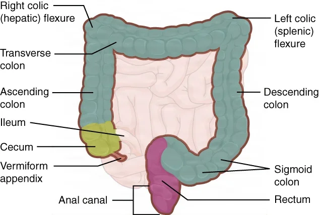
- Cecum: a blind pouch, about 6 cm long, that hangs below the ileocecal valve and receives what comes from the ileum.
- Appendix (vermiform appendix): a narrow, winding tube, about 7 to 10 cm long and closed at its tip, attached to the cecum below the ileocecal junction by its own small mesentery, the mesoappendix. It holds lymph tissue but has no clear digestive job.
- Ascending colon: rises up the right side to the underside of the liver, where it turns sharply left at the hepatic flexure, also called the right colic flexure.
- Transverse colon: crosses the abdomen below the stomach to just below the spleen, where it turns down at the splenic flexure, also called the left colic flexure.
- Descending colon: runs down the left side.
- Sigmoid colon: the S-shaped part in the pelvis, which curves toward the midline and leads into the rectum.
The ascending colon, the descending colon and the rectum lie in the retroperitoneum, fixed to the back wall. The transverse colon and the sigmoid colon hang on their own mesentery, the mesocolon, so they move more freely.
Landmarks you can see
Three features set the colon apart from the small intestine at surgery.
- Teniae coli: three long bands of smooth muscle, the outer muscle layer gathered into strips.
- Haustra: the pouches the teniae bunch the colon into, which give it its puckered look.
- Epiploic appendices, or appendages: small fat-filled tags of peritoneum along the teniae, with no known job.
Where the teniae spread out and disappear, at the level of the sacral promontory, the rectum begins.
What it does
The large intestine has no villi and makes no digestive enzymes. Its glands make mucus, which eases the passage of feces and protects the wall. Its jobs:
- Absorbs water and electrolytes: it takes most of the water left in the residue and turns liquid into semisolid feces. Each day about 500 mL of residue reaches the cecum, and only about 150 mL of it is passed as feces.
- Houses bacteria: trillions of bacteria of more than 700 kinds live in the colon, and most do no harm while they stay inside it. They ferment leftover carbohydrates, making gas, or flatus, up to 1500 mL a day. They make vitamin K and several B vitamins, such as biotin, which the colon absorbs. They also break down bilirubin from the bile into stercobilin, the pigment that colors stool brown.
- Finishes digestion: since the colon makes no enzymes, the last digestion of food residue is the bacteria's work.
- Compacts and stores: the residue stays 12 to 24 hours while water is drawn out, then moves on toward the rectum.
It moves in three ways: slow haustral contractions push the residue from one pouch to the next; peristalsis, slower here than higher up; and strong mass movements, usually three or four times a day during or just after a meal, which sweep the contents toward the rectum. Food entering the stomach sets them off, the gastrocolic reflex.
Feces are water, undigested residue such as plant fiber, bacteria, worn-out lining cells and salts.
Appendicitis starts when the appendix's narrow lumen is blocked. It swells, becomes infected, and can burst within a day, spilling germs into the abdomen. The pain usually begins around the navel and settles in the right lower quadrant.
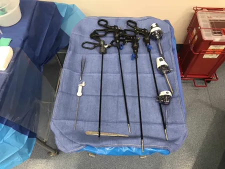
A right hemicolectomy removes the end of the ileum, the cecum, the ascending colon and part of the transverse colon, for disease of the cecum, ascending colon or hepatic flexure. Diverticulitis, cancer or other disease lower down can need a sigmoid colectomy.
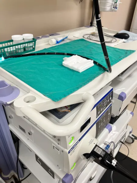
A colonoscopy looks at the whole large intestine from the rectum to the cecum. Most colon cancers grow from polyps, benign growths of the lining, and polyps found during a colonoscopy can be removed before they turn into cancer.
11The rectum and anal canal
The rectum
The rectum follows the sigmoid colon down in front of the sacrum and coccyx. It is not straight: three bends to the side make three folds inside, the rectal valves, which help keep gas apart from feces. Its lower end, the rectal ampulla, widens to hold feces until they are passed. It is about 10 to 12.5 cm (4 to 5 inches) long; some anatomy references give about 20 cm.
The anal canal
The anal canal, about 3.8 to 5 cm (1.5 to 2 inches) long, is the last part of the tube. It lies in the perineum, below the pelvic cavity, and opens to the outside at the anus. Its two sphincters stay closed except during defecation.
- The internal anal sphincter is smooth muscle and works without your control.
- The external anal sphincter is skeletal muscle under your voluntary control.
Inside the canal, the pectinate (dentate) line marks where the lining of the gut meets skin. Above it the lining feels little pain; below it, pain is sharp.
The canal also holds cushions of blood vessels. When their supporting tissue weakens, or when they swell, they become hemorrhoids: external ones are covered with skin, and internal ones lie under the lining.
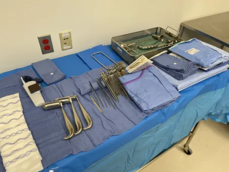
A low anterior resection removes the lower sigmoid colon and the upper rectum, often for cancer, and keeps the anal sphincter so the patient can still control the bowel.
Storing and passing stool
When a mass movement pushes feces into the rectum, the stretch of its wall sets off the defecation reflex: the sigmoid colon and rectum contract and the internal sphincter relaxes. The brain then chooses whether to open the external sphincter and pass the stool or to hold it for later. Bearing down, by closing the glottis and tightening the diaphragm and the abdominal muscles (the Valsalva maneuver), helps push.
Timing changes the stool.
- If defecation keeps being put off, more water is drawn out and the stool hardens. Constipation is passing stool less often than normal, with hard, dry stool that is difficult to pass.
- If residue moves through too fast, too little water is absorbed. Diarrhea is the passing of loose, unformed stool.
- Fecal incontinence is the loss of bowel control, so that stool, gas or mucus leaks out without the person meaning it to.
Normal habits vary widely, from two or three bowel movements a day to three or four a week.
12The liver
Where it sits and how it is built
The liver is the largest gland in the body, about 3 pounds in an adult. It fills most of the right upper quadrant and reaches into the left upper quadrant, just under the diaphragm, where the lower ribs shield it.
- A tough fibrous capsule, the Glisson capsule, wraps it, and the visceral peritoneum covers almost all of that.
- It has a large right lobe and a smaller left lobe, divided in front by the falciform ligament. Two small lobes, the quadrate and the caudate, sit on its underside; references differ on whether to count them with the right lobe or the left.
- Surgeons also divide it into eight segments, each with its own branch of the portal vein, hepatic artery and bile duct, so a segment can be removed on its own.
- The ligamentum teres, or round ligament, runs in the free lower edge of the falciform ligament from the navel to the liver. It is what remains of the umbilical vein, which brought blood from the placenta before birth.
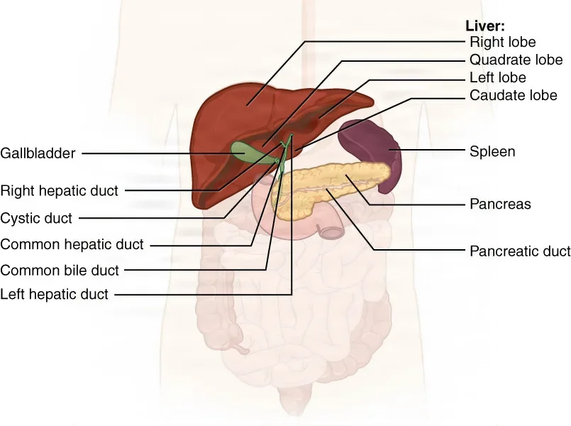
Blood reaches the liver two ways, at the porta hepatis on its underside. The hepatic artery brings blood rich in oxygen. The hepatic portal vein brings blood from the stomach, intestines, spleen and pancreas, full of absorbed nutrients, and also of any drugs and toxins that were swallowed. Blood leaves through the hepatic veins into the inferior vena cava. Because all the blood from the gut passes through the liver first, the liver is the most common place for cancers of the gut to spread. Chapter 5 traces these vessels.
Inside, the liver is built of tiny lobules. In each one, plates of liver cells, the hepatocytes, line wide blood channels called sinusoids, where they take up and process what the blood brings. Kupffer cells in the sinusoids are macrophages that eat bacteria, worn-out blood cells and debris. Bile made by the hepatocytes drains the opposite way, through tiny canals to the bile ducts.
What the liver does
The liver carries out hundreds of jobs. These are the ones you are expected to know.
| Job | What it means |
|---|---|
| Makes bile | About 600 mL to 1 liter a day; its bile salts break fat into tiny droplets so it can be digested and absorbed |
| Stores glucose as glycogen | Turns extra glucose into glycogen and releases glucose again when the blood sugar falls, a steady supply of fuel for the brain and body |
| Converts ammonia to urea | Breaking down amino acids makes ammonia, which is toxic; the liver turns it into urea, which the kidneys pass out in urine |
| Regulates the blood's amino acids | Makes the amino acids the body can build for itself and breaks down extra ones for energy |
| Makes plasma proteins | Albumin, which holds water inside the blood vessels, and most of the other plasma proteins, though not the antibodies |
| Regulates clotting | Makes fibrinogen, prothrombin and most of the other clotting factors; several need vitamin K, which is absorbed with fats and so depends on bile |
| Makes cholesterol | Builds cholesterol, the raw material for bile salts and steroid hormones, and the lipoproteins that carry fats in the blood |
| Filters drugs and toxins | Its enzymes break down drugs, alcohol, ammonia and other harmful substances into less harmful forms |
| Removes bacteria | Kupffer cells clear bacteria and old red blood cells from the portal blood |
| Makes immune factors | Makes complement proteins, part of the body's defense against germs |
| Stores vitamins and minerals | Holds iron, copper, vitamin B12 and vitamins A, D, E and K |
Hemoglobin itself is made by developing red blood cells in the red bone marrow. The liver's part is to store the iron those cells need and to clear away the bilirubin left over when old hemoglobin is broken down.
A diseased liver shows at the field. It may make too few clotting factors, so the patient bleeds more easily, and it may break down anesthetic drugs differently.
Bile, bilirubin and jaundice
Bile is a yellow-green to yellow-brown, slightly alkaline fluid: water, bile salts, bile pigments, cholesterol, lecithin and other fats, and electrolytes. The bile salts are what help digestion. Working like dish soap, they split large globules of fat into tiny droplets, a process called emulsification, which gives the fat-digesting enzymes far more surface to work on. They then carry the products of fat digestion to the lining to be absorbed. Most bile salts are absorbed again in the ileum and sent back to the liver to be used again.
Bilirubin is the main pigment in bile. When old red blood cells are broken down, in the spleen and also in the liver and the bone marrow, the heme from their hemoglobin becomes bilirubin. The liver puts the bilirubin into bile, and it leaves the body in the feces. It gives bile its color, and bacteria in the colon change it into the brown pigment of stool.
Jaundice is a yellow color of the skin, the whites of the eyes (the sclera) and the mucous membranes, caused by bilirubin building up in the blood. There are three kinds of cause: too much bilirubin, from red blood cells breaking down; a liver too sick to process it, as in hepatitis or cirrhosis; or a blocked bile duct, from a gallstone or a tumor, that stops bile from leaving. When bile cannot reach the intestine, the stool turns pale, because no pigment reaches it, and fat is poorly absorbed, because no bile salts reach it.
13The gallbladder and the biliary tree
The gallbladder
The gallbladder is a pear-shaped muscular sac, about 8 to 10 cm long, that sits in a shallow hollow on the underside of the right lobe of the liver. It holds about 40 to 70 mL of bile.
- It has three parts: the wide fundus, which juts out past the lower edge of the liver; the body; and the narrow neck, which bends into the cystic duct.
- The cystic duct, only 1 to 2 cm long, joins the gallbladder to the common hepatic duct. Bile travels both ways through it: in for storage and out when it is needed.
- The cystic artery, its blood supply, usually branches from the right hepatic artery, though it can come off other arteries nearby.
- Its wall has no submucosa. The lining draws water and salts out of the bile, concentrating it as much as tenfold, and the smooth muscle of the wall squeezes it out. The visceral peritoneum holds it against the liver.
The Calot triangle is bordered by the cystic duct, the common hepatic duct and the cystic artery. A closely related area, the hepatocystic triangle, has the underside of the liver as its upper border. These small spaces matter in every gallbladder operation, because the common bile duct runs right beside them; chapter 32 explains what the surgeon must see before clipping anything.
The biliary tree
Bile takes this path from the liver to the duodenum.
- Tiny canals between the liver cells collect it and drain into bile ducts.
- The bile ducts join into the right and left hepatic ducts.
- These two join to form the common hepatic duct.
- The cystic duct from the gallbladder joins the common hepatic duct, and from that point on it is the common bile duct.
- The common bile duct runs down behind the first portion of the duodenum and through the head of the pancreas. In the wall of the second portion of the duodenum it meets the main pancreatic duct, and they open together at the ampulla of Vater, also called the hepatopancreatic ampulla, on a small mound, the major duodenal papilla.
- The sphincter of Oddi, also called the hepatopancreatic sphincter, is a ring of smooth muscle at the ampulla. It opens to let bile and pancreatic juice pass into the duodenum and closes to hold them back.
Storing and releasing bile
Between meals the sphincter of Oddi stays closed, so bile from the liver backs up through the cystic duct into the gallbladder, which stores and concentrates it. When fatty chyme reaches the duodenum, the lining there releases cholecystokinin (CCK): the gallbladder contracts, the sphincter relaxes, and bile flows down the common bile duct into the duodenum.
The biliary tree carries waste as well as digestive juice. Bilirubin and other waste the liver puts into bile travel down the ducts and leave the body in the feces.
Gallstones (cholelithiasis) form when bile holds too much cholesterol or too few bile salts. A stone stuck in the cystic duct inflames the gallbladder (cholecystitis). A stone in the common bile duct blocks bile and causes jaundice, and one that moves down to the ampulla can cause pancreatitis.
14The pancreas
Where it lies and how it is built
The pancreas is a long, soft gland that lies across the back of the upper abdomen, behind the stomach, in the retroperitoneum.
- Head: the wide part, tucked into the C-shaped curve of the duodenum.
- Neck: the short part beside the pylorus that joins the head to the body.
- Body: lies across the spine, over the superior mesenteric artery and vein.
- Tail: tapers to the left and reaches the hilum of the spleen.
The main pancreatic duct, the duct of Wirsung, runs the length of the gland and joins the common bile duct at the ampulla of Vater. Many people also have a second, smaller duct, the accessory duct or duct of Santorini, which opens into the duodenum on its own, about an inch above the ampulla.
The pancreas is two glands in one.
- Its exocrine part, most of the gland, is made of grape-like clusters of cells (acini) that make pancreatic juice and send it down the ducts to the duodenum.
- Its endocrine part is the islets of Langerhans, small clusters of cells scattered through it that release hormones straight into the blood.
Pancreatic juice
The pancreas makes more than a liter of pancreatic juice a day. It is clear and mostly water, with salts, sodium bicarbonate and digestive enzymes.
- Sodium bicarbonate makes the juice slightly alkaline. It neutralizes the acid chyme arriving from the stomach, stops pepsin working, and gives the enzymes of the small intestine the pH they need.
- Enzymes break down all three kinds of food, as the table shows.
| Enzyme | Breaks down | Into |
|---|---|---|
| Pancreatic amylase | Starches and other carbohydrates | Smaller sugars |
| Pancreatic lipase | Fats (triglycerides), once bile has emulsified them | Fatty acids and monoglycerides |
| Trypsin, chymotrypsin, carboxypeptidase and elastase | Proteins | Peptides and amino acids |
| Nucleases | DNA and RNA | Nucleotides |
The enzymes that digest protein leave the pancreas in an inactive form, such as trypsinogen. In the duodenum an enzyme on the lining switches trypsinogen into trypsin, and trypsin then switches on the others. If these enzymes become active inside the pancreas, they digest the gland itself: that is pancreatitis. Heavy alcohol use and gallstones are its common causes.
Hormones from the duodenum control the flow: secretin brings out the bicarbonate, and CCK brings out the enzymes.
The islets
Each islet holds several kinds of cells.
- Alpha cells, about 20 percent of the islet, make glucagon, which raises a low blood sugar.
- Beta cells, about 75 percent, make insulin, which lowers a high blood sugar.
- Delta cells, about 4 percent, make somatostatin, which slows the secretions of the stomach and the pancreas and the absorption of nutrients.
Chapter 9 teaches these hormones and diabetes.
A pancreaticoduodenectomy, or Whipple procedure, is done to cure pancreatic cancer. It removes the head of the pancreas together with the duodenum that wraps around it, part of the jejunum, the lower stomach, the end of the common bile duct and the gallbladder. Chapter 32 teaches it.
15Nutrients and metabolism
The six kinds of nutrients
Nutrients are the substances in food and drink that the body needs to survive. There are six kinds. Carbohydrates, fats and proteins supply energy and, with water, are needed in large amounts; vitamins and minerals are needed in very small amounts.
| Nutrient | What the body uses it for | Absorbed as |
|---|---|---|
| Carbohydrates | The quickest fuel, about 4 calories a gram; extra glucose is stored as glycogen in the liver and muscles | Simple sugars: glucose, fructose and galactose |
| Fats (lipids) | The body's main store of energy, about 9 calories a gram; a few fats also build parts of cells | Fatty acids and monoglycerides |
| Proteins | Built from amino acids into enzymes, hormones and the structure of tissues, and used for energy too, about 4 calories a gram | Amino acids |
| Vitamins | Organic compounds the body's chemical reactions need, some working as helpers for enzymes (coenzymes); the fat-soluble A, D, E and K and the water-soluble B vitamins and C | Absorbed with dietary fats or with water |
| Minerals (mineral salts) | Inorganic substances, such as sodium, potassium, calcium, phosphorus, magnesium and chloride, that the body cannot make and must get from food | Absorbed as ions |
| Water | Up to 70 percent of an adult's body weight; it supplies no energy | Absorbed as it is, mostly in the small intestine |
Fiber is the part of plant foods the body has no enzymes to digest. It supplies no nutrients, but it helps move food through the tube and softens the stool.
Metabolism
Metabolism is the sum of all the chemical reactions in the body. It has two halves, running side by side all the time.
- Catabolism breaks large molecules into smaller ones and releases energy. About 40 percent of that energy is captured in ATP, the cell's energy currency, and the rest is given off as heat. Breaking down food for fuel is catabolism.
- Anabolism uses that energy to build large molecules from small ones, such as proteins from amino acids or glycogen from glucose, and to build new tissue and repair damaged tissue.
When the body takes in more energy than it uses, it stores the extra as fat; when it takes in less, it draws on its stores.
16Words for digestion and elimination
The Core Curriculum lists these terms, and the cert exam uses them. Most appear above; here they are together.
| Term | Meaning |
|---|---|
| Ingestion | Taking food into the mouth |
| Digestion | Breaking food down into molecules small enough to be absorbed |
| Absorption | Taking the products of digestion from the gut into the blood or lymph |
| Bolus | The soft ball of chewed food that is swallowed |
| Swallowing (deglutition) | Moving food from the mouth to the stomach, in three phases |
| Peristalsis | Waves of muscle tightening and relaxing that push the contents along the tube |
| Chyme | The soupy mix of food and digestive juices made in the stomach |
| Reflux | Flow backward, as of stomach contents up into the esophagus |
| Regurgitation | Flow back up into the mouth, as of food or fluid from the stomach; reverse peristalsis causes it |
| Flatus | Gas in the intestine, passed through the anus |
| Defecation | Passing feces out through the anus |
| Constipation | Stools that come less often than normal and are hard, dry and difficult to pass |
| Diarrhea | Loose, unformed stools |
| Incontinence | Loss of control; in fecal incontinence, stool, gas or mucus leaks out without the person meaning it to |
| Jaundice | Yellow skin, eyes and mucous membranes from too much bilirubin in the blood |
| Ulcer | A crater-like sore that breaks through a membrane; in the gut, usually from acid |
| Metabolism, anabolism, catabolism | All the body's chemical reactions; the ones that build up; the ones that break down |
17Where surgery meets the gut
The layers and the anastomosis
When the surgeon joins two pieces of gut, an anastomosis, the stitches or staples must hold the right layers.
- The submucosa is the strongest layer of the bowel wall, thanks to its collagen, and every anastomosis must include it.
- In a two-layer hand-sewn joint, the inner stitches catch the mucosa, the submucosa and the muscle, and the outer stitches join the serosa of the two ends.
- The esophagus has no serosa and gets its blood in segments, so a join there is less forgiving than one in the bowel.
Chapter 25 teaches the sutures and staplers, and chapter 32 the steps of each operation.
the submucosa holds the stitches, the serosa covers the outside, and the mesentery carries the blood.
Germs inside the lumen
The bowel's bacteria do no harm inside it, but in the wound they can cause infection. Once the bowel is opened, the risk of a surgical site infection remains even after a bowel prep and antibiotics, so the team uses isolation technique, also called bowel technique: instruments and items that touch the open bowel are kept apart from the rest of the field. Chapters 29 and 32 teach the steps, chapter 10 the germs, and chapter 11 the wound classes.
The mesentery comes with the specimen
Blood, nerves and lymph reach the intestine through its mesentery, so when a segment comes out, its mesentery and the vessels inside it are clamped and divided with it. The lymph from the bowel drains through nodes beside the mesentery, the same path cancer cells can follow, which is why a cancer operation also removes the lymph nodes in that part of the mesentery.
After surgery
Surgery slows the bowel down. After abdominal or pelvic surgery, peristalsis may be slowed for at least a day, and after bowel surgery for several days. Fluids and early walking help it return; the nurse listens for bowel sounds and checks the abdomen for swelling, and constipation is common.
Disease of the liver, the bile ducts or the pancreas reaches the field too. A patient with a blocked bile duct or liver disease may clot poorly, because the liver makes the clotting factors and needs bile to absorb vitamin K, and may handle anesthetic drugs differently.
Lo que te llevas de este capítulo
- Digestion breaks food into molecules small enough to absorb. Six processes do it: ingestion, propulsion (swallowing and peristalsis), mechanical digestion, chemical digestion, absorption and defecation.
- The parietal peritoneum lines the abdominal wall and the visceral peritoneum covers the organs, with the peritoneal cavity between. Most of the duodenum, the pancreas, the ascending and descending colon and the rectum are retroperitoneal. The omenta, the mesentery and the mesocolon hold the organs and carry their vessels.
- The wall of the tube is mucosa, submucosa, muscularis and serosa. The esophagus has an adventitia instead of a serosa, the stomach adds an oblique muscle layer, and the submucosa is the layer that holds an anastomosis.
- Saliva from the parotid (Stensen duct), submandibular (Wharton duct) and sublingual (ducts of Rivinus) glands starts starch digestion and forms the bolus. Swallowing has a voluntary phase and two automatic phases, and the lower esophageal sphincter keeps acid out of the esophagus.
- The stomach (cardia, fundus, body and pylorus, with its two curvatures) stores food, churns it into chyme, starts protein digestion with acid and pepsin, absorbs little, and makes intrinsic factor for vitamin B12.
- The small intestine (duodenum, jejunum from the ligament of Treitz, ileum to the ileocecal valve) finishes digestion and absorbs nearly everything through its folds, villi and microvilli. The large intestine (cecum and appendix, then ascending, transverse, descending and sigmoid colon, with its hepatic and splenic flexures, teniae coli, haustra and epiploic appendices) absorbs water, holds bacteria that make vitamin K, and forms and stores feces.
- The liver makes bile, clotting factors and plasma proteins, stores glycogen, turns ammonia into urea and filters drugs and toxins. Bile runs from the hepatic ducts to the common hepatic duct, joins the cystic duct to become the common bile duct, and enters the duodenum at the ampulla of Vater through the sphincter of Oddi. The Calot triangle is the cystic duct, the common hepatic duct and the cystic artery.
- The pancreas sends bicarbonate and enzymes (amylase for starch, lipase for fat, trypsin and chymotrypsin for protein) to the duodenum, and its islets make glucagon (alpha cells), insulin (beta cells) and somatostatin (delta cells).
Ponte a prueba5 preguntas rápidas
Gratis para todos. Elige una respuesta para cada una y luego revísalas todas juntas.
En el quirófanoDónde vas a usar esto
Todo el banco de preguntas del capítulo en series de 10, con la respuesta explicada después de cada una.
Parte del programa para educadoresLas palabras del capítulo en tarjetas de estudio: las que fallas vuelven antes.
Parte del programa para educadoresLo asigna tu docente. Cada grupo saca sus propias 25 preguntas del banco del capítulo.
Parte del programa para educadores
Sugerir una corrección
¿Algo está mal, confuso o desactualizado en este capítulo? Avísanos y lo corregimos para todos. Tu correo solo se usa para responderte.