Kidneys, bladder and reproduction
The kidneys that clean the blood, the tubes and bladder that carry and hold urine, and the female and male reproductive organs: the anatomy under every urology, gynecology and obstetric case.
1Why it matters
The urinary system makes urine and carries it out of the body. The reproductive system makes the sex cells and sex hormones and, in females, carries and feeds a pregnancy. They share a crowded space in the pelvis, and in males one tube, the urethra, carries both urine and semen, so surgical texts often teach the male reproductive organs with the urinary system.
You meet these organs in urology, gynecology and obstetrics cases, and in some general surgery cases, such as a groin hernia repair.
- The kidneys guard the whole body. They clean the blood, set its water, salt and acid levels, help control blood pressure and clear many drugs. When they fail, drugs can build up, so doses change.
- Urine output is a window on the patient. The team measures urine during surgery to judge fluid balance; a normal adult makes about 0.5 to 1 mL of urine per kilogram of body weight per hour. Chapter 31 teaches urinary catheters and measuring urine.
- Pelvic organs lie close together. The bladder sits just in front of the uterus, and each ureter passes under a uterine artery beside the cervix, so in pelvic surgery the surgeon finds them before anything is clamped.
- Scopes go in through the urethra. Through it the urologist reaches the bladder, the prostate, the ureters and the inside of the kidney.
This chapter teaches the structure and work of both systems and where surgery meets them. Other chapters hold the rest: the renal arteries and veins as part of the circulation in chapter 5; the adrenal glands, and the hormones of the pituitary, ovaries and testes as endocrine glands, in chapter 9; urinary tract infections in chapter 10; how tumors and disease develop in chapter 11; diuretics in chapter 12; urinary catheters, measuring urine and the lithotomy position in chapter 31; hernia repair and breast surgery in chapter 32; the obstetric and gynecologic operations in chapter 33; the genitourinary operations, with diseases such as kidney stones and an enlarged prostate, in chapter 34; and breast reconstruction in chapter 38.
2The urinary system at a glance
The urinary system has four kinds of organs.
- Two kidneys filter the blood and make urine.
- Two ureters carry urine from the kidneys to the bladder.
- The bladder stores urine until it is a good time to empty it.
- The urethra carries urine from the bladder out of the body.
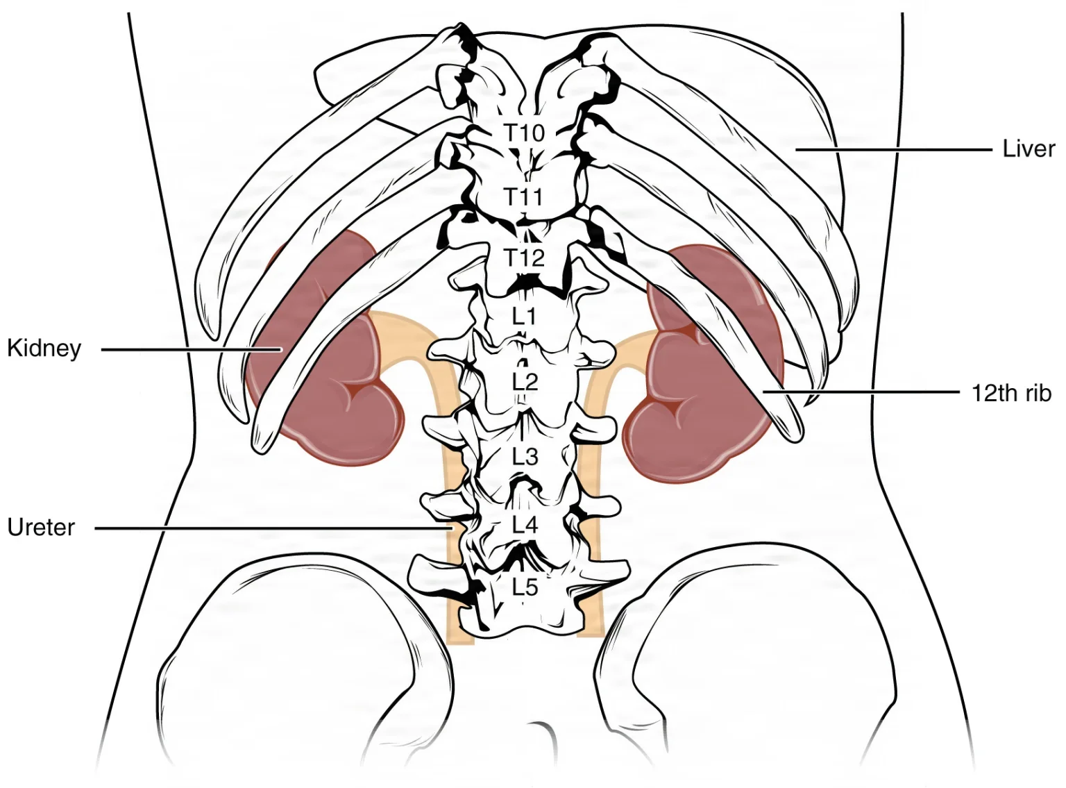
What the kidneys do
Urine is the by-product. The kidney's real work is keeping the makeup of the blood steady, and it does several jobs at once.
- Filters out wastes: it removes the waste products of the body's chemistry, such as urea, and excess water, and sends them out in the urine.
- Balances fluid and electrolytes: it adjusts how much water and how much sodium, potassium and other salts it keeps, which holds the blood's volume steady. This is homeostasis of fluid volume.
- Helps control blood pressure: it releases the enzyme renin, which starts a chain that raises blood pressure (see below).
- Activates vitamin D and helps control calcium: it makes calcitriol, the active form of vitamin D, which lets the gut absorb calcium.
- Drives red blood cell production: it makes most of the body's erythropoietin (EPO), the hormone that tells the bone marrow to make red cells.
- Keeps the blood's pH in range: it can get rid of extra acid or extra base.
- Clears drugs: many water-soluble drugs leave the body in the urine.
When the kidneys fail, every one of these jobs suffers. The patient may have weakness, shortness of breath, widespread swelling (edema), anemia, too much acid and potassium in the blood, and heart rhythm problems.
3The kidneys
Where they sit
The kidneys are two bean-shaped organs, each about the size of a fist: about 11 to 14 cm long, 6 cm wide and 4 cm thick.
- They lie against the back wall of the abdomen, one on each side of the spine, in the retroperitoneal space, behind the peritoneum.
- They reach from about the 12th thoracic to the 3rd lumbar vertebra (T12 to L3). The right kidney sits a little lower than the left, because the liver lies above it.
- The 11th and 12th ribs shield their upper parts.
- An adrenal gland sits on top of each one; chapter 9 teaches these glands.
The kidneys get a large share of the blood: about a fifth to a quarter of everything the heart pumps at rest.
Three layers wrap and hold each kidney.
- The renal capsule, a tough fibrous layer, covers the kidney itself and helps hold its shape.
- A cushion of fat, the renal fat pad, surrounds the capsule and absorbs shocks.
- The renal fascia encloses the fat and the kidney and anchors them to the back wall. It is also called Gerota fascia.
Because the kidneys lie behind the peritoneum, the surgeon can reach them from the flank or through the abdomen. Today a simple nephrectomy, the removal of one kidney, is done through the abdomen with the patient in a modified lateral position, most often by laparoscopy or with a robot. Chapter 34 teaches the operations.
an open flank incision near the 12th rib, with the patient lying on the side, operative side up. Several references still walk through this open operation step by step for a simple nephrectomy.
Inside the kidney
Cut a kidney from top to bottom and it shows two zones and a set of collecting funnels.
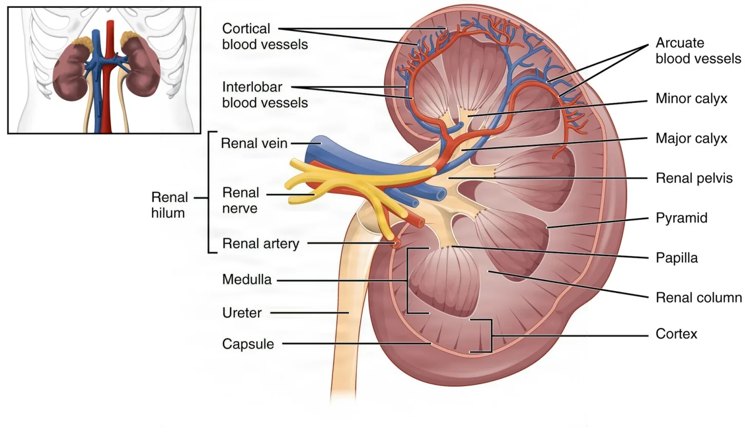
- Renal cortex: the outer zone, lighter in color. It holds the filtering parts of every nephron.
- Renal medulla: the inner zone. It holds the renal pyramids, with their wide bases against the cortex and their tips, the renal papillae, pointing toward the center. The papillae are bundles of collecting ducts.
- Renal columns: bands of tissue that reach down from the cortex between the pyramids and carry blood vessels.
- Minor calyces (singular calyx): small cups that catch urine dripping from the papillae.
- Major calyces: larger cups formed where minor calyces join.
- Renal pelvis: a funnel formed where the major calyces meet. Smooth muscle in its wall pushes urine into the ureter.
- Renal hilum: the notch on the inner (medial) side where the renal artery, renal vein, nerves, lymph vessels and the renal pelvis enter and leave. The renal pelvis lies behind the blood vessels there.
urine flows from the papillae into the minor calyces, then the major calyces, then the renal pelvis and out through the ureter.
Stones often lodge in the renal pelvis. In a percutaneous nephrolithotomy (PCNL), the surgeon passes a nephroscope through the skin into the kidney and breaks up and removes them.
The renal artery and vein
Each kidney has one main artery and one main vein. Chapter 5 traces them with the rest of the circulation.
- The renal artery branches straight off the abdominal aorta.
- The renal vein carries the cleaned blood straight into the inferior vena cava.
Inside the kidney, the artery divides again and again, into segmental, interlobar, arcuate and cortical radiate arteries, and finally into tiny afferent arterioles, one to each nephron.
The renal artery, the renal vein, their nerves and lymph vessels together are called the pedicle of the kidney, and the surgeon controls the pedicle when a kidney is removed.
The left renal vein is longer than the right, so a living donor usually gives the left kidney. In a kidney transplant, the new kidney goes into the recipient's iliac fossa, low in the pelvis on one side, and its renal artery is joined to the recipient's iliac artery.
The nephron
The nephron is the kidney's working unit, the smallest part that can make urine. Each kidney has about a million of them.
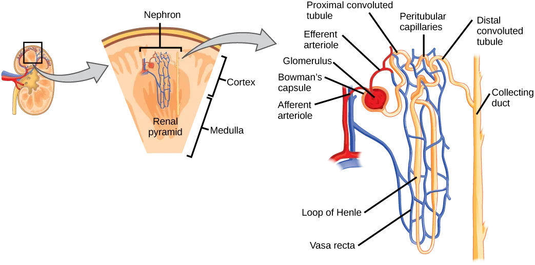
Each nephron has a filter at its start and a long tube after it.
- Glomerulus: a tuft of capillaries under high pressure, fed by an afferent arteriole and drained by an efferent arteriole.
- Bowman's capsule (glomerular capsule): the cup at the start of the tubule that wraps the glomerulus and catches the fluid filtered out of it. The glomerulus and Bowman's capsule together are the renal corpuscle.
- Renal tubule: the tube that carries the filtered fluid away and refines it. It has three parts.
- The proximal convoluted tubule (PCT), a twisting stretch whose lining cells have a brush border of microvilli. It does more reabsorbing than any other part.
- The loop of Henle, a hairpin that dips down toward or into the medulla (descending limb) and comes back up (ascending limb). About 15 percent of nephrons have long loops that reach deep into the medulla.
- The distal convoluted tubule (DCT), a shorter twisting stretch back in the cortex.
- Collecting duct: each one gathers fluid from several nephrons and carries it down through a pyramid to a papilla. Strictly, it is not part of the nephron.
After the glomerulus, the efferent arteriole forms a second set of capillaries, the peritubular capillaries and the vasa recta, that wrap the tubule and take back what it reabsorbs.
The juxtaglomerular apparatus (JGA) sits where the distal convoluted tubule touches the arterioles of its own glomerulus.
- The macula densa, a patch of cells in the wall of the distal tubule, senses the fluid flowing past.
- The juxtaglomerular cells, modified muscle cells in the wall of the afferent arteriole, adjust the blood flow into the glomerulus and release renin.
How urine is made
The nephron makes urine in three steps.
- Filtration: blood pressure in the glomerulus pushes part of the plasma through the capillary wall into Bowman's capsule. The filter works mostly by size: water, salts, glucose, amino acids, vitamins and wastes pass, but blood cells, platelets and large proteins such as albumin stay in the blood.
- Reabsorption: as the fluid moves along the tubule, the tubule takes back what the body needs and returns it to the blood. About two thirds of the water, sodium and potassium, and nearly all of the glucose, amino acids and vitamins, are taken back in the proximal tubule. The loop of Henle, the distal tubule and the collecting ducts take back more.
- Secretion: the tubule also moves some substances from the blood into the fluid, such as hydrogen ions (acid), potassium, creatinine and some drugs, such as penicillin.
The numbers show how much reabsorption matters. The kidneys filter about 125 mL of fluid a minute in a male and 105 mL in a female, roughly 150 to 180 liters a day, and they send about 99 percent of it back into the blood. What is left, about 1 to 2 liters a day, is urine. The rate of filtration is the glomerular filtration rate (GFR).
Filtration depends on blood pressure. Muscle in the arteriole walls keeps it fairly steady through ordinary swings, but if the mean arterial pressure falls below about 60 mm Hg, as in shock, the kidneys can no longer filter properly.
A blood test shows how well the kidneys filter. Creatinine, a waste made by muscle, is filtered and hardly reabsorbed, so a rising serum creatinine means the kidneys are clearing less; the GFR is estimated from it.
filtration pushes fluid out of the glomerulus, reabsorption takes the useful parts back, and secretion adds wastes the filter missed.
The kidneys and blood pressure
When blood pressure falls, the kidneys help raise it again through the renin-angiotensin-aldosterone system (RAAS).
- The juxtaglomerular cells sense the drop and release the enzyme renin into the blood.
- Renin turns angiotensinogen, a protein made by the liver, into angiotensin I.
- An enzyme from the lungs, angiotensin-converting enzyme (ACE), turns angiotensin I into active angiotensin II.
- Angiotensin II narrows blood vessels all over the body, which raises the pressure within moments.
- Angiotensin II also makes the adrenal cortex release aldosterone, which makes the kidneys keep sodium. Water follows the sodium, so blood volume and pressure stay up for longer.
The kidney does not release angiotensin itself: its renin starts the chain that makes it.
A second hormone, antidiuretic hormone (ADH, also called vasopressin), comes from the posterior pituitary. When blood volume falls by 5 to 10 percent, ADH opens water channels in the collecting ducts so the kidneys keep more water, and it also narrows blood vessels.
Chapter 9 teaches these hormones, chapter 11 shock, and chapter 12 the drugs that act on this system, such as diuretics and ACE inhibitors.
Vitamin D, calcium and red cells
- Vitamin D: the liver changes vitamin D into an inactive form, and the kidney adds the final step that makes calcitriol, the active form. Parathyroid hormone switches this step on when blood calcium is low. Calcitriol makes the gut absorb more calcium.
- Calcium: the kidney also takes back more calcium from the urine when parathyroid hormone calls for it.
- Red cells: the kidneys make about 85 percent of the body's EPO, more when the body needs oxygen, such as after heavy bleeding. Patients with kidney failure lose this and become anemic.
4The ureters
The ureter is a muscular tube that carries urine from the renal pelvis to the bladder. There are two, one from each kidney.
Its course
Each ureter follows a gentle S-shaped path.
- It starts where the renal pelvis narrows, at the ureteropelvic junction (UPJ).
- It runs down behind the peritoneum, lying on the psoas muscle.
- It crosses over the iliac vessels at the brim of the pelvis.
- In the pelvis it sweeps out along the side wall, then turns in toward the bladder.
- It pierces the back of the bladder wall at a slant and opens into the bladder at the ureterovesical junction (UVJ).
The slanted path through the bladder wall acts as a one-way valve: as the bladder fills and squeezes, it presses the tunnel shut, so urine cannot flow back up toward the kidney. Backward flow is called reflux; a child whose ureter lacks this slant is prone to reflux and serious urinary tract infections.
Size and wall
- In an adult, each ureter is about 25 to 30 cm long and about 4 to 5 mm across.
- Its lining is transitional epithelium, which can stretch, with scattered goblet cells that make protective mucus.
- Its middle layer is smooth muscle. Waves of contraction, peristalsis, push urine down the ureter without help from gravity.
- A loose outer coat of collagen and fat anchors it to the back wall.
The ureter is narrowest in three places, which is where kidney stones tend to stick and cause pain and blockage: the UPJ, the spot where it crosses the iliac vessels, and the UVJ.
In a ureteroscopy, the urologist passes a thin scope up through the urethra and bladder into the ureter and renal pelvis to look, take a biopsy, or break up and remove stones.
5The bladder
Where it sits
The bladder (urinary bladder) is a hollow muscular sac that stores urine.
- It lies in the pelvis just behind the pubic symphysis.
- In a female, the uterus and the vagina lie behind it. In a male, the rectum lies behind it and the prostate below it.
- Like the kidneys and ureters, it lies outside the peritoneal cavity: peritoneum covers only its top, the dome. As the bladder fills, the dome rises into the abdomen.
Because the bladder lies below the peritoneum, the surgeon can reach it through a suprapubic incision, just above the pubic bone, without entering the peritoneal cavity.
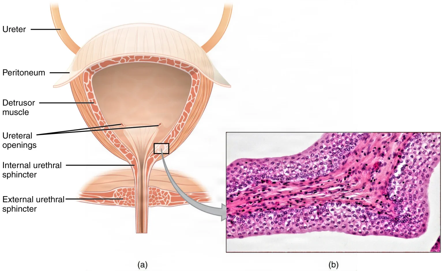
Its parts
- Dome: the top, which rises as the bladder fills.
- Trigone: a smooth triangle on the floor of the bladder. Its three corners are the two ureteral openings, about 2.5 cm apart, and the opening of the urethra. The surgeon looks for these openings at cystoscopy.
- Bladder neck: the funnel at the bottom where the urethra begins. Muscle fibers of the bladder wall gather here to form the internal sphincter.
The wall
The wall has layers suited to stretching.
- Mucosa: the mucous membrane lining, made of transitional epithelium. When the bladder is empty it lies in folds (rugae), and its surface cells are cube-shaped; when the bladder stretches, the folds flatten and the surface cells flatten too.
- Submucosa: connective tissue under the lining.
- Detrusor muscle: crisscrossing bands of smooth muscle that squeeze the bladder to empty it.
- Outer covering: peritoneum (serosa) over the dome, and a connective tissue adventitia elsewhere.
The Core Curriculum's list names the layers as the mucous membrane, the muscle and the wall.
Storing and emptying urine
An adult bladder holds from nearly nothing up to about 500 to 600 mL; references give its full capacity as anywhere from 350 to 700 mL.
Two rings of muscle keep it closed.
- The internal urethral sphincter, at the bladder neck, is smooth muscle under automatic (autonomic) control.
- The external urethral sphincter, lower down where the urethra passes through the pelvic floor, is skeletal muscle that you control. Its nerve is the pudendal nerve, from the sacral spinal cord (S2 to S4).
Emptying the bladder is called micturition, urination or voiding.
- While the bladder fills, sympathetic nerves keep the detrusor relaxed and the external sphincter stays tight.
- Stretch receptors in the wall send signals to the sacral spinal cord. At about 150 mL, you feel the first urge, which is easy to ignore.
- To void, parasympathetic nerves make the detrusor contract and the internal sphincter relax, and you relax the external sphincter.
- As the bladder keeps filling, the urge grows; at about 300 to 400 mL, control may be lost.
The ability to hold urine is continence. Losing control of urine is incontinence; being unable to empty the bladder is urinary retention. In late pregnancy the growing uterus presses on the bladder, so it holds less and must be emptied more often. With age, the bladder loses muscle tone and may not empty completely.
Stress urinary incontinence is a leak of urine when pressure in the abdomen rises, such as with a cough or a sneeze. A sling placed under the urethra supports it at those moments and is now the usual operation for stress incontinence in females.
the Marshall-Marchetti-Krantz (MMK) procedure, which one reference still teaches: through a low transverse incision, stitches placed beside the urethra are fixed to the back of the pubic bone to suspend the bladder neck and urethra. The sling has largely replaced it and the similar Burch procedure.
6The urethra
The urethra carries urine from the bladder out of the body. It is the one urinary organ that differs clearly between females and males.
In both, it starts at the bladder neck, at the lower corner of the trigone, and passes through the internal and then the external sphincter. Its lining starts as transitional epithelium, like the bladder's, and ends as stratified squamous epithelium near the opening. Mucous glands along it protect it from the acid or alkali in urine.
The female urethra
- It is short, about 4 cm long (references give 3 to 5 cm), and runs just behind and below the pubic symphysis.
- It lies against the front wall of the vagina.
- Its opening, the external urinary meatus, lies in the vestibule below the clitoris and above the vaginal opening, between the labia minora.
- The small Skene glands (paraurethral glands) open beside the meatus.
- It carries only urine.
Because it is short and close to the vagina and anus, germs reach the bladder more easily, which is the main reason urinary tract infections are more common in females. Chapter 10 teaches these infections.
The male urethra
The male urethra is about 20 cm long (references give 20 to 25 cm). It runs from the bladder neck through the prostate, through the floor of the pelvis and the length of the penis to the tip. It carries urine, and during ejaculation it carries semen. It has four parts:
- Preprostatic urethra: a very short part within the wall of the bladder neck.
- Prostatic urethra: about 3 cm long and the widest part, running through the middle of the prostate. On its floor is a small mound, the verumontanum, where the two ejaculatory ducts open.
- Membranous urethra: the shortest part, about 2.5 cm, passing through the muscles of the pelvic floor, where the external sphincter surrounds it.
- Spongy (penile) urethra: running inside the corpus spongiosum of the penis to the external urethral meatus at the tip of the glans.
Because the prostate wraps around the urethra, an enlarged prostate can squeeze it and make urination difficult. Chapter 31 explains why this matters when a catheter is passed.
In a cystoscopy, the urologist passes a scope through the urethra to look at the urethra, the bladder lining and the ureteral openings; in a male, the verumontanum, the bladder neck and the lobes of the prostate are checked too.
7Urine
What normal urine is made of
A healthy adult makes about 1 to 2 liters of urine a day. It is about 95 percent water and 5 percent dissolved substances.
| Part | What it is |
|---|---|
| Water | Most of the volume; how much depends on how much the person drinks and loses in other ways |
| Mineral salts | Electrolytes the kidney does not need to keep, such as sodium, potassium and chloride |
| Nitrogen wastes | From the breakdown of proteins: mostly urea, which the liver makes from toxic ammonia, with a little ammonium and uric acid, and creatinine from muscle |
| Pigment | Urochrome, a yellow pigment that comes from the breakdown of the heme in old red blood cells |
Fresh normal urine has little or no smell, and its color runs from pale yellow to deep amber. Dark, concentrated urine, sometimes with a slight smell of ammonia, often means the person is short of water. The smell of ammonia in a stale sample comes from bacteria breaking down urea after it leaves the body.
| Measure | Normal range |
|---|---|
| Volume | About 750 to 2000 mL a day |
| pH | 4.5 to 8.0, depending a lot on diet |
| Specific gravity | About 1.010 to 1.025; some references give a wider range, about 1.003 to 1.032 |
Specific gravity compares the weight of urine with the weight of the same amount of pure water, which is 1.000. Urine is always heavier than water because of what is dissolved in it, so the number shows how concentrated it is: high when the patient is dehydrated, low when the urine is dilute.
The amount matters too. Oliguria is too little urine, about 300 to 500 mL a day or less; the body needs to make at least about 500 mL a day to get rid of its wastes. Anuria is almost no urine, and polyuria is too much, more than about 2.5 to 3 liters a day.
What should not be in urine
| Finding | What it may point to |
|---|---|
| Albumin and other plasma proteins (proteinuria) | Only a trace of protein normally passes the glomerulus, so more than that usually means a damaged glomerulus |
| Glucose (glycosuria) | Blood sugar so high that the tubules cannot take it all back, usually from untreated or poorly controlled diabetes |
| Ketones, such as acetone | The body is burning fat for fuel instead of glucose, as in uncontrolled diabetes or a diet very short of carbohydrate |
| Blood (hematuria) | Infection, stones, cancer or injury of the urinary tract or kidney; a stone or tumor can turn urine pink or red |
| White blood cells (pus cells) and bacteria | Infection; leukocyte esterase, made by white cells, and nitrite, made by many bacteria, are signs of it on the dipstick |
| Casts | Tiny cylinders of cells and protein, molded in the shape of the tubule where they formed, seen in many kidney conditions |
| Crystals | Can appear after some drugs, such as sulfa drugs, especially when the patient is short of fluid; minerals settling out of the urine are also what form kidney stones |
Blood in a sample taken during a menstrual period is contamination, not a sign of disease.
The urinalysis
A urinalysis checks the urine for both normal and abnormal substances, and it is a routine test before many operations.
- Looking at it: the color, clarity and odor.
- Dipstick: a strip coated with chemicals that change color to show albumin (protein), bilirubin, glucose, ketones, leukocytes, blood, nitrite and urobilinogen; the pH and specific gravity are read too.
- Microscope: the sample is spun in a centrifuge, and the sediment at the bottom is checked for cells, germs, crystals and casts.
- Culture: when infection is suspected, a culture finds the exact germ, and a sensitivity test shows which antibiotics will work. Chapter 10 teaches culture and sensitivity.
The sample may be caught as the patient voids or taken through a catheter, and some tests use all the urine made over 24 hours.
8The female reproductive system
The female reproductive organs make egg cells and hormones, receive sperm, carry and nourish a pregnancy, and deliver the baby. Most of them lie inside the pelvis.
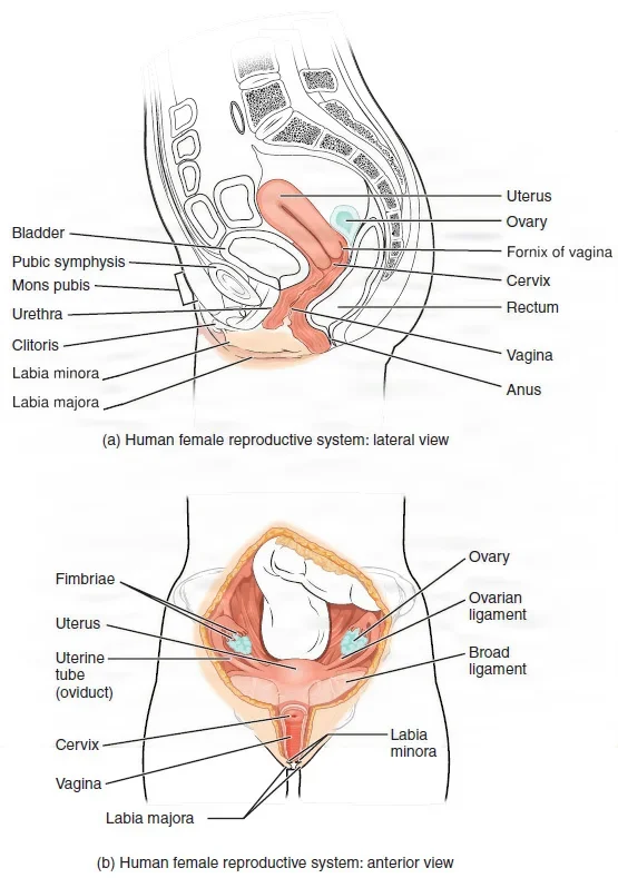
The external genitalia
The external organs together are called the vulva.
- Mons pubis: a pad of fat over the pubic bone, covered with hair after puberty.
- Labia majora: two outer folds of skin over fat, covered with hair, that run back from the mons and protect the structures between them.
- Labia minora: two thinner, hairless folds inside the labia majora that protect the urethral and vaginal openings. In front, they join to form a hood over the clitoris, the prepuce.
- Clitoris: a small, very sensitive organ of erectile tissue at the front where the labia minora meet. It develops from the same tissue as the penis but does not contain the urethra.
- Vestibule: the area enclosed by the labia minora. It holds the urinary meatus in front and the vaginal opening, the introitus, behind it.
- Hymen: a thin fold of tissue that may partly surround the vaginal opening.
- Perineum: the area between the vaginal opening and the anus. Under its skin lie strong muscles of the pelvic floor. In childbirth it may be cut to keep it from tearing as the baby's head comes out, an episiotomy.
Two pairs of small glands open into the vestibule.
- The Bartholin glands (greater vestibular glands) lie on each side of the vaginal opening, at about the 5 and 7 o'clock positions. They make mucus that keeps the area moist, and they can become infected and need to be opened and drained. They match the bulbourethral glands in the male.
- The Skene glands (paraurethral glands) lie beside the urethra and open next to the urinary meatus.
The vagina
The vagina is a muscular canal, about 10 cm long, that runs from the vestibule to the cervix.
- Its front wall, about 6 to 8 cm long, lies against the bladder and urethra; its back wall, about 7 to 10 cm, lies against the rectum.
- The cervix pokes into its top. The recesses around the cervix are the fornices: front, back and one on each side. The back (posterior) fornix touches the peritoneum of the posterior cul-de-sac, the pouch of Douglas (see the uterus, below).
- Its wall has a lining of stratified squamous epithelium with folds (rugae) that let it stretch, a middle layer of smooth muscle, and an outer fibrous layer.
- Helpful bacteria, mostly Lactobacillus, keep it acidic, below pH 4.5, which holds back harmful germs.
- It receives the penis and semen during intercourse, lets menstrual blood out, and is the last part of the birth canal.
The ovaries
The ovaries are the female gonads, each about the size and shape of an almond, about 2 to 4 cm long, one on each side of the uterus.
- What holds them: each ovary hangs from the back of the broad ligament by a fold of peritoneum, the mesovarium. The ovarian ligament ties it to the uterus, and the suspensory ligament, also called the infundibulopelvic ligament, carries its blood vessels from the side wall of the pelvis.
- How they are built: a thin layer of epithelium, not peritoneum, covers each ovary. Its outer cortex holds the follicles, each an egg cell with its supporting cells; its inner medulla holds the blood vessels, lymph vessels and nerves.
- Oocyte formation: the egg cells, called oocytes, form before birth. A newborn has about 1 to 2 million, about 400,000 remain at puberty, and almost none are left by the end of menopause. Usually one is released each cycle.
- Hormones: the growing follicles make estrogen, which builds up the lining of the uterus and brings on the body changes of puberty. After an egg is released, the empty follicle becomes the corpus luteum and makes progesterone, which readies the lining for a pregnancy and keeps it going. The ovaries also make a little testosterone.
The uterine tubes, the ovaries and their ligaments together are called the adnexa.
The uterine (fallopian) tubes
The uterine tubes, also called the fallopian tubes or oviducts, carry the egg from the ovary to the uterus. Each fallopian tube is about 10 to 13 cm long.
From the uterus outward, each tube has four parts.
- Interstitial (intramural) part: about 1 cm long, inside the wall of the uterus.
- Isthmus: the narrow part next to the uterus.
- Ampulla: the wide, winding middle, more than half the tube's length, where the egg is usually fertilized.
- Infundibulum: the trumpet-shaped outer end, which opens into the peritoneal cavity and is fringed with finger-like fimbriae.
The tube does not touch the ovary. At ovulation, the fimbriae sweep over the ovary and draw the egg in, and then beating cilia and waves of muscle in the tube move it toward the uterus. A fold of the broad ligament, the mesosalpinx, holds each tube.
Because the tube opens into the peritoneal cavity, germs that climb up through the vagina and uterus can reach the pelvis; an infection there is pelvic inflammatory disease, which can scar the tubes. A pregnancy that implants outside the uterus is an ectopic pregnancy, and most of them grow in a tube.
The uterus
The uterus is a hollow, thick-walled, pear-shaped organ that holds and nourishes a pregnancy. When not pregnant, it is about 7 cm long and 5 cm wide. It sits in the pelvis between the bladder in front and the rectum behind, and it usually tips forward.
It has three regions.
- Fundus: the rounded top, above the openings of the uterine tubes.
- Body (corpus): the main part. A slight narrowing, the isthmus, separates it from the cervix.
- Cervix: the narrow neck, about 2 to 3 cm long, that pokes into the vagina. Its canal runs from the internal os, inside, to the external os, which opens into the vagina; the os opens wide in labor.
Its wall has three layers.
- Perimetrium: the outer serous layer, the visceral peritoneum that covers the uterus.
- Myometrium: the thick middle layer of smooth muscle, most of the wall. Its fibers run in every direction, so it can contract strongly in labor and push out menstrual blood.
- Endometrium: the inner lining. Its deep basal layer stays put; its thicker functional layer grows each month under estrogen and progesterone and is shed in menstruation.
As the peritoneum drapes over the uterus, it dips into two pouches.
- In front, it folds forward from the uterus onto the bladder. This fold is the bladder flap, and the shallow pouch beneath it is the anterior cul-de-sac.
- Behind, it dips down between the uterus and the rectum to form the posterior cul-de-sac, the pouch of Douglas.
The uterine ligaments
Several ligaments hold the uterus in place, along with the muscles of the pelvic floor.
| Ligament | Where it runs | What it does |
|---|---|---|
| Broad | A double fold of peritoneum from each side of the uterus to the side wall of the pelvis; the uterine tube runs in its upper free edge | Hangs the uterus from the pelvic walls |
| Round | From the uterus near the tube, forward between the layers of the broad ligament and through the inguinal canal to the labia majora | Helps hold the uterus tipped forward |
| Cardinal | At the base of each broad ligament, beside the cervix; also called the paracervical or Mackenrodt ligament | Gives the uterus strong support and carries the uterine artery and vein |
| Uterosacral | From the back of the cervix, around each side of the rectum, to the sacrum | Holds the uterus from behind |
The cardinal ligaments give the uterus its main support; one reference also calls the broad ligament a primary support.
The uterine arteries, which run in the cardinal ligaments, and the ovarian arteries, which run in the suspensory ligaments, supply the uterus, tubes and ovaries.
An abdominal hysterectomy removes the uterus, and a total hysterectomy takes the cervix with it. A salpingo-oophorectomy removes a uterine tube and its ovary; bilateral means both sides. Chapter 33 teaches these operations.
9The breast
The breasts are accessory organs of the female reproductive system. Their job is to make milk for a baby, which is called lactation.
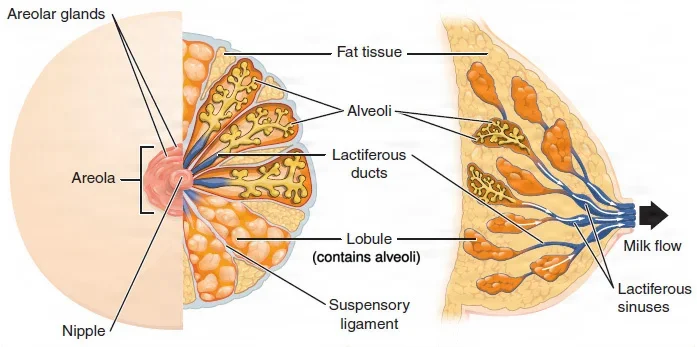
- Where it lies: each breast lies on the pectoralis major muscle, from about the 2nd to the 6th rib, with a tail of tissue, the tail of Spence, reaching toward the armpit.
- Mammary glands: the milk-making glands are modified sweat glands, arranged in about 15 to 20 lobes. Each lobe holds lobules of tiny sacs, the alveoli, that make milk.
- Mammary ducts: each lobe drains through a lactiferous duct, which widens into a lactiferous sinus just before it opens on the nipple.
- Nipple and areola: the nipple is the raised tip where the ducts open. The areola is the pigmented ring around it, with small areolar glands that make a lubricating fluid to protect the nipple during nursing.
- Fat and ligaments: fat around the lobes sets the breast's size, and bands of connective tissue, the suspensory (Cooper) ligaments, attach the breast to the skin.
Most of the breast's lymph drains to the lymph nodes in the armpit, which is why surgeons check them for the spread of breast cancer. A lumpectomy removes the mass with margins free of cancer cells and keeps the rest of the breast, a form of breast-conserving surgery. Chapter 5 teaches the sentinel node, chapter 32 breast surgery, and chapter 38 breast reconstruction.
10The menstrual cycle
From the first period (menarche) until periods stop (menopause), the ovaries and uterus go through a repeating cycle, usually about 28 days long, though 21 to 32 days is common. Day 1 is the first day of bleeding. Hormones from the hypothalamus and pituitary drive the cycle; chapter 9 teaches them.
Two cycles run side by side: one in the ovary, one in the lining of the uterus.
In the ovary
- Follicular phase (about days 1 to 14): pituitary hormones make several follicles grow and release estrogen. Usually only one, the dominant follicle, survives.
- Ovulation (about day 14 of a 28-day cycle): the dominant follicle makes so much estrogen that the pituitary releases a surge of luteinizing hormone (LH). The surge makes the follicle burst and release its egg into the peritoneal cavity, where the fimbriae catch it.
- Luteal phase (the rest of the cycle): the empty follicle becomes the corpus luteum and makes progesterone. If no pregnancy starts within about 10 to 12 days, the corpus luteum breaks down and its progesterone falls.
In the uterus
The Core Curriculum names four phases. Other references count three, with ovulation as the line between the proliferative and secretory phases.
| Phase | About when, in a 28-day cycle | What happens |
|---|---|---|
| Menstrual phase (menses) | Days 1 to 5 (it can last 2 to 7 days) | Progesterone has fallen, so the functional layer of the endometrium dies and is shed with blood through the vagina |
| Proliferative phase | From the end of bleeding to ovulation | Rising estrogen from the growing follicles rebuilds the endometrium |
| Ovulation | About day 14 | The egg is released; this ends the proliferative phase |
| Progestational (secretory) phase | From ovulation to the next period | Progesterone from the corpus luteum readies the thick lining: its glands make a nourishing fluid and its spiral arteries grow; if no pregnancy starts, the lining breaks down and the next period begins |
An egg lives only about a day after ovulation, but sperm can survive in the tubes for 3 to 5 days, so intercourse a few days before ovulation can still lead to pregnancy.
11Words for pregnancy
The Core Curriculum lists these terms.
| Term | Meaning |
|---|---|
| Fertilization | A sperm joins an egg, usually in the ampulla of the uterine tube; the single new cell, with genes from both parents, is the zygote |
| Implantation | The ball of cells from the fertilized egg, now a blastocyst, embeds itself in the endometrium, starting at the end of the first week, usually in the fundus or back wall of the uterus |
| Embryo | The developing human from week 3 to week 8 after fertilization, while the organs form |
| Fetus | The developing human from week 9 until birth |
| Gestation | The time the baby develops in the uterus, about 38 weeks from fertilization; the due date is counted as about 40 weeks from the first day of the last menstrual period, in three trimesters of about 13 weeks |
| Amnion | The thin inner sac that fills with amniotic fluid and surrounds the embryo and fetus, cushioning it |
| Chorion | The outer membrane around the whole pregnancy; its finger-like villi grow into the endometrium and become the baby's side of the placenta |
| Placenta | The organ, made of the chorion and the pregnant patient's endometrium, through which the fetus gets oxygen and nutrients and gives up wastes; the two bloodstreams never mix. The umbilical cord joins it to the fetus, with two arteries and one vein |
| Gravidity | The total number of times a patient has been pregnant, including a current pregnancy; a pregnant patient is a gravida |
| Parity | The number of births after 20 weeks of gestation (or of a baby weighing at least 500 g), alive or not; twins count as one birth. A patient who has given such a birth is a para. One reference counts births from 24 weeks |
| Premature | Born before the end of the 37th week of gestation; a normal pregnancy lasts about 40 weeks |
| Abortion | The end of a pregnancy before the fetus can live outside the uterus; the legal age of viability, usually 20 to 24 weeks, differs from place to place |
| Spontaneous abortion | One that happens on its own; also called a miscarriage |
| Induced abortion | One ended on purpose, by medicine or by a procedure that empties the uterus |
A chart may write gravidity and parity as G and P: a patient who is pregnant now and has given birth once before, after 20 weeks, is G2 P1.
Some references, the Core Curriculum's list among them, write gravidity as "gravity".
gravidity counts every pregnancy, the current one included; parity counts only births after 20 weeks.
A cesarean section delivers the baby through incisions in the abdomen and the uterus. Chapter 33 teaches it.
12The male reproductive system
The male reproductive organs make sperm and the hormone testosterone, and they carry sperm out of the body in semen. Unlike the female organs, many of them lie outside the pelvis.
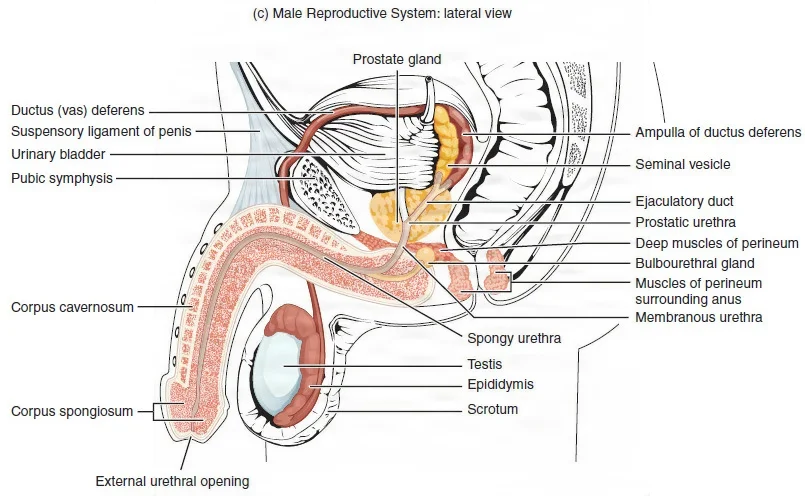
The scrotum and testes
The scrotum is a sac of skin and muscle that hangs behind the penis and in front of the anus. A wall down its middle, the scrotal septum, divides it into two compartments, one for each testis.
Sperm develop best at about 2 to 4 °C below body temperature, and the scrotum adjusts to keep them there. In the cold, the dartos muscle under its skin and the cremaster muscles around each testis pull the testes up against the body; in the warmth, they relax and let the testes hang lower.
The testes (singular testis), or testicles, are the male gonads: two ovals, each about 4 to 5 cm long, that make sperm and testosterone.
- Tunica vaginalis: a smooth serous sac with two layers around each testis, holding a little clear fluid. A hydrocele is too much fluid collecting in it.
- Tunica albuginea: a tough white layer of dense connective tissue that covers the testis itself and sends walls inward, dividing it into 300 to 400 lobules.
- Seminiferous tubules: tightly coiled tubes in each lobule, where sperm are made. The Sertoli cells in their walls support and protect the developing sperm.
- Leydig cells (interstitial cells): cells in the spaces between the tubules that make testosterone.
The making of sperm, spermatogenesis, starts at puberty and goes on for life. One round, from stem cell to finished sperm, takes about 64 days, and the testes make about 100 to 300 million sperm a day. Finished sperm leave the tubules through a network of small ducts into the epididymis.
Testosterone keeps sperm production going and builds muscle and bone, brings on the body changes of puberty, and maintains sex drive. Luteinizing hormone from the pituitary controls how much is made; chapter 9 teaches this.
Before birth, during the seventh month, each testis moves down through the abdominal wall into the scrotum. A testis that has not come down by birth is an undescended testis (cryptorchidism); chapter 34 teaches the operation that brings it down.
In a hydrocelectomy, the surgeon removes the swollen, fluid-filled sac of tunica vaginalis.
The ducts
Sperm travel through a chain of ducts on their way out.
- Epididymis: a tightly coiled tube along the back of each testis, about 6 m long if it were straightened out. Sperm take about 12 days to pass through it, and on the way they mature and become able to swim. They are stored in its tail until ejaculation, and its lining adds fluid that carries them along.
- Ductus deferens, also called the vas deferens: a thick, muscular tube that carries sperm from the epididymis up inside the spermatic cord, through the inguinal canal into the pelvis, and over and behind the bladder, where it widens into the ampulla. Its muscle pushes the sperm along by peristalsis.
- Ejaculatory duct: a short duct, one on each side, formed where the ductus deferens joins the duct of the seminal vesicle. It passes through the prostate and opens into the prostatic urethra at the verumontanum.
- Urethra: carries the semen out through the penis.
The Core Curriculum's list writes this duct as the "vastus ductus deferens"; its name is the ductus deferens, or vas deferens.
Because the ductus deferens runs near the skin in the scrotum, it is easy to reach. In a vasectomy, the surgeon cuts and seals a short piece of each one, which stops sperm from leaving and is a lasting form of birth control.
The spermatic cord and the inguinal canal
The spermatic cord is the bundle that runs from the abdominal wall to each testis and carries what it needs. It holds:
- the ductus deferens and its small artery;
- the testicular artery;
- the testicular veins, which form a net called the pampiniform plexus;
- the cremasteric artery and vein;
- nerves and lymph vessels;
- a remnant of the processus vaginalis, a pouch of peritoneum.
The cremaster muscle wraps the cord and the testis. When the veins of the pampiniform plexus swell, they make a varicocele; it is more common on the left, because the left testicular (gonadal) vein drains into the left renal vein at a right angle, which puts more back pressure on it.
The inguinal canal is a tunnel through the lower abdominal wall, near the inguinal ligament. It runs from the deep inguinal ring, an opening in the transversalis fascia, to the superficial inguinal ring. It carries the spermatic cord in a male and the round ligament of the uterus in a female. The weak area of the groin is larger in males, which matches the higher rate of inguinal hernias in males. Chapter 32 teaches hernias and their repair.
The glands and semen
Three kinds of glands add the fluid that makes up most of the semen.
- Seminal vesicles: two glands behind the bladder that make about 60 percent of the semen. Their fluid is rich in fructose, a sugar that fuels the sperm.
- Prostate: a walnut-sized gland, weighing about 20 to 30 g, that sits under the bladder in front of the rectum and wraps around the prostatic urethra. It adds a milky, alkaline fluid that first thickens the semen and then thins it again after ejaculation. It can be felt in a rectal exam.
- Bulbourethral glands, or Cowper glands: two small glands just below the prostate, beside the urethra. With arousal, before ejaculation, they release a thick mucus that lubricates the urethra and neutralizes acid left by urine.
Semen is the fluid that comes out in ejaculation, about 2 to 5 mL at a time. Sperm make up only about 5 percent of it; the rest comes from the glands.
The prostate grows again from about age 25. In many older patients it grows enough to squeeze the urethra, causing a frequent, urgent need to void, a weak stream and a bladder that never feels empty. This is benign prostatic hypertrophy (BPH), also called benign prostatic hyperplasia. A transurethral resection of the prostate (TURP) removes the obstructing tissue through the urethra with a resectoscope.
For prostate cancer, a radical prostatectomy removes the whole prostate with the seminal vesicles. The nerves for erection run close beside the prostate, so when the tumor is still inside the prostate's capsule the surgeon may try to spare them; erectile dysfunction and loss of urine control are known risks of the operation.
The penis
The penis carries the urethra and delivers semen.
- Shaft: three long columns of spongy erectile tissue, which fill with blood in an erection. The two larger corpora cavernosa (singular corpus cavernosum), side by side, make up most of it. The smaller corpus spongiosum, along the underside, surrounds the spongy urethra.
- Glans penis: the rounded tip, formed by the end of the corpus spongiosum and rich in nerve endings. The external urethral meatus opens on it.
- Prepuce (foreskin): the fold of skin that covers the glans. Circumcision removes it.
A suspensory ligament holds the penis to the pubic symphysis.
Words for male reproduction
| Term | Meaning |
|---|---|
| Erection | The penis stiffens because its arteries widen and fill the erectile tissue with blood, and the filled tissue presses the veins shut so the blood cannot drain away |
| Flaccid | Soft and relaxed, the penis's usual state, as during urination |
| Ejaculation | The forceful release of semen from the urethra |
| Retrograde ejaculation | Semen goes backward into the bladder instead of out through the penis; it can follow prostate surgery or come from some drugs |
| Erectile dysfunction | Being unable to get or keep an erection firm enough for intercourse; it has many causes, such as diabetes, vascular disease, some drugs and prostate problems |
| Priapism | An erection that will not go away, lasting more than 4 hours; it is an emergency that can damage the penis for good |
| Semen | The fluid released in ejaculation, sperm plus the secretions of the glands |
The Core Curriculum's list uses the older word impotence; today the term is erectile dysfunction.
13Where surgery meets these organs
The ureter in pelvic surgery
The ureter runs close to the organs of the female pelvis. It comes down into the pelvis behind the ovary, near the ovarian vessels in the suspensory (infundibulopelvic) ligament, and then, beside the cervix, passes under the uterine artery. A clamp, tie or cut meant for those vessels can catch the ureter, so the surgeon identifies it before dividing them. A memory aid: water (the ureter) runs under the bridge (the uterine artery).
For some pelvic and abdominal operations, the urologist first places ureteral catheters or stents, so the surgeon can feel the ureters and is less likely to cut or tie them.
The bladder in lower abdominal surgery and cesarean birth
The bladder lies right behind the lower abdominal wall, and it rises when it is full.
- Before a cesarean, an indwelling urinary catheter is put in to drain it.
- When the peritoneum is opened, the surgeon lifts it and feels it first to make sure no bowel, omentum or bladder is caught in the cut.
- In a cesarean, the surgeon divides the bladder flap, the fold of peritoneum between the uterus and the bladder, and a bladder retractor (a DeLee or a Balfour bladder blade) holds the bladder down, out of the way, before the uterus is opened.
The inguinal canal in hernia repair
In an open groin hernia repair in a male, the spermatic cord lies in the field. The surgeon frees it and loops a small Penrose drain around the testicular vessels and ductus deferens to lift it gently out of the way. In a female, the canal holds the round ligament instead. Chapter 32 teaches the repairs.
Through the urethra
The urethra is the doorway for the urologist's scopes. Cystoscopy looks into the bladder, a ureteroscope goes on up the ureter, and a resectoscope cuts away prostate tissue in a TURP. A TURP is done with the patient in the lithotomy position, and chapter 34 teaches these cases.
Lo que te llevas de este capítulo
- The kidneys lie behind the peritoneum at about T12 to L3. Inside, urine flows from the papillae of the medulla's pyramids into the minor calyces, the major calyces, the renal pelvis and the ureter. The renal artery comes from the aorta and the renal vein drains to the inferior vena cava, through the hilum.
- The nephron starts with a glomerulus inside Bowman's capsule; its tubule runs from the proximal convoluted tubule through the loop of Henle to the distal convoluted tubule, which drains to a collecting duct. It makes urine by filtration, reabsorption and secretion, sending about 99 percent of the filtered fluid back to the blood.
- Besides removing wastes, the kidneys balance water and salts, raise blood pressure through renin (renin, angiotensin, aldosterone), activate vitamin D for calcium, and make EPO for red cells.
- Each ureter crosses the iliac vessels, passes under the uterine artery in a female, and enters the bladder at a slant that stops reflux. The bladder's trigone lies between the two ureteral openings and the urethra; its detrusor muscle empties it, and an internal (involuntary) and external (voluntary) sphincter hold it shut.
- The female urethra is about 4 cm and carries only urine; the male urethra is about 20 cm (preprostatic, prostatic, membranous and spongy parts) and carries urine and semen. Normal urine is water, salts, nitrogen wastes and pigment; protein, glucose, ketones, blood, pus cells, casts and crystals are abnormal findings on a urinalysis.
- The vulva holds the labia, clitoris and vestibule (urinary meatus and introitus), with the Bartholin and Skene glands; the perineum lies between the vagina and the anus. The ovaries make eggs, estrogen and progesterone; the uterine tubes (interstitial part, isthmus, ampulla, infundibulum with fimbriae) carry the egg; the uterus has a fundus, body and cervix, three layers (perimetrium, myometrium, endometrium) and four main ligaments (broad, round, cardinal, uterosacral).
- The menstrual cycle runs through the menstrual, proliferative, ovulation and progestational (secretory) phases. Know the pregnancy terms: fertilization, implantation, embryo, fetus, gestation, amnion, chorion, placenta, gravidity, parity, premature and abortion, spontaneous or induced.
- Sperm are made in the seminiferous tubules of the testes and testosterone in the Leydig cells; they pass through the epididymis, ductus deferens (inside the spermatic cord, through the inguinal canal) and ejaculatory duct to the urethra, gaining fluid from the seminal vesicles, prostate and bulbourethral glands.
Ponte a prueba5 preguntas rápidas
Gratis para todos. Elige una respuesta para cada una y luego revísalas todas juntas.
En el quirófanoDónde vas a usar esto
Todo el banco de preguntas del capítulo en series de 10, con la respuesta explicada después de cada una.
Parte del programa para educadoresLas palabras del capítulo en tarjetas de estudio: las que fallas vuelven antes.
Parte del programa para educadoresLo asigna tu docente. Cada grupo saca sus propias 25 preguntas del banco del capítulo.
Parte del programa para educadores
Sugerir una corrección
¿Algo está mal, confuso o desactualizado en este capítulo? Avísanos y lo corregimos para todos. Tu correo solo se usa para responderte.