Hormones: the endocrine system
The glands that send chemical messages through the blood, how the body keeps each message in balance, and what surgery on the neck, the adrenal glands and the pituitary can do to them.
1Why it matters
Your body has two systems that carry messages. The nervous system sends fast electrical signals along nerves, and its effects are over quickly. The endocrine system sends chemical messengers, called hormones, through the blood. Hormones take longer to work, but their effects usually last longer.
Hormones run growth, metabolism, the balance of water and salts, the body's answer to stress, and reproduction. Each one acts on particular cells, and each one is kept within a narrow range, because too much or too little causes disease.
You meet this system at the surgical field in four ways.
- Surgeons operate on the glands. They remove all or part of the thyroid, the parathyroid glands, the adrenal glands, the pituitary and the thymus. These glands sit close to nerves and vessels that must be protected, such as the nerve that moves the vocal cords.
- Every operation is a stress, and the body answers it with hormones. Cortisol and epinephrine pour out, the heart speeds up and the blood sugar rises, even in a patient without diabetes.
- Many patients live with a gland disorder. Diabetes, thyroid disease and long-term steroid use change how a patient heals, how well they fight infection and how they handle the stress of surgery.
- Some hormones are drugs on the field. The hormones most often given in surgery are epinephrine, oxytocin and the steroids called glucocorticoids.
This chapter explains how hormones work and how the body keeps them in balance, then goes gland by gland, and ends with what surgery and gland disease mean for the patient on the table. Other chapters teach what it leaves out: the hypothalamus as part of the brain in chapter 4; the thymus as lymphoid tissue in chapter 5; the pancreas as a digestive organ, with its enzymes, in chapter 7; the ovaries and testes and the kidneys' own hormones in chapter 8; immunity in chapter 10; diabetes as it affects wound healing and infection in chapter 11; insulin, steroids and other hormone drugs in chapter 12; blood sugar, temperature and malignant hyperthermia under anesthesia in chapter 13; the thyroid and parathyroid operations and the Whipple procedure in chapter 32; adrenal surgery in chapter 34; the thymectomy in chapter 41; and pituitary surgery in chapter 42.
2How hormones work
Endocrine and exocrine glands
A gland is one cell or a group of cells that makes and releases a chemical substance. Glands come in two kinds.
- Endocrine glands have no ducts. They release their hormones into the fluid around their cells, and the blood carries the hormones through the body.
- Exocrine glands release their products through ducts, onto a surface or into a cavity. Sweat glands and the oil (sebaceous) glands of the skin are exocrine glands.
The pancreas is both. Most of it is exocrine and sends digestive juice down a duct into the small intestine, but small clusters of cells scattered through it, the islets, release hormones into the blood.
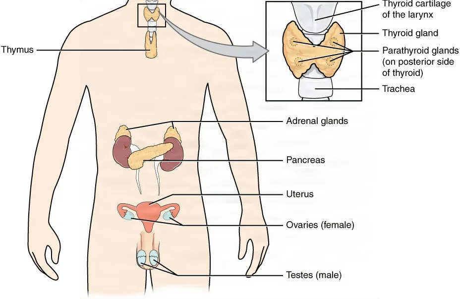
The main endocrine glands are the pituitary, pineal, thyroid, parathyroid and adrenal glands, the islets of the pancreas, the thymus, and the ovaries and testes. Many other organs also have cells that make hormones, including the hypothalamus, the heart, the kidneys, the stomach and small intestine, the liver, the skin and fat tissue.
Target cells and receptors
A hormone travels everywhere the blood goes, but it acts only on its target cells: the cells that carry a receptor for it. A receptor is a protein shaped to fit one hormone, the way a lock fits one key. When the hormone binds, the cell changes what it is doing, for example by making a new protein, changing how it uses nutrients, or making a hormone or other product of its own.
Hormones fall into two chemical groups, and the group decides how a hormone works.
- Steroid hormones are made from cholesterol. Cortisol, aldosterone, estrogen, progesterone and testosterone are steroids. They dissolve in fat, not water, so they ride in the blood on carrier proteins. They pass straight through the cell membrane to a receptor inside the cell and switch genes on. Riding on their carriers, they last much longer in the blood than most other hormones.
- Nonsteroid hormones are built from amino acids. The smallest are amines, made from a single amino acid, such as epinephrine, norepinephrine and the thyroid hormones. Peptides, such as antidiuretic hormone and oxytocin, are short chains of amino acids, and proteins, such as growth hormone, insulin and glucagon, are longer chains. Most of them cannot cross the cell membrane, so they bind a receptor on the cell's surface, which sets off a chain of signals inside. The thyroid hormones are the exception: they enter the cell.
The difference shows in how long a hormone lasts. Half of a dose of cortisol is gone from the blood in about 60 to 90 minutes, but half of a dose of epinephrine is gone in about a minute.
Hormones also work on each other. Some pairs are antagonists, with opposite effects: insulin lowers the blood sugar and glucagon raises it. Others are synergists, which together give a stronger effect than either alone.
Negative feedback keeps levels in range
Chapter 2 introduced negative feedback, the body's main way of keeping a value steady. Most hormones are controlled this way: once the blood holds enough of a hormone, its release slows, so the level stays in a narrow range.
Three kinds of trigger start a gland releasing its hormone.
- A change in the blood: a rise in blood sugar makes the pancreas release insulin, and blood that is too concentrated makes the pituitary release antidiuretic hormone.
- Another hormone: the hypothalamus releases hormones that control the pituitary, and the pituitary releases hormones that control other glands.
- A nerve signal: in danger, sympathetic nerves make the adrenal glands release epinephrine and norepinephrine within seconds.
Positive feedback, which pushes a change further, is rare. During labor, oxytocin makes the uterus contract, the baby's head stretches the cervix, and the stretch brings out more oxytocin, until the baby is born.
The hypothalamus and the pituitary: the command center
The hypothalamus, part of the brain (chapter 4), and the pituitary gland hanging below it work together as the command center of the endocrine system. Many signals that reach the nervous system pass through them and are turned into hormones.
The hypothalamus controls the pituitary in two ways.
- The anterior pituitary answers to releasing hormones and inhibiting hormones made by the hypothalamus. They travel down the pituitary stalk in a short chain of vessels, the hypophyseal portal system, which carries them straight to the anterior pituitary before they reach the rest of the body.
- The posterior pituitary is wired to the hypothalamus. Nerve cells in the hypothalamus make two hormones, send them down their axons in the stalk, and store them in the posterior pituitary until they are released.
Many glands sit at the bottom of a chain of three. The hypothalamus releases a hormone that tells the anterior pituitary to release a second hormone, which tells a target gland to release a third. As the third hormone rises in the blood, it slows the hypothalamus and the pituitary, and the chain winds down. The thyroid is the classic example, and the adrenal cortex works the same way.
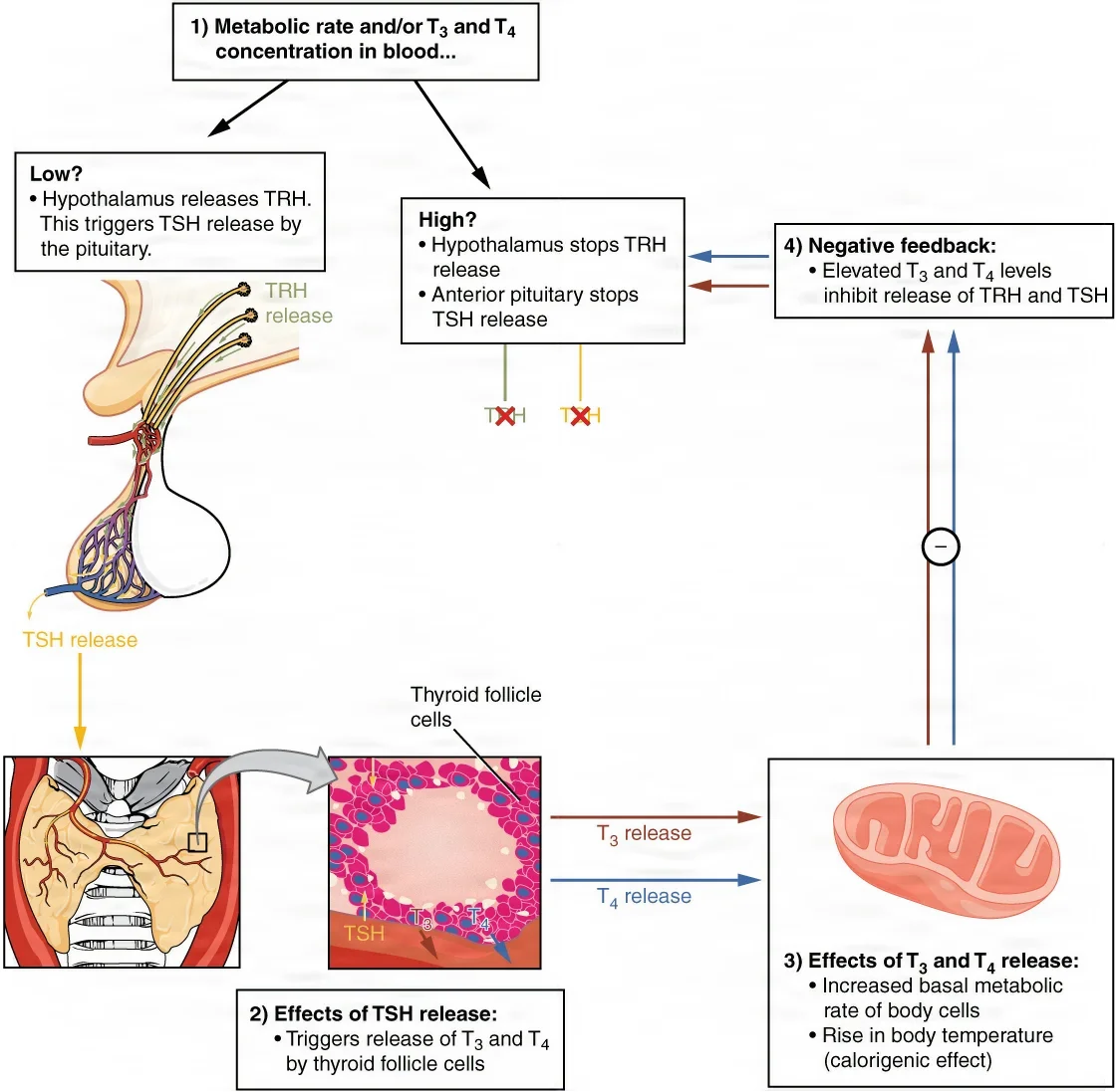
Hypothalamus, then pituitary, then gland, then hormone in the blood. When the gland's hormone runs high, it switches off the hypothalamus and the pituitary; when it runs low, they switch back on.
3The pituitary gland
Where it sits
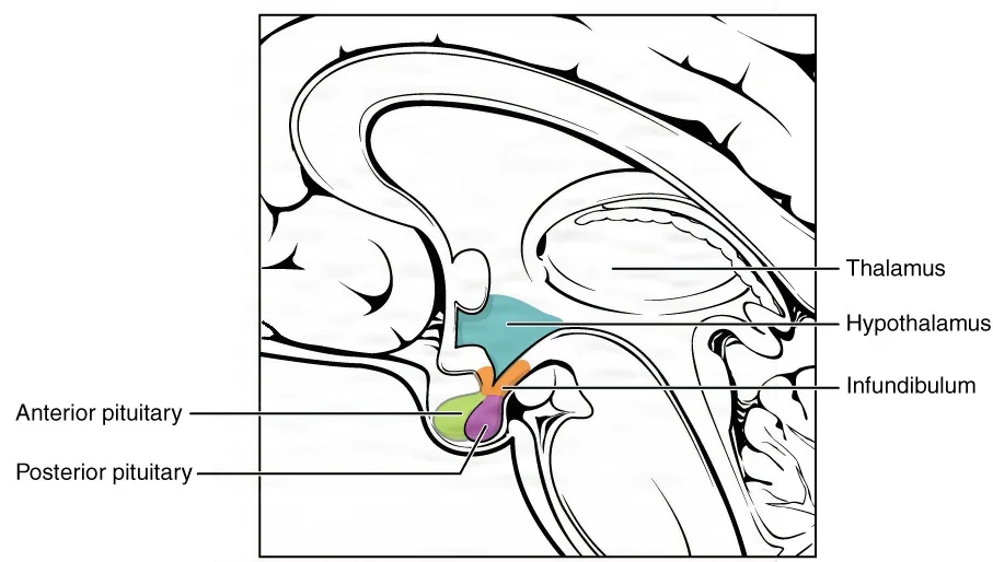
The pituitary gland, or hypophysis, is about the size of a pea. It hangs from the underside of the hypothalamus on a stalk called the infundibulum, and it sits in a small hollow on top of the sphenoid bone in the floor of the skull, the sella turcica.
Its neighbors explain how a pituitary tumor shows itself and how surgeons reach it.
- The optic chiasm, where the two optic nerves cross, lies just above it. A tumor that presses up on the chiasm changes the field of vision, classically taking away the outer half of what each eye sees.
- The cavernous sinuses lie on each side, holding cranial nerves III to VI and the internal carotid arteries.
- The sphenoid sinus lies in front of and below it, so surgeons can reach the gland through the nose and the sinus (chapter 6).
The pituitary is often called the master gland, because its hormones control the thyroid, the adrenal cortex and the ovaries and testes.
It has two lobes, which grow from different tissue before birth and work differently.
- The anterior lobe, or adenohypophysis, is glandular tissue. It makes its own hormones.
- The posterior lobe, or neurohypophysis, is nerve tissue. It makes no hormones; it stores and releases two hormones made in the hypothalamus.
Anterior lobe hormones
The anterior lobe makes six hormones that you need to know.
| Hormone | Target | What it does |
|---|---|---|
| Growth hormone (GH), also called somatotropin | Most tissues, especially muscle, bone and cartilage; it also makes the liver release growth factors | Builds protein and tissue; frees fat for fuel and raises the blood sugar |
| Thyroid-stimulating hormone (TSH), also called thyrotropin | The thyroid gland | Makes the thyroid release its hormones, T4 and T3 |
| Adrenocorticotropic hormone (ACTH), also called corticotropin | The adrenal cortex | Makes the adrenal cortex release cortisol and its other steroids |
| Follicle-stimulating hormone (FSH) | The ovaries and testes | Makes eggs and sperm develop; in the ovaries, makes follicles grow and release estrogens |
| Luteinizing hormone (LH) | The ovaries and testes | Triggers ovulation and the ovaries' release of estrogens and progesterone; makes the testes release testosterone |
| Prolactin (PRL) | The breasts | Builds the milk glands in pregnancy and makes milk after birth |
TSH, ACTH, FSH and LH are called tropic hormones ("turning" hormones), because they turn other glands on and off. Some references spell the word "trophic," and the Core Curriculum lists them that way. FSH and LH together are the gonadotropins, the hormones that run the ovaries and testes; chapter 8 shows where the surge of LH falls in the menstrual cycle.
The hypothalamus has a releasing hormone for each one: TRH brings out TSH, CRH brings out ACTH, GnRH brings out FSH and LH, and GHRH brings out growth hormone. Growth hormone also has an inhibiting hormone, GHIH, also called somatostatin.
The anterior lobe also releases beta endorphin, one of the brain's own painkilling chemicals, and cells between the two lobes release melanocyte-stimulating hormone, which can darken the skin.
When growth hormone is out of balance, the signs depend on age. Too much in a child, before the bones stop growing, causes gigantism. Too much in an adult, once growth has stopped, causes acromegaly: the bones of the face, hands and feet grow larger. Too little in a child stunts growth (pituitary dwarfism, or growth hormone deficiency).
Posterior lobe hormones
The posterior lobe is really an extension of the brain. Nerve cells in the hypothalamus make antidiuretic hormone and oxytocin, pass them down their axons, store them in their endings in the posterior lobe, and release them into the blood when the cells fire. That is the "production of neural transmitter" in the Core Curriculum's list: hormones made and released by nerve cells.
Antidiuretic hormone (ADH), also called vasopressin, saves water.
- Sensors in the hypothalamus watch how concentrated the blood is. When it is too concentrated, as in dehydration or after a salty meal, the posterior lobe releases ADH.
- ADH makes the tubules of the kidneys take back more water, so less urine is made and the blood is diluted back to normal. Then the release slows, by negative feedback.
- At high levels, ADH also narrows blood vessels and raises the blood pressure, which is where the name vasopressin comes from.
- Alcohol blocks its release, which is why drinking alcohol makes you pass more urine.
Oxytocin acts on the uterus and the breasts. It makes the uterus contract during childbirth, through the positive feedback loop above, and it makes the milk ducts squeeze milk out when a baby nurses (the let-down reflex). It also seems to play a part in bonding.
Too little ADH causes diabetes insipidus: the kidneys cannot hold on to water, so the patient passes large amounts of dilute urine, is always thirsty, and can become dehydrated. It is treated with a drug form of vasopressin.
At the field, oxytocin is also a drug. After a cesarean birth, the anesthesia provider gives it into the vein to help the uterus contract and slow the bleeding, and the surgeon may ask for it to be injected straight into the uterine muscle. The scrub draws it up from a vial held by the circulator and labels the syringe before passing it. Chapter 12 teaches drugs on the field.
4The pineal gland
The pineal gland is a tiny gland deep in the brain, below and behind the thalamus. It lies behind the pituitary.
Its cells make the hormone melatonin, built from serotonin. Light controls how much they make.
- In daylight, light striking the retinas sends signals through the hypothalamus to the pineal gland, which then makes less melatonin, and you stay awake.
- As darkness falls, the pineal gland makes more melatonin, and you grow drowsy.
So melatonin helps set the body's daily sleep and wake cycle, its circadian rhythm, which also shapes appetite and body temperature. Jet lag comes from crossing time zones faster than melatonin can reset, and some travelers take melatonin pills to sleep. Children have higher levels than adults, which may help hold back puberty.
A tumor in the region of the pineal gland often causes hydrocephalus, a buildup of cerebrospinal fluid in the brain (chapter 4).
5The thyroid gland
Its shape and its neighbors
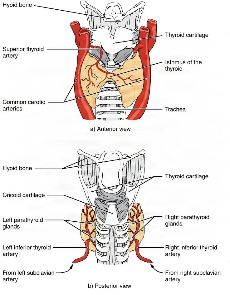
The thyroid gland is shaped like a butterfly. It lies low in the front of the neck, on the front of the trachea, just under the larynx. Its right and left lobes are joined by a narrow middle band, the isthmus, which crosses the trachea between the second and fourth tracheal rings. It weighs about 20 grams and is the largest of the endocrine glands. It has a very rich blood supply.
About 30 percent of patients have a pyramidal lobe, a thin strip of thyroid tissue that runs upward from the isthmus. It is left over from the duct along which the thyroid moved down from the base of the tongue before birth.
The thyroid is crowded by structures the surgeon must protect.
- It lies under the thin strap muscles of the neck, on the trachea, and its lobes rest against the larynx, the trachea and the esophagus. A band of tissue, the ligament of Berry, fixes it to the cricoid cartilage and the trachea.
- The common carotid artery, the internal jugular vein and the vagus nerve run up the neck beside each lobe.
- Two pairs of arteries feed it: the superior thyroid arteries, from the external carotid arteries, enter the upper poles, and the inferior thyroid arteries, from the thyrocervical trunks of the subclavian arteries, enter the lower part of the gland. Now and then a single extra artery, the thyroidea ima, rises from the aortic arch or the brachiocephalic artery to the bottom of the isthmus. Three pairs of veins drain it.
- The recurrent laryngeal nerve, a branch of the vagus nerve, runs up in the groove between the trachea and the esophagus, behind the thyroid, and works the muscles that move the vocal cords. Cutting one nerve leaves the voice hoarse; injuring both can close the airway.
- The external branch of the superior laryngeal nerve runs near the superior thyroid artery and works one small muscle of the larynx, the cricothyroid. Injury to it makes it hard to shout or to sing high notes.
- The parathyroid glands lie on the back of the lobes.
Thyroid hormones and metabolism
The thyroid is built of tiny hollow sacs, the follicles, each full of a thick fluid, the colloid. The cells lining each follicle build the thyroid hormones in the colloid, and they need iodine from food to do it. That is why iodine is added to table salt.
- Thyroxine (T4) carries four iodine atoms. It is the main hormone the gland makes.
- Triiodothyronine (T3) carries three. It is the stronger of the two, and many body cells turn T4 into T3 by removing one iodine.
The hormones wait in the colloid until TSH releases them into the blood. There, more than 99 percent of them ride on carrier proteins, and only the small free share enters the cells.
T4 and T3 set the body's metabolism, the pace of the chemical work in its cells.
- They raise the basal metabolic rate, the energy the body burns at rest, so cells use more oxygen and fuel and give off more heat.
- They are needed for growth and for the development of the brain and nerves before birth and in childhood.
- They make the heart and blood vessels more sensitive to epinephrine and norepinephrine, so too much thyroid hormone speeds the heart, strengthens the beat and raises the blood pressure.
The TRH, TSH and thyroid chain in the feedback figure controls the output. A blood test for TSH is the single best test of thyroid function. Because of negative feedback, a high TSH means the gland is underactive, and the pituitary is pushing it harder; a low TSH means the gland is overactive.
When the diet lacks iodine, the thyroid cannot make enough hormone, the pituitary keeps sending more TSH, and the gland swells. An enlarged thyroid is called a goiter. A goiter can also come with an overactive gland.
Calcitonin
Scattered between the follicles are the parafollicular cells, or C cells. When the blood calcium rises, they release calcitonin, which lowers it: it slows the bone cells that break down bone (osteoclasts), cuts the absorption of calcium in the gut and increases the calcium lost in the urine.
Calcitonin usually does little to keep the calcium steady; parathyroid hormone does most of that work. A drug form of calcitonin is used for weak, thin bones (osteoporosis), and a high blood level can mark a medullary thyroid cancer.
6The parathyroid glands and calcium
Where they are
The parathyroid glands are small, oval, yellow to brownish glands, each about the size of a lentil, on the back of the thyroid lobes. A thick capsule of connective tissue separates them from the thyroid, and fat often covers them.
- Most people have four: two upper glands near the upper poles of the thyroid and two lower glands near the lower poles. The lower ones vary more in where they sit.
- Some people have more, and glands can sit away from the thyroid, lower in the neck or in the upper chest (the superior mediastinum). The Core Curriculum lists four to six glands; one reference gives a range of 2 to 12, with about 90 percent of people having four.
- Both the upper and the lower glands get their blood from the inferior thyroid artery.
They are hard to tell apart from a lobule of fat, a lymph node or a small thyroid nodule, and during thyroid surgery they can be damaged, removed by accident or cut off from their blood supply.
Parathyroid hormone and blood calcium
Calcium is needed for muscles to contract, for nerves to send signals and for blood to clot, so its level in the blood is held in a narrow range.
When the blood calcium falls, the chief cells of the parathyroid glands release parathyroid hormone (PTH), also called parathormone. It raises the calcium in three ways.
- Bone: it makes osteoclasts break down bone matrix and free calcium into the blood, and it slows the cells that lay calcium down in bone.
- Kidneys: it makes the kidneys take back more calcium from the urine, and it makes them produce calcitriol, the active form of vitamin D.
- Gut: calcitriol makes the intestine absorb more calcium from food.
As the calcium rises, PTH release slows, by negative feedback. PTH and calcitonin pull in opposite directions, and together they keep the calcium steady.
| Parathyroid hormone (PTH) | Calcitonin | |
|---|---|---|
| Made by | The chief cells of the parathyroid glands | The C (parafollicular) cells of the thyroid |
| Released when | Blood calcium falls | Blood calcium rises |
| Effect on blood calcium | Raises it | Lowers it |
| Effect on bone | Frees calcium from bone | Slows the release of calcium from bone |
Too much or too little parathyroid hormone
- Hyperparathyroidism: when the trouble starts in the glands themselves, it is most often a single benign tumor of one gland, an adenoma (80 to 85 percent of these cases), less often an enlargement of all four glands, and rarely a cancer. The high calcium thins the bones so they break easily, forms kidney stones, and causes muscle weakness, tiredness, belly pain and changes in mood.
- Hypoparathyroidism: most often the result of thyroid surgery that injures the parathyroid glands or cuts off their blood supply. The low calcium makes nerves and muscles fire too easily: numbness and tingling, twitching, cramps and muscle spasms, a state called tetany. Severe cases can bring on seizures and spasm of the larynx that blocks breathing, and can be fatal.
7The adrenal glands
Where they sit
The two adrenal glands sit on top of the kidneys, which is why they are also called the suprarenal glands. They lie behind the peritoneum, under the diaphragm, inside the same fascia as the kidneys (Gerota fascia). The right gland is triangular and lies against the inferior vena cava. The left gland is crescent-shaped and lies behind the stomach and the pancreas.
They have one of the richest blood flows in the body. Arteries reach them from the inferior phrenic arteries, the aorta and the renal arteries. The right adrenal vein is short and drains straight into the inferior vena cava; the left drains into the left renal vein.
Each gland is two glands in one.
- The adrenal cortex, the outer layer, is glandular tissue that makes steroid hormones.
- The adrenal medulla, the inner core, is modified nerve tissue that makes catecholamines.
The adrenal cortex
The cortex has three zones, and each makes its own group of steroids.
| Zone, from outside in | Hormone group | Main hormone | What it does |
|---|---|---|---|
| Zona glomerulosa | Mineralocorticoids | Aldosterone | Makes the kidneys keep sodium and let go of potassium; water follows the sodium, so blood volume and blood pressure rise |
| Zona fasciculata | Glucocorticoids | Cortisol | Raises the blood sugar by breaking down stored glycogen, fat and muscle protein for fuel; helps the body through long stress; dampens inflammation and the immune response |
| Zona reticularis | Androgens | Weak sex hormones | Turned into testosterone or estrogens in the tissues; after menopause they are the main source of estrogens |
ACTH from the anterior pituitary drives the release of cortisol and the androgens. The chain runs CRH from the hypothalamus, ACTH from the pituitary, then cortisol, and a high cortisol level switches the chain off. Aldosterone is also released when the potassium is high, the sodium is low or the blood pressure falls; a fall in pressure works through a hormone chain that starts in the kidneys (chapter 8).
Steroid drugs such as prednisone, dexamethasone and hydrocortisone copy cortisol. They are used to calm inflammation, in a joint, in the lungs or on the skin, and to prevent the rejection of a transplanted organ. Taken for a long time, they shut down the patient's own adrenal cortex, a point that matters on the day of surgery. Chapter 12 teaches them as drugs.
The adrenal medulla
The medulla is really part of the sympathetic nervous system (chapter 4). Its large chromaffin cells release epinephrine (adrenaline) and norepinephrine (noradrenaline) into the blood, about four parts epinephrine to one of norepinephrine. These hormones are catecholamines, a group that also includes dopamine.
Sympathetic nerves fire the medulla in danger or sudden stress, and the hormones arrive within seconds. This is the fight-or-flight response.
- The heart beats faster and harder, and the blood pressure rises.
- The airways widen, and more blood goes to the muscles, the heart and the brain.
- Blood vessels to the gut, the kidneys and the skin narrow, and digestion slows.
- The liver turns glycogen into glucose, so the blood sugar rises. The pupils widen and the mouth goes dry.
Epinephrine is also a drug you will handle at the field. A little of it is mixed with local anesthetics such as lidocaine; it narrows the small vessels, so the numbing drug stays where it was injected and lasts longer, and the tissue bleeds less. Chapter 12 teaches its safe use.
8The pancreatic islets and blood sugar
Most of the pancreas makes digestive juice (chapter 7). Scattered through it are tiny clusters of hormone-making cells, the pancreatic islets, or islets of Langerhans, which release their hormones into the blood.
Each islet holds four kinds of cells.
- Alpha cells, about 20 percent of the islet, make glucagon.
- Beta cells, about 75 percent, make insulin.
- Delta cells, about 4 percent, make somatostatin, which holds back the release of both insulin and glucagon. The hypothalamus, the stomach and the intestine make somatostatin too.
- PP cells, about 1 percent, make pancreatic polypeptide, which seems to play a part in appetite.
Glucose is the body's preferred fuel, and the brain depends on a steady supply. The body keeps the blood glucose, or blood sugar, at about 70 to 100 mg/dL, and it stores extra glucose in the liver and the muscles as glycogen. Insulin and glucagon work as a seesaw to hold it there.
- When the blood sugar rises, as after a meal, the beta cells release insulin. Insulin lets most body cells, above all muscle and fat cells, take glucose in; it makes the liver store glucose as glycogen; and it helps the body build fat and protein. The blood sugar falls. The brain, the liver, the kidneys and red blood cells take up glucose without needing insulin.
- When the blood sugar falls, as during fasting or hard exercise, the alpha cells release glucagon. Glucagon makes the liver break glycogen back down into glucose and build new glucose from amino acids and glycerol. The blood sugar rises.
Each hormone is held in check by negative feedback: a falling blood sugar turns off insulin, and a rising one turns off glucagon. Cortisol, epinephrine and growth hormone also raise the blood sugar, which is why stress pushes it up.
Beta cells make insulin, which lowers the blood sugar. Alpha cells make glucagon, which raises it.
Diabetes mellitus
In diabetes mellitus ("honey-sweet"), the blood sugar stays too high, a state called hyperglycemia, because the body makes too little insulin or its cells do not respond to it.
- Type 1: the immune system destroys the beta cells, so the body makes little or no insulin. The patient needs insulin by injection or pump for life. It usually starts young and makes up a small share of cases.
- Type 2: the cells resist insulin, the pancreas makes more to keep up, and in time the beta cells wear out. It makes up about 90 to 95 percent of cases and is linked to excess weight and too little activity. Diet, exercise and pills often control it, and some patients need insulin.
The early signs are passing large amounts of urine (polyuria), great thirst (polydipsia) and hunger (polyphagia): the extra glucose spills into the urine and pulls water with it.
Over the years, a high blood sugar damages blood vessels and nerves. That leads to heart attacks and strokes, kidney failure, blindness, poor blood flow to the legs, and loss of feeling in the feet (neuropathy). Together these raise the risk of injury, infection and tissue death, and lead to many toe, foot and leg amputations.
Without insulin, cells burn fat for fuel, and acids called ketones build up in the blood. This ketoacidosis can lead to coma and death if it is not treated.
The opposite problem, hypoglycemia, a blood sugar that is too low, most often comes from too much insulin. The patient becomes anxious, sweaty, shaky, hungry and weak, with a fast heart rate; as the brain runs short of glucose, they become confused and may have seizures or lose consciousness. A very high and a very low blood sugar can look alike, so the cause is checked quickly and treated.
9The thymus and other hormone makers
The thymus
The thymus lies in the mediastinum, in the upper front of the chest behind the sternum. It is large and active in infancy and childhood and shrinks with age.
Its hormones, the thymosins, help T lymphocytes, the immune cells that mature in the thymus, to develop. Chapter 5 teaches the thymus as lymphoid tissue, and chapter 10 teaches immunity.
A thymectomy removes the thymus. It is done for a tumor of the thymus, a thymoma, and for myasthenia gravis, an autoimmune disease that weakens the muscles; thymomas are often found together with it. The classic approach is through the split sternum (a median sternotomy), and less invasive approaches go through a partial sternotomy, through the neck, or through small chest incisions with a thoracoscope or a robot. Chapter 41 teaches it.
Other organs that make hormones
| Organ | Main hormones | What they do |
|---|---|---|
| Ovaries | Estrogens and progesterone | Female sex characteristics, the menstrual cycle and pregnancy (chapter 8) |
| Testes | Testosterone | Male sex characteristics and sperm production (chapter 8) |
| Placenta | Human chorionic gonadotropin (hCG), estrogens and progesterone | Keep a pregnancy going |
| Heart | Atrial natriuretic peptide (ANP), from the walls of the atria | Makes the kidneys let go of sodium and water, lowering blood volume and pressure |
| Kidneys | Renin, erythropoietin (EPO) and calcitriol | Raise blood pressure, drive red blood cell production and help absorb calcium (chapter 8) |
| Stomach and small intestine | Gastrin, secretin and cholecystokinin (CCK) | Control the juices of digestion (chapter 7) |
| Fat tissue | Leptin | Gives the feeling of fullness after a meal |
10When glands make too much or too little
Most endocrine diseases come from a gland making too much of its hormone (hypersecretion) or too little (hyposecretion). The prefix "hyper-" means above normal and "hypo-" means below normal, so hyperthyroidism is an overactive thyroid and hypothyroidism an underactive one. Too little is often treated by replacing the hormone; too much, with drugs that lower it or by removing the gland.
| Gland | Too much | Too little |
|---|---|---|
| Anterior pituitary (growth hormone) | Gigantism in a child; acromegaly in an adult | Stunted growth in a child (pituitary dwarfism) |
| Posterior pituitary (ADH) | Syndrome of inappropriate antidiuretic hormone (SIADH): the kidneys hold back too much water, so the blood is diluted and its sodium falls | Diabetes insipidus: large amounts of dilute urine, thirst |
| Thyroid | Hyperthyroidism, most often Graves disease, an autoimmune disease: fast heart rate, heat intolerance, sweating, weight loss, tremor, nervousness, bulging eyes, sometimes a goiter | Hypothyroidism, most often Hashimoto thyroiditis, an autoimmune disease, or after the thyroid is removed: tiredness, feeling cold, weight gain, slow heart rate, constipation, slowed thinking |
| Parathyroids | Hyperparathyroidism: high calcium, thin bones, kidney stones | Hypoparathyroidism: hypocalcemia and tetany |
| Adrenal cortex | Cushing syndrome (too much cortisol): high blood sugar, a round "moon" face, a fat pad on the back of the neck, skin that bruises easily, poor wound healing, infections; from a pituitary tumor making too much ACTH (Cushing disease), an adrenal tumor, or long-term steroid drugs | Addison disease (too little cortisol and aldosterone): weakness, weight loss, low blood pressure, low blood sugar and low sodium; most often autoimmune |
| Adrenal medulla | Pheochromocytoma, a tumor of the chromaffin cells: bouts of very high blood pressure, fast heart rate, sweating, anxiety, high blood sugar | |
| Pancreatic islets | Too much insulin (most often an insulin overdose): hypoglycemia | Diabetes mellitus: hyperglycemia |
11Hormones and surgery
The stress of surgery
The body treats an operation as an injury. Nerve signals from the cut tissue reach the hypothalamus, which turns on the pituitary and the adrenal glands, and the blood fills with cortisol, catecholamines, glucagon and growth hormone.
The result is the stress response.
- The heart rate and blood pressure rise.
- The liver pours out glucose while the cells respond less to insulin, so the blood sugar rises, even in patients without diabetes. The bigger the operation, the greater this insulin resistance.
- Muscle protein is broken down for fuel, which can slow healing.
- Cortisol held high for a long time weakens the lymphocytes that fight infection.
Bleeding adds to it. In a heavy bleed, the autonomic nervous system releases vasopressin, glucagon, cortisol, epinephrine and norepinephrine. At first they help, by raising the cardiac output and narrowing the vessels so blood goes to the heart and brain. Chapter 11 teaches shock.
Enhanced recovery after surgery (ERAS) pathways are built to lower the body's stress response to an operation. The CDC's guideline for preventing surgical site infection calls for controlling the blood sugar around surgery, with a target below 200 mg/dL, in patients with and without diabetes.
A 2022 update from three US infection prevention societies (SHEA, IDSA and APIC) sets a lower target: keep every patient's blood sugar between 110 and 150 mg/dL in the first day or two after surgery, with or without diabetes.
The patient with a gland disorder
Diabetes, thyroid disorders and adrenal insufficiency (Addison disease) are among the endocrine disorders that change how a patient goes through surgery.
Diabetes. A high blood sugar slows wound healing and raises the risk of a surgical site infection, so the patient's blood sugar is checked before, during and after surgery. Surgery upsets both the patient's meals and their insulin doses. A patient with diabetes should be the first case of the day, so they can get back to their usual meals as soon as possible. In a long case, the team pads pressure points with extra care, because poor circulation and lost feeling put the skin at risk.
An overactive thyroid. Before planned surgery, drugs bring the thyroid hormone levels back to normal (a euthyroid state). These patients have a higher risk of heart rhythm problems, such as atrial fibrillation.
The danger is thyroid storm (thyrotoxic crisis): a sudden release of a large amount of thyroid hormone, which can be set off by a stress such as surgery. The patient develops a high fever, a very fast heart rate, high blood pressure, vomiting or diarrhea, agitation and confusion, and may have seizures or slip into a coma. A calm, quiet room helps prevent it. If it happens, the team gives oxygen, fluids and drugs and cools the patient.
An underactive thyroid. These patients are at risk of breathing failure around surgery. A rare and dangerous form, myxedema coma, mostly affects people older than 75.
A large goiter or thyroid nodule can press on the trachea or push it aside and make the breathing tube hard to place, so the difficult airway cart is brought into the room before induction.
Long-term steroids. A patient who has taken steroid drugs for more than about 3 weeks may have a shut-down adrenal cortex that cannot make the extra cortisol the stress of surgery calls for. The result can be an adrenal crisis, with a dangerous fall in blood pressure. So the patient's steroid use is found out before surgery. The usual dose is kept up, and whether an extra stress dose of steroid is added is decided for each patient, from how much steroid they take and for how long, and from the type and length of the operation. A stress dose for every patient on steroids is no longer the routine.
Thyroid and parathyroid surgery
Surgeons remove all or part of the thyroid for a known or suspected cancer, for a goiter that presses on the airway or the esophagus, and for some patients with an overactive gland, such as in Graves disease. Parathyroid tissue is removed for hyperparathyroidism. When the disease starts in the glands themselves, its cause is most often a single adenoma.
Chapter 32 teaches the operations: the thyroidectomy, the hemithyroidectomy, which removes one lobe with the isthmus, and the parathyroidectomy. The anatomy in this chapter explains their three chief complications: injury to the recurrent laryngeal nerve, a low blood calcium and a hematoma in the neck.
- The recurrent laryngeal nerve. The surgeon finds the nerve and protects it, often with a nerve monitor built into the breathing tube. Because the nerve controls the voice, the patient's voice is checked before surgery as a baseline. A nerve injured on one side leaves the voice hoarse; injury to both sides can block the airway and call for an emergency tracheotomy.
- Low calcium. If the parathyroid glands are removed or lose their blood supply, PTH falls and so does the blood calcium. After a total thyroidectomy or a parathyroidectomy, the calcium reaches its lowest point 48 to 72 hours after surgery. Early signs are numbness and tingling of the fingers, toes and lips. Tapping over the facial nerve makes the face muscles twitch (a positive Chvostek sign), and a blood pressure cuff pumped up on the arm makes the hand and wrist spasm (a positive Trousseau sign). Tetany can follow, with spasms of the hands and feet, seizures and stridor, the harsh, high-pitched sound of a narrowed airway, which can be fatal.
- A neck hematoma. Bleeding under the closed wound can press on the trachea. That is why the back table stays sterile until the patient has been extubated and has left the room, and a tracheotomy tray stays close by.
If a parathyroid gland is found on the removed thyroid, it is saved and reimplanted (chapter 32). A patient whose whole thyroid is removed takes thyroid hormone, levothyroxine, for life.
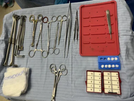
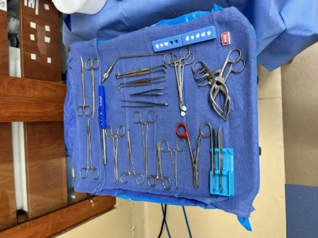
The recurrent laryngeal nerve is the voice; the parathyroids are the calcium. Hoarseness after thyroid surgery points to the nerve; tingling and twitching point to low calcium.
Adrenal surgery
An adrenalectomy removes one or both adrenal glands, for a tumor that makes too much hormone, such as a pheochromocytoma, a tumor making cortisol (Cushing syndrome) or one making aldosterone, or for cancer. It is usually done laparoscopically, which means less harm and a shorter hospital stay than an open operation; for one gland, the patient lies on the side or on the back. Chapter 34 teaches it.
A pheochromocytoma makes the case dangerous.
- Before surgery, the patient takes drugs that block the effects of the catecholamines, the alpha blocker first and then the beta blocker.
- During surgery, the anesthesia provider watches the blood pressure continuously through an arterial line. Handling the tumor can release a flood of catecholamines and send the blood pressure soaring, so the surgeon ties off the adrenal vein early.
- Once the tumor is out, the catecholamines fall away. The blood pressure can drop sharply, and so can the blood sugar, so the patient goes to intensive care to be watched.
After a tumor that made too much cortisol is removed, the other adrenal gland may have been shut down and cannot keep up, so steroids are given during and after surgery and tapered slowly. A patient who loses both adrenal glands needs glucocorticoid and mineralocorticoid replacement for life.
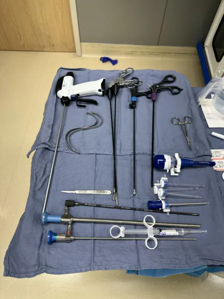
Pituitary surgery
Most pituitary tumors are removed through the nose and the sphenoid sinus, a transsphenoidal hypophysectomy, with an endoscope or a microscope. A large or complex tumor may need a craniotomy instead. The sella may be packed with a small graft of fat or muscle so cerebrospinal fluid cannot leak. Chapter 42 teaches it.
The gland's work explains what the team watches for afterward.
- Diabetes insipidus: the loss of ADH, or swelling around the gland, can cause a flood of dilute urine.
- Vision: a hematoma can press on the optic nerves or the optic chiasm.
- A leak of cerebrospinal fluid.
- Lost hormones: when the whole gland is removed, the patient needs thyroid hormone, sex hormones and glucocorticoids for life.
The pancreas at surgery
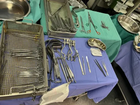
Removing part of the pancreas, as in a Whipple procedure for pancreatic cancer (chapter 32), can leave the patient making too little insulin, so the blood sugar must be watched. For some patients with type 1 diabetes, a pancreas transplant or a transplant of islets alone can let the body make insulin again; during a pancreas transplant, the anesthesia provider checks the blood sugar throughout.
Lo que te llevas de este capítulo
- Endocrine glands are ductless and release hormones into the blood; exocrine glands use ducts. A hormone acts only on target cells with a receptor for it. Steroid hormones, made from cholesterol, enter the cell; most hormones made from amino acids bind a receptor on its surface.
- Most hormones are held in range by negative feedback. The hypothalamus controls the anterior pituitary with releasing and inhibiting hormones, and the pituitary's tropic hormones (TSH, ACTH, FSH, LH) control other glands.
- The pituitary sits in the sella turcica under the optic chiasm. Its anterior lobe (adenohypophysis) makes GH, TSH, ACTH, FSH, LH and prolactin. Its posterior lobe (neurohypophysis) stores and releases ADH (vasopressin) and oxytocin, made in the hypothalamus. Too little ADH is diabetes insipidus. The pineal gland, behind the pituitary, makes melatonin for the sleep and wake cycle.
- The thyroid's two lobes and isthmus lie on the trachea below the larynx, beside the recurrent laryngeal nerves. T4 and T3 need iodine and set the metabolic rate; calcitonin lowers the blood calcium. TSH is the best single test of thyroid function.
- Usually four parathyroid glands sit on the back of the thyroid and get their blood from the inferior thyroid arteries. PTH (parathormone) raises the blood calcium through bone, kidneys and gut.
- The adrenal cortex makes mineralocorticoids (aldosterone), glucocorticoids (cortisol) and androgens; the medulla's chromaffin cells make the catecholamines epinephrine and norepinephrine for fight or flight. In the pancreatic islets, beta cells make insulin, which lowers the blood sugar, alpha cells make glucagon, which raises it, and delta cells make somatostatin. The thymus, in the mediastinum, makes thymosins for T cells.
- Surgery sets off a stress response that raises cortisol, catecholamines and the blood sugar. Patients with diabetes need their blood sugar watched; patients on long-term steroids may need a stress dose; an overactive thyroid can flare into thyroid storm.
- Thyroid and parathyroid surgery risk the recurrent laryngeal nerve (hoarseness), the parathyroids (low calcium, tetany) and a neck hematoma. A pheochromocytoma can swing the blood pressure up during surgery and down after it, and pituitary surgery can cause diabetes insipidus.
Ponte a prueba5 preguntas rápidas
Gratis para todos. Elige una respuesta para cada una y luego revísalas todas juntas.
En el quirófanoDónde vas a usar esto
Todo el banco de preguntas del capítulo en series de 10, con la respuesta explicada después de cada una.
Parte del programa para educadoresLas palabras del capítulo en tarjetas de estudio: las que fallas vuelven antes.
Parte del programa para educadoresLo asigna tu docente. Cada grupo saca sus propias 25 preguntas del banco del capítulo.
Parte del programa para educadores
Sugerir una corrección
¿Algo está mal, confuso o desactualizado en este capítulo? Avísanos y lo corregimos para todos. Tu correo solo se usa para responderte.