Microbiology
The germs that cause infection, how they spread from one person to the next, and how the body fights back: the science under every rule of asepsis and every precaution you take in the OR.
1Why it matters
Germs live everywhere: in the air, in water and soil, on surfaces, and on and inside your body. Most of them do no harm, and many are useful. Only a small share cause disease, and a germ that can cause disease is called a pathogen.
Surgery cuts through the skin, the body's best barrier, and lets germs in. Most surgical site infections start with germs the patient already carries on the skin or in the bowel. Others come from outside the patient: the team, the room's air and the instruments and supplies.
Every rule you follow in the OR traces back to this chapter: why you scrub, why sterilizers are tested with spores, why a dropped clamp never goes back on the field, why you wear eye protection on every case. When you know how germs live, grow and spread, those rules stop being habits and become reasons.
The chapter has two halves. The first is the science: cells, microscopes and stains, how germs are grown and named, and the main groups of microbes. The second is infection: the germs the OR worries about most, the body's defenses, how infection spreads, and the precautions that protect the patient and you.
Other chapters hold the rest: the organelles of human cells and membrane transport in chapter 2; blood cells and the lymph system in chapter 5; inflammation, wound healing, wound classes and surgical site infection in chapter 11; antibiotics and vaccines as drugs in chapter 12; anesthesia emergencies, anaphylaxis among them, in chapter 13; electrosurgery and surgical smoke in chapter 20; asepsis and sterile technique in chapter 26; disinfection, sterilization and prion reprocessing in chapter 27; attire, the hand wash and the steps for putting on and taking off protective equipment in chapter 28; collecting cultures and other specimens at the field, and passing sharps, in chapter 29; and room cleaning, isolation turnovers, spills and medical waste in chapter 30.
2A short history
People guessed long before microscopes that tiny unseen creatures might cause disease. Proof took centuries.
- 1670s: Antonie van Leeuwenhoek, a Dutch cloth merchant, made lenses strong enough to see single-celled organisms, the "animalcules" Leeuwenhoek drew and described. The drawings show that some of them were bacteria.
- 1796: Edward Jenner gave the first vaccination, against smallpox.
- 1847: Ignaz Semmelweis saw that mothers on wards run by doctors and medical students died of fever after childbirth far more often than mothers cared for by midwives. The students went straight from autopsies to examining patients without washing their hands. Once Semmelweis made them wash in chlorinated lime water, deaths fell to the midwives' rate.
- 1850s and 1860s: Louis Pasteur showed that microbes from the air, not spontaneous generation, spoil food, and proposed that microbes also cause infection: the germ theory of disease. Pasteur also invented pasteurization and later made vaccines, among them the first rabies vaccine in 1885.
- 1867: Joseph Lister, a surgeon, brought the germ theory into the operating room. Lister insisted on clean hands and sprayed carbolic acid (phenol) during surgery to kill germs. Wound infections fell, and antiseptic surgery became standard practice.
- 1876 and 1882: Robert Koch proved with anthrax that a specific microbe causes a specific disease, and later set out Koch's postulates, the steps for proving which germ causes a disease. With them, Koch and colleagues identified the bacteria of tuberculosis and cholera.
- 1882 and 1884: Paul Ehrlich developed the acid-fast stain and Hans Christian Gram the Gram stain. Labs still use both every day.
- 1928: Alexander Fleming discovered penicillin. Antibiotics came into use in the 1940s and opened the era of modern surgery.
Microbiology today
Microbiology is now many fields: bacteriology (bacteria), virology (viruses), mycology (fungi), parasitology (parasites) and immunology (the immune system). Medical microbiology studies the germs that cause disease, and epidemiology studies how disease moves through groups of people.
Labs now identify many germs by reading their DNA or RNA, which also shows how closely one germ is related to another.
Two problems drive much of the field. Emerging diseases are new diseases, or old ones that were never a public health threat before, that are now becoming one. And germs keep learning to resist antibiotics: in the United States, about 2 million people a year get a serious antibiotic-resistant infection.
3The chemistry of life
Every living thing is built from the same few kinds of molecules. Knowing them explains how heat and chemicals kill germs.
- Water: up to 70 percent of an adult's body weight. Water dissolves salts, sugars and many other substances, so it is called the universal solvent, and almost all of life's chemical reactions take place in it.
- Carbohydrates: sugars and starches, the cell's quick fuel and its stored energy. They also build structures, such as the cell walls of fungi.
- Lipids: fats, used to store energy and to build cell membranes.
- Proteins: chains of amino acids folded into exact shapes. Proteins form structures and act as enzymes, which run the cell's chemical reactions.
- Nucleic acids: DNA and RNA, which store and pass on the genetic instructions. Chapter 2 teaches them in detail.
A protein works only while it keeps its shape. Heat, acids, bases and some chemicals make proteins lose that shape, or denature, and a denatured protein can no longer do its job. Many ways of killing germs work like this: steam under pressure, for example, coagulates the proteins and nucleic acids inside microbes.
4Cells: prokaryotes and eukaryotes
Cell theory says that the cell is the basic unit of life, that all living things are made of cells, and that all cells come from other cells.
Every cell has a plasma (cell) membrane, cytoplasm, genetic material in the form of DNA, and ribosomes, which build proteins. Beyond that, cells fall into two great groups.
| Prokaryotic cell | Eukaryotic cell | |
|---|---|---|
| Found in | Bacteria and archaea | Humans and other animals, plants, fungi, protozoa and algae |
| Nucleus | None: the DNA lies loose in an area called the nucleoid | A true nucleus wrapped in a membrane |
| Chromosomes | Usually one, circular | Several, rod-shaped |
| Organelles wrapped in membranes | None | Many, such as mitochondria, the endoplasmic reticulum and the Golgi apparatus |
| Ribosomes | Smaller (70S) | Larger (80S) in the cytoplasm |
| Size | About 0.5 to 1 micrometer | About 5 to 20 micrometers |
| How it divides | Binary fission | Mitosis, and meiosis to make sex cells |
Viruses are not cells at all, so they belong to neither group.
The difference matters in treatment. Many antibiotics attack parts only bacteria have, such as the bacterial cell wall or the smaller 70S ribosome, so they kill bacteria and leave human cells alone. Fungi and protozoa are eukaryotes like us, which is why drugs that kill them without harming the patient are harder to make.
Inside a eukaryotic cell
Eukaryotic cells, including yours, do their work in compartments called organelles. Chapter 2 teaches them in the human cell; here is the short list the Core Curriculum names.
- Cell (plasma) membrane: a double layer of fatty molecules with proteins set in it, controlling what enters and leaves.
- Cell wall: found in fungi (made of chitin), plants (cellulose) and many algae, but never in human or other animal cells.
- Cytoplasm: the jellylike fluid inside the membrane that holds the organelles. In some single-celled organisms, such as amoebas, the cytoplasm has a firmer outer gel layer, the ectoplasm, around a more fluid endoplasm.
- Nucleus: holds the DNA.
- Endoplasmic reticulum: a network of folded membranes; rough ER is dotted with ribosomes.
- Golgi apparatus: stores, changes and ships large molecules.
- Mitochondria: make ATP, the energy that runs the cell.
- Ribosomes: build proteins.
The parts of a bacterial cell
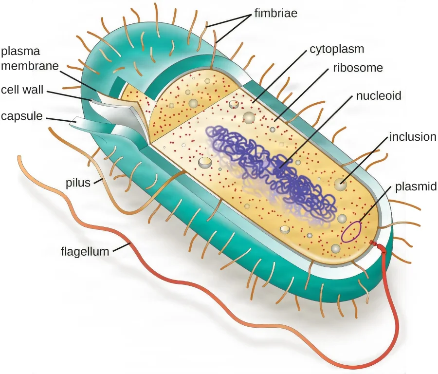
- Capsule: a sticky coat outside the wall, usually made of sugars or proteins. A looser coat that washes off easily is a slime layer. A capsule helps the bacterium cling to surfaces and makes it hard for white blood cells to swallow, so a capsule makes a germ more dangerous; Streptococcus pneumoniae is a well-known example.
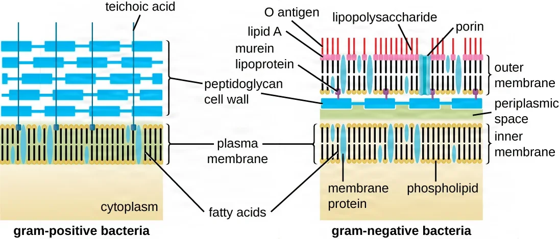
- Cell wall: a rigid layer that gives the cell its shape and keeps it from bursting when water rushes in. Bacterial walls are made of peptidoglycan, a meshlike material found only in bacteria. Penicillin and its relatives work by blocking it. How thick the wall is decides the Gram stain result, below.
- Plasma membrane: the thin membrane just inside the wall that lets some substances through and keeps others out. Every cell has one.
- Cytoplasm: the gel of water and dissolved chemicals that fills the cell.
- Nucleoid: the area where the single, circular chromosome lies coiled, with no membrane around it.
- Plasmids: small rings of extra DNA outside the chromosome. They often carry genes for resisting antibiotics, and bacteria can pass them to one another, which is one way resistance spreads.
- Ribosomes: the small 70S ribosomes where proteins are made.
- Inclusions: stores of nutrients, such as sugars or phosphate, kept in the cytoplasm.
- Flagella (singular, flagellum): long whiplike filaments that spin like propellers to drive the cell along. A cell may have one or many.
- Pili and fimbriae: hairlike strands of protein. Fimbriae are short and numerous and let the cell stick to tissue and surfaces, an early step in an infection and in building a biofilm. Pili are longer and fewer; one kind, the sex pilus, passes DNA from one bacterium to another.
Moving things across a membrane
A membrane is choosy about what crosses it. Substances move in two ways; chapter 2 teaches them in the human cell.
- Passive transport uses no energy. In diffusion, particles spread from where they are crowded to where they are scarce. Osmosis is the diffusion of water across a membrane. In filtration, pressure pushes fluid and small particles through a membrane.
- Active transport spends the cell's energy (ATP) to pump substances the other way, from where there is little to where there is more.
- Endocytosis is active transport in which the membrane folds around material and pinches off a pocket to bring it inside. In phagocytosis ("cell eating") the cell engulfs large particles, even whole bacteria; this is how white blood cells destroy germs. In pinocytosis ("cell drinking") it takes in fluid and small dissolved particles. Only eukaryotic cells can do this.
- Exocytosis is the reverse: a sac inside the cell joins the membrane and empties its contents outside, getting rid of waste or sending out chemical signals.
The cell wall protects a bacterium from osmosis. In plain water, water rushes into a cell; a cell without a wall swells and can burst, while the wall holds a bacterium together for longer.
5Seeing and identifying microbes
To treat an infection, the team needs to know which germ is causing it. The lab finds out by looking at the germ under a microscope, staining it, growing it and testing it.
Microscopes
A simple microscope has a single lens, like a magnifying glass; Leeuwenhoek's microscopes were simple ones. A compound microscope passes light through two or more sets of lenses; the everyday lab microscope is a compound one.
- Light microscopes use light. The brightfield microscope, the everyday lab microscope, shows a dark specimen on a bright background; darkfield, phase-contrast and fluorescence microscopes add contrast in other ways.
- Electron microscopes use a beam of electrons instead of light and magnify far more, enough to show viruses and the inside of a cell. A transmission electron microscope (TEM) sends electrons through a very thin slice; a scanning electron microscope (SEM) shows a surface in three dimensions. Neither can be used on living specimens.
- Scanning probe microscopes trace the surface of a specimen with a fine probe and magnify enough to show single atoms on a surface.
Total magnification is the power of the eyepiece times the power of the objective. A 10x eyepiece and a 40x objective give 400x, enough to see bacteria. A brightfield microscope tops out at about 1000x, which is still not enough to see a virus.
A compound microscope has these parts:
- Ocular (eyepiece): the lens you look through, usually 10x. A binocular microscope has two.
- Body tube: links the eyepiece to the objective lenses.
- Arm: joins the tube to the base. You carry the microscope by the arm and the base.
- Nosepiece: the rotating disc that holds the objectives and turns one into place over the specimen.
- Objective lenses: usually 4x or 10x, 40x and 100x. The 100x lens is an oil immersion lens: a drop of oil between the slide and the lens keeps the light from scattering and sharpens the image.
- Stage: the flat platform that holds the slide, with clips and knobs that slide it from side to side.
- Coarse and fine focus knobs: the coarse knob for large moves at low power, the fine knob for small moves, above all at 40x and 100x.
- Rack stop: keeps the objective from striking the slide.
- Illuminator: the light source under the stage.
- Condenser and diaphragm (iris): the condenser, under the stage, focuses the light on the specimen, and the diaphragm opens or closes to let more or less light through.
- Base: the heavy foot that keeps the microscope steady.
Treat a microscope gently. Carry it with both hands, by the arm and the base; clean lenses only with lens paper; start on the lowest power; never use the coarse knob with the 40x or 100x objective; use oil only on the oil immersion lens and wipe it off afterward; and store the microscope covered.
Labs use microscopes to see a germ's shape, size and staining, and pathologists use them to examine tissue. In surgery, the operating microscope, a compound microscope on a stand, magnifies the field for delicate work such as eye and ear surgery; chapter 23 covers it.
Stains
Most microbes are nearly clear, so they are stained to stand out. The specimen is spread thin on a slide (a smear) and fixed, usually with gentle heat, which attaches it to the slide and kills it.
- A simple stain uses one dye and colors every cell the same.
- A differential stain uses more than one dye, so different kinds of cells end up different colors. The Gram, acid-fast, endospore, capsule and flagella stains are all differential stains.
The Gram stain sorts bacteria into two groups by their cell walls. Labs run it routinely, and it is one of the most used stains.
- Crystal violet, a purple dye, colors every cell.
- Gram's iodine locks the purple dye into thick walls.
- Alcohol (the decolorizer) washes the purple out of cells with thin walls.
- Safranin, a pink counterstain, colors the cells that lost the purple.
Gram-positive bacteria have a thick wall of peptidoglycan and stay purple. Gram-negative bacteria have a thin layer of peptidoglycan covered by an outer membrane; they lose the purple and stain pink. That outer membrane carries endotoxin, and it keeps some antibiotics and disinfectants out, which makes gram-negative germs harder to kill.
- Acid-fast stain: picks out bacteria with a waxy layer of mycolic acid in their walls, above all Mycobacterium tuberculosis. A red dye, carbolfuchsin, is pushed into the cells, often with heat, and holds fast through an acid-alcohol wash, so acid-fast cells stay red against a blue background. A stained smear of sputum is often the first step in diagnosing tuberculosis.
- Endospore stain: malachite green is heated into spores, which stay green while the rest of the cell is counterstained pink. It identifies spore-formers such as Bacillus and Clostridium.
- Capsule stain: capsules do not take up most dyes, so the background is stained instead (a negative stain, such as India ink), and each capsule shows as a clear halo around its cell.
- Flagella stain: makes the very thin flagella visible.
Growing microbes: culture
To identify a bacterium and find which drugs kill it, the lab grows it outside the body. This is a culture. A small sample of tissue or fluid is placed in or on a culture medium, a liquid or gel that feeds microbes, and kept warm, usually for one to several days, until colonies grow.
Media come in several types.
- General (all-purpose) media grow a wide range of organisms.
- Complex media contain extracts of meat, yeast or plants whose exact chemistry is not known. Defined media contain only measured, known ingredients.
- Enriched media add the extra nutrients that fussy (fastidious) germs need.
- Selective media let some organisms grow and hold others back, for example with bile salts that stop gram-positive bacteria.
- Differential media make different bacteria look different, for example by changing color when a germ ferments a certain sugar.
- Anaerobic media and methods grow bacteria that oxygen kills.
- Transport media keep a specimen alive on its way to the lab, to be cultured there.
Preserving cultures. Refrigeration slows growth, so some cultures keep for a time in the refrigerator. For long-term storage, cultures are mixed with glycerol and frozen to about −80°C, or freeze-dried (lyophilized) into a powder.
Some germs need special techniques to grow:
- Low oxygen: anaerobes grow in an anaerobic jar or chamber, where chemical packs remove the oxygen. Germs that like extra carbon dioxide grow in a candle jar, where a burning candle uses up most of the oxygen.
- Enrichment: an enrichment culture favors one wanted germ that makes up only a small part of a sample, so it can be found and studied.
- Cell and animal culture: viruses grow only inside living cells, so labs grow them in flasks of human or animal cells, in fertilized eggs or in live animals. Most flu vaccine is made in hens' eggs.
Culture and sensitivity (C and S). Once the germ grows, it is identified, and then it is tested against antibiotics. In one common method, paper discs soaked in different antibiotics are set on a plate covered with the germ. Where a drug works, a clear ring with no growth forms around its disc, and the germ is called sensitive to that drug. The results tell the doctor which antibiotic will work.
A culture should be taken before antibiotics are started, because antibiotics given first make the culture results inaccurate. In surgery, tissue, fluid or pus suspected of infection is often sent for culture; chapter 29 covers how cultures and pelvic washings are taken and passed off at the field.
6Naming microbes
Taxonomy
Taxonomy is the science of sorting living things into groups. It began with Carolus Linnaeus, and today's system has eight ranks, from largest to smallest: domain, kingdom, phylum, class, order, family, genus and species.
All life now falls into three domains, set out in the 1970s from differences in the cells' genes:
- Bacteria: prokaryotes, including every bacterium that causes disease.
- Archaea: prokaryotes that look like bacteria but differ in their genes, membranes and walls. Medicine deals mostly with bacteria.
- Eukarya: all eukaryotes: animals, plants, fungi, protozoa and algae.
Viruses are not cells, so they sit outside the three domains and have a naming system of their own.
Binomial names
Every organism has a two-part scientific name: the genus first, then the species, as in Homo sapiens for humans or Escherichia coli. The genus starts with a capital letter, the species name is all lowercase, and both are printed in italics, or underlined when written by hand. After the first mention, the genus is shortened to its first letter: E. coli, S. aureus.
One species can include strains that behave very differently. Most E. coli live harmlessly in the gut, but the strain O157:H7 causes bloody diarrhea and kidney failure.
7Microbes and their hosts
Normal flora
Your body is home to vast numbers of microbes, the normal flora (also called the normal microbiota). They live on the skin and in the mouth, nose, throat, bowel and vagina, the parts of the body open to the outside world. All other tissues are normally sterile.
- Resident flora live in a body area all the time. On the skin they live deep in creases, hair follicles and oil glands, where hand washing and skin preps cannot reach all of them.
- Transient flora are picked up from the people and things you touch and stay only a short time. Some are pathogens, and hand washing removes them easily.
Normal flora protect you. They take up the space and food a pathogen would need, and some make substances that kill invaders. Lactobacilli in the vagina, for example, make lactic acid that keeps yeast in check.
Symbiosis
Organisms that live closely together are in symbiosis. There are three main kinds.
| Relationship | The microbe | The host | Example |
|---|---|---|---|
| Commensalism | Benefits | Not affected | Staphylococcus epidermidis living on the skin's dead cells |
| Mutualism | Benefits | Benefits | Gut bacteria that digest plant fiber we cannot, and strains of E. coli that make vitamin K |
| Parasitism | Benefits | Harmed | Every infectious disease, such as tetanus or tuberculosis |
The lines blur, and place matters. E. coli is useful in the bowel, but if a perforated bowel spills it into the abdomen, or it travels into the bladder, it causes serious infection.
Opportunistic pathogens
A primary pathogen can cause disease even in a healthy person. An opportunistic pathogen causes disease only when the host's defenses are down: a break in the skin, a weak immune system, or normal flora wiped out by antibiotics.
People at risk include the very young and the old, pregnant people, patients on chemotherapy or with immune diseases such as AIDS, and patients recovering from surgery or with burns or large wounds.
Two examples you will meet often. S. epidermidis, harmless on the skin, can grow on catheters and implants and cause serious infection. The yeast Candida, normally kept in check by the bacteria around it, overgrows when antibiotics kill those bacteria, causing thrush or a vaginal yeast infection.
surgery creates opportunity. An incision breaks the skin, implants and catheters give germs a surface to cling to, and a patient recovering from surgery is more open to infection. That is why the patient's own harmless flora cause most surgical site infections.
8Bacteria
Bacteria are single-celled prokaryotes, and they live almost everywhere. Most never harm anyone, and only a small share of species cause disease, but bacteria cause more surgical site infections and other healthcare-associated infections than any other group of germs.
Shapes and arrangements
Bacteria are first described by their shape, seen under the microscope after staining.
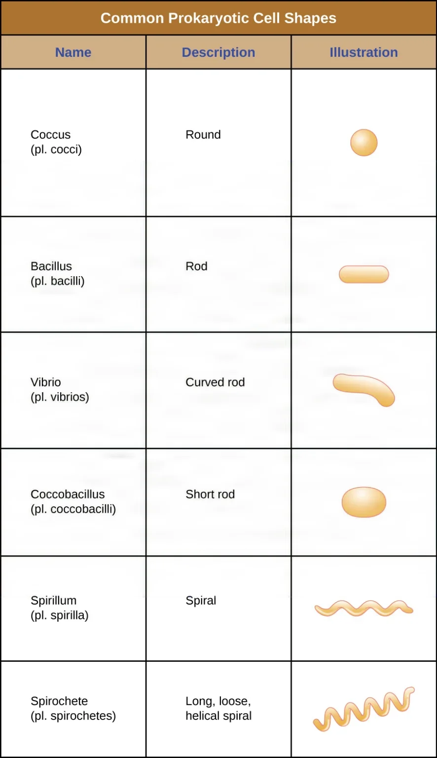
- Cocci (singular, coccus): round cells.
- Bacilli (singular, bacillus): rod-shaped cells. Short rods are called coccobacilli.
- Spiral forms: a vibrio is a curved, comma-shaped rod; a spirillum is a spiral; a spirochete is a long, loose, flexible spiral, such as the germ of syphilis.
Cells of one species often stay together after they divide, in a pattern that helps identify them.
- Diplococci: pairs, such as Neisseria meningitidis, a cause of meningitis.
- Streptococci: chains, like beads on a string; the name comes from the Greek for a twisted chain.
- Staphylococci: clusters, like bunches of grapes.
- Other arrangements include squares of four (tetrads) and chains of rods (streptobacilli).
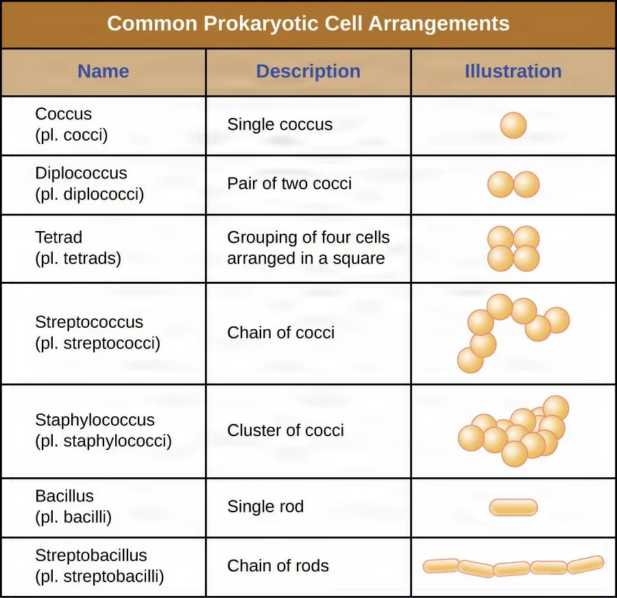
The name often tells you the shape: Staphylococcus means grapelike clusters of cocci, Streptococcus chains of cocci, and Bacillus a rod. A lab report of "gram-positive cocci in clusters" points to staphylococci.
What bacteria need to grow
- Oxygen. Obligate (strict) aerobes, such as Mycobacterium tuberculosis, cannot grow without oxygen. Obligate (strict) anaerobes are killed by it. Facultative anaerobes grow with or without oxygen; E. coli and its relatives, the enteric bacteria, are facultative. Microaerophiles need a little oxygen, far less than air holds.
- Anaerobes and wounds. Strict anaerobes thrive in the bowel and in dead tissue with no blood supply (tissue death is called necrosis). That is why deep puncture wounds and dead tissue breed tetanus and gas gangrene.
- Temperature. The bacteria that infect people are mesophiles: they grow best at moderate temperatures, including body temperature, 37°C (98.6°F). Cold slows them, and high heat kills them.
- pH. Most bacteria grow best near neutral pH, about 7, and stomach acid kills most of the germs you swallow. A few, such as Helicobacter pylori, survive in the stomach.
- Moisture. Most bacteria need water, and drying kills many of them. Some resist drying, such as the mycobacteria of tuberculosis, which survive in dried sputum.
- Nutrients. Bacteria absorb food from their surroundings. They need carbon, oxygen, nitrogen, hydrogen, phosphorus, sulfur and potassium. Dead tissue and foreign material in a wound help even a few bacteria grow into an infection.
How bacteria multiply
Bacteria reproduce by binary fission: the cell copies its DNA, grows, and splits into two identical daughter cells. The time a population takes to double, its generation time, can be as short as 20 minutes for E. coli in the lab, while other bacteria need days. Because the number doubles again and again, a few cells can become millions within hours.
In a closed culture, growth runs through four phases:
- Lag phase: the cells adjust and get ready but do not divide yet.
- Log (exponential) phase: fast, steady doubling.
- Stationary phase: food runs short and waste builds up, so growth levels off.
- Death phase: more cells die than divide, and the colony dies out.
Endospores
A few bacteria, mainly of the genera Bacillus and Clostridium, can form an endospore, usually just called a spore, when conditions turn bad. The cell seals its DNA inside a thick, dried-out coat and goes dormant: no growth and no metabolism.
A spore survives boiling, drying, freezing, radiation and many chemicals, some for decades or longer. When conditions improve, it germinates and becomes an active cell again. A spore is a resting form for survival, and it does not grow or divide.
The spore-formers that matter in medicine include Bacillus anthracis (anthrax), Clostridium tetani (tetanus), C. perfringens (gas gangrene), C. botulinum (botulism) and Clostridioides difficile (C. diff).
Spores are the reason sterile means free of all living microorganisms, spores included, and the reason sterilizers are tested with spores: a process that kills the toughest spores has killed everything else that lives. Only prions resist more. Chapter 27 covers these tests.
Biofilms
Most bacteria do not drift alone. They stick to a surface and wrap themselves in a slimy matrix they make, forming a biofilm, an organized community that often holds several species. The plaque on teeth is a biofilm.
Biofilms form on catheters, implants, artificial joints and contact lenses, and on instruments that are not cleaned well. Germs deep in a biofilm resist antibiotics, disinfectants and the body's defenses far better than free-floating germs, and a biofilm resists routine cleaning.
That is one reason instruments are cleaned at the point of use before blood and debris can dry on them, as chapters 27 and 30 explain.
How bacteria cause disease
Pathogenicity is a germ's ability to cause disease. Virulence is how strongly it can, a scale that runs from harmless to deadly.
To cause disease, a pathogen must get in (exposure), hold on (adhesion), spread into the tissue (invasion) and multiply there (infection).
- Adhesion: fimbriae, capsules and biofilm let germs cling to tissue and devices.
- Invasion: enzymes break down tissue so the germ can spread; Clostridium perfringens, for example, digests the collagen that holds tissue together.
- Escaping defenses: a capsule keeps white blood cells from swallowing the germ, and some bacteria kill white blood cells outright.
- Toxins: poisons that damage the host.
| Exotoxin | Endotoxin | |
|---|---|---|
| What it is | A protein the living bacterium releases | Part of the gram-negative outer membrane: the lipid A of lipopolysaccharide |
| Made by | Mostly gram-positive bacteria, some gram-negative | Gram-negative bacteria only |
| When it is released | While the bacterium lives and grows | Mainly when the cell dies and breaks apart |
| What it does | Attacks particular cells; very powerful, even in tiny amounts | Fever and inflammation; large amounts cause a dangerous drop in blood pressure (septic shock) and organ failure |
| Heat | Most are destroyed by heat | Survives heat |
| Examples | The toxins of tetanus, botulism, diphtheria and cholera | Any gram-negative germ, such as E. coli or Salmonella |
The dose matters too. The number of germs on a surface or an item is called its bioburden, and the number needed to start an infection depends on the germ: as few as 10 to 200 cells of some, a million or more of others. For a surgical wound, the risk of infection rises sharply once there are more than 100,000 germs in each gram of tissue, but far fewer can do it when dead tissue or foreign material such as suture or an implant is present.
Resistance to antibiotics
When an antibiotic kills most of a germ population, the few that happen to resist it survive and multiply, and they can hand their resistance genes to other bacteria on plasmids. Using antibiotics when they are not needed, or stopping them too early, speeds this up.
A multidrug-resistant organism (MDRO) resists several antibiotics, and some resist nearly all of them. An infection with an MDRO looks like any other infection, but it is harder to treat, keeps the patient in the hospital longer and is more likely to kill.
Hospitals must run antibiotic stewardship programs to use antibiotics wisely. The surgical team supports this by giving preventive antibiotics only when they are ordered and at the right time (chapter 12).
9Viruses
A virus is not a cell. It is a tiny particle, far smaller than a bacterium, that can reproduce only inside a living host cell. Outside a cell it is inactive.
- Structure: a core of genetic material, either DNA or RNA but never both, inside a protein coat called a capsid, which is built from subunits called capsomeres. Some viruses also wear an envelope, a piece of membrane taken from the host cell as the virus buds out, often studded with spikes that latch on to new cells. A complete virus particle is a virion.
- Size: about 20 to 900 nanometers, too small for a light microscope, so viruses are seen with an electron microscope.
- Shape: helical (rod-shaped), polyhedral (many-sided, often a 20-sided shape) or complex.
- Classification: by their genetic material (DNA or RNA, single- or double-stranded), whether they have an envelope, their shape and how they replicate. Herpesviruses, for example, are double-stranded DNA viruses with an envelope, and HIV is an enveloped virus with single-stranded RNA.
- Hosts: every kind of living thing has its viruses, but each virus infects only certain species and certain cells. Viruses that infect bacteria are called bacteriophages, or phages.
How viruses replicate
A virus takes over the cell's machinery to copy itself, in steps:
- Attachment: the virus binds to receptors on the host cell.
- Penetration: it gets inside. A phage injects its genes; animal viruses enter by endocytosis or by fusing with the cell membrane.
- Uncoating: the capsid comes off and frees the viral genes.
- Biosynthesis: the cell is made to build viral genes and proteins.
- Maturation: new virions are assembled.
- Release: the new virions burst out, killing the cell, or bud off through its membrane.
Phages show two patterns, which the Core Curriculum names.
- Lytic cycle: the phage replicates at once and bursts (lyses) the cell, killing it.
- Lysogenic cycle: the phage's DNA joins the bacterium's chromosome and is copied along with it every time the cell divides. Stress, such as ultraviolet light or toxic chemicals, can later switch it into the lytic cycle.
Animal viruses can hide in a similar way, called latency. Herpes simplex virus, varicella-zoster virus (chickenpox, which can come back years later as shingles) and Epstein-Barr virus lie dormant in the body for years and can reactivate when the person is stressed or immunosuppressed. Others, such as HIV and hepatitis C virus, cause chronic infections that go on making virus for years. HIV is a retrovirus: it carries an enzyme, reverse transcriptase, that copies its RNA into DNA.
Phages are being tested as a treatment that kills harmful bacteria.
Viruses and cancer
Some viruses can change a cell's genes so that it grows out of control. Human papillomavirus (HPV) is linked to cervical, anal, and head and neck cancers; hepatitis B and C viruses to liver cancer; Epstein-Barr virus to Burkitt lymphoma; and human herpesvirus 8 to Kaposi sarcoma, a cancer common in AIDS.
Why viruses matter in the OR
Antibiotics do not kill viruses. Some can be treated with antiviral drugs, and many can be prevented with vaccines. Viruses spread through the air, in food or water, and in blood and body fluids, and some are carried by insects. Viruses with an envelope, such as HIV and herpes, are among the easiest germs to kill with a disinfectant, while small viruses without one, such as poliovirus, are tougher. At the field, the viruses that threaten you most are the bloodborne ones: hepatitis B, hepatitis C and HIV.
Viroids
A viroid is simpler still: a short, circular strand of RNA with no protein coat. The viroids found so far cause diseases of crop plants, such as potatoes and tomatoes.
The hepatitis D virus works in a related way. It is incomplete and can multiply only in a person who also has hepatitis B.
10Fungi, protozoa and algae
These three groups are all eukaryotes.
Fungi
Fungi include molds, yeasts and mushrooms. They are neither plants nor animals: they absorb their nutrients from living or dead material, and many break down dead matter. Of more than 70,000 species, only about 300 cause disease.
- Yeasts are single cells that reproduce by budding. Molds grow as long filaments called hyphae, which tangle into a mat called a mycelium.
- Labs identify a fungus by growing it and looking at its colony's shape, pattern and color, then staining its cells for the microscope.
- Dimorphic fungi switch forms, growing as a mold at room temperature and as a yeast at body temperature; Histoplasma and Candida are examples.
- Fungal cell walls contain chitin, and their membranes hold ergosterol in place of cholesterol, a difference many antifungal drugs aim at.
- Fungi reproduce sexually or asexually, mostly by making spores. Fungal spores resist heat, cold and drying and float in the air; in a hospital, fungi can grow in the air ducts and release spores.
- A fungal infection is a mycosis. Superficial (cutaneous) mycoses affect the skin, hair and nails, such as ringworm and athlete's foot, and pass by contact. Deep (systemic) mycoses start in the lungs or through broken skin and can be fatal; they strike mostly people with weak immune systems.
Protozoa
Protozoa are single-celled eukaryotes that move about and feed like tiny animals. Many live free in fresh water and the sea; others are parasites.
- They move with cilia (many tiny hairs), flagella (whiplike tails) or pseudopods ("false feet," flowing extensions of the cell, as in an amoeba).
- Many have a feeding stage, the trophozoite, and some can seal themselves inside a tough cyst to survive outside a host.
- Some, such as amoebas, have two layers of cytoplasm: an outer gel, the ectoplasm, and an inner fluid, the endoplasm.
- They usually enter through an insect bite, in food or water, or by sexual contact, and cause diseases such as malaria (carried by mosquitoes), giardiasis and cryptosporidiosis (from water), toxoplasmosis and trichomoniasis.
Algae
Algae are protists that make their own food by photosynthesis. They range from single cells to large seaweeds, make much of the oxygen in the world's waters, and are the source of agar, the gel used in culture media. Algae seldom cause human disease, but some make toxins. In a red tide, for example, shellfish that feed on toxic algae can poison the people who eat them.
11Prions
A prion is an infectious protein: a normal brain protein folded into the wrong shape. It has no DNA or RNA. When it meets normal copies of the protein, it makes them misfold too, and the damage spreads through the brain.
Prions cause rare brain diseases called transmissible spongiform encephalopathies, because the brain fills with spongelike holes. The main one in humans is Creutzfeldt-Jakob disease (CJD); a variant form (vCJD) is linked to mad cow disease in cattle. CJD causes rapidly worsening dementia and is always fatal.
Most cases of CJD appear for no known reason or run in families. A few have been spread by medical care: contaminated brain instruments and electrodes, transplanted corneas and dura mater, and hormone extracts. The incubation period can last decades.
Prions gather in the brain, spinal cord and eye. The immune system does not react to them, they cannot be grown in a lab, and they resist the disinfection and sterilization that kill every other germ. A patient with known or suspected CJD therefore needs special handling of instruments, with single-use instruments whenever possible; chapters 27 and 30 give the steps.
12The germs that matter in surgery
The Core Curriculum lists the causative agents a surgical technologist should know. This section starts with the ones the OR worries about most, then gives the rest in short tables.
Where surgical site infections come from
Most surgical site infections come from the patient's own flora on the skin, in the mucous membranes or in a hollow organ such as the bowel. They are usually gram-positive cocci, especially staphylococci. When the bowel is opened or the incision is near the groin, bowel flora join in: gram-negative rods such as E. coli, enterococci, and anaerobes such as Bacteroides fragilis. Germs can also come from outside the patient: the surgical team, the room's air, and the instruments and supplies.
The staphylococci
- *Staphylococcus aureus is the most common cause of surgical site infections. Up to one person in three carries it on the skin or in the nose without being sick. In a wound it makes thick pus, a suppurative* infection, and abscesses, and it can spread to the heart valves (endocarditis), to bone (osteomyelitis) and into the blood. Its toxins also cause food poisoning and toxic shock syndrome.
- *Staphylococcus epidermidis* is normal skin flora and an opportunist. It grows as a biofilm on catheters, heart valves and orthopedic implants and is one of the most frequent causes of healthcare-associated infection.
- MRSA (methicillin-resistant *S. aureus*) resists methicillin and the antibiotics related to it. It spreads mainly by contact with hands, equipment and supplies, and healthcare workers can carry it in the nose without being ill. It infects incisions, burns and devices such as catheters and chest tubes, and it now spreads in the community too. About 2 people in 100 carry it. Many hospitals screen surgical patients for it and treat carriers before surgery to clear it.
- MRSE (methicillin-resistant *S. epidermidis*) is the same problem in S. epidermidis and a cause of wound infection.
- VRSA and VISA are strains of S. aureus that resist vancomycin, the drug long used for MRSA, fully (VRSA) or partly (VISA). Other antibiotics can treat them, but they can be fatal in very sick patients.
The streptococci and enterococci
- *Streptococcus pyogenes* (group A strep) causes strep throat, scarlet fever and impetigo. In a wound it can cause a fast-spreading infection, including necrotizing fasciitis, the "flesh-eating" infection. Streptococcal wound infections have been traced to team members who carried the germ.
- *Streptococcus pneumoniae* is a leading cause of pneumonia and middle ear infection (otitis media), and also of meningitis. A thick capsule protects it from white blood cells, and a vaccine covers some strains.
- *Streptococcus agalactiae* (group B strep) lives normally in the vagina and can infect a baby during birth.
- *Streptococcus mutans* causes tooth decay.
- *Enterococcus lives in the bowel and the female genital tract. It causes urinary tract infections, wound and abdominal infections and endocarditis. VRE (vancomycin-resistant enterococci)* spreads by contact with stool, urine or blood, with equipment and furniture, and on the hands of staff, and it strikes patients who are very ill, have had surgery or have already been treated with vancomycin.
Gram-negative rods
- *Escherichia coli* is normal bowel flora. It is the most common cause of urinary tract infections, and when the bowel is perforated or opened it can infect the abdomen and the wound. In the bloodstream it can spread to other organs and kill. Some strains, such as O157:H7, cause bloody diarrhea when eaten in contaminated food.
- *Klebsiella pneumoniae* causes pneumonia and healthcare-associated infections. Some strains are carbapenem-resistant, part of a group called CRE (carbapenem-resistant Enterobacterales) that resists even last-line antibiotics.
- *Pseudomonas aeruginosa* lives in water, soil and sewage and in the gut. It is a dangerous hospital germ, above all for burn patients and anyone who has lost the skin's barrier. It forms biofilms, resists many antibiotics and some disinfectants, and can cause pneumonia, wound and urinary infections, bone infection and sepsis.
- *Acinetobacter* causes wound and body-wide infections, mostly in patients with weak immune systems, and many strains are multidrug-resistant.
- *Bacteroides fragilis* is an anaerobe of the normal bowel flora. When the bowel is opened during surgery, it is one of the typical causes of wound infection, and infections that start in the colon can spread into abdominal abscesses and peritonitis.
The clostridia
All clostridia are spore-forming, rod-shaped anaerobes.
- *Clostridioides difficile (C. diff, formerly Clostridium difficile*) causes severe diarrhea and colitis. Antibiotics upset the normal bowel flora and let it take over, so recent antibiotic use is the main risk. Its spores survive on surfaces and are not killed by alcohol hand rubs, so caregivers wash with soap and water, the patient is on contact precautions, and the room is cleaned with a spore-killing product (chapter 30).
- *Clostridium perfringens causes gas gangrene* in deep wounds cut off from oxygen, such as deep, penetrating combat wounds: its toxins and enzymes destroy tissue fast, and gas forms in the dead muscle. It also causes food poisoning.
- *Clostridium tetani lives in soil and in human and animal intestines. It enters through a puncture or dirty wound, and its toxin travels along the nerves, causing the severe muscle spasms of tetanus*. A toxoid vaccine prevents it.
- *Clostridium botulinum makes a toxin so strong that a tiny amount can kill. It blocks the nerve signals to muscles, causing the limp paralysis of botulism*.
Mycobacteria
- *Mycobacterium tuberculosis causes tuberculosis (TB)*. It is an acid-fast rod with a waxy wall that resists drying and disinfectants. It spreads through the air when a person with active TB of the lungs or larynx coughs out tiny droplet nuclei, and as few as about 10 germs can start an infection. Contaminated respiratory or anesthesia equipment can spread it too. In the lungs, germs the body cannot kill are walled off in small nodules (tubercles), where they can stay dormant and become active again if the immune system weakens.
- MDR-TB (multidrug-resistant TB) resists the two main TB drugs, isoniazid and rifampin. The rare XDR-TB (extensively drug-resistant TB) also resists key second-line drugs and is especially deadly for people with HIV. Patients with active TB of any kind need airborne precautions.
- *Mycobacterium leprae* causes Hansen's disease (leprosy), a chronic infection of the peripheral nerves.
The bloodborne viruses
Bloodborne pathogens are germs carried in the blood that can cause disease in people. Three viruses carry most of the risk for the surgical team: HIV (human immunodeficiency virus), and the viruses of hepatitis B and hepatitis C (HBV and HCV). They travel in blood and in other body fluids: semen, vaginal secretions, and cerebrospinal, synovial, pleural, peritoneal and amniotic fluid, as well as any fluid with blood in it. At the field they pass through a needlestick or other sharps injury, or a splash onto a mucous membrane such as the eye or mouth.
- Hepatitis B is the most infectious of the bloodborne diseases. The virus can survive on a surface for up to 7 days and is about 100 times more infectious than HIV. It can cause chronic hepatitis, cirrhosis, liver cancer and death. For a person with no immunity, a needlestick from an infected patient carries a risk of infection of roughly 23 to 62 percent, depending on how much virus the patient's blood holds. A safe, effective vaccine exists, and a vaccinated person who has developed immunity has no risk.
- Hepatitis C spreads mainly through blood, for example by shared needles. Many infected people develop chronic hepatitis and cirrhosis, it causes liver cancer, and it is a leading reason for liver transplants. There is no vaccine, but most infections can now be cured with antiviral drugs. The risk of infection after a needlestick from an infected patient is about 0.2 percent, by the CDC's 2020 estimate; older references give about 1.8 percent.
- HIV destroys the immune system's helper T cells. A person can carry and spread it for years without symptoms. Untreated, it leads to AIDS (acquired immunodeficiency syndrome), diagnosed when opportunistic infections or certain cancers appear. There is no cure, but drugs can control it. Casual contact does not spread it, and the risk after a needlestick from an infected patient is about 0.3 percent, or 1 in 300.
hepatitis B is the bloodborne virus most likely to infect you after a needlestick, and the only one of the three with a vaccine. Employers must offer it free to workers exposed to blood, so if you are not immune, ask about it before you start clinical work.
The rest of the bacteria on the list
| Bacterium | What it causes |
|---|---|
| Bacillus anthracis | Anthrax. A spore-former whose spores can be breathed in, swallowed or enter through the skin, and a possible bioterrorism agent |
| Bartonella | Cat-scratch disease, from the bite or scratch of a cat, and trench fever, carried by lice and fleas |
| Bordetella pertussis | Whooping cough (pertussis), a life-threatening disease in children; prevented by vaccine; spread by droplets |
| Campylobacter jejuni | Diarrhea (gastroenteritis); can set off Guillain-Barré syndrome |
| Chlamydia trachomatis | Chlamydia, a sexually transmitted infection; can infect a newborn's eyes during birth |
| Corynebacterium diphtheriae | Diphtheria, from a toxin that damages the throat, heart and nerves; prevented by a toxoid vaccine |
| Gardnerella vaginalis | Bacterial vaginosis |
| Haemophilus influenzae | Meningitis, pneumonia and bloodstream infection. Despite its name, it does not cause the flu, which is a virus |
| Helicobacter pylori | Chronic gastritis and stomach ulcers; survives stomach acid |
| Lactobacillus | Normal flora, the main bacteria of the vagina, whose lactic acid keeps yeast in check |
| Legionella pneumophila | Legionnaires' disease, a dangerous pneumonia caught from contaminated water systems such as faucets, cooling towers and fountains |
| Listeria monocytogenes | Listeriosis, from food; can cross the placenta and harm the fetus |
| Moraxella catarrhalis | Middle ear infection, sinusitis, bronchitis and pneumonia |
| Mycoplasma hominis | Pelvic inflammatory disease and bacterial vaginosis. Mycoplasmas have no cell wall, so the Gram stain does not show them |
| Neisseria gonorrhoeae | Gonorrhea, a sexually transmitted disease; can blind a baby infected at birth, which antibiotic eye drops prevent |
| Neisseria meningitidis | Bacterial meningitis, highly contagious and dangerous; droplet precautions |
| Porphyromonas gingivalis | Gum (periodontal) disease |
| Rickettsia rickettsii | Rocky Mountain spotted fever, carried by ticks |
| Salmonella enterica | Food poisoning (salmonellosis); one type causes typhoid fever |
| Treponema pallidum | Syphilis, a sexually transmitted spirochete that can cross the placenta |
| Vibrio cholerae | Cholera: severe, watery diarrhea from contaminated water |
Fungi, the prion and the protozoan
| Agent | What it causes |
|---|---|
| Blastomyces dermatitidis | Blastomycosis. A dimorphic fungus that lives in soil and is breathed in as spores; usually a mild lung infection, but in people with weak immune systems it can spread, above all to the skin |
| Candida albicans | Candidiasis. A yeast of the normal flora that overgrows after antibiotics or in weak hosts, causing thrush, vaginal yeast infection and skin infection, and sometimes a deep, spreading infection that can be fatal. A related yeast, Candida auris, resists many antifungal drugs and has caused outbreaks in hospitals |
| Coccidioides | Coccidioidomycosis (valley fever), a lung infection from breathing fungal spores from soil; common in California's San Joaquin Valley |
| Histoplasma capsulatum | Histoplasmosis, a lung disease from breathing spores in soil contaminated with bird or bat droppings |
| Pneumocystis jirovecii | Pneumocystis pneumonia (PCP), a leading cause of pneumonia in AIDS and in other patients with weak immune systems |
| The prion of Creutzfeldt-Jakob disease | A fatal brain disease; see Prions, above |
| Trichomonas vaginalis | Trichomoniasis, a sexually transmitted infection caused by a protozoan |
The rest of the viruses on the list
| Virus | What it causes |
|---|---|
| Cytomegalovirus (CMV) | Usually no symptoms, or an illness like mononucleosis; serious in AIDS, in transplant patients and in newborns. Spread by saliva, urine, sex, breastfeeding, blood transfusions and transplanted organs |
| Enteroviruses, including poliovirus | Polio: most often a mild intestinal infection, but in a small share of cases it reaches the nervous system and causes paralysis. Spread mainly by the fecal-oral route; prevented by vaccine |
| Hepatitis A virus (HAV) | Liver infection spread by the fecal-oral route, in food and water; rarely fatal; vaccine available |
| Hepatitis D virus (HDV) | Infects only people who also have hepatitis B, and makes the liver damage worse; bloodborne |
| Hepatitis E virus (HEV) | Acute liver disease from contaminated water or undercooked meat; does not become chronic |
| Herpes simplex viruses 1 and 2 (HSV-1, HSV-2) | HSV-1 usually causes cold sores around the mouth and HSV-2 genital herpes, though HSV-1 can also infect the genitals. Both hide in nerve cells and flare again |
| Epstein-Barr virus (EBV, human herpesvirus 4) | Infectious mononucleosis; stays latent and is linked to Burkitt lymphoma |
| Human papillomavirus (HPV) | Genital warts and cancers of the cervix, the anus, and the head and neck; sexually transmitted; vaccine available |
| Lyssavirus (rabies virus) | Rabies, spread by the bite or saliva of an infected animal; nearly always fatal |
| Morbillivirus (measles virus) | Measles (rubeola): fever, cough, runny nose, red eyes and rash. Very contagious and airborne; prevented by the MMR vaccine |
| Rubella virus | German measles, a mild rash illness; infection during pregnancy can cause congenital rubella syndrome in the baby; vaccine available |
| SARS-CoV-2 | COVID-19, a respiratory illness spread mainly by droplets, and through the air during procedures that make aerosols, such as intubation. People with no symptoms can spread it |
| Varicella-zoster virus | Chickenpox. It hides in nerve ganglia and can return many years later as shingles; airborne; vaccine available |
| Variola virus | Smallpox, once one of history's great killers. Worldwide vaccination wiped it out by 1977, and it is now a bioterrorism concern |
13The body's defenses
The body defends itself in layers, often called three lines of defense. The first two make up innate (nonspecific) immunity: present from birth, working the same way against any invader, and with no memory. The third is acquired (adaptive) immunity, which targets one particular germ and remembers it.
Surgery breaks through the first line, and sometimes more, which is why surgical patients are so open to infection.
The first line: barriers
- Intact skin: a tough outer layer of dead cells packed with keratin, which keep shedding and carry germs away with them. Fatty acids from sweat and oil glands keep the surface dry, salty and acidic. A cut or a burn opens the door.
- Mucous membranes line the nose, mouth, lungs and the urinary and digestive tracts. Their sticky mucus traps germs. In the airway, tiny hairs called cilia sweep the mucus up and out, a system called the mucociliary escalator, and coughing helps clear it.
- Flushing: tears, saliva, urine and the movements of the gut wash germs away.
- Chemicals: enzymes such as lysozyme, in saliva and mucus, break down bacterial walls, and stomach acid kills most swallowed germs.
- Normal flora crowd out invaders.
The second line: inflammation, phagocytes and fever
When germs get past the barriers, the innate defenses inside the body take over.
- Inflammation: injured tissue releases chemicals such as histamine. Blood vessels widen and leak, bringing fluid and white blood cells to the spot. The signs are redness, swelling, heat, pain and loss of function. Chapter 11 teaches inflammation and healing in depth.
- Phagocytes: white blood cells that swallow and digest germs by phagocytosis. Neutrophils arrive first, within about 90 minutes. Macrophages, which grow from monocytes, follow within hours to two days and clear away large numbers of bacteria. Pus is a collection of dead cells, white cells and living and dead germs.
- Fever: substances called pyrogens, including the endotoxin of gram-negative bacteria, reset the brain's thermostat, the hypothalamus, to a higher temperature that helps the body fight infection.
- Other helpers: natural killer cells, another kind of white blood cell, kill body cells taken over by viruses, and defensive proteins in the blood help destroy germs.
The third line: acquired immunity
Acquired immunity is run by two kinds of white blood cells called lymphocytes: B cells and T cells. Both start in the bone marrow; B cells mature there, and T cells mature in the thymus.
An antigen is anything the immune system recognizes as foreign, usually a protein or sugar on the surface of a germ, a grain of pollen or a transplanted organ. An antibody, also called an immunoglobulin, is a Y-shaped protein made to fit one antigen, like a key in a lock.
Humoral (antibody) immunity belongs to the B cells. When a B cell meets its antigen, it multiplies and turns into plasma cells that pour out antibodies. Antibodies neutralize germs and toxins, clump germs together, coat them so phagocytes can eat them, and switch on the defensive proteins in the blood. They work against germs in the blood and other body fluids.
Antibodies come in five classes. The one to know in the OR is IgE, which sits on mast cells and sets off allergic reactions, true latex allergy among them.
Cell-mediated immunity belongs to the T cells. It goes after germs hiding inside the body's own cells, such as viruses. Helper T cells (CD4 cells) direct the whole response, sending chemical signals that switch on B cells and other T cells. Cytotoxic T cells (CD8 cells) find and kill infected and cancerous cells. HIV destroys helper T cells, which is why AIDS cripples both kinds of acquired immunity.
Memory. After a first infection, some B and T cells stay on as memory cells. If the same germ returns, they answer faster and harder and destroy it quickly. That is why you catch some diseases only once, and it is why vaccines work.
Types of acquired immunity
| Natural | Artificial | |
|---|---|---|
| Active: your body makes its own antibodies and memory cells, and protection lasts years or for life | Catching the disease and recovering | A vaccine |
| Passive: you receive ready-made antibodies, which protect at once but only for a while | Antibodies from the mother, through the placenta or breast milk | An injection of antibodies, such as immune globulin, tetanus antitoxin or hepatitis B immune globulin (HBIG) |
Immunization
A vaccine trains the immune system with a harmless form or piece of a germ, so the body makes antibodies and memory cells without going through the disease.
- Live attenuated vaccines use a weakened living germ, as in the measles, mumps and rubella (MMR) vaccine, the varicella vaccine and the oral polio vaccine. They give strong, lasting immunity, but they are not given to people whose immune systems cannot fight even the weakened germ.
- Inactivated vaccines use killed germs, such as the injected polio vaccine and the hepatitis A vaccine. They cannot cause the disease but usually need several doses or boosters.
- Subunit and recombinant vaccines use only key pieces of a germ, often made by recombinant gene technology (genetic engineering), such as the hepatitis B and HPV vaccines.
- Toxoid vaccines use a bacterial toxin that has been made harmless, the toxoid, and protect against diseases caused by toxins, such as tetanus and diphtheria.
Passive immunization gives ready-made antibodies for fast protection after an exposure, such as hepatitis B immune globulin (HBIG), rabies immune globulin or varicella-zoster immune globulin.
Vaccinating healthcare workers protects both the workers and their patients. Facilities follow the CDC's immunization recommendations, and OSHA requires employers to offer the hepatitis B vaccine at no cost to workers exposed to blood.
When the immune system goes wrong
Hypersensitivity is an immune response that causes harm. An allergy is one kind, and the substance that sets it off is an allergen. The first exposure sensitizes the person, and the reaction comes with a later exposure.
| Type | What happens | Examples |
|---|---|---|
| I: immediate (allergic) | IgE on mast cells sets off a release of histamine within minutes | Hives, hay fever, asthma and latex allergy; at its worst, anaphylaxis |
| II: cytotoxic | Antibodies attack cells that carry a foreign antigen | A transfusion reaction from mismatched blood; hemolytic disease of the newborn |
| III: immune complex | Clumps of antigen and antibody settle in vessels and tissues and set off inflammation | Serum sickness; the Arthus reaction at an injection site |
| IV: delayed (cell-mediated) | T cells, not antibodies, cause inflammation hours to days after contact | Contact dermatitis, such as poison ivy or a reaction to the chemicals in rubber gloves; a positive tuberculin (TB) skin test |
Anaphylaxis is a whole-body type I reaction. Histamine and other chemicals widen the blood vessels and make them leak, so the blood pressure falls fast, while the airway swells and the bronchial tubes clamp down. Death can follow within minutes. Under anesthesia the most common signs are in the heart and circulation and in the skin, but a rash is hard to see under the drapes and the patient cannot say what they feel, so watch for falling blood pressure, a racing heart and bronchospasm. The most common causes in surgery are muscle relaxants (neuromuscular blocking drugs), latex and antibiotics.
If it happens, the team acts at once: the cause is stopped, epinephrine is handed to the anesthesia provider or circulator to inject, the IV is opened wide, and the airway is kept open with 100 percent oxygen. The scrub protects the sterile field and may need to pack or close the wound quickly. Chapter 13 covers the emergency in detail.
Autoimmune diseases happen when the immune system loses its tolerance of "self" and attacks the body's own tissue. Some strike one organ, such as type 1 diabetes, Graves disease and Hashimoto thyroiditis. Others are systemic, such as rheumatoid arthritis and systemic lupus erythematosus.
Immunodeficiency means the immune system cannot defend the body properly, so opportunistic infections take hold.
- Primary (inborn) immunodeficiencies are genetic, such as severe combined immunodeficiency (SCID), in which both B and T cells fail.
- Acquired (secondary) immunodeficiencies develop later in life and can often be reversed if the cause is fixed. Causes include HIV and AIDS, diabetes, cancer and its treatment with chemotherapy or radiation, high doses of steroids, severe malnutrition (the most common cause worldwide), and long, critical illness after infection, surgery or trauma.
Latex allergy
Natural rubber latex comes from the sap of the rubber tree. It is found in many medical products: gloves, catheters, drains, tourniquets and Esmarch bandages, blood pressure cuffs, bulb syringes, tubing and syringe plungers. Latex can cause three kinds of reaction.
- Irritant contact dermatitis: dry, red, itchy, cracked skin from gloves, soaps and glove powder. It is the most common reaction, and it is not an allergy.
- Allergic contact dermatitis: a type IV reaction to the chemicals used to make rubber, not to the latex itself. A rash like poison ivy appears hours to days after contact.
- True latex allergy: a type I reaction, through IgE, to the proteins in latex. Within minutes the person may develop hives, itching, nose and eye symptoms, wheezing, swelling of the lips and throat, a racing heart and falling blood pressure, up to anaphylaxis and cardiac arrest. Latex that touches a mucous membrane during surgery is especially dangerous, and latex proteins also ride through the air on glove powder.
Patients at highest risk are those with spina bifida, those who have had many operations (especially as children), those who need repeated urinary catheters, and people with asthma or other allergies, including some food allergies. Healthcare workers are the second-highest-risk group.
Caring for a patient with a latex allergy:
- The allergy is flagged on the chart, on an allergy wristband and on signs on the stretcher and the OR doors.
- The patient is scheduled as the first case of the morning, because latex proteins can hang in a room's air for at least an hour after latex gloves are used.
- Every item is latex-free, often from a latex allergy cart kept for these cases, and anything not marked latex-free is checked before it is opened.
- Everyone on the team wears non-latex gloves, both pairs when double gloving, and latex-free hair covers.
- Watch for a reaction, which can take 15 to 60 minutes to appear when latex touches the skin or a mucous membrane.
To protect yourself, use non-latex gloves or low-allergen, powder-free latex gloves (the FDA banned powdered medical gloves in 2017), avoid oil-based hand lotions under latex gloves because they break the latex down, wash and dry your hands after taking gloves off, and learn the signs of latex allergy. Gloves labeled "hypoallergenic" do not lower the risk of true latex allergy.
14How infection happens
Epidemiology
Epidemiology is the study of how disease starts and spreads through a population, with the aim of preventing outbreaks and controlling them. It includes etiology, the study of what causes a disease.
Frequency of disease is measured in a few ways.
- Morbidity means being ill. The morbidity rate is the number of sick people out of a set number, such as per 100,000 people. Mortality is death from a disease.
- Incidence is the number of new cases in a period of time. Prevalence is the total number of people who have the disease at one point in time.
Patterns of disease
- Sporadic: occasional, scattered cases, such as tetanus.
- Endemic: always present at a steady, often low, level in an area, as malaria is in parts of Brazil.
- Epidemic: many more cases than expected in an area in a short time, as in an unusually bad flu season.
- Pandemic: an epidemic that spreads worldwide, such as HIV/AIDS or COVID-19.
Epidemiological studies look for the causes of disease and test treatments. In an observational study, researchers watch and measure without changing anything, for example comparing people who have a disease with people who do not; such studies show links but cannot prove a cause. In an experimental study, such as a clinical trial, researchers change something, such as giving a drug, and compare the results. The first known epidemiological study was John Snow's tracing of the London cholera outbreaks of 1848 and 1854 to contaminated drinking water.
Public health: in the United States, the main public health agency is the CDC. Some diseases are notifiable (reportable): every case must be reported to public health authorities, who track them across the country.
Healthcare facility epidemiology: every hospital must run an infection prevention and control program. Its infection preventionists watch for healthcare-associated infections such as surgical site infections (surveillance), report them, including to the CDC's National Healthcare Safety Network (NHSN), share the results with the teams, and train staff and check that infection control rules are followed.
Contamination, colonization and infection
- Contamination: germs are present on or in something, such as an instrument, a surface or a wound.
- Colonization: germs live and multiply on or in the body without causing disease, though a colonized person can still pass them on.
- Infection: germs invade body tissue, multiply and cause injury.
- Carrier: a person who harbors a pathogen and can spread it without being sick. A passive carrier, such as a worker with unwashed hands, only carries the germ along; an active carrier is infected. Hepatitis B, herpes and HIV often spread from carriers with no symptoms.
Classifying infectious diseases
- Communicable diseases spread from person to person, and contagious ones spread easily, as measles does. Noncommunicable infections do not spread between people.
- Acute diseases come on fast and run a short course, like the flu. Chronic diseases last months or years, like hepatitis B in some people. In latent diseases the germ goes dormant and can flare again, like herpes and shingles.
- A local infection stays in one small area. In a focal infection, germs or their toxins spread from one site to a second. A systemic infection spreads through the whole body.
- A primary infection is the first one; a secondary infection is a new one that takes hold because the first left the body weakened.
- Iatrogenic diseases result from medical care, such as an infected surgical site. Nosocomial (healthcare-associated) diseases are caught in a healthcare facility. Zoonotic diseases come from animals, as rabies does.
The chain of infection
For an infection to pass from its source to a new person, six links must connect. Break any one link and the infection stops. Every precaution in this chapter breaks at least one.
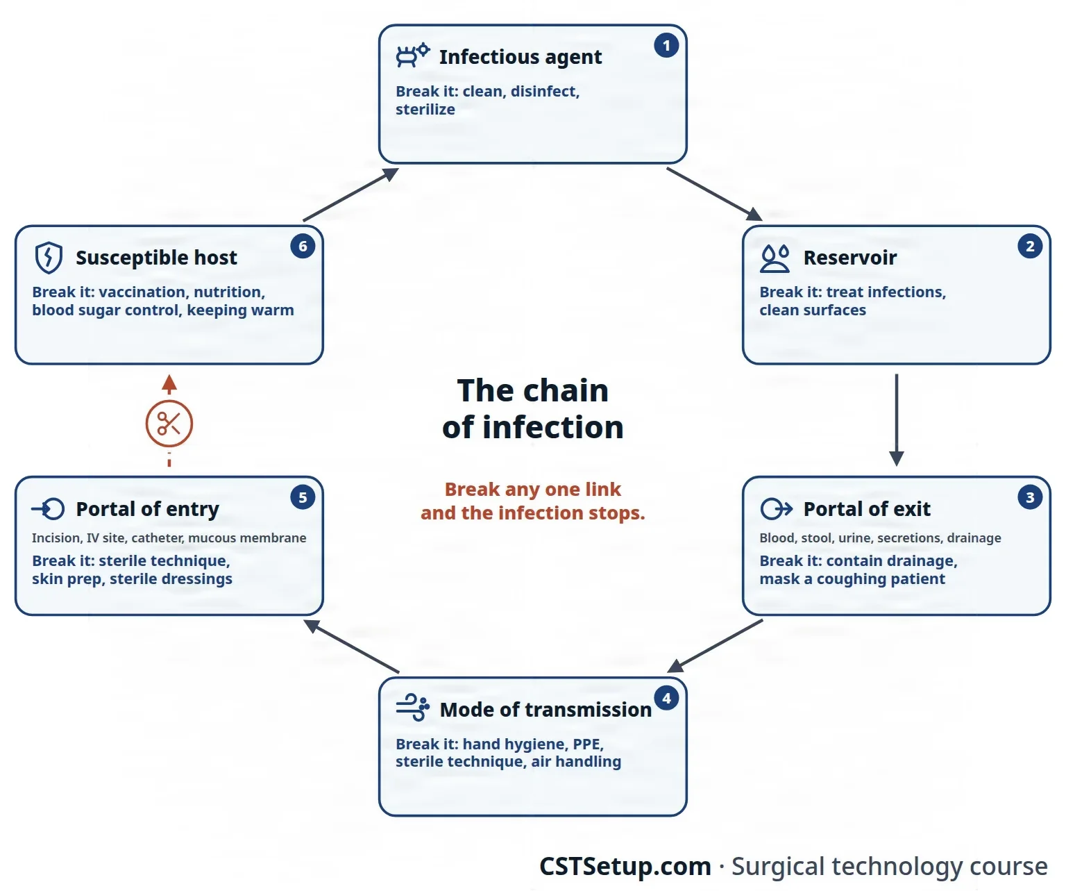
- Infectious agent: the germ. Its kind, its virulence and how many there are decide how likely an infection is. Break it by cleaning, disinfecting and sterilizing.
- Reservoir: where the germ lives and multiplies. Reservoirs can be humans (sick people and carriers), animals, or nonliving places such as soil, water, food and surfaces. S. aureus lives on people, while C. tetani lives in soil. Break it by treating infections and cleaning the environment.
- Portal of exit: how the germ leaves the reservoir, such as in blood, stool, urine, respiratory secretions or wound drainage. Break it by containing drainage and secretions and masking a coughing patient.
- Mode of transmission: how the germ travels to the new host, described in the next section. Break it with hand hygiene, PPE, sterile technique and clean air handling.
- Portal of entry (entry site): how the germ gets in: through broken skin and wounds, mucous membranes, the respiratory, digestive or urinary tract, the placenta, or straight into tissue by injection or incision. In the OR, the incision, IV sites and catheters are all portals of entry. Break it with sterile technique, skin prep and sterile dressings.
- Susceptible host: a person who cannot fight the germ off. Old age, poor nutrition, chronic disease such as diabetes, a weak immune system and recovering from surgery make a host susceptible, while immunity from a vaccine or past infection protects. Break it with vaccination, good nutrition, control of blood sugar and keeping the patient warm.
Modes of transmission
Germs travel from host to host in several ways.
- Direct contact (direct transmission): touching an infected person or their blood or body fluids, kissing or sex. Germs can also reach tissue directly, as when a perforated bowel spills its contents into the abdomen. Passing an infection from parent to baby during pregnancy, birth or breastfeeding is vertical transmission.
- Droplet: large droplets sprayed out by coughing, sneezing, talking, or procedures such as suctioning and intubation land in the eyes, nose or mouth of someone nearby. They are larger than 5 micrometers and fall to the ground within about 3 to 6 feet. Influenza, pertussis and meningococcal disease spread this way.
- Indirect contact: a contaminated object, a fomite, carries the germ, such as an instrument, a catheter, bed linen, a dressing or a doorknob. Unwashed hands carry germs the same way. Fomites are a major cause of healthcare-associated infections.
- Vehicle: contaminated food or water carries the germ.
- Airborne: tiny droplet nuclei, also called aerosol droplets (the dried remains of droplets, 5 micrometers or smaller), or dust carrying germs hang in the air for a long time and travel far on air currents. Tuberculosis, measles and chickenpox spread this way, and procedures such as intubation can make other germs airborne too.
- Vector: a living carrier, usually an insect. A mechanical vector, such as a fly, carries germs on its body; a biological vector, such as a mosquito or tick, is itself infected and passes the germ on through a bite.
What decides whether infection happens
Meeting a pathogen does not always mean infection. The risk depends on a balance: the dose of germs times their virulence, weighed against the resistance of the host.
- The germ: how many there are, how virulent they are, and whether they resist antibiotics.
- Its surroundings: whether the germ finds the moisture, food, temperature and oxygen it needs. Dead tissue, blood clots, foreign material and poor blood flow all help germs grow.
- The patient: older age, poor nutrition, diabetes, obesity, smoking, alcohol or drug misuse, a weak immune system from disease, chemotherapy or steroids, an infection elsewhere in the body, and a long hospital stay before surgery.
- The operation: a long, complex procedure, a contaminated wound, tissue damaged by rough handling, a low body temperature, low oxygen, blood transfusion, poor hair removal or skin prep, preventive antibiotics given at the wrong time, breaks in sterile technique, and heavy traffic or poor air flow in the room.
The team works on these factors on every case: strict sterile technique, gentle tissue handling, keeping the patient warm, clipping hair instead of shaving it, antibiotics on time, little traffic in the room, and careful cleaning and sterilization. A patient whose defenses are strong heals better, and chapter 11 shows how.
The course of an infectious disease
An infectious disease follows a typical course of five periods.
- Incubation period: from the time the germ enters until symptoms begin. The germ is multiplying, but there are too few to cause signs. It can last a day or two, or months to years; for hepatitis B it is about 10 to 12 weeks.
- Prodromal period: vague, general symptoms appear, such as tiredness, a mild fever and aches.
- Period of illness (the acute phase): the germ is at its strongest, and the signs and symptoms are at their worst.
- Period of decline: the number of germs falls and symptoms ease, but the weakened patient is open to a secondary infection.
- Period of convalescence: the body recovers and returns to normal, though some diseases leave lasting damage.
A person can spread an infection in any of these periods, sometimes before feeling sick and after feeling well again.
Three words describe what the disease looks like.
- A sign is objective: something that can be seen or measured, such as a fever, a rash, a fast pulse or a lab result.
- A symptom is subjective: something only the patient feels, such as pain, nausea or tiredness.
- A syndrome is a group of signs and symptoms that occur together in a particular disease, such as AIDS.
Healthcare-associated infections
A healthcare-associated infection (HAI) is one a patient catches while being cared for in a healthcare facility; the older name is nosocomial infection. About 1 hospital patient in 25 gets one.
The most common are tied to devices and procedures: urinary tract infections from catheters, bloodstream infections from central lines, pneumonia in patients on ventilators, and surgical site infections. Many are caused by bacteria that resist at least one antibiotic.
A surgical site infection is an infection of the incision, or of the organ or space that was operated on; chapter 11 teaches its types, signs, time limits and treatment.
Spread of germs from one person or object to another is cross-contamination. Carrying a patient's own germs from one part of the body to another is self-infection (autoinfection). Germs that come from the patient are endogenous; germs from outside the patient, such as the team, the air and the instruments, are exogenous.
The single most important way to stop the spread of infection in healthcare is hand hygiene.
15Standard Precautions
From universal precautions to Standard Precautions
In 1987, as the risk of HIV and hepatitis B to healthcare workers became clear, the CDC introduced universal precautions: treat the blood and certain body fluids of every patient as if they were infected, whether or not the patient is known to carry a disease. A second system, body substance isolation, applied the same idea to all moist body substances. In 1996 the CDC combined the two into Standard Precautions, used with every patient, all the time, in every place care is given.
The idea is simple: any patient may carry a germ nobody knows about, so the same precautions apply to everyone. Standard Precautions cover blood; every body fluid, secretion and excretion except sweat, whether or not it contains visible blood; broken skin; and mucous membranes.
OSHA's Bloodborne Pathogens Standard still uses the term universal precautions, and it accepts Standard Precautions in their place as long as every other part of the standard is met. You will hear both terms.
What Standard Precautions include
Standard Precautions are a set of practices used together.
- Hand hygiene before and after every patient, after touching blood, body fluids or contaminated items, and right after taking off gloves. Gloves never replace it. An alcohol hand rub is the routine choice; wash with soap and water when your hands are visibly soiled or after caring for a patient with C. difficile, whose spores alcohol does not kill. A medical hand wash takes at least 20 seconds of rubbing, and chapter 28 gives the steps.
CDC's 2002 hand hygiene guideline, which some references still repeat, calls for rubbing for at least 15 seconds.
- Personal protective equipment (PPE) chosen for the exposure you expect: gloves to touch blood, body fluids, mucous membranes or broken skin; a fluid-resistant gown when your arms or clothes could be soiled; and a mask with eye protection, or a face shield, when splashes, sprays or droplets could occur.
- Respiratory hygiene and cough etiquette: anyone who is coughing covers the mouth and nose, throws tissues away at once, cleans their hands and wears a mask if they can.
- Safe injection practices: a sterile, single-use needle and syringe for every injection, single-dose vials whenever possible, and never one syringe or IV bag shared among patients.
- Masks for spinal procedures: a face mask for anyone injecting into the spinal or epidural space, as in spinal anesthesia, or placing a catheter there.
- Sharps safety: never recap a used needle by hand, never bend or break needles, and drop used sharps into a puncture-resistant container close to where they were used.
- Patient care equipment: reusable equipment is cleaned and disinfected or sterilized before it is used on the next patient, and heavily soiled items are contained.
- The environment and linen: surfaces are cleaned and disinfected routinely, and soiled linen is handled with gloves, held away from your body and bagged.
In the OR, Standard Precautions apply to every case. Every patient's blood is treated as infectious, so you wear PPE at the field, at breakdown and while cleaning, on every case.
PPE as a barrier
OSHA requires the employer to provide PPE at no cost, and it counts as protection only if blood and other infectious material cannot pass through it to your clothes, skin, eyes, mouth or other mucous membranes. Chapter 28 covers which items you wear and how to put them on and take them off; here is what the rules require.
- Gloves whenever your hands may touch blood or body fluids. Disposable gloves are changed as soon as they are contaminated and are never washed and reused. The scrubbed team double gloves for surgery: a second pair greatly cuts the number of holes in the inner glove, and a colored inner glove (a perforation indicator system) makes a puncture easy to see.
- Gowns whenever your arms or clothes could be soiled. A sterile gown's fluid barrier is rated from level 1 (minimal) to level 4 (high), and the level is matched to the blood and fluid expected.
- Eye protection and a mask whenever splashes, sprays, spatter or droplets of blood may occur, which in surgery means every case. AST's first choice is goggles that are indirectly vented, coated against fogging and snug across the brow. Prescription glasses do not protect, even with side pieces, so wear goggles over them. A face shield covers more of the face, but it is backup protection: the mask and goggles are still worn under it.
Other references still count glasses with solid side shields, or a mask with an attached eye shield, as eye protection, as the Core Curriculum's list does; AST's 2017 guideline treats neither as the best protection.
- Respirators whenever protection from airborne germs is needed: a NIOSH-approved, fit-tested N95 respirator or better, used under the facility's respiratory protection program. A surgical mask is not a respirator, because air leaks in around its edges. An N95 works only if it seals to the face, so you are fit-tested before first use and every year after, you check the seal each time you put one on, and no facial hair may lie under the seal.
- Fluid-resistant hoods, and shoe covers or boots, whenever heavy contamination is expected, as in orthopedic surgery.
- Taking PPE off: remove it as soon as possible after it is soiled, and before you leave the work area, put it in its proper container, and clean your hands.
Surgical smoke from electrosurgery and lasers can carry viruses and fragments of blood; chapter 20 covers smoke evacuation and the respirator worn with it.
16Transmission-based precautions
For a patient known or suspected to carry certain germs, transmission-based precautions are added on top of Standard Precautions. They are chosen by how the germ spreads, and one patient may need more than one kind.
| Precaution | Used for | What it adds |
|---|---|---|
| Contact | Germs spread by touching the patient or the things around them: MRSA, VRE and other MDROs, C. difficile, draining wounds and abscesses, herpes simplex, impetigo | A gown and gloves for any contact with the patient or the room, put on when entering and taken off before leaving; equipment kept for that patient or cleaned before reuse; a private room |
| Droplet | Germs in large respiratory droplets: influenza, pertussis, Neisseria meningitidis, Haemophilus influenzae type b, group A strep (for the first 24 hours of antibiotics), rubella | A mask, usually put on when entering the room; a private room, or at least 3 feet between patients; the patient wears a mask when moved |
| Airborne | Germs that float in the air: tuberculosis, measles, and chickenpox or widespread shingles | A private airborne infection isolation room at negative pressure, door closed; a fit-tested N95 respirator put on before entering; the patient wears a mask when moved; staff who are not immune kept out of measles and chickenpox rooms |
One reference still teaches a mask only within 3 feet of a droplet patient, and respiratory protection only within 3 feet of an airborne patient; current CDC guidance puts the mask on at the door of a droplet room and the N95 on before entering an airborne room.
When an isolation patient needs surgery
Patients with highly contagious infections spread by droplets almost never come to the OR unless the surgery cannot wait.
The OR is kept at positive pressure to protect the sterile field, so a patient on airborne precautions, such as one with active TB, needs extra steps.
- The patient wears a surgical mask and comes straight to the OR.
- The case is done near the end of the day if possible, with as few people as possible and little traffic in and out.
- The team wears fit-tested N95 respirators.
- A portable HEPA filter unit sits near the patient's head during intubation, or the patient is intubated in a negative-pressure room first.
- The patient recovers in a negative-pressure room.
For a patient on contact precautions, the infected area is covered or contained for transport, and the team wears gowns and gloves. After the case the room is cleaned and disinfected, with a spore-killing product after a C. difficile case (chapter 30).
17The OSHA Bloodborne Pathogens Standard
OSHA's Bloodborne Pathogens Standard (29 CFR 1910.1030), issued in 1991, protects workers who may come into contact with blood or other potentially infectious materials. The Needlestick Safety and Prevention Act of 2000 added the duty to use safer needles and sharps devices. Under the standard, employers must:
- Write an exposure control plan, review and update it every year, and involve front-line workers in choosing safer devices.
- Use engineering controls that remove the hazard, such as safety needles, blunt suture needles, needleless IV connectors and sharps containers, and work practice controls, such as the neutral zone and never recapping a needle by hand.
- Provide PPE at no cost and make sure workers use it.
- Offer the hepatitis B vaccine at no cost to exposed workers. A worker who declines signs a declination form and can change their mind later.
- Provide a confidential medical evaluation and follow-up right away after any exposure.
- Train every exposed worker when hired and at least once a year after that.
- Label hazards with fluorescent orange or orange-red biohazard labels on containers of regulated waste and on refrigerators and containers that hold blood; red bags can be used instead of labels.
- Keep records, including a sharps injury log that protects the injured worker's privacy.
- Keep food and drink out of work areas where blood or other infectious material may be present.
Regulated medical waste goes into red bags or labeled containers, and sharps into puncture-resistant containers; chapter 30 covers waste and spills.
Sharps safety
A sharp is any object that can pierce the skin. Sharps injuries are the most common way bloodborne pathogens reach healthcare workers. They have been linked to more than 60 different germs, though hepatitis B, hepatitis C and HIV are the main concern.
The sharps at the field include suture and hypodermic needles, scalpel blades, needle-point electrosurgery tips, trocars, sharp instruments such as skin hooks and scissors, guide wires, and orthopedic drill bits, pins, wires and saw blades.
The risky moments are passing and receiving a scalpel, loading and passing sutures, two people reaching for the same sharp, putting a blade on or taking it off, holding tissue back by hand, and suturing. Fatigue, rushing, distraction and noise all raise the risk.
You defend against sharps injuries in a few ways.
- Use safety devices and blunt suture needles where you can.
- Never recap a used needle by hand; if recapping cannot be avoided, use a one-handed scoop or a recapping device. Never bend, break or remove a needle by hand.
- Pass sharps hands-free through a neutral zone, load blades with an instrument and remove them with a single-handed blade remover, and keep sharps contained on the field; chapter 29 teaches the technique.
- Double glove.
- Drop used sharps into an upright, puncture-resistant container close to where they are used, and replace it before it gets too full.
After an exposure
An occupational exposure is contact with a hazard at work, such as blood or another infectious body fluid. For bloodborne pathogens it means a needlestick or cut, or blood or body fluid splashed into the eyes, nose or mouth or onto broken skin. Act at once.
- Wash a wound or the skin with soap and water. Flush a splash in the mouth or nose with water, and rinse the eyes with clean water, saline or sterile irrigation fluid. In the OR, step away from the field as soon as possible and let a teammate take over.
- Report it to your supervisor right away, and go to employee health or the emergency department.
- Test: the source patient is asked to agree to testing for HIV, hepatitis B and hepatitis C, and you are tested confidentially at once, as a baseline.
- Postexposure prophylaxis (PEP), if it is needed:
- For HIV: three or more antiviral drugs for 4 weeks, started as soon as possible, ideally within 2 hours, and within 72 hours at the latest. It is treated as an emergency.
- For hepatitis B: nothing more if you are vaccinated and immune. If you are not, hepatitis B immune globulin (HBIG), which is about 75 percent effective, within a week, along with the vaccine series.
- For hepatitis C: there is no PEP; follow-up testing finds an infection early so that it can be treated.
- Follow up with repeat testing at about 6 weeks, 3 months and 6 months, counseling, and checks for side effects of the drugs.
- Document the exposure in detail, for your own records, your employer and workers' compensation.
PEP is voluntary, and no one can be forced to take it.
One reference says HIV prophylaxis works best when started within 24 hours and uses two or three drugs for about a month.
if you are not sure whether blood reached a cut or your eye, report it anyway. Every exposure must be reported, and HIV prophylaxis works best when it starts within hours.
Lo que te llevas de este capítulo
- Most microbes are harmless and many help, but surgery breaks the skin barrier and lets in opportunists, above all the patient's own flora, the main source of surgical site infections.
- Bacteria are prokaryotes with no nucleus. Their cell wall decides the Gram stain: gram-positive bacteria stay purple (thick peptidoglycan), gram-negative bacteria stain pink (a thin wall under an outer membrane that carries endotoxin).
- Spores and biofilms make bacteria hard to kill. Sterilization means killing spores, and only prions resist more.
- Viruses are not cells, need a host cell to replicate and are not killed by antibiotics. Hepatitis B, hepatitis C and HIV are the bloodborne viruses that threaten you at the field, and only hepatitis B has a vaccine.
- The body defends itself with barriers, then inflammation, phagocytes and fever, then B cells (antibodies) and T cells with memory. A vaccine gives active immunity; ready-made antibodies give passive immunity.
- There are four types of hypersensitivity. True latex allergy is type I and can cause anaphylaxis, so a latex-allergic patient gets latex-free supplies, a team in non-latex gloves and the first case of the day.
- Infection needs all six links of the chain to connect, from the germ and where it lives, through how it leaves, travels and gets in, to a host who cannot fight it off. Break one link and it stops.
- Standard Precautions apply to every patient; contact, droplet and airborne precautions are added by how a germ spreads. After a sharps injury or a splash, wash, report at once and get evaluated for PEP within hours.
Ponte a prueba5 preguntas rápidas
Gratis para todos. Elige una respuesta para cada una y luego revísalas todas juntas.
En el quirófanoDónde vas a usar esto
Todo el banco de preguntas del capítulo en series de 10, con la respuesta explicada después de cada una.
Parte del programa para educadoresLas palabras del capítulo en tarjetas de estudio: las que fallas vuelven antes.
Parte del programa para educadoresLo asigna tu docente. Cada grupo saca sus propias 25 preguntas del banco del capítulo.
Parte del programa para educadores
Sugerir una corrección
¿Algo está mal, confuso o desactualizado en este capítulo? Avísanos y lo corregimos para todos. Tu correo solo se usa para responderte.