How disease and injury work
What happens in the body when disease or injury strikes, from inflammation, shock and cancer to trauma, and how a wound heals, is classed and can become infected.
1Why it matters
Much of surgery treats disease and injury. A surgeon removes a tumor, drains an abscess, stops bleeding from an injured organ or corrects a defect a baby was born with. To follow an operation, and to see what comes next, you need to know what has gone wrong in the body and why. Pathology is the study of disease.
Every operation is also an injury. The incision is a wound, and the body heals it the same way it heals a cut or a scrape. How well it heals depends on the patient's health, on what the surgeon finds inside, and on how the team handles the tissue, which is the part you control.
This chapter starts with the words used for disease, then the body's first answer to any injury (inflammation), problems with fluids and blood flow, including shock, and tumors and cancer. Then it turns to wounds: their types, how they heal, how surgical wounds are classed, how they become infected, what helps or slows healing, and what can go wrong. It ends with trauma and with disorders present at birth.
Other chapters teach what it leaves out: cells, tissues and growth changes such as hyperplasia and dysplasia in chapter 2; the layers of the skin and how a broken bone heals in chapter 3; germs, the immune system and how infection spreads in chapter 10; fluids, blood products and drugs in chapter 12; shock, blood loss and body temperature under anesthesia, and malignant hyperthermia, in chapter 13; sutures, staples, drains and wound closure in chapter 25; asepsis in chapter 26; setting up for a trauma case in chapter 28; bowel technique in chapters 29 and 32; dressings in chapter 30; preventing pressure injuries during positioning in chapter 31; damage control surgery in chapter 32; and the diseases of each body system, with their operations, in chapters 32 to 42.
2The words of disease
Doctors, nurses and the chart use a small set of words to describe any disease. You will hear them at handoffs and read them in the history and physical.
Where a disease comes from
- Etiology means cause. "Etiology unknown" in a chart means nobody knew the cause when the patient was examined. A disease can have one cause or many, from the environment and from inside the body.
- Idiopathic describes a disease whose cause is unknown, one that seems to arise on its own.
- Pathogenesis is how a disease begins and develops.
How a disease shows itself
- Sign: evidence someone else can see or measure, such as a fever, a rash, swelling, an injury or loss of function.
- Symptom: what the patient feels and reports, such as pain or "my vision has been getting worse for five days."
- Syndrome: a group of signs that go together and point to a particular disease.
- Diagnosis: identifying the disease or injury a patient has, and its cause. It starts with an assessment, and tests and procedures confirm a diagnosis or rule it out.
- Invasive procedure: a test or treatment that breaks the skin or a mucous membrane, or puts a device into a body cavity. A noninvasive one touches only the skin, or nothing at all.
- Definitive diagnosis: a diagnosis confirmed by the usual investigations, such as imaging. In trauma, treatment often starts before one is made.
The course of a disease
- Acute: comes on suddenly. An acute illness is usually short, about 3 weeks or less.
- Chronic: long term, lasting weeks, months or years.
- Complication: a second problem that happens along with the main one or because of it, and usually makes the main problem more serious. An infection after an operation is a complication.
- Exacerbation: a worsening. A sudden flare of a chronic disease is called an acute exacerbation.
- Remission: the disease subsides, and the symptoms lessen or disappear.
- Relapse: the disease comes back after a remission.
- Prognosis: the expected outcome of a disease or a treatment, usually given in words such as excellent, good or poor.
- Terminal: an illness that will end in death.
- Morbidity and mortality: the rates of illness and of death in a population. A common cold has high morbidity and low mortality. You will also hear that an operation carries a higher morbidity and mortality, meaning more complications and more deaths.
Treatment words
- Curative treatment is meant to cure the problem.
- Palliative treatment makes the condition easier to live with, by easing symptoms such as pain, without curing it.
3Inflammation: the body's first answer to injury
Whatever hurts tissue, whether germs, chemicals, a blow, heat or radiation, the body answers in the same way: with inflammation. Inflammation limits the damage, removes some or all of the cause, and starts the repair. It is part of the body's built-in (innate) defense.
The five signs
The four classic signs were written down in Roman times, and a fifth, loss of function, may join them. You may hear the old Latin names.
| Sign | Latin name | Why it happens |
|---|---|---|
| Redness | rubor | Nearby vessels widen (vasodilation), and more blood flows into the area |
| Heat | calor | The extra blood flow warms the area |
| Swelling (edema) | tumor | Leaky capillaries let fluid and white cells into the tissue |
| Pain | dolor | Swollen tissue presses on pain receptors, and chemicals from injured cells, such as prostaglandins, set off pain nerves |
| Loss of function | (the fifth sign) | Swelling and pain make the part hard to use, like a swollen ankle |
Notice that "tumor" here simply means swelling. Later in this chapter the word means a new growth.
What happens in the tissue
- Right after an injury, the small vessels at the site clamp down for a moment. This slows bleeding and keeps any germ toxins from spreading.
- Then the vessels widen. Injured cells release chemical signals, and mast cells release histamine, which opens the vessels further, so the area turns red and warm.
- The capillary walls become leaky. Plasma seeps into the tissue, which dilutes toxins and helps the blood clot, but it also causes swelling and pain.
- White blood cells move out of the vessels into the tissue. Neutrophils arrive within about 90 minutes, and macrophages within about 5 to 48 hours. Both engulf and digest germs and debris, a process called phagocytosis.
- Pus may collect: a mix of dead cells, white cells and living and dead germs.
Acute and chronic inflammation
Acute inflammation settles as the tissue heals. When it does not settle, it becomes chronic and causes disease of its own, as in arthritis or tuberculosis. The suffix -itis means inflammation of a part: peritonitis is inflammation of the peritoneum, and meningitis of the meninges, the membranes around the brain and spinal cord.
When macrophages cannot destroy something foreign, they surround and wall it off. The small mass that forms is a granuloma. Granulomas form in tuberculosis and some fungal infections, and around foreign material such as a suture.
Inflammation and the surgical wound
A wound cannot heal without inflammation: it clears away bacteria, foreign material and dead tissue. In the first few days, the edges of a healing incision look a little swollen and red, and that is normal.
Inflammation that goes on too long slows healing. Many chronic wounds "stall" in this stage, and a film of bacteria on a wound (a biofilm) can keep it inflamed. Increased inflammation with fluid draining from the wound is one of the first signs of infection. Drugs that damp inflammation, such as steroids and nonsteroidal anti-inflammatory drugs (NSAIDs), can slow healing too.
When the wound becomes infected
A wound always has some bacteria on it, but it becomes infected when the body's defenses can no longer keep the germs in check. An infected wound is painful, red and swollen, and drains pus; the drainage increases or changes color, and the patient may run a fever.
Infections differ in how far they spread:
- Abscess: a pocket of pus in one place, walled off by macrophages, fibrin and granulation tissue. Staphylococcus aureus, including MRSA, is a common cause. A small abscess at an incision is usually easy to treat.
- Cellulitis: a spreading bacterial infection of the skin and the tissue under it, usually caused by streptococci or staphylococci. Unlike an abscess, it has no single pocket of pus to drain.
- Spread to a cavity or the blood: infection that reaches a body cavity inflames its lining, as in peritonitis, and infection that reaches the bloodstream (septicemia) can damage organs throughout the body. Either can be fatal.
Infection slows healing in several ways. Fighting germs uses up the energy the body needs for repair, and bacteria release toxins that kill cells. Sutures break down faster in an infected wound, so the wound can split open. That is why an infected wound is usually not sutured closed but left open to heal.
4Fluid and blood flow disorders
The body's fluids and its blood flow are kept in a narrow range. When they slip out of it, every tissue suffers, the healing wound included.
Water and electrolytes
Body fluids hold electrolytes: substances that split into electrically charged particles. Positive ones (cations) include sodium, potassium, calcium and magnesium; negative ones (anions) include chloride, bicarbonate and phosphate. Sodium is the main cation in the fluid outside the cells, and potassium the main one inside them.
Dehydration means the blood and tissues hold too little water. Repeated vomiting, diarrhea and not drinking enough all cause it, and an infant can become dangerously dehydrated very fast. Severe dehydration is an emergency: the person can lose consciousness, the kidneys can fail, and the person can die without fluids.
Surgical patients lose fluid in many ways: bleeding, fasting before surgery, fever and sweating, vomiting, diarrhea, suction through a nasogastric tube, a bowel blockage or a fistula, and evaporation from an open abdomen. The very young and the very old are affected fastest. A large loss (more than about 500 mL), or a fast one, brings on shock and needs fluid replaced at once.
Edema is extra water in the tissues, most often in the arms and legs. Fluid leaks out of the capillaries because of an underlying disease (such as heart or kidney failure), some drugs, pregnancy, a local injury or an allergic reaction. Swollen skin feels tight, and a finger pressed into it may leave a dent that lasts a few seconds, called pitting. Edema in a wound slows its healing.
Sodium controls how much water the body holds and how it moves between the cells and the fluid around them. Normal blood sodium is about 135 to 145 mEq/L.
- Hyponatremia (low sodium, below 135) can follow long vomiting or diarrhea, some diuretics, suction through a nasogastric tube, and surgery itself. In a transurethral resection of the prostate (TURP), the patient can absorb so much irrigating fluid that the blood sodium is diluted.
- Hypernatremia (high sodium) often comes from too little fluid replacement. It can cause confusion, lethargy and seizures.
Potassium is needed for the nerve signals that make skeletal, smooth and heart muscle contract. Normal blood potassium is about 3.5 to 5.0 mEq/L.
- Hypokalemia (low potassium, below 3.5) can follow vomiting and diarrhea, heavy tissue injury, shock, diuretics and heavy suctioning of body fluids. It causes heart rhythm problems, muscle weakness and a bowel that stops moving (ileus).
- Hyperkalemia (high potassium, above 5.0) can follow a massive transfusion of stored blood, kidney failure, low blood volume, acidosis, crush injuries, burns and some drugs, such as succinylcholine. It can stop the heart.
Normal ranges differ a little from one lab to another.
Blood in the wrong place
- Hyperemia: more blood than usual in a part. It is active when more blood flows in, as in inflammation, and passive when blood cannot drain away (congestion).
- Hemorrhage: bleeding, usually bleeding that goes on for more than a few minutes, starves tissue of blood or threatens life. Bleeding from an artery, inside the body or inside the skull is the most dangerous. The risk is highest after major trauma and in patients on anticoagulants or with a clotting disorder. In surgery, kidney failure, liver disease and disseminated intravascular coagulation (DIC) can cause catastrophic bleeding. Exsanguinating hemorrhage is bleeding that could drain the patient's whole blood volume.
- Hematoma: a mass of blood, usually clotted, trapped in a tissue, organ or space after a vessel breaks.
- Thrombosis: a blood clot (thrombus) forming inside the blood vessels. A clot is lifesaving when it seals a bleeding vessel, but dangerous anywhere else, because it can block the blood supply to an organ.
- Embolism: a sudden blockage of a vessel by something carried in the blood (an embolus). An embolus can be a clot, a piece of plaque, bacteria, cancer cells, amniotic fluid, fat from a broken bone or an air bubble.
The classic embolism of surgery starts as a deep vein thrombosis (DVT): a clot forms in a deep vein, most often in the leg, breaks loose and lodges in the lungs as a pulmonary embolism (PE). Three things set up a clot, together called Virchow's triad: injury to the lining of a vessel, blood pooling in the legs, and blood that clots too easily (hypercoagulability), as after surgery, burns or a stroke and with cancer, sepsis, pregnancy, estrogen use or diabetes. Blood that pools in an unmoving patient can clot. Trauma, fractures, burns, hip surgery that flexes and rotates the hip, a pneumatic tourniquet, obesity and circulatory disease all raise the risk. Antiembolism stockings, sequential compression devices (SCDs) and drugs help prevent it.
When blood cannot get through, tissue suffers in steps:
- Ischemia: too little blood flow to a tissue or organ. Tissue that is retracted, clamped or handled roughly can become ischemic during surgery.
- Necrosis: the death of cells or tissue, from a poor blood supply, germs, trauma, radiation or chemicals. A heart attack (myocardial infarction) is necrosis of heart muscle caused by ischemia.
- Gangrene: necrosis of a body part, usually because its blood supply has failed. Gangrene with an infection by bacteria that grow without oxygen also decomposes the tissue.
Shock
Shock (circulatory shock) is a life-threatening state in which the circulation cannot deliver enough blood, and so enough oxygen and nutrients, to keep the cells working. It has nothing to do with emotional shock. Blood is shunted away from the organs, and cells begin to die.
A patient in shock usually has a fast heart rate, low blood pressure, fast breathing, cool and clammy skin and little urine, and may become confused or lose consciousness. Early on, the blood pressure can stay normal while the body compensates, so a fast pulse, cool skin and falling urine output may be the first clues. Shock feeds on itself: left uncorrected, each change makes the next one worse, until the patient dies.
Shock is named by its cause:
- Hypovolemic shock: too little volume in the vessels. In adults it is usually caused by bleeding (hemorrhagic shock), but vomiting, diarrhea, burns and large fluid shifts after trauma cause it too. Hemorrhagic shock is the most common cause of death in trauma.
- Cardiogenic shock: the heart cannot pump enough blood, most often after a heart attack.
- Obstructive shock: a blockage, such as a pulmonary embolism, stops the blood flow; some references group it with cardiogenic shock.
- Distributive shock (also called vascular shock): the vessels lose their tone and widen, so their resistance drops and blood pools. Its three main forms are the next three in this list; adrenal insufficiency can also cause it.
- Septic shock, a distributive shock: a severe infection, most often bacterial, that sets off an inflammatory response in the whole body. It can kill quickly, and it can lead to DIC.
- Anaphylactic shock, a distributive shock: a severe allergic reaction releases histamine, and vessels widen throughout the body.
- Neurogenic shock, a distributive shock: the autonomic nervous system can no longer hold the vessels' tone, after a spinal cord or brain injury, some drugs or anesthesia.
Treatment aims at the cause, and in every case at restoring circulation, oxygen to the tissues and the electrolyte balance, with fluids, blood products and drugs. Chapter 13 covers shock and blood loss during anesthesia.
5Tumors and cancer
Surgery is used to diagnose cancer, to find out how far it has spread and to treat it, so the words of cancer come up in the OR all the time.
The words
- A neoplasm, or tumor, is an abnormal new growth. A tumor is either benign or malignant. Any word ending in -oma names a tumor, such as osteoma, leiomyoma or lymphoma.
- Benign describes a tumor that cannot spread to other parts of the body. Its cells are of one tissue type and look like the tissue they came from.
- Malignant describes tissue that grows in a disorganized, uncontrolled way and can invade and spread. A malignant tumor is a cancer.
- The primary tumor is the one at the site where the cancer started.
- Metastasis is the spread of cancer cells to a nearby or distant part of the body, where they start new tumors. Cancer cells usually travel in the lymph or the blood.
| Benign tumor | Malignant tumor | |
|---|---|---|
| How it grows | Slowly | Fast and invasively |
| Its cells | One tissue type, much like the tissue it came from | Disorganized, often several cell types |
| Its edges | Usually encapsulated; does not invade the healthy tissue or use up its nutrients | Invades the tissue around it, takes its nutrients and blood supply, and grows its own disorganized network of vessels (angiogenesis) |
| Spread | Does not spread | Can metastasize through the lymph or the blood |
| Harm | Can press on nerves, vessels or organs and stop them working | Destroys tissue and, untreated, usually kills |
Cancers are named by the tissue they start in:
- Carcinoma: from epithelial cells, the tissue that covers surfaces (chapter 2).
- Sarcoma: from connective tissue such as bone or muscle.
- Leukemia: cancer of the white blood cells.
- Lymphoma: cancer of the lymphocytes, the white cells of the lymph system.
- Melanoma: cancer of the skin's pigment cells (melanocytes), often starting in a mole, and able to spread aggressively.
Carcinoma in situ means abnormal cells that have stayed in the place they started ("in situ" is Latin for "in place") and have not spread. They can become an invasive cancer.
What causes cancer
Cancer starts when a cell's DNA is damaged. Normally, a damaged cell repairs itself or dies on schedule (apoptosis). When repeated damage leaves an error the cell does not repair, the cell keeps dividing instead of dying, and if the immune system fails to find and destroy it, a malignant growth begins.
A carcinogen is a substance that causes cancer. The causes fall into three groups that can work together: problems inside the body, such as immune dysfunction or long-lasting inflammation; things in the environment; and genes.
- Chemicals: tobacco smoke is the classic example, full of carcinogens. Alcohol, a poor diet and work exposures such as asbestos, benzene and lead raise the risk too.
- Radiation: sun exposure and ionizing radiation, such as X-rays.
- Viruses: hepatitis B, the human papillomavirus (HPV), the Epstein-Barr virus and HIV. HPV causes the cell changes in the cervix that a Pap test looks for.
- Genes: some mutations are inherited from a parent and present from birth, such as the BRCA1 and BRCA2 mutations linked to breast and ovarian cancer. Others are acquired over a lifetime.
Inactivity and obesity add to the risk. Some cancers have no risk factor anyone can find.
Finding cancer and sizing it up
Many cancers can be treated if they are found early, which is why screening tests exist. Cutting down on the known risks, such as tobacco, alcohol and a diet high in fat and processed food, staying active, keeping a healthy weight and keeping vaccines up to date all lower the risk of cancer.
- Tumor markers: substances that cancer cells put on their surface or release into the blood, such as prostate-specific antigen (PSA) in prostate cancer and CA-125 in some ovarian cancers. Some benign tumors release markers too, so a marker cannot prove cancer; markers are most useful for tracking how well treatment is working.
- Nuclear medicine: a radioactive substance given by vein, by mouth or into the tissue, then traced to show the structure and work of an organ or tissue.
- Biopsy: removing tissue, cells or fluid to be examined for disease. Tissue can be cut out (an excisional biopsy), taken as a core with a hollow needle or trocar, through the skin or during surgery, or swept from a passage with a small brush. A cell washing or a smear can be a biopsy too.
- Frozen section: tissue frozen and examined under the microscope while the patient is still on the table. Its answer can decide whether the surgeon goes on to a larger operation. A permanent section is fixed on slides and read later. Chapter 29 covers handling specimens.
Once cancer is found, two systems describe it:
- Grading describes how abnormal the cancer cells look under the microscope. Well-differentiated cells still look like the tissue they came from; poorly differentiated and undifferentiated cells do not, and they tend to grow and spread fast. Grades run from G1 to G4 (some references use grades I to IV), with GX when the grade cannot be judged. The higher the grade, the more abnormal the cells and the more serious the disease.
- Staging describes how far the cancer has spread.
Most solid tumors are staged with the TNM system, which has three parts:
- T (tumor): the size and extent of the main tumor, from T1 to T4; Tis is carcinoma in situ, T0 means no primary tumor is found, and TX means it cannot be assessed.
- N (nodes): whether the cancer has reached the nearby lymph nodes; N0 means no, N1 to N3 mean more and more nodes.
- M (metastasis): whether it has spread to distant organs, such as the brain or liver; M0 means no, M1 means yes.
The T, N and M results are combined into a stage. For example, a bladder cancer that is T1 N0 M0 (an early tumor, no nodes, no distant spread) is stage I. Each kind of cancer has its own staging rules, published by the American Joint Committee on Cancer, and blood cancers such as leukemia are not staged with TNM. Staging guides the choice of treatment and the prognosis.
The nodes are often checked during surgery with a sentinel lymph node biopsy, which removes the first node the tumor drains to; chapter 5 explains how it is found, and chapter 32 the operation.
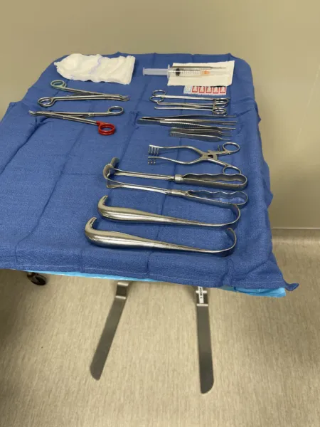
Treating cancer
The treatment plan depends on the kind of cancer and its stage, on the patient's overall health, and on the goal of treatment. Most plans combine several treatments.
Surgery can serve several goals:
| Goal | What the surgery does | Example |
|---|---|---|
| Prevention (prophylactic) | Removes tissue at high risk of becoming cancer | Removing a mole from skin exposed to the sun |
| Diagnosis | Removes a suspicious lesion to be examined | Biopsy of a lump |
| Cure (curative) | Removes all the cancerous tissue | Removing a tumor that has not spread |
| Control (debulking) | Removes part of a tumor so other treatments work better | Shrinking a tumor that cannot all be removed |
| Palliation | Eases symptoms and improves the quality of life | Removing a tumor that causes pain |
| Reconstruction | Restores function and appearance after cancer treatment | Rebuilding a breast after a mastectomy |
A benign tumor may need nothing more than its removal. A malignant one needs prompt treatment to keep it from spreading.
Where the tumor is, its grade and its stage decide how big the operation is. In breast cancer, for example, a small tumor is often removed with a margin of normal tissue (breast-conserving surgery), followed by radiation, but breast-conserving surgery is not usually an option when cancer is found in two or more separate parts of the breast. In a modified radical mastectomy the whole breast is removed, with a sentinel node biopsy or an axillary node dissection.
Chemotherapy and radiation, which the Core Curriculum calls adjunct treatments, are often added to surgery:
- Chemotherapy: drugs that kill cancer cells. It can aim to cure, to shrink a tumor and stop it spreading, or to ease symptoms. Chemotherapy given after surgery to destroy cells left behind is adjuvant therapy; chemotherapy given before surgery to shrink the tumor is neoadjuvant therapy.
- Radiation therapy: ionizing radiation that destroys tumor tissue. It can be aimed at the body from outside, as for the whole breast after breast-conserving surgery, or placed inside it (brachytherapy) as radioactive seeds left in the tumor, needles or a catheter loaded with radioactive pellets, as in breast and prostate cancer. A single dose can also be given during surgery (intraoperative radiation therapy). Clips the surgeon leaves in the tumor bed help the radiation oncologist aim later.
- Targeted and biologic therapy: drugs that kill cancer cells while sparing normal cells, and treatments that help the immune system kill cancer cells.
Chapter 12 covers drugs on the field, and chapter 22 radiation safety.
What cancer does to the whole body
A cancer harms more than the organ it grows in.
- Anemia: from bleeding inside the body and the body's failure to replace red cells.
- Bleeding: a cancer of the blood-forming cells, such as leukemia, can leave too few platelets, so the patient bruises and bleeds easily.
- Infections: such a cancer can also leave too few working white cells, and chemotherapy and radiation lower the white cell count too.
- Blood clots: the tumor can release clotting factors or press on vessels, so clots (thrombosis) form more easily.
- Pain: from tumor pressing on and destroying tissue, and from chemicals the tumor releases.
- Paraneoplastic syndromes: effects of a cancer far from the tumor, caused by hormones or other chemicals the tumor cells release. Lung cancers, especially small cell lung cancer, can cause a high calcium level, too much antidiuretic hormone or Cushing syndrome this way, sometimes before the cancer is even diagnosed.
- Weight loss and cachexia: cachexia is wasting of the body. The tumor takes the nutrients healthy tissue needs and changes the body's metabolism.
These effects follow the patient into the OR. A wasted (cachectic) patient loses heat fast, and cancer is one of the risk factors for a wound that splits open.
6Wounds
A wound is any tissue damaged by surgery or by injury.
The words of wound healing
The Core Curriculum lists the terms below. Several also appear elsewhere in this chapter.
| Term | Meaning |
|---|---|
| Abrasion | Skin or mucous membrane scraped away, as when skin rubs against a hard surface |
| Adhesion | A band of scar tissue that binds together parts that are normally separate, such as loops of bowel and the peritoneum |
| Avulsion | Tissue torn away, partly or completely; a skin avulsion is also called degloving |
| Collagen | The strong, fibrous protein of connective tissue (skin, tendons, fascia, bone); new collagen gives a healing wound its strength |
| Contracture | Scar or fibrous tissue that shortens and stops a joint or skin from moving normally |
| Contusion | A bruise |
| Cicatrix | A scar, especially the one left after a wound heals by filling and contracting |
| Dead space | A gap between tissue layers that should lie together, where blood and serum can collect |
| Debridement | Removing dead tissue or foreign material from a wound, usually by cutting it away with sharp instruments such as a scalpel and scissors |
| Edema | Extra fluid in the tissues (swelling) |
| Extravasation | Fluid, such as blood, bile or an IV drug, escaping from its normal space into the tissue around it |
| Exudate | Fluid that seeps out of tissue, rich in protein and cells; it can be serous, bloody (hemorrhagic) or pus (purulent) |
| Fistula | An abnormal passage between two organs or vessels, or between an organ and the skin |
| Gangrene | Death of tissue (necrosis), usually because its blood supply failed |
| Granulation tissue | New connective tissue full of tiny blood vessels that fills a wound from its base |
| Granuloma | A small mass of immune cells walling off something they cannot destroy, such as a suture |
| Hematoma | A collection of blood, usually clotted, trapped in tissue |
| Hemostasis | Stopping bleeding, by the body or by the surgical team |
| Ischemia | Too little blood flow to a tissue |
| Keloid | A thick, raised scar that grows beyond the edges of the original wound |
| Laceration | An irregular cut or tear in the flesh |
| Nosocomial infection | An infection picked up in a healthcare facility, also called a hospital-acquired or healthcare-associated infection |
| Perforation | A hole through a structure, such as a perforated bowel |
| Proud flesh | Too much granulation tissue, which bulges above the wound and keeps the skin from growing over it |
| Scar | The fibrous tissue that replaces injured tissue; it has none of the original tissue's special work |
| Seroma | A pocket of serous fluid (serum) that collects in a wound |
| Serosanguineous | Containing both serum and blood, as some wound drainage does |
| Sinus tract | A channel that leads from a pocket inside the body out to the surface, usually from an infection; a tract running under the edge of a wound is also called tunneling |
| Tissue tensile strength | How much pulling force tissue can take before it pulls apart; a healing wound slowly gains it back |
Types of wounds
The Core Curriculum sorts wounds into three groups by how they came about.
Intentional (surgical) wounds are planned. An incision or an excision is made under sterile conditions, for a treatment, with clean edges that can be brought together, bleeding under control, and closure right after the procedure.
Unintentional (traumatic) wounds come from an accident or an assault. They are not made under sterile conditions, their edges are irregular, they may involve tissue loss and several injuries at once, and bleeding may not be under control. Traumatic wounds are described in several ways, and one wound can fit more than one:
- Closed or open: in a closed wound the skin stays intact over damaged tissue; in an open wound the skin is broken.
- Simple or complicated: a simple wound has no tissue loss and nothing left in it; a complicated wound has lost tissue or holds a foreign body.
- Clean or contaminated: a clean wound has edges that can be brought together and is expected to heal by first intention; a dirty object makes a wound contaminated, and it can become infected within a short time.
A traumatic wound comes from a mechanical force (a mechanical injury), from heat or cold, from chemicals, or from electricity or radiation. By its cause, it can be:
- Blunt: a force that does not break the skin, such as a blow, a fall or a crash, causing contusions and internal injuries.
- Penetrating: an object that goes through the skin, such as a knife (a puncture or laceration) or a bullet.
- Chemical: damage that depends on how long the chemical touched the tissue, how strong it was, how much tissue it reached and how it acts.
- Thermal: damage from heat or cold.
- Electrical and radiation injuries are covered with burns, below.
Chronic wounds last because of an underlying condition, such as pressure or poor circulation, or an infection. A chronic wound fails to move through the phases of healing on time; in the United States, a wound that has not closed or improved within 30 days is called chronic. It tends to have jagged edges and a higher risk of infection.
- A pressure injury is damage to a patch of skin and the tissue beneath it, usually over a bony prominence, caused by pressure or pressure combined with shear. Older names include pressure ulcer, pressure sore (the term in the Core Curriculum's wound outline), decubitus ulcer and bedsore. Pressure injuries are staged: Stage 1 is intact skin with redness that does not fade when pressed; Stage 2 is a partial-thickness loss of skin with the dermis showing; Stage 3 is a full-thickness skin loss with fat showing; Stage 4 exposes fascia, muscle, tendon, cartilage or bone; and an injury covered by dead tissue so its depth cannot be seen is unstageable. A deep tissue pressure injury shows as a purple or maroon patch of intact skin, or a blood-filled blister, over tissue damaged beneath it. A pressure injury that starts during surgery may not show until several days later. Chapter 31 covers preventing pressure injuries during positioning.
- An ulcer is an open sore on a skin or mucous membrane surface. Venous ulcers, arterial ulcers and diabetic foot ulcers are common chronic wounds.
A partial-thickness wound involves the epidermis, the dermis or both; a full-thickness wound goes through every layer of the skin and can reach fascia or bone.
Burns and other injuries from heat, cold, chemicals and radiation
Burns can be caused by heat (flames, hot liquids, steam, hot metal or tar), chemicals, electricity, radiation and breathing in smoke or hot gases. An electrical burn can look small on the skin while the current has destroyed much more tissue inside, all along its path through the body to the exit wound; doctors call this the iceberg effect.
Burns are graded by how deep they go:
- Superficial (first-degree): the epidermis only; red and painful, like a sunburn. It heals on its own in a few days (about 3 to 6).
- Superficial partial-thickness (second-degree): the whole epidermis and part of the dermis, with painful blisters. It heals in about 10 to 21 days.
- Deep partial-thickness (second-degree): deeper into the dermis. Partial-thickness burns often heal by second intention.
- Full-thickness (third-degree): all the way through the epidermis and dermis, destroying the tissue and damaging the nerve endings. These burns need debridement and skin grafts.
- Deep full-thickness (fourth-degree): into the muscle and bone underneath.
The size of a burn is given as a percentage of the body's surface area. A quick estimate, the rule of nines, gives the head 9%, each arm 9%, the front and the back of the trunk 18% each, each leg 18% and the genital area 1%. An adult with more than 15% of the body burned (10% in a child) is likely to go into hypovolemic shock, because a burn shifts so much fluid out of the circulation. Chapter 38 covers burn surgery.
Other injuries in this group:
- Frostbite: tissue frozen by extreme cold. Ice forms inside the cells and the blood supply is blocked. Ears, cheeks, nose, fingers and toes are hit most often.
- Hypothermia: a body temperature that is too low. In surgery, AST defines it as a core temperature below 36°C. A general medical definition sets the line lower, at 35°C.
- Hyperthermia: a body temperature that is too high, as in heat stroke or a severe infection. Chapter 13 covers malignant hyperthermia, a reaction to anesthesia drugs.
- Radiation injury: from long exposure to the sun or to radiation sources such as X-rays and radiation therapy. Radiation therapy can damage the blood vessels of the treated area, which slows healing there.
The skin's other diseases in the Core Curriculum's list, such as moles (nevi), warts, keratoses and the skin cancers (basal cell carcinoma, squamous cell carcinoma and melanoma), are taught with plastic and reconstructive surgery in chapter 38.
7How wounds heal
After an injury, the body either regenerates the tissue, adding new cells of the same kind, or fills the gap with scar. Some cells, such as those of muscle (heart muscle included) and nerves, cannot divide to replace themselves, so their injuries heal with scar tissue, which fills the gap but cannot do their special work.
The three phases
Healing moves through three phases that overlap. The Core Curriculum calls them the lag, proliferative and maturation phases; you will also hear them called the inflammatory, proliferative and remodeling phases. Some references count stopping the bleeding (hemostasis) as a phase of its own; others include it in the first.
1. Lag (inflammatory) phase. It starts within minutes of the injury and lasts about 3 to 5 days; one reference gives 1 to 4 days.
- Bleeding stops: vessels constrict, platelets clump, and a clot forms.
- An exudate of blood, lymph and fibrin clots in the wound and loosely glues the cut edges together, and a scab dries over it to keep fluid in and germs out.
- Inflammation clears the wound: white cells eat bacteria, fibrin and dead cell pieces for about 3 to 4 days, and macrophages stay on, releasing growth factors and calling in fibroblasts.
- The wound has almost no tensile strength yet. The skin edges look a little swollen and red.
2. Proliferative phase. It begins around day 3 to 5 and lasts until about day 20.
- Fibroblasts build collagen, and new capillaries grow into the wound (angiogenesis). Together they form granulation tissue.
- Healthy granulation tissue is pink or red, uneven in texture and does not bleed easily. Granulation tissue that is dark and bleeds easily (friable) can be a sign of infection.
- Epithelial cells grow in from the edges to cover the wound, and the scab falls off. They can travel only about 3 cm, so a large open wound may need a skin graft.
- The wound begins to contract about day 5, most strongly at about 2 weeks. A firm ridge of new tissue can be felt under a healing incision.
- By the end of this phase, the wound has about 25 to 30 percent of its original strength, and skin sutures are usually removed.
3. Maturation (remodeling) phase. It begins at about 2 to 3 weeks and can last a year or longer.
- Collagen is broken down and rebuilt, above all where the wound takes stress, and it becomes denser and better organized.
- Fewer blood vessels remain, so the scar fades from red to pale, and it flattens and shrinks.
- The wound slowly gains strength. It reaches about 70 to 80 percent of its original strength by about the third month, and it never gets back more than about 80 percent.
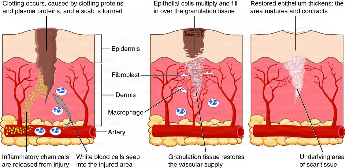
A healed wound is never as strong as the tissue it replaced: at best about 80 percent, and only after months. Until then, sutures and the patient's care, such as no heavy lifting, hold it together.
First, second and third intention
A wound heals in one of three ways, depending on whether its edges are brought together and when. Chapter 25 covers these as a closing choice; here is what the tissue does in each.
- First intention (primary intention, or primary union): the edges of a clean wound are brought together with sutures, staples, tape or skin adhesive soon after it is made. Little tissue is lost, no dead space is left, and drainage is minimal. Epidermal cells seal the surface within 24 to 48 hours, little granulation tissue is needed, and the scar is thin and flat.
- Second intention (secondary intention, or granulation and contraction): the edges cannot be brought together, or are left apart, so the wound stays open and heals from its base upward. Granulation tissue slowly fills it, it contracts, and epithelium grows over the top. Healing is slower, the scar (cicatrix) is wider, and infection is more likely. Infected and dirty wounds, chronic wounds such as pressure injuries, and traumatic wounds with a lot of tissue loss heal this way, and a large one may need a skin graft.
- Third intention (tertiary intention, or delayed primary closure): a contaminated or infected wound is cleaned and left open, often packed, for about 3 days or more. Once no infection shows and clean granulation tissue has formed, the surgeon brings the two granulating surfaces together. The result has middling strength and scarring. It is used for heavily contaminated (Class III or IV) wounds and for trauma patients too unstable for the wound to be closed during the operation.
A wound that heals by second intention can grow too much granulation tissue, called proud flesh, which bulges above the edges and blocks the skin from closing over it. Large gaps healed this way leave a weak area that can become a hernia.
Scars
A new scar is red or purple and later takes on the color of the skin around it. Scar tissue is less elastic than normal tissue and has none of its special functions.
- Hypertrophic scar: a scar with too much tissue, red or raised. It usually improves after about 6 months and may fade over years.
- Keloid: a scar that keeps growing and spreads beyond the edges of the original wound, made of a swirling mass of collagen. Keloids form most often on the shoulders, chest and back, develop more often in people with dark skin, and often come back after treatment.
- Contracture: a scar that shortens as it matures and pulls the skin or a joint so it cannot move normally. Large burns on the back, the buttocks or the back of the neck are prone to it.
Bone heals by a similar process, only more slowly. Chapter 3 covers how a broken bone heals.
8Wound classes
Every surgical wound is given a class at the end of the operation, based on how contaminated it became and so how likely it is to become infected. The class guides how the wound is managed after surgery. The four classes come from the CDC.
| Class | Name | What puts a wound in it | Examples |
|---|---|---|---|
| I | Clean | No infection and no inflammation is found, and the respiratory, alimentary (digestive), genital and uninfected urinary tracts are not entered. The wound is closed primarily, with closed drainage if a drain is needed. An incision made after blunt trauma belongs here if it meets the same rules | Total hip replacement, coronary artery bypass graft, craniotomy, breast biopsy, vitrectomy |
| II | Clean-contaminated | One of those tracts is entered under control, without unusual contamination. Operations on the biliary tract, appendix, vagina and oropharynx belong here when there is no infection and no major break in technique | Laparoscopic cholecystectomy, appendectomy, tonsillectomy, cystoscopy, gastric bypass |
| III | Contaminated | An open, fresh, accidental wound; a major break in sterile technique; gross spillage from the gastrointestinal tract; or acute inflammation without pus | Open fracture, a colon resection with gross spillage of bowel contents, penetrating trauma, removing metal fragments after an explosion |
| IV | Dirty or infected | An old traumatic wound with dead tissue left in it, an infection already present, or a perforated organ (viscus). The germs that may cause infection were in the wound before the operation began | Incision and drainage of an abscess, removal of a perforated (ruptured) appendix |
The class follows what actually happens, so it can change during the case. Opening the bowel makes a clean case at least Class II, gross spillage of bowel contents makes it Class III, and so does a major break in sterile technique, such as using a nonsterile instrument in the wound. The final class is decided at the end of the procedure, discussed in the team's debriefing and recorded on the intraoperative record.
A laparoscopic cholecystectomy is Class II, because the biliary tract is entered. It becomes Class III if bile spills or the gallbladder is acutely inflamed without pus, and Class IV if infection, such as pus, is already present. The Core Curriculum's outline and one surgical technology textbook list an uncomplicated case as Class I, and Class II only when a cholangiogram is done.
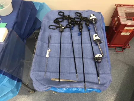
An appendectomy is Class II when the appendix is not perforated, and Class III when it is acutely inflamed without pus. A perforated (ruptured) appendix, or one with pus around it, is Class IV, because the germs were in the abdomen before the operation began. Some references class a perforated appendix as Class III.
The higher the class, the higher the risk of infection. One reference estimates the risk at about 1 to 5 percent for a clean wound and more than 27 percent for a dirty or infected one. Preventive antibiotics are given for clean-contaminated operations, and for clean ones when a prosthetic joint or prosthetic material in a blood vessel goes in or an infection would be a catastrophe; patients with contaminated and dirty wounds are often already on antibiotics to treat an infection.
The class also shapes how you handle instruments. Instruments that touch the open bowel, or a tumor that could shed cancer cells, are kept apart from the clean setup so they cannot carry germs or cells to the rest of the wound. In the same way, during an abdominal hysterectomy, instruments that touch the cervix and vagina are taken off the field so they cannot contaminate the abdominal incision. Chapters 29 and 32 teach this isolation (bowel) technique, and chapter 33 the hysterectomy.
Entering the respiratory, digestive, genital or urinary tract makes a wound at least Class II. A burst organ or pus already in the wound makes it Class IV.
9Surgical site infection
A surgical site infection (SSI) is an infection of the incision, or of the organ or space that was operated on, after surgery. It is one of the most common infections patients pick up in a hospital, and the most common cause of delayed healing in surgical patients. About 2 to 4 percent of operations are complicated by one.
The three depths
The CDC's National Healthcare Safety Network sorts SSIs by how deep they go:
- Superficial incisional SSI: involves only the skin and the subcutaneous tissue of the incision. It counts if it starts within 30 days after the operation.
- Deep incisional SSI: involves the deep soft tissue of the incision, the fascia and muscle layers. It counts if it starts within 30 or 90 days, depending on the type of operation.
- Organ or space SSI: involves any part of the body, deeper than the fascia and muscle, that was opened or handled during the operation, such as an abscess inside the abdomen, an infection of a joint or of bone (osteomyelitis), or mediastinitis, an infection in the middle of the chest. It too counts within 30 or 90 days, depending on the operation.
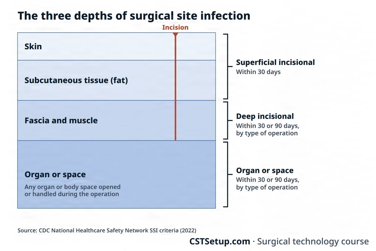
Each depth has signs that confirm it, such as pus draining from the incision or from a drain placed in the organ or space; germs grown from fluid or tissue taken from the site; pain, tenderness, swelling, redness or heat with the incision opened by the surgeon; a deep incision that splits open, with a fever above 38°C (100.4°F); or an abscess found on examination or imaging.
Deep and organ or space infections together make up one third to one half of all SSIs, and they usually keep the patient in the hospital or bring them back. A superficial SSI is often found after the patient has gone home.
Where the germs come from
- From the patient (endogenous): the patient's own germs, from the skin, the mucous membranes and the hollow organs such as the bowel, are the main source of SSI germs. Most are staphylococci from the skin, but an incision near the groin or perineum, or an opened bowel, adds bowel germs. Skin prep lowers these germs but cannot get rid of them, which is why drapes wall off the rest of the patient's skin. Germs can also seed the wound from an infection somewhere else in the patient's body, especially when an implant is placed.
- From outside the patient (exogenous): the surgical team, the OR environment and its air, and every tool, instrument and material brought onto the sterile field.
The germs found most often in SSIs are Staphylococcus aureus, coagulase-negative staphylococci, Enterococcus and Escherichia coli, and more and more of them resist antibiotics, such as methicillin-resistant S. aureus (MRSA).
Whether contamination becomes an infection depends on how many germs reach the wound, how dangerous they are, and how well the patient can resist them. Foreign material, such as sutures and implants, lets far fewer germs start an infection, which is one reason they raise the risk.
What makes an SSI more likely
Some patients start with a higher risk:
- older age, because the immune system responds less and tissue heals more slowly
- poor nutrition, which leaves out the proteins needed for healing and defense
- diabetes, which harms the circulation that the immune system depends on
- substance or alcohol abuse, which often comes with malnutrition, liver damage and a weakened immune system
- a weakened immune system from disease, cancer treatment or high doses of steroids
- a long stay in the hospital before surgery, which lets the patient pick up hospital germs, including resistant ones
The operation itself adds risk when the wound class is higher and when the procedure is long, because a long case means more time exposed to germs in the air and on devices.
What the team does about it
The CDC's 2017 guideline focuses on a few measures:
- Preventive antibiotics: given only when guidelines call for them, and timed so the drug is already in the blood and tissues when the incision is made. In clean and clean-contaminated operations, no more doses are given after the incision is closed, even when a drain is left in. Chapter 12 covers the drugs.
Some references still continue preventive antibiotics for up to 24 hours after surgery.
- Blood sugar control: high blood sugar harms healing, immunity and the blood vessels. The CDC's target around surgery is below 200 mg/dL, in patients with and without diabetes.
A 2022 update from three infection prevention societies (SHEA, IDSA and APIC) sets a lower target: keep every patient's blood sugar between 110 and 150 mg/dL in the first day or two after surgery, whether or not they have diabetes.
- Normal body temperature (normothermia): a cold patient's vessels constrict, so less oxygen reaches the wound and infection becomes more likely.
- Oxygen to the tissues: helped by keeping the patient warm and the blood volume replaced.
- An antiseptic bath: the patient showers or bathes with soap or an antiseptic at least the night before surgery.
Beyond these, every SSI rule you learn elsewhere works toward the same goal: skin prep (chapter 31), sterile technique (chapter 26), drapes and attire (chapter 28), and hand hygiene by everyone who is not scrubbed.
When an SSI happens
Infection shows first as more inflammation than expected and drainage from the wound. A superficial infection is treated at its first signs: a sample of the drainage is sent for culture, and antibiotics may be started. A deep infection may not show for days and brings pain and fever. If it does not settle, the surgeon reopens the wound to drain pus and remove dead tissue (incision and drainage, I&D), and the wound may be irrigated and left open to heal.
10What helps or slows healing
The Core Curriculum sorts what helps or slows healing into three groups: the patient's own condition, outside factors such as drugs and smoking, and surgical technique, which is how the team handles the tissue and keeps the field sterile. Together they affect how fast a wound heals, how strong it becomes and whether it gets infected.
The patient
- Age: an older patient's cells renew more slowly, the collagen is less elastic, blood flow is poorer and the immune system is weaker, and many older patients are short of nutrients or fluids. Very young and very old patients may also have a poorer blood supply or weak muscle tone.
- Disease and comorbidities: diabetes harms the circulation that brings oxygen and nutrients to the wound, and high blood sugar weakens the white cells that fight bacteria. Heart and lung disease, poor circulation, anemia, cancer and infection all slow healing too, and so does a weakened immune system.
- Nutrition, when the patient is malnourished: new tissue is built from protein, and a shortage of vitamins C and A, zinc or iron is linked to infection and weaker wounds. A thin patient with little body fat also loses heat more easily.
- Nutrition, when the patient is obese: fat tissue has a poor blood supply, so it heals slowly, and its weight strains the closure, which makes the wound more likely to split open or become an incisional hernia.
- Fluid and electrolyte balance: low blood volume makes the vessels constrict and starves the wound of the oxygen and glucose it needs, so keeping the blood volume replaced is part of preventing infection. Edema in the wound slows healing as well.
- Oxygen: low oxygen in the wound slows the fibroblasts and the collagen they build, so the wound is weaker, and it leaves the wound more open to infection.
Outside factors
- Chemotherapy: it can slow or stop the growth of the new cells a wound needs, and it weakens the immune system.
- Prescription medications: steroids damp the inflammatory phase, and long-term steroid use raises the risk that a wound will split open. NSAIDs can interfere with the inflammatory stage, anticoagulants raise the risk of bleeding and of the wound splitting open, and long courses of antibiotics can let a second infection take hold.
- Illicit drugs and alcohol: patients who abuse them are often malnourished, may have liver damage that limits how the body makes energy for its cells, and often have a weakened immune system.
- Smoking: nicotine constricts the vessels and cuts the oxygen reaching the wound, and the cough it brings strains a fresh incision. Patients are urged to stop.
- Radiation: tissue treated with radiation has a poorer blood supply, radiation lowers the white cell count by depressing the bone marrow, and earlier radiation is a risk factor for a wound splitting open.
Surgical technique
This is the group the surgical team controls, and the scrub has a hand in every part of it.
- Principles of asepsis: any germs that get into the wound can cause infection. Chapter 26 teaches sterile technique.
- Bowel technique: keeping instruments that touched the open bowel away from the clean closure, taught in chapters 29 and 32.
- Dissection technique: cutting tissue cleanly and precisely, sharp or blunt, and saving as many blood vessels as possible, because tissue without a blood supply cannot heal.
- Duration of surgery: a long operation leaves the wound exposed longer to germs in the air and on devices, so working efficiently matters.
- Hemostasis: small bleeding vessels form clots and hematomas that block healing and invite infection. Chapter 29 covers the methods of controlling bleeding.
- Tissue approximation: the edges are brought together accurately and without tension. Tension on a suture line squeezes the capillaries at the wound edge and causes swelling and pain, and sutures tied too tightly strangle the tissue.
- Tissue handling: tissue is handled as little and as gently as possible, retractors press no harder than they must, and exposed tissue is kept moist. Rough handling bruises tissue and causes swelling, bleeding, ischemia and necrosis.
- Tissue perfusion: tissue heals only with a good blood supply. Cold, pain, fear, nicotine and low blood volume all constrict the vessels, so keeping the patient warm helps.
- Drains: dead space left in a wound fills with blood and serum and can become infected. Careful suturing closes it, and a drain keeps fluid from collecting where it cannot be closed. Chapter 25 covers drains.
- Irrigation: keeps exposed tissue moist and washes debris and germs out of a contaminated wound.
Many of these are Halsted's principles of surgery, which chapter 25 teaches in full.
11When healing goes wrong
Wound complications can come at any point in healing, but most show up in the first week after surgery. Some stay at the surface; others reach deep tissue and can break the wound open.
Infection
Infection, covered above under surgical site infection, is the most common cause of delayed healing in surgical patients. Its germs come from the patient (endogenous) or from outside the patient (exogenous).
Bleeding, hematoma and seroma
- Hemorrhage: some bleeding from a wound is normal; heavy bleeding is not. It can come from a clot that breaks loose, a suture that slips or a vessel worn through, and dressings are checked often in the first 48 hours. Bleeding at the surgical site is an abnormal finding you report.
- Hematoma: blood collects in the wound because bleeding was not fully stopped during surgery or small vessels keep oozing. It raises the risk of infection, a large one presses on vessels and starves the tissue around it, and it may need another operation to drain.
- Seroma: serous fluid collects in the wound, from tissue injured during surgery or bleeding into it, often where dead space was left. It sits between the wound edges and keeps them from healing, and it too may need surgery.
Necrosis
Tissue that has lost its blood supply dies. Dead tissue blocks healing and harbors bacteria, so it is removed by debridement.
Tissue disruption
- Dehiscence: the layers of a wound separate, partly or completely. It happens when the pull on the wound overcomes the strength of the suture or the knot, and it often comes between the 5th and 10th days after surgery. Serosanguineous fluid may drain from the wound first, and the patient may feel that "something has given way." Obesity, older age, male sex, lung disease, coughing, cancer, earlier radiation, trauma, poor nutrition and long-term steroids raise the risk, and so do infection, too much tension, sutures placed too close to the edge or to each other, sutures tied too tight, and knots that are loose or cut too short.
- Evisceration: dehiscence of an abdominal wound with the abdominal contents, such as bowel or omentum, pushing out through it. It is an emergency. The organs are covered with a sterile towel soaked in sterile saline to keep them moist, the patient lies with the head slightly raised and the knees bent to ease the pressure on the wound, nothing is given by mouth, and the surgeon is called, because the patient goes back to the OR.
- Herniation: a scar that formed after a wound broke down is much weaker than healthy tissue, and contaminated or infected wounds are more likely to leave a hernia. Abdominal contents can bulge through the weak spot, an incisional hernia, usually found 2 to 3 months after surgery, and bowel can become trapped (incarcerated) in it.
- Re-injury: a wound that is still weak can be torn open again, which is why patients restrict their activity, avoid heavy lifting and protect the site after surgery.
- Rejection: tissue or an organ taken from another person can be attacked by the patient's immune system as foreign. A graft of the patient's own tissue (an autograft) is not rejected. An implant brings a risk of its own: as foreign material, it lets far fewer germs start an infection.
Suture complications and tensile strength
A suture holds the wound until the wound can hold itself, and a wound gains strength slowly. If the suture breaks, a knot slips, or the suture loses its strength in an infected wound, the wound can split open. Suture is also foreign material: it can form a granuloma, and it lowers the number of germs needed to start an infection. Chapter 25 covers choosing a suture that keeps its strength long enough for the tissue.
Adhesions, fistulas and sinus tracts
- Adhesions: bands of scar tissue that join abdominal or pelvic organs to each other or to the peritoneum. They follow infection, repeated operations, rough handling of tissue, and foreign bodies such as lint or glove powder. They can cause pain or a bowel obstruction, and they make the next operation riskier.
- Fistula: an abnormal passage from one organ or vessel to another, or to the skin.
- Sinus tract: a draining channel from an infection inside the body out to the surface.
Never leave eviscerated organs uncovered or let them dry out. Cover them with a sterile towel soaked in sterile saline, stay with the patient and get the surgeon.
12Trauma
Trauma means injury. It is the leading cause of death for people from about 1 to 45 years old, and the third leading cause at all ages. Most trauma patients arrive outside regular working hours or on a weekend, when fewer staff are working, so hospitals organize their care as a system.
How injuries happen
The way an injury happened, its mechanism of injury, predicts what injuries the patient is likely to have. Crashes, falls and assaults produce injury patterns that are fairly predictable.
- Blunt injury: a force that damages deep tissue without breaking the skin. Most blunt injuries come from motor vehicle crashes, falls and assaults. Speeding up, slowing down, shearing and crushing forces tear tissue inside; in a crash, the aorta can tear where it is fixed in the chest as the body stops and the aorta keeps moving. Because blunt injuries are hidden, they can be harder to find, and deadlier, than penetrating ones. A contusion is a bruise.
- Penetrating injury: something pierces the skin and goes into the body, or the body is thrown onto something sharp, opening a wound with deeper injuries under it. Gunshots and knife attacks cause most of them. A high-speed bullet damages far more tissue than a slow one, and the exit wound is usually larger than the entry. A wound at or below the nipple line can reach both the chest and the abdomen. An impaled object stays in until the patient is in the OR, where the bleeding it may be holding back can be controlled.
- Blast injury: the pressure wave of an explosion injures gas-filled organs such as the lungs, bowel and middle ear; flying debris causes penetrating wounds; and the blast throws the victim.
- Burns and electrical and inhalation injuries, covered above, can come from an explosion or on their own.
- An occult injury is one the normal assessment does not find, such as hidden bleeding.
An open traumatic wound is contaminated. A fresh one is Class III, and an old one with dead tissue left in it is Class IV; some references use about 4 hours after the injury as the line between the two. Traumatic wounds were not made under sterile conditions, so they are usually debrided and irrigated, and many are left open to heal by second or third intention.
Trauma systems
In the United States, hospitals equipped to treat severe injuries are designated as trauma centers by the state or local government and verified by the American College of Surgeons (ACS), whose Committee on Trauma sets the criteria along with state laws. The levels vary from state to state, running from Level I, the highest, to Level IV or, in some systems, Level V.
- Level I: total care for every kind of injury, from prevention through rehabilitation, with specialists and equipment ready 24 hours a day.
- Level II: care for all injuries, but without some of the specialists and resources a Level I center has.
- Level III: prompt evaluation, resuscitation, emergency surgery and stabilization, then transfer to a higher level.
- Level IV: advanced trauma life support before transfer, often in a rural clinic or hospital.
The criteria include the number and kinds of trauma specialists, the equipment, fast transport, real-time communication with rescue teams and teaching the community about injury prevention. Emergency medical services (EMS) decide where to take a patient by field triage: for example, a patient whose Glasgow Coma Scale score is 13 or lower, whose systolic blood pressure is under 90 mm Hg, or whose breathing rate is under 10 or over 29 goes to the highest-level center available.
The American College of Surgeons also runs Advanced Trauma Life Support (ATLS), the course that teaches physicians its protocols for trauma care. Only licensed physicians can be certified, though other staff may audit it in some places.
What severe injury does to the body
Deaths after injury come in three waves. Some patients die at the scene, within minutes, from tears of the heart or aorta or injuries to the brain stem or spinal cord. Others die in the first hours, about half of them from bleeding, often from the spleen, liver or lungs: these are the patients fast, aggressive care in the golden hour can help most. The rest die days or weeks later, in intensive care, from complications and organ failure. Overall, bleeding is the main cause of death after injury.
Hemorrhagic shock is the failure of the circulation caused by heavy, prolonged bleeding. As blood is lost, the vessels of the limbs, muscles and gut clamp down to keep blood flowing to the heart, brain and kidneys, and hormones such as epinephrine, norepinephrine, vasopressin and cortisol pour out. At first this works, and the blood pressure can stay normal, but the starved tissues swell, small clots form and cells die. Losing about 20 percent of the blood volume causes mild shock, 20 to 40 percent moderate shock, and 40 percent or more severe shock.
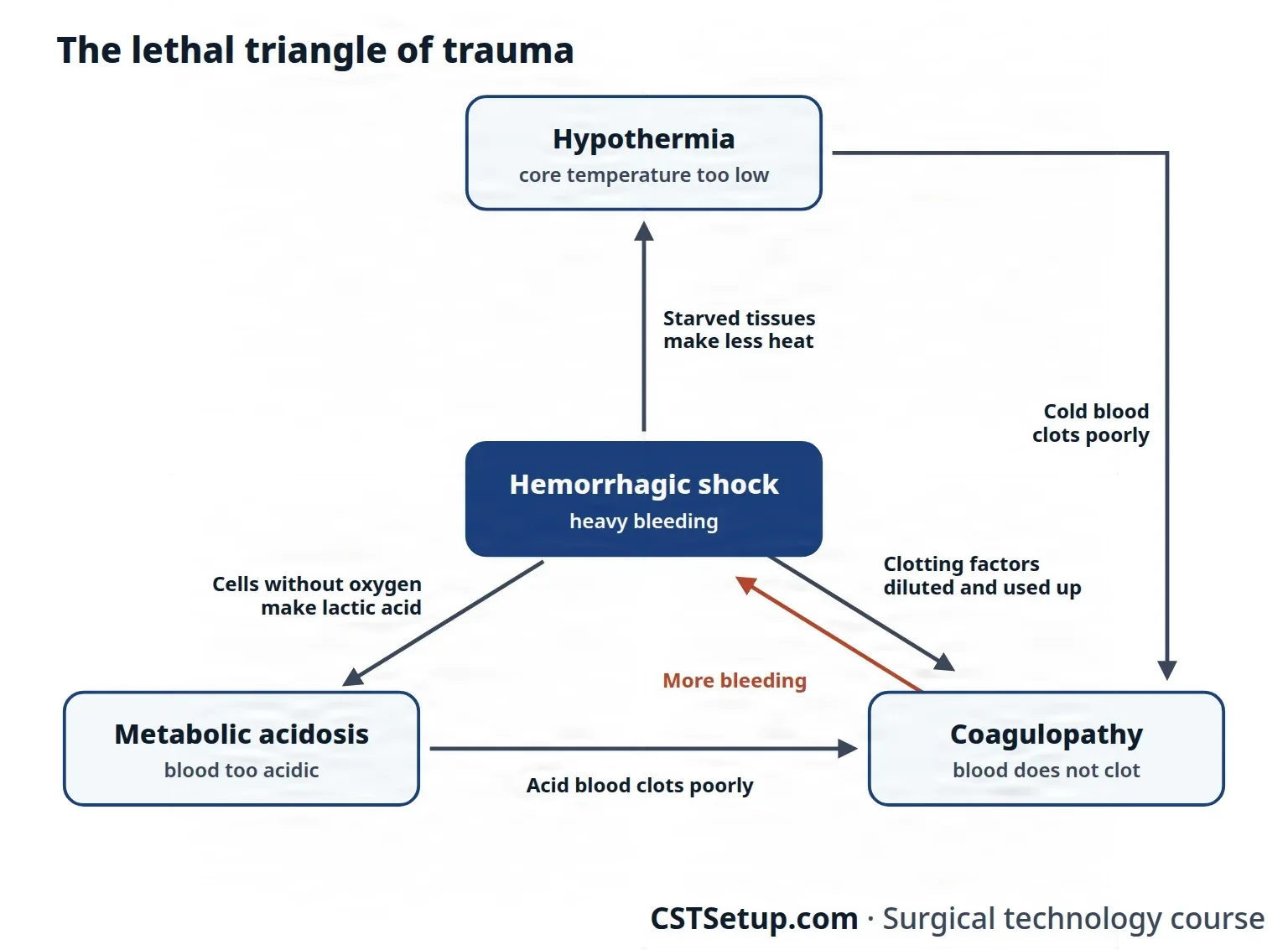
The lethal triangle
Patients who survive the first hour face three conditions that the trauma team watches above all, called the lethal triangle (or lethal triad, or triangle of death):
- Hypothermia: a core temperature that stays too low. Tissues starved of blood stop making heat, and the patient loses more heat when clothing is removed, during transport and on the table. Nearly every critically injured patient is cold by the time care begins.
- Metabolic acidosis: blood that is too acidic (a low pH). Cells without oxygen switch to a way of making energy that leaves lactic acid, and without good circulation the acid builds up.
- Coagulopathy: the blood's clotting system fails. Cold and shrinking capillaries shift fluid that dilutes the blood, IV fluids dilute the platelets and clotting factors further, and the clotting factors get used up in the organs (disseminated intravascular coagulation, DIC), so the patient keeps bleeding even after the main bleeding is stopped.
Each one makes the others worse: a cold patient clots poorly, acid blood clots poorly, and more bleeding deepens the shock that causes the cold and the acid. That is why the team warms the patient and treats the triad along with replacing blood.
Compartment syndrome
Injured tissue swells. When it swells inside a closed space, such as a muscle group wrapped in fascia, the abdomen or the skull, the pressure rises until blood can no longer get in, and the tissue dies. This is compartment syndrome. It is most common in the limbs, the abdomen and brain injury.
- In a limb, the signs are severe pain, especially when the muscles are moved passively, numbness or tingling, and weakness. Normal pressure in a compartment is under 20 mm Hg; above 30 mm Hg, the surgeon cuts the fascia open to relieve it, a limb-saving operation called a fasciotomy.
- In the abdomen, swollen bowel and organs raise the pressure until organs begin to fail (abdominal compartment syndrome). The treatment is opening the abdomen (decompressive laparotomy), and the swelling can leave it too full to close.
ATLS principles of trauma care
Trauma care follows its own rules:
- The problem most likely to kill the patient is treated first.
- Treatment starts even before a definitive diagnosis is made.
- Treatment may start without a detailed history.
Hospitals use algorithms, set treatment pathways, so care is systematic and consistent, but a badly injured patient often forces the team to step outside them.
The golden hour is the first critical hour after an injury, when fast, definitive care does the most to prevent death and disability. It starts at the scene, where EMS judges how badly the patient is hurt, begins treatment and chooses the hospital.
The primary survey finds and treats the threats to life, in order, following ABCDE. First responders begin it in the field, and the trauma team repeats it on arrival. Each threat is corrected before the next step.
- A, airway with cervical spine protection: the airway is secured, and the neck is held still in a rigid collar until an exam and X-rays rule out a neck injury.
- B, breathing: the chest is checked for injuries that stop breathing, such as a pneumothorax, a hemothorax (blood in the chest) or a flail chest.
- C, circulation: bleeding is controlled early, to prevent coagulopathy and exsanguination, and IV lines are placed for blood tests, fluids and drugs.
- D, disability: a quick neurologic check for a brain injury.
- E, exposure and environment: the clothing is removed to see every injury, and the patient is kept warm.
The order matters. A patient with a penetrating wound and bowel outside the abdomen still gets the airway and breathing secured first, because an evisceration can wait minutes and a blocked airway cannot.
Neck injuries, blunt, penetrating or from a near-hanging, can damage the airway, the larynx and the great vessels. The neck is treated as broken until cleared, and the airway is secured first, by rapid sequence intubation through the mouth or, when that is not possible, a surgical airway. In the OR the patient lies supine, prepped and draped as for a wide thyroidectomy, with vascular instruments and sutures as for carotid surgery (chapter 40), fine dissecting instruments and thyroid and carotid retractors. Vessels are repaired directly or with a patch, and the airway is repaired, often with a tracheostomy (chapter 36).
The FAST exam (focused assessment with sonography for trauma) adds a bedside ultrasound to the primary survey in an unstable patient with blunt trauma. It looks for blood around the heart and free fluid in the abdomen, and if it finds fluid in such a patient, they go straight to the OR for an exploratory laparotomy.
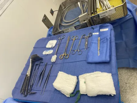
Resuscitation means restoring the body's balance after severe injury: the blood volume, the flow to the tissues and the tone of the vessels. It begins as soon as help arrives and goes on through the emergency department, the OR and intensive care. It is a balancing act: too much IV fluid dilutes the blood and its clotting factors, and raising the blood pressure too far speeds bleeding and can knock loose the clots already formed. At least two large IV lines are placed, and fluids and blood are given through a pump that warms them. Many hospitals have a massive transfusion protocol that gives red cells, plasma and platelets in equal (1:1:1) amounts. Chapters 12 and 13 cover blood products and anesthesia care.
The secondary survey comes once the patient is stable enough to move. It is a head-to-toe exam for less obvious injuries, with imaging such as ultrasound, X-rays and MRI, more IV lines, a urinary catheter and a nasogastric tube, and a history from the patient or witnesses. A patient who needs surgery to save their life may go straight to the OR after the primary survey, and every patient with multiple injuries recovers in intensive care.
A definitive procedure is a planned operation that repairs injured structures and restores their function. A patient in the lethal triangle may be too unstable for one, and gets damage control surgery instead: stop the bleeding and the contamination, and leave the repair for later. Chapter 32 teaches damage control and staged closure of the abdomen, chapter 28 setting up for a trauma case, and chapters 39 to 42 the injuries of the bones, vessels, chest and nervous system.
In trauma, the most lethal problem comes first, treatment starts without a definitive diagnosis, and warmth is part of the treatment. A cold, acidotic patient keeps bleeding.
13Disorders present at birth
A congenital disorder is present at birth; it develops during fetal life. Much of pediatric surgery corrects structural defects that formed before birth.
- A genetic abnormality is inherited, passed down in the genes.
- An acquired abnormality develops in fetal life because of something in the environment.
The body's organs form in embryonic life, the first 8 weeks of development; after that the developing baby is called a fetus. The first 60 days are the most vulnerable, because tissues are forming so fast.
- A teratogen is anything that harms the developing embryo or fetus, such as alcohol, environmental mercury, some drugs, radiation and infections such as rubella, cytomegalovirus and syphilis.
- A mutagenic substance (a mutagen) changes the genes themselves. X-rays are both mutagenic and teratogenic.
- Diet matters too: too little folic acid in the mother's diet causes neural tube defects such as spina bifida.
Some genetic disorders affect the whole body: cystic fibrosis involves several systems and needs long-term care, and muscular dystrophy causes muscle weakness that gets worse over time. In Down syndrome, an extra chromosome 21 gives the person 47 chromosomes instead of the usual 46.
Surgery treats many congenital defects, and each is taught with its specialty: pyloric stenosis and omphalocele in chapter 32; an undescended testis (cryptorchidism) in chapter 34; choanal atresia in chapter 36; cleft lip and palate in chapter 37; syndactyly in chapter 38; clubfoot in chapter 39; heart defects, esophageal atresia and pectus excavatum in chapter 41; and spina bifida (myelomeningocele) and hydrocephalus in chapter 42.
Lo que te llevas de este capítulo
- Disease has its own vocabulary: etiology is the cause, signs are measured and symptoms are reported, acute means sudden and chronic means long term, and palliative care eases what curative care would fix.
- Inflammation, with its redness, heat, swelling, pain and loss of function, is the body's first answer to any injury, and a wound cannot heal without it; inflammation that lasts too long slows healing.
- Shock is a circulation that cannot feed the cells. It is named by its cause: hypovolemic (hemorrhagic), cardiogenic, obstructive or distributive (septic, anaphylactic, neurogenic).
- A benign tumor stays put; a malignant one invades and metastasizes. Grading describes how abnormal the cells look, staging (TNM) how far the cancer has spread, and together they guide the operation and the adjunct treatments.
- Wounds heal in three overlapping phases (lag or inflammatory, proliferative, maturation), by first, second or third intention, and never regain more than about 80 percent of their original strength.
- Wound classes run from I (clean) to IV (dirty or infected): entering a tract makes a wound at least Class II, and pus or a perforated organ makes it Class IV.
- Surgical site infections are superficial, deep or organ or space; most of their germs come from the patient's own skin and organs, and asepsis, gentle tissue handling, hemostasis, closing dead space and a warm patient prevent them.
- In trauma, the most lethal problem is treated first, following ABCDE, and the lethal triangle of hypothermia, metabolic acidosis and coagulopathy is fought from the first minutes.
Ponte a prueba5 preguntas rápidas
Gratis para todos. Elige una respuesta para cada una y luego revísalas todas juntas.
En el quirófanoDónde vas a usar esto
Todo el banco de preguntas del capítulo en series de 10, con la respuesta explicada después de cada una.
Parte del programa para educadoresLas palabras del capítulo en tarjetas de estudio: las que fallas vuelven antes.
Parte del programa para educadoresLo asigna tu docente. Cada grupo saca sus propias 25 preguntas del banco del capítulo.
Parte del programa para educadores
Sugerir una corrección
¿Algo está mal, confuso o desactualizado en este capítulo? Avísanos y lo corregimos para todos. Tu correo solo se usa para responderte.