Chest and heart surgery
The lungs, the heart and the machine that stands in for both: how chest and heart cases are opened, set up and closed, and how the scrub keeps a crowded, fast-moving field safe.
1What chest and heart surgery covers
Cardiothoracic surgery treats the organs inside the chest. Thoracic surgery works on the lungs, the bronchi and the smaller airways, the pleura, the chest wall and the mediastinum; cardiac surgery works on the heart and the great vessels, for disease present at birth and disease acquired later in life. A thoracic surgeon may also operate on the esophagus and the thymus, or a general surgeon may, helped by a thoracic surgeon.
Heart surgery builds on what you learn in general, vascular and thoracic surgery. Much of what makes it complex is the number of different devices and how they fit together, which you learn by practice, study, mentors and in-service training.
The team:
- The surgeon and assistants, one of whom often takes the leg vein.
- The anesthesia provider, who places a double-lumen breathing tube so one lung can be collapsed, runs the monitoring lines, and often watches the heart with a transesophageal echocardiogram (TEE) during the case.
- The perfusionist, who sets up and runs the heart-lung machine and is responsible for its safe operation, oxygenating the blood outside the body while the heart is stopped, and who by AmSECT's standards is board certified or equally qualified.
- The scrub and the circulator, who keep two back tables and several Mayo stands in order in a room crowded with monitors, the bypass machine and solution basins.
Heart rooms are large, with ceiling booms that carry equipment and gases off the floor, and some are hybrid rooms that add a catheterization lab's imaging; chapter 17 teaches room design and chapter 22 the hybrid room.
As in every procedure chapter, operations that share instruments, positions and steps are taught together as co-related procedures, and any approach other than open counts as minimally invasive surgery (MIS). The Core Curriculum lists four approaches for the chest (open, endoscopic, robot-assisted and video-assisted thoracic surgery) and, for the heart, sternotomy or thoracotomy, robot-assisted and percutaneous (through the skin).
Other chapters hold the pieces around this one: the heart and vessels (chapter 5), the lungs (6), the thymus as a gland (9), the drugs (12), the monitors (13), scopes and the robot (21), the heart-lung machine, cell saver and slush machine (23), drains (25), hemostatic agents at the field (29), positioning (31), and the saphenous vein's open harvest and all peripheral vascular work (40); the endoscopic vein harvest for a heart bypass is taught here, and airway endoscopy by ENT surgeons in chapter 36.
2Anatomy that matters
The chest wall and mediastinum
The sternum has three parts: the manubrium at the top, the body (gladiolus) and the xiphoid process at the bottom. Twelve pairs of ribs form the sides, and chapter 3 teaches how they join the sternum and the spine.
Intercostal muscles fill the spaces between the ribs, and an intercostal artery, vein and nerve run with each one. The nerve lies in the groove on the lower edge of its rib, so surgeons take care not to injure it, and when it must be disturbed they may inject a local anesthetic around it to ease pain afterward. Inside the front chest wall run the internal mammary (internal thoracic) arteries, which chapter 5 teaches as coronary grafts.
The mediastinum is the space between the two lungs. It holds the heart, the great vessels, the trachea and main bronchi, the esophagus and the thymus. It is divided into compartments: the anterior mediastinum, behind the sternum, holds the thymus, lymph nodes and fat; the middle holds the heart, the pericardium and the roots of the great vessels; and the posterior lies between them and the spine. The thymus is large in childhood and shrinks in adults, its tissue slowly replaced by fat; chapters 9 and 10 teach its role.
The diaphragm closes the chest off from the abdomen, and the phrenic nerves, which run along the sides of the pericardium, make it contract.
The lungs and breathing
There are three lobes in the right lung and two in the left. Each lung divides into about 10 segments, each with its own bronchus and its own branches of the pulmonary artery and vein, which is what lets a surgeon remove one segment alone. The bronchus, the pulmonary vessels, the nerves and the lymph vessels enter each lung at its hilum, on the side facing the mediastinum; the apex of each lung rises just above the collarbone, and its base rests on the diaphragm.
The pleura is a double membrane. The parietal pleura lines the chest wall and the visceral pleura covers the lung, with a thin film of fluid, about 50 mL, between them. The space between them holds a negative pressure. If the chest wall or pleura is opened, air rushes in and the lung collapses, which is why an opened chest is closed with a drainage system that restores the pressure. Chapter 6 teaches the respiratory system in full.
Three words describe the work of breathing:
- Ventilation: moving air into and out of the lungs. The diaphragm contracts and the ribs rise during inspiration (breathing in), and air flows out passively during expiration (breathing out).
- Diffusion: oxygen crossing from the air sacs (alveoli) into the blood.
- Perfusion: oxygen delivered to and taken up by the tissues.
The heart
Chapter 5 teaches the heart, its valves, the coronary arteries and the great vessels in full; here are the terms and landmarks heart surgery uses. The wall has three layers, the epicardium, the myocardium (the muscle) and the endocardium, and the apex is the lower left tip of the left ventricle. The tricuspid and mitral valves are tied to papillary muscles by the chordae tendineae, and the pulmonary and aortic valves guard the exits. One full heartbeat is the cardiac cycle: the ventricles contract in systole and relax and fill in diastole.
The beat starts in the conduction system. A node is a group of specialized cells that make and pass on the electrical signal: it begins in the sinoatrial (SA) node, the heart's pacemaker, travels to the atrioventricular (AV) node and down the bundle of His, the bundle branches and the Purkinje fibers. In valve and septal surgery the surgeon keeps stitches away from the conduction tissue near the AV node, because injuring it blocks the signal.
The coronary arteries, the first branches of the aorta, feed the heart muscle. On the left, the recurrent laryngeal nerve leaves the vagus nerve and curves around the ductus arteriosus, or its remnant, beside the aortic arch, and the phrenic and vagus nerves cross the field, so surgeons protect them in lung, ductus and transplant operations.
3Tests before surgery
The lungs
- Imaging: chest X-rays show fluid or air in the pleural space, tumors and deformities; CT and MRI study a mass more closely; PET helps tell a cancerous nodule or lymph node from a harmless one; pulmonary angiography looks for a pulmonary embolism when CT cannot settle it. The chest X-ray should be in the room during surgery.
- Pulmonary function tests (PFTs): breathing tests that measure how well the lungs work, such as the tidal volume, the vital capacity, the total lung capacity and the forced expiratory volume in 1 second (FEV1). An FEV1 below 1 liter marks a patient at extremely high risk of lung complications after surgery.
- Arterial blood gases (ABGs): oxygen, carbon dioxide and pH in arterial blood, among the most important blood tests for the lungs. A complete blood count, cultures of sputum and tumor markers are also used.
- Ventilation and perfusion studies show how much each part of the lung breathes and receives blood, which helps the team keep gas exchange working during the operation.
- Endoscopy for tissue: bronchoscopy, mediastinoscopy and thoracoscopy, each with biopsies, are the three procedures that confirm a diagnosis. They are taught below.
The heart
- Noninvasive tests: the resting and exercise electrocardiogram (stress test), chest X-ray, echocardiography from the chest wall (TTE) or the esophagus (TEE), nuclear scans, CT, PET and MRI or MR angiography.
- Cardiac catheterization: a catheter passed from a peripheral vessel into the heart. Left heart catheterization, through the femoral, radial or brachial artery, studies the coronary arteries, the aortic and mitral valves and the left ventricle; right heart catheterization, through a femoral, subclavian or internal jugular vein, studies the right heart and the pulmonary artery. Contrast injected into the coronary openings outlines blockages, and a ventriculogram shows how well the ventricles pump.
- Ejection fraction: the share of the blood in the ventricle at the end of filling that it pumps out with each beat, a key measure of heart function.
- Electrophysiology (EP) studies map the heart's electrical pathways.
- Blood tests: a full clotting profile, because aspirin and some other drugs weaken the platelets; troponin and other markers of heart muscle damage; liver and kidney function; blood glucose. Blood is typed, tested for cold antibodies that could clump when the patient is cooled, and kept in a monitored blood refrigerator, with packed red cells ready at once.
4Diseases of the lungs, pleura and chest wall
- Lung cancer is a common reason for a thoracotomy. Its types include squamous cell carcinoma and small cell carcinoma, and it is staged by the TNM system: the tumor's size and spread, the lymph nodes involved and any metastasis to distant organs. Lymph nodes are numbered by station for staging, so every node specimen must be labeled with its exact station. Cancer that spreads to the lung from elsewhere is also removed.
- Chronic obstructive pulmonary disease (COPD) is lasting inflammation of the airways, caused by cigarette smoke, with limited airflow, mucus and scarring. In emphysema the lung loses its elasticity, the air sacs are destroyed and air is trapped, so gas exchange fails and the patient has dyspnea and hypoxia; overstretched areas called blebs form in the worst tissue. In chronic bronchitis the lining is inflamed and thickened, with mucus and pus blocking the airways.
- Bronchiectasis is lasting widening of the bronchi, usually after infection.
- Pleural effusion is extra fluid in the pleural space, such as serous fluid, pus or blood; it is a sign of another disease or an injury. A cancer can cause one (a malignant effusion).
- Empyema is pus in the pleural space, after pneumonia, a chest injury or a chest operation. It must be drained, or scar tissue forms and divides the space into pockets.
- Pleuritis is inflammation of the pleura.
- Pneumothorax is air in the pleural space, which keeps the lung from expanding, and hemothorax is blood there, usually from an injury; both are taught with chest injuries below.
- Atelectasis is collapse of all or part of a lung.
- Aspiration is breathing an object, particle or liquid into the airway. An inhaled object most often goes to the right lung, because the right bronchus is straighter.
- Pulmonary embolism is a clot, usually from a deep vein of the leg, that lodges in the lung's arteries and can kill lung tissue and the patient.
- Cystic fibrosis is a genetic disease that thickens the secretions of the lungs, pancreas, liver and intestines; it is one reason for a double lung transplant.
- Infections of the lungs include bronchitis, pneumonia, tuberculosis and Legionnaires' disease; chapter 10 teaches their germs.
- Mesothelioma, a cancer of the pleura, is treated by removing the pleura (pleurectomy).
- Thymoma is a tumor of the thymus, the most common tumor of the anterior mediastinum, and it is often linked with myasthenia gravis, an autoimmune disease in which antibodies block the muscles' acetylcholine receptors and the muscles tire.
- Chest wall deformities: in pectus excavatum (funnel chest) the sternum is sunken; in pectus carinatum (pigeon chest) it sticks out.
The signs to know: dyspnea is difficult breathing, hypoxia a lower than normal oxygen level, cyanosis a bluish tint of the skin from poor oxygenation, and hemoptysis coughing up blood.
5Diseases of the heart and great vessels
Coronary artery disease
Arteriosclerosis is hardening of the arteries with loss of their elasticity. Atherosclerosis, its most common form, is the buildup of cholesterol and fatty deposits in the artery lining. When it narrows the coronary arteries, it is coronary artery disease (CAD).
- Ischemia is too little blood supply to an area because its vessels are blocked.
- Angina is chest pain from ischemia of the heart muscle, most often on exertion.
- Infarction is death of tissue from lost blood flow; in the heart muscle it is a myocardial infarction (MI), a heart attack.
Its risk factors include age, male sex and family history, which cannot be changed, and high blood fats, high blood pressure, smoking, obesity, diabetes, stress and inactivity, which can.
Heart failure and heart muscle disease
Heart failure (congestive heart failure) means the heart cannot pump enough, from damage or disease of its muscle. When the left side fails, blood backs up into the lungs; when the right side fails, fluid collects in the limbs and the abdominal organs become congested. The New York Heart Association's Classes I to IV grade how far the symptoms limit ordinary activity.
Cardiomyopathy is any disease of the heart muscle that weakens the heart's performance. In hypertensive heart disease, years of high blood pressure make the heart muscle thicken and enlarge, and like other cardiomyopathies it can end in heart failure.
Valve disease
A diseased valve either narrows (stenosis) or leaks backward (regurgitation, also called insufficiency); chapter 5 explains both.
- Mitral stenosis: in low-income countries rheumatic fever is a frequent cause. The left atrium dilates, the pressure backs up into the lungs (pulmonary hypertension), atrial fibrillation is not unusual, and clots can form in the left atrial appendage.
- Mitral regurgitation comes from leaflets that do not close, as when they prolapse, a chord ruptures or infection perforates a leaflet.
- Aortic stenosis comes from rheumatic fever, a valve born with two cusps instead of three (bicuspid), or calcification, and aortic insufficiency from rheumatic fever, a widened aortic root, infection or trauma; in time aortic stenosis brings fatigue, breathlessness, angina, fainting and heart failure.
- Rheumatic heart disease is the long-term result of rheumatic fever, a streptococcal disease, and it usually deforms the valves.
- A murmur is a sign of valve disease: the sound of blood flowing roughly through the valve, heard with a stethoscope.
Inflammation
Endocarditis, myocarditis and pericarditis are inflammation or infection of the heart's lining (and valves), its muscle and the sac around it. A thick, scarred pericardium that squeezes the heart (constrictive pericarditis) is removed by a pericardiectomy.
Rhythm disorders
An arrhythmia (dysrhythmia) is an abnormal heartbeat, sometimes from an extra conduction pathway that can be ablated, as in Wolff-Parkinson-White syndrome.
- Bradycardia is a slow heart rate, usually under 60 beats a minute in an adult; tachycardia is a fast one; chapter 5 gives the numbers.
- Fibrillation is uncoordinated quivering of the heart muscle instead of pumping. Atrial fibrillation (AF), the most common lasting arrhythmia, robs the ventricles of the atria's last push of blood and lets blood pool in the atria, where clots can form and travel to the brain or lungs. Ventricular fibrillation is chaotic activity of the ventricles that pumps no blood.
- Heart block is a partial or complete failure of the conduction cells to start or carry the beat. Complete heart block and slow rhythms are the most common reasons for a permanent pacemaker.
Aneurysm and dissection
An aneurysm is a weak spot in an artery wall, or a heart chamber, that balloons out and can rupture. A saccular aneurysm bulges from one side; a fusiform one involves the whole circumference. A thoracoabdominal aortic aneurysm runs from the chest into the abdomen.
In an aortic dissection, a tear in the inner lining lets blood into the middle layer of the wall, where it forms a false channel that can block the branches or rupture. Two classification systems, DeBakey and Stanford, group dissections by where they start and how far they reach: a Type A (Stanford) dissection involves the ascending aorta, wherever it starts, and a Type B does not. Blood pressure drugs are usually the first treatment until repair.
A left ventricular aneurysm most often follows an MI: thin scar replaces part of the muscle and bulges, and it can rupture or throw clots.
Congenital heart disease
A congenital condition is present at birth. Rubella early in pregnancy, the mother's diabetes, alcohol and some chromosome disorders raise the risk; atrial septal defects, for example, are common in children with Down syndrome.
Heart defects are grouped by whether the child is cyanotic and how the defect changes blood flow to the lungs:
- Acyanotic defects with too much lung blood flow, such as a patent ductus arteriosus (PDA) and an atrial or ventricular septal defect (ASD, VSD), let blood cross from the high-pressure left side to the low-pressure right side and overload the right heart and lungs.
- Obstructive defects, such as pulmonary stenosis and coarctation of the aorta, make the chamber behind the narrowing work harder.
- Cyanotic defects, such as tetralogy of Fallot, send oxygen-poor blood to the body.
The ductus arteriosus is a normal vessel in the unborn child that joins the pulmonary artery to the descending aorta and should close soon after birth. Coarctation is a narrowing of the aorta present from birth. A shunt is a path that bypasses a structure or carries fluid from one place to another, whether made by disease or by the surgeon. Each defect is taught with its repair below.
6Incisions, position, prep and drape
Incisions
| Incision | Where it runs | Position | Used for |
|---|---|---|---|
| Posterolateral thoracotomy | A curve from below the nipple along the ribs toward the back, below the tip of the shoulder blade; the rib space chosen depends on the target | Lateral | Lobectomy, pneumonectomy, first rib resection; the most common chest incision, though used less since VATS |
| Anterolateral thoracotomy | A curve under the pectoral muscle out to the axillary line | Supine with a pad under the operative side, the arm on that side supported | Minimally invasive coronary bypass, trauma to the front of the heart, a repeat operation |
| Lateral thoracotomy | Along the costochondral junction in front to the back edge of the shoulder blade | Lateral | Lung biopsy, first rib resection, lobectomy |
| Median sternotomy | Midline, from the sternal notch to below the xiphoid, the sternum split with a saw | Supine | Most heart operations, and both lungs at once |
| Mini-sternotomy, mini-thoracotomy | A partial upper or lower sternal split, or a small incision of about 5 cm between the ribs | Supine, a small roll under the side | Minimally invasive and robotic valve, coronary and rhythm surgery |
| Clamshell (transsternal bilateral anterior thoracotomy) | Under both breasts, side to side between the anterior axillary lines, crossing the sternum at the fourth rib space | Supine | Lung transplant; emergency access to the heart when no sternal saw is available; both internal mammary arteries are cut |
| Subxiphoid | Vertical, from over the xiphoid down about 10 cm | Supine | Draining or biopsying the pericardium, attaching pacemaker electrodes |
| Thoracoabdominal | Low on the left chest, continued down the abdomen | Chest tilted 45 degrees, abdomen supine | Thoracoabdominal aneurysm |
Thoracotomy means any incision into the chest cavity, and sternotomy an incision into the sternum. VATS uses ports instead (below).
Position
- Lateral, with the operative side up, for a thoracotomy and VATS, as chapter 31 teaches; for the chest, the arm on the operative side is brought up above the chest on a double armboard or an overhead board, under 90 degrees, so it does not block the incision, and the cervical spine, brachial plexus and ulnar nerves are at risk.
- Supine for a sternotomy, with the arms tucked at the sides and the legs slightly frog-legged so the groins and the saphenous veins can be reached; the head is repositioned during long cases.
- Supine with the operative side raised on a roll for an anterolateral or mini-thoracotomy, the arm on that side supported.
- Semilateral for a thoracoabdominal aneurysm, to reach both the chest and the abdominal aorta.
Prep and drape
- Chest (thoracotomy and VATS): from the neck to the iliac crest and from one side of the bed to the other; the upper arm may be prepped and held in an overhead cradle. Drape with towels, an incise drape and a fenestrated thoracotomy drape. In the lateral position, lay a magnetic instrument pad on the top drape as a neutral zone and use the drape's side pockets so instruments cannot slide to the floor.
- Heart, with a leg vein harvest: from the jaw to the toes, the chest and abdomen from bedline to bedline, and both legs and feet all the way around. The perineum and the feet are covered, and a towel folded lengthwise across the umbilicus joins the side drapes. Towels laid over the legs after the vein is taken, and over other exposed areas during rewarming, cut heat loss.
- Heart, without a vein harvest: down to below the knees, so a femoral artery can be reached for pressure monitoring, a balloon pump or cannulation.
Prevent prep solution from pooling at the bedline and in skin folds, because it can burn the skin, and an alcohol-based prep that pools is a fire risk. For a repeat sternotomy, adhesive defibrillator pads go on the left side of the chest and the right upper back before the drapes, since the heart cannot be reached with internal paddles until it is freed.
7Trays, instruments and supplies
Chest instruments
Open thoracic surgery uses a general or laparotomy set with long instruments, at least 9 inches (22.5 cm) for most adults, plus chest wall, lung and bronchus instruments, staplers and some vascular clamps.
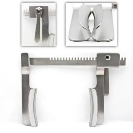
- Rib retractors (rib spreaders): the Finochietto is a self-retaining frame with a blade on the end of each arm, opened with a ratcheted crank; it is opened slowly, over moist laps that protect the wound edges.
- Scapula retractor, such as the Davidson, to lift the shoulder blade.
- Rib instruments: periosteal elevators and rib strippers (such as the Alexander periosteotome and the Matson stripper) to free the periosteum, rib shears (such as the Bethune) to cut a rib, and Sauerbruch rib shears or a rongeur to trim the cut end.
- Rib approximator (rib contractor), such as the Bailey, to draw the ribs together at closure.
- Lung instruments: atraumatic lung-grasping forceps, such as the Duval and the Lovelace, and lung retractors, such as the Allison and the Semb.
- Bronchus clamps, such as the Sarot: large right-angled clamps that close off a main bronchus, their jaws stippled to grip its cartilage.
- Right-angle (Mixter) clamps to pass around vessels and bronchi, and sponge dissectors.
- Staplers, both open and endoscopic, to divide lung tissue and close the bronchus.
- A chest tube passer to place the tubes.
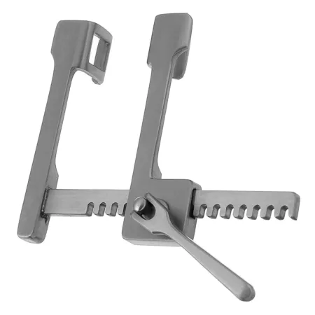
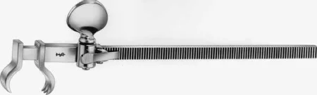
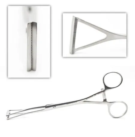
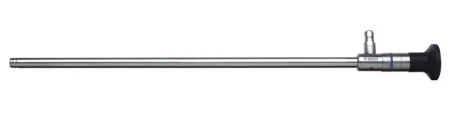
Thoracoscopic (VATS) instruments have two pivot points instead of one, so they open within the small space of a port without pressing on the tissue around it, which keeps them clear of the chest's major nerves.
Heart instruments
A heart case uses a general set with cardiac instruments, thoracic instruments and staplers, and lung instruments if needed; coronary, valve, aneurysm and chest wall sets are added for the operation planned.
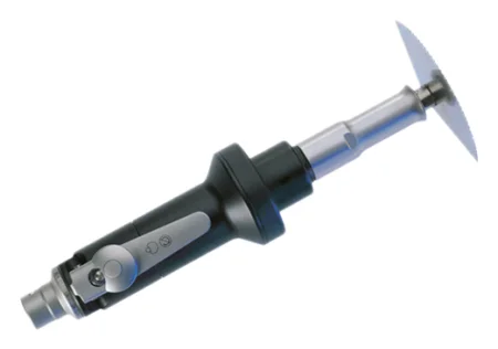
- The sternal saw splits the sternum. An oscillating saw is used for a repeat sternotomy, because it lets the surgeon cut through gradually and avoid the scar tissue behind the bone. A Lebsche sternal knife and mallet are kept on the setup for when a power saw cannot be used, as in a power failure.
- Sternal retractors, some with attachments that hold the left atrium open for mitral surgery, and a retractor that lifts one side of the sternum to expose the internal mammary artery.
- Vascular clamps come as cross-clamps, which close a vessel completely, and partial occlusion (side-biting) clamps, such as the Satinsky and Lambert-Kay, which close off only part of the wall. Their jaws carry fine rows of teeth or serrations made to hold a vessel without crushing its delicate inner lining, so they must be kept in good condition. Small bulldog clamps close smaller vessels.
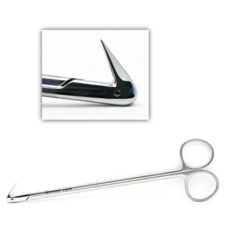
- Fine scissors, such as the angled Potts and the Diethrich coronary scissors, to open and extend vessel incisions.
- Fine forceps, such as the DeBakey and the Gerald, and fine needle holders, such as the Ryder and the Castroviejo.
- Coronary dilators to size a coronary artery, an aortic punch to cut a round hole for a vein graft, and sternal wire twisters and cutters.
- Valve instruments: retractors that expose the valve, valve sizers, holders and suture holders.
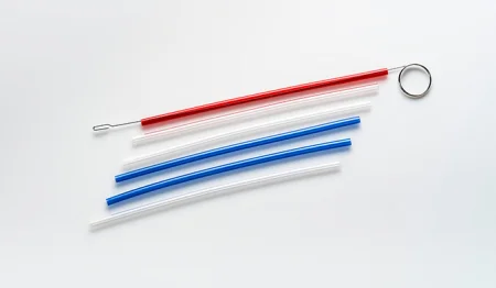
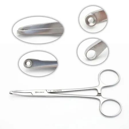
- The Rummel tourniquet: a short length of tubing, bought or cut from a straight catheter, slid down over a cannulation stitch or a vessel tape to tighten it, its stylet pulling the strands through and a hemostat holding them.
- Internal defibrillator paddles stay on the field, ready. When the heart fibrillates, the surgeon places one on each side of it and the circulator sets the charge; everyone stands clear of the patient during a shock.
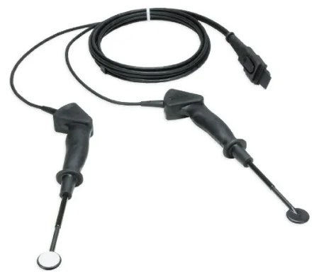
Other equipment in the room includes the heart-lung machine, the cell saver, a defibrillator, an irrigation cooling and warming machine, a warming mattress or forced-air warmer, pacemaker generators, a headlight, a balloon pump, assist devices and ablation sources; chapter 23 teaches the machines.
Supplies, grafts and implants
The Core Curriculum's cardiothoracic supply list includes the aortic punch, the balloon pump, a blower (mister), bulldogs, catheters, defibrillator paddles, the bypass circuit, an off-bypass (stabilizer) system, pacemakers, pledgets, the Rummel tourniquet, shunts, suture boots, a thoracentesis kit, umbilical tape, valve prostheses, vascular clamp inserts, a vein harvesting system and vessel loops. Its vascular items, the Doppler probe, the valvulotome, the vein stripper and the interventional sheaths, guidewires, stents, tunnelers and ports, are taught in chapter 40.
- Suture boots (rubber shods) slip over hemostat tips so they hold suture without damaging it; small dissecting sponges (peanuts or "pill" sponges), tubing, umbilical tapes and vessel loops identify and lift vessels.
- A thoracentesis kit holds a needle passed through the chest wall into the pleural space, usually to remove fluid.
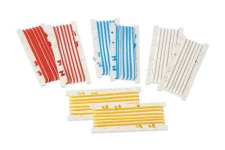
- Sutures: nonabsorbable polypropylene, polyester and similar synthetics, mostly double armed (a needle on each end); size 3-0 is common on large vessels and 5-0 or 6-0 on smaller ones, and finer still on the coronary arteries. Use alternately colored strands and numbered suture holders to keep a valve's many stitches in order.
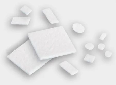
- Pledgets: small pads of Teflon felt on a stitch that reinforce a suture line. Prepare one by clamping a mosquito across its middle and passing the needle through it on each side of the clamp, or pass the clamp and pledget to the surgeon to load.
- Sternal closure: stainless steel wire, sizes 4 to 7, or cables, bands or plates for a frail sternum.
- Grafts: tubes of Dacron, Teflon or PTFE, straight or branched. Knitted grafts are soft and porous; woven grafts have a tight weave that leaks less blood, so they are used when the patient is fully heparinized. Grafts soaked through with collagen need no preclotting, and arch grafts come with side branches for the head vessels. Graft sizers find the size, and only that size is opened.
- Patches of Teflon, PTFE, Dacron, bovine pericardium or the patient's own pericardium close holes and reinforce suture lines.
Every implant (graft, patch, valve, ring or device) is recorded with its type, size, lot and serial number, as the Safe Medical Devices Act requires.
Running a heart field
Expect to keep at least two back tables and three Mayo stands, each set up the same way every time so you always know where an instrument is and have room to prepare supplies; the room is crowded, so plan the space.
- The saphenous vein is usually taken by a surgical assistant at the same time as the chest is opened, with its own Mayo and its own light: the two ceiling lights are on the chest, so a third light or a headlight is needed for the leg.
- Keep instruments used on the groin or leg apart, and change gown and gloves when moving from the leg to the chest, or after holding the heart and moving to the other side of the table.
- Wipe calcium and debris off instruments with a moist sponge, and keep a small basin for clots and debris, so nothing drops back into the heart and becomes an embolus.
- Keep the back table and Mayo sterile and stay scrubbed until the patient has left the room, in case the chest must be reopened.
Drugs and solutions
Chapter 12 teaches these as drugs; here is how each is used at the field.
- Heparin prevents clotting in the bypass circuit: 300 to 400 units per kilogram is injected before bypass, through a central line or straight into the right atrium. Heparinized saline rinses vessels and grafts.
- Protamine sulfate reverses heparin after bypass, given slowly into a vein by the anesthesia provider; it can cause an allergic reaction. When protamine starts, the pump suckers (cardiotomy suction) are turned off, so that blood that is losing its heparin is not returned into the circuit, where it would clot.
- Papaverine relaxes vessel muscle and prevents spasm in the internal mammary and radial artery grafts. A vein solution, such as a balanced salt solution with heparin and papaverine, distends vein grafts, so the surgeon can check them for twists and leaks.
- Topical antibiotic irrigation, such as bacitracin in saline. It must never be suctioned into the cell saver, because in a vein it can harm the nerves and kidneys (chapter 12).
- Hemostatic agents: gelatin sponge with thrombin, oxidized cellulose and collagen for oozing; bone sealant or bone wax on the cut sternum and ribs; fibrin sealant to stop air leaking from the lung or a bronchial join; and adhesives such as albumin with glutaraldehyde to seal aortic suture lines. Gelatin works poorly in a fully heparinized patient.
- Cold and warm solutions: cold saline and slush cool the heart during arrest, and warm saline is used before and after it.
- Label every drug and solution, as chapter 29 teaches; heparinized solutions, papaverine, vein solutions and topical hemostatics matter most.
- Inotropes and antiarrhythmics are kept on hand, and the team helps give them when they are needed.
Cardioplegia, the solution that stops the heart, is taught with bypass below.
Monitoring
Chapter 13 teaches the monitors, and a heart case uses nearly all of them. Heart and chest cases add sensors on the forehead that measure the oxygen in the brain (cerebral oximetry), TEE that also finds air left in the heart and guides cannulas and balloon pumps, and, with one lung collapsed, frequent blood gases, because blood passing through the airless lung picks up no oxygen.
Counts
An initial instrument count is advised for every procedure in which an instrument could be left behind, as in an opened chest. Count small items that could be lost in the chest, such as peanuts, Kittner dissectors and umbilical tapes, with the sponges; count vessel loops and rubber shods too, and the laps used to stop prep from pooling at the neck. When there are two incisions, such as the chest and the leg, each closure gets its own count. Follow the count times in chapters 28 and 29.
8Chest drainage
After the chest or pleura is opened, closed chest drainage removes air, blood and fluid from the pleural space or the mediastinum and restores the negative pressure, so the lung can expand again; chapter 25 teaches the chest tube and the three-chamber drainage unit with its water seal. The unit is set up by its maker's instructions. After a lung resection, tubes of 28 to 30 French leave through stab wounds near the eighth or ninth rib space at the anterior axillary line; after heart surgery, tubes drain the mediastinum and the pericardium, and the pleura if it was opened. If a tube comes out, cover the hole at once with the sterile petroleum gauze kept with the patient for this purpose, so air cannot rush in.
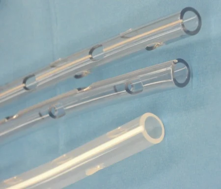
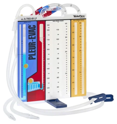
lift the drainage unit to the level of the chest or tip it over, even for a transfer. It stays upright and below the chest at all times, because fluid that runs back can collapse the lung, or compress the heart.
9Lung and chest procedures
A diagnostic endoscopy often comes first, and thoracic surgery can then be done open, by VATS or with the robot. Most lung operations, 80 to 90 percent, are now done by VATS, but the open instruments stay ready, often just outside the room, in case the surgeon must convert.
Bronchoscopy and endobronchial ultrasound
What and why: a bronchoscopy looks directly at the trachea and bronchi to find a lesion and its extent, take biopsies, brushings and washings, and treat: removing a foreign body, opening a narrowing, cutting away a tumor or placing a stent. A rigid scope, under general anesthesia with the patient ventilated through it, takes large instruments, as for a foreign body in a child; a flexible scope reaches the smaller bronchi while the patient breathes around it; both may be used in one session.
Two newer methods help find and stage lung cancer: navigational bronchoscopy steers a guide along a map built from the CT scan to a small suspicious area of the lung for biopsy, and endobronchial ultrasound (EBUS) finds lymph nodes in the mediastinum and the hilum for a needle biopsy.
Key points: chapter 36 teaches the rigid and flexible setups, the Lukens trap and the specimens step by step. When cancer is suspected, the surgeon examines the healthy lung first, so cancer cells are not carried into it. Guide instruments into the channel when asked, and confirm the source of each specimen with the circulator and surgeon. Afterward the patient has nothing by mouth until the gag reflex returns, which may take 2 to 3 hours; the complications are injury to the airway or the lung, a torn blood vessel near the bronchi, and infection.
Wound class: II, the airway is entered through its own opening (as chapter 36 explains).
Mediastinoscopy
What and why: a lighted rigid scope passed through a small incision above the sternal notch, to see and biopsy lymph nodes and tumors around the carina and the upper lobe bronchi. It stages lung cancer before a thoracotomy and can biopsy the thymus. EBUS has largely replaced it.
Position, prep and drape: general anesthesia; supine with the neck extended on a shoulder roll; a thyroid-type prep and drape.
Setup: a minor set, the mediastinoscope with its light cord, suction and aspirating tubes, biopsy forceps, an ESU and a smoke evacuator, and a long aspirating needle. A sterilized scope is preferred to one that is only high-level disinfected. A thoracotomy tray stays in the room in case a biopsy causes heavy bleeding.
Key steps:
- A short incision, about 2 cm, above the sternal notch, down to the fascia in front of the trachea.
- The surgeon tunnels along the front of the trachea with a finger, then passes the scope down the tunnel.
- Each node is first punctured with the needle and syringe to prove it is not a blood vessel; then cup biopsy forceps take tissue. Lift each piece off the forceps onto a saline-moistened Telfa pad or into its container.
- Bleeding is controlled with the ESU, a gelatin sponge or pressure from a small sponge on a carrier, and the incision is closed.
After surgery: many patients go home the same day; bleeding and infection are the complications.
Wound class: I, no tract is entered.
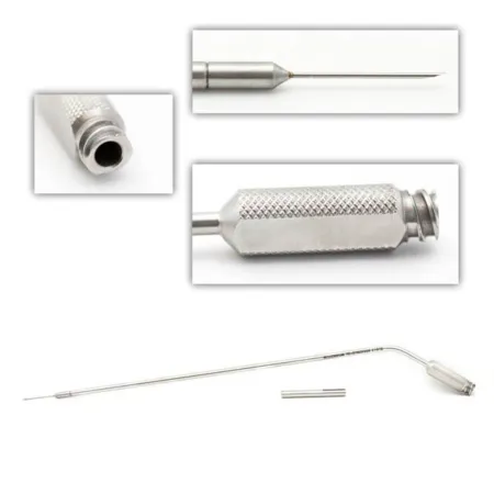
Thoracotomy: opening and closing the chest
Open chest operations generally open and close the same way.
Opening:
- The skin, fat and muscle are cut with the knife and the ESU.
- A scapula retractor lifts the shoulder blade, and the intercostal muscles are cut to enter the chest.
- Sometimes a rib is removed: the surgeon cuts the periosteum, strips it off with an elevator or rib rasp, cuts the rib with rib shears and trims the sharp ends with a rongeur.
- Moist laps cover the wound edges and the rib spreader goes in, opened slowly and gently.
Closing:
- Chest tubes are placed and the instruments come out.
- Four to six heavy pericostal sutures are passed around the ribs above and below, or through holes drilled in them, and tagged with hemostats.
- A rib approximator draws the ribs together while the sutures are tied.
- The muscles, fat and skin are closed in layers, and the tubes are connected to the drainage unit.
A thoracotomy is painful. An epidural, a local anesthetic catheter in the chest wall or freezing of the intercostal nerves may be used, along with other pain drugs.
VATS and robotic chest surgery
What and why: video-assisted thoracic surgery (VATS) works inside the chest through ports, with a rigid scope and long thoracotomy instruments. It spares the muscles a thoracotomy cuts, has the same goals as the open operation, and needs a full surgical team under general anesthesia. It is used to treat pleural disease, such as blebs and effusions, to biopsy masses, and for wedge resection, lobectomy, lung volume reduction, removal of the pericardium, sympathectomy, stopping bleeding and removing clots. Many authors call it thoracoscopy, though one textbook keeps that word for a smaller look at the pleura under sedation. Chapter 21 teaches the scopes and ports.
Position, prep and drape: lateral with the operative side up and a chest prep, with a double-lumen tube so the lung on the operated side collapses; the surgeon marks the shoulder blade and the port sites on the skin before starting.
Setup: a 5 or 10 mm thoracoscope, 0 or 30 degree, camera and light source, endoscopic graspers, dissectors, scissors, clip appliers and staplers, with open thoracic trays at hand. Insufflation equipment is in the room; as chapter 21 explains, gas is used only when the anesthesia provider wants help collapsing the lung.
Ports and team: three or four ports are placed in a "baseball diamond" pattern so the instruments do not cross inside the chest, or one larger port (uniportal VATS) carries them all. The first port usually goes in through a cut of 2 to 3 cm somewhere from the fifth to the seventh rib space, for a 10 or 12 mm trocar. In VATS the surgeon and assistant stand at the patient's front, while in an open thoracotomy they stand at the back; the scrub stands at the back at the level of the legs, with the Mayo over the legs.
Wedge resection or lung biopsy (the model VATS case):
- Ports are placed and the scope goes in.
- The lung is held with a grasper and a wedge of tissue, or a small lesion, is cut and sealed with an endoscopic stapler; lymph nodes may be taken too.
- The surgeon fills the chest with warm saline while the lung is inflated and watches the staple line for bubbles of air; leaks are sewn or sealed.
- A chest tube goes in through the lowest port, and the ports are closed.
After surgery: a hospital stay of about 4 to 7 days; the complications include infection, bleeding, atelectasis, pneumonia, a pneumothorax and air under the skin.
A nodule may be marked beforehand with methylene blue dye so the surgeon can find it. When a bronchoscopy comes first, set it up on its own table, then cover that table and move it away before the VATS begins. Through the scope the surgeon can also put talc into the pleural space, which irritates the pleura so the layers stick together and fluid or air cannot collect again (pleurodesis), or cut the sympathetic nerve chain beside the spine for severe sweating of the hands (sympathectomy).
Robotic chest surgery: robots are used for lobectomy, removal of a thymoma, esophagectomy and sympathectomy, with the patient lateral on a double-lumen tube and the team ready to convert quickly to an open thoracotomy in an emergency.
Wound class: II for a wedge resection or lung biopsy. The lungs are part of the lower respiratory tract, so cutting and stapling across lung tissue enters that tract under controlled conditions; one surgical technology textbook lists VATS lung cases as Class I.
Lobectomy and smaller resections
What and why: a lobectomy removes one or more lobes of a lung, most often for lung cancer confined to a lobe; also for spread of cancer from elsewhere, bronchiectasis, giant blebs, benign tumors, fungal and other localized infections, cysts and injury. It can be open or by VATS; the principles are the same.
Position, prep and drape: lateral for a posterolateral thoracotomy, or supine for some upper and middle lobe resections; a thoracotomy prep and drape.
Setup: a major thoracotomy set and a general set, bronchus instruments, deep vascular clamps and forceps, vessel loops, umbilical tapes, clips, linear staplers and fibrin sealant.
Key steps:
- The chest is opened, and the surgeon examines the whole lung and the mediastinum to make sure the disease has not spread further.
- The lobe is held with lung-grasping forceps, the pleura over the hilum is opened, and the pulmonary artery and vein branches to the lobe are freed with sponge dissectors and right-angle clamps.
- Each branch is tied twice and divided, or divided with a stapler.
- The bronchus is clamped and divided, and closed with sutures or a stapler. Keep suction ready while the bronchus is open, so blood and secretions cannot drain into the other lung; you may be asked to run it.
- Out comes the lobe, and pleura is sewn over the bronchial stump.
- The chest is irrigated and the lung that is left is inflated, so any air leak shows; chest tubes are placed and the wound is closed.
Smaller resections: a segmentectomy removes one or more segments and spares the healthy lung around them; before cutting, the remaining lung is inflated to show the boundary of the diseased segment. A wedge resection removes a wedge of lung holding a small, peripheral lesion, without following the segments.
Specimen: the lobe, and lymph nodes labeled by station.
After surgery: the complications include infection, bleeding, atelectasis, pneumonia, a pneumothorax, a pulmonary embolism, air under the skin and a lasting air leak.
Wound class: II, because the bronchus is opened. One surgical technology textbook lists a lobectomy as Class I and notes that opening the bronchial tree changes the class.
Pneumonectomy
What and why: a pneumonectomy removes a whole lung, usually for cancer; also for widespread bronchiectasis or abscess in one lung, or to debulk a tumor pressing on vital structures.
Position and setup: lateral, with a thoracotomy prep and drape and the same sets as a lobectomy: the major thoracotomy and general sets, bronchus instruments, deep vascular clamps, vessel loops, umbilical tapes, clips and staplers.
Key steps:
- Through a thoracotomy, the lung is held away with lung forceps or retractors, adhesions are freed and the dissection is carried to the hilum.
- The vagus, phrenic and, on the left, recurrent laryngeal nerves are held out of the way with vessel loops or moist umbilical tapes.
- The pulmonary artery and the superior and inferior pulmonary veins are clamped, divided and tied with heavy silk, oversewn with fine suture or stapled.
- The main bronchus is clamped near the carina, divided and closed with sutures or staples, and the lung is removed.
- Saline fills the chest while the anesthesia provider inflates the lung with positive pressure, so leaks at the bronchial stump show as bubbles. A flap of pleura or intercostal muscle may cover the stump.
- Chest tubes may be placed, and the wound is closed.
After a pneumonectomy, the chest tubes are not connected to suction. Besides the complications of a lobectomy, the middle of the chest can shift toward the empty side (mediastinal shift), and fluid can flood the remaining lung (acute pulmonary edema).
Wound class: II, the main bronchus is opened; one textbook lists Class I.
Lung volume reduction
For severe emphysema, the worst, overinflated parts of the lung are cut away with linear staplers through a sternotomy or VATS, so the healthier lung can work better. Strips of PTFE or bovine pericardium, or a sealant, reinforce the fragile staple lines; the lung is reinflated under saline to look for leaks, and the chest tubes go to water seal without suction.
Wound class: II, as for a wedge resection.
Acute pulmonary embolectomy
For a pulmonary embolism that causes life-threatening circulatory failure, the clots are removed through a median sternotomy on bypass, usually without stopping the heart, which stays warm. Have Rummel tourniquets ready for the venae cavae, and suction ready before the artery is opened. The pulmonary trunk is opened between two traction sutures with a #11 blade and Potts scissors, and the clots are removed with forceps and suction while the lungs are squeezed to push out smaller ones; keep every clot as a specimen. The artery is closed before bypass ends, and some surgeons add a vena cava filter (chapter 40).
Wound class: I; the pulmonary artery is opened, but no tract is entered.
Decortication and empyema drainage
What and why: long-lasting inflammation, infection (empyema or tuberculosis) or a tumor lays a fibrous peel over the lung that traps it. Decortication strips the peel away so the lung can expand; it can bleed heavily and injure the lung.
Position and setup: lateral, with a thoracotomy prep and drape; the thoracotomy and general sets, bronchus instruments, deep vascular clamps, vessel loops and umbilical tapes, with curettes as an extra, and culture tubes for the fluid.
Key steps:
- A posterolateral thoracotomy, usually at the fifth or sixth rib, part of which may be removed.
- Adhesions are divided, and pus or fluid is drained and sent for culture.
- The anesthesia provider inflates the lung from time to time to show where peel is still holding it down, and the surgeon peels it off with sponge dissectors and sharp dissection, handling the lung gently.
- Chest tubes are placed and the chest is closed.
After surgery: physical therapy goes on for months so the lung expands fully. The complications are infection, bleeding, a peel not fully removed, so breathing stays hard, and paralysis of the diaphragm if the phrenic nerve is injured.
Empyema drainage: an early empyema may be drained by a closed thoracostomy, a chest tube through a rib space under local anesthesia, usually outside the OR; a chronic one may need a rib resection with a large drain, or an open thoracostomy, in which part of a rib is removed and the cavity packed open; take aerobic and anaerobic cultures, and keep suction ready so the pus does not spill.
Wound class: IV when infection is already present, as in an empyema; one textbook lists a decortication as Class I, which fits only when no infection is found.
Thymectomy
What and why: a thymectomy removes the thymus, for a tumor of the gland (thymoma) or for myasthenia gravis; chapter 9 describes the gland. The classic approach splits the sternum, and the gland can also be removed through the neck, by VATS or with the robot.
Before surgery: a patient with myasthenia gravis may have trouble breathing afterward and may need to stay intubated or have a tracheostomy; the drugs for myasthenia, such as neostigmine, are agreed with the anesthesia provider and kept at hand, and a tracheostomy tray is in the room.
Position, prep and drape: supine; the prep runs from the chin to below the umbilicus; laps tucked at the sides of the neck stop the solution pooling and are then removed (they are counted); a thyroid sheet.
Setup: basic, vascular and thoracotomy trays, a sternal saw with a sternal knife and mallet, rake retractors, clip appliers, bone wax, peanut or Kittner dissectors and umbilical tape (both counted), and a chest tube with its drainage unit if a pleura is opened.
Key steps:
- A median sternotomy; bone wax controls bleeding from the cut sternum.
- The thymus is freed from the fat of the anterior mediastinum and the pericardium, keeping clear of the great vessels; its vessels are clamped, tied or clipped and divided.
- A drain is placed, or chest tubes if a pleura was opened, and the sternum is closed with heavy wire.
Wound class: I.
Lung transplant
What and why: one or both lungs are replaced with donor lungs. A single lung is used for restrictive lung disease, emphysema, pulmonary hypertension and other end-stage lung diseases without infection; both lungs are replaced for cystic fibrosis or chronic infection. The recipient starts immunosuppression and infection prevention before surgery.
Setup: the donor and recipient are two patients, each with their own plan and setup. Thoracotomy instruments, cold perfusion solution and staplers are used, with bypass ready if the patient becomes unstable, and TEE watches the heart.
Key steps (recipient):
- Lateral for a single lung, supine for both; a wide prep from the nipple line to the knees.
- A thoracotomy for one lung; for both, two thoracotomies, a sternotomy or a clamshell incision.
- The lung to be removed is collapsed and its artery clamped; if the patient cannot tolerate it, bypass (sometimes from the groin) supports the circulation.
- The diseased lung is removed, leaving the tissue around the bronchus so its blood supply is kept.
- Three joins are made: bronchus to bronchus, pulmonary artery to pulmonary artery, and the recipient's pulmonary veins to a cuff of the donor's left atrium. The bronchial join may be wrapped with a flap of omentum or intercostal muscle.
- The new lung is inflated and inspected, chest tubes are placed and the chest closed. A bronchoscopy at the end clears secretions and checks the bronchial join.
After surgery: drugs against rejection for life; the complications include infection, bleeding, atelectasis, pneumonia, fluid in the lungs and rejection of the new lung.
Wound class: II, the airway is opened; one textbook lists Class I.
Pectus excavatum and carinatum repair
What and why: in pectus excavatum the sternum and costal cartilages are pushed in, giving a sunken chest. It is often noticed in early adolescence and can worsen during growth. Repair is most often cosmetic; fewer patients have it for impaired heart and lung function, with complaints such as chest pain, shortness of breath on exercise or rhythm disturbances. In pectus carinatum the sternum pushes out; it is usually treated first with a brace, and open repair is for a deformity the brace does not correct.
The Nuss procedure (minimally invasive): one or more curved metal bars are placed behind the sternum to push it out, and they stay for about 3 years. It is most often done between about 10 and 16 years of age.
- Position: supine, the upper chest raised on a soft roll, with the arms placed out of the way of the sides of the chest as the surgeon prefers.
- Setup: a 5 mm thoracoscope, the pectus set with bars, a bender, stabilizers and accessories, a minor set and umbilical tape. A titanium bar is used if the patient is allergic to nickel.
Key steps:
- The chest is measured and a template bent to the shape wanted; the bar is bent to match with the plate bender.
- Two incisions are made on the sides of the chest in line with the deepest part of the defect, and pockets are made under the skin.
- The thoracoscope goes in through a small port to watch the next steps.
- An introducer is passed under the sternum, across the front of the mediastinum and out the other side, then used to pull an umbilical tape back through the tunnel.
- The tape pulls the bar through with its curve facing back, and the bar is flipped over to push the sternum out.
- Stabilizers are fixed to the bar ends with wire or heavy suture, and the bar is sewn to the chest wall muscle so it cannot move.
- The wounds are closed and a chest X-ray checks for a pneumothorax.
After surgery: the patient stays supine in recovery so the bar does not shift, may have an epidural, stays in hospital for 3 to 4 days, and resumes limited activity after about 6 weeks. Complications include bar movement, a pneumothorax and fluid around the lungs or heart.
Open repair: the modified Ravitch operation removes only short lengths of the deformed cartilage, keeping its covering (the perichondrium), and fixes the sternum in a better position; it is now kept for carinatum that bracing did not correct and for mixed deformities.
Wound class: I, no tract is entered.
Esophageal atresia and tracheoesophageal fistula
What and why: in esophageal atresia the esophagus is interrupted, its upper part ending in a blind pouch. It often comes with a tracheoesophageal fistula (TEF), a channel that should not exist joining the trachea to the esophagus, which lets saliva and milk, or stomach secretions, reach the lungs. It can come with other defects, such as septal defects, tetralogy of Fallot and neural tube defects. The esophagus is joined at once when the two ends reach and the infant is well enough; otherwise the repair is staged or delayed, and a segment of colon can lengthen the esophagus.
Position, prep and drape: the infant lies in the thoracotomy position with the head of the table slightly raised; the prep runs from the neck to the iliac crest, front and back. Chapter 13 teaches how to keep an infant warm.
Key steps:
- A posterolateral thoracotomy; a Finochietto retractor goes in and the azygos vein is tied and divided.
- The anesthesia provider advances a tube already in the upper pouch (a Replogle tube) so the pouch can be seen. The fistula is looped with a vessel loop and divided.
- The hole in the trachea is closed with fine absorbable sutures and tested under warm saline.
- Both ends of the esophagus are freed, held with Babcocks, and joined in one layer with very fine absorbable suture, the tube passed across the join.
- A small chest tube may be placed and the wound closed in layers.
After surgery: the airway is suctioned often until the baby can swallow, and feeding starts 2 to 6 days later, once a contrast study shows the join is sound; a leak, infection or a recurrent fistula are the complications.
Wound class: II, the esophagus and trachea are opened.
10Chest injuries
Blunt injury to the chest causes at least a fifth of all trauma deaths, and motor vehicle crashes cause most chest injuries. A penetrating wound at or just below the nipple line, or the tips of the shoulder blades, may involve both the chest and the abdomen. Chapter 11 teaches trauma care in general, and chapter 28 setting up for a trauma case.
The injuries that need surgery at once are a tension pneumothorax, an open pneumothorax, a flail chest, a massive hemothorax and pericardial tamponade. A thoracotomy may be started in the emergency department to relieve a tamponade or stop bleeding from the heart, lungs or great vessels, and the patient then goes straight to the OR.
Setting up a trauma thoracotomy, in order of need:
- Exposure: at least two suctions running, sponges, and a Finochietto retractor and Deaver retractors from the start.
- Removing blood and clots: a large basin for clots, and 10 to 15 laps dipped in warm saline and wrung out, with at least 30 more in reserve.
- Cardiovascular sutures by the surgeon's card, or 2-0 and 3-0 polypropylene if none is known; fibrin sealant may also be used.
- The chest wall is closed at the end, often in one layer, over chest tubes.
An autotransfusion system should be ready for lung injuries. As chapter 12 explains, contamination or sepsis is not an absolute bar to cell salvage once the wound is washed with antibiotic irrigation, though makers still advise against it with a cancer, and the surgeon and anesthesia provider decide; pleural fluid is suctioned off with a separate suction first, and only the blood that collects afterward is salvaged.
Chest wall
- An open chest wound that lets air in is sealed at once, often with petrolatum gauze, and the air is let out with a needle (needle thoracostomy) or a chest tube; a large wound needs a ventilator.
- Flail chest: two or more neighboring ribs each broken in two or more places, so a segment of chest wall floats free. It moves the wrong way, sinking in when the patient breathes in and bulging out on breathing out, and the lung beneath it is usually bruised. One reference defines it as three or more neighboring ribs. The patient may need a breathing tube and a ventilator.
Lungs and pleura
- Simple pneumothorax: air in the pleural space, with no opening to the outside air.
- Open (communicating) pneumothorax: the wound opens the pleura to the air, the "sucking chest wound." The lung on that side collapses, and the middle of the chest shifts and squeezes the other lung.
- Tension pneumothorax: air keeps entering the pleural space and cannot leave, so the pressure climbs, the mediastinum, lung and great vessels are pushed to the other side, blood return to the heart falls and the patient goes into shock. It can kill quickly and is relieved with a needle or a chest tube.
- Hemothorax: blood in the pleural space. A large chest tube, 36 to 40 French, is placed in the emergency department; when 1,000 to 1,500 mL of blood drains, the chest is explored in the OR.
- Pulmonary contusion, a bruised lung that bleeds into its air sacs, is the most common chest injury in children. A torn lung may be clamped in the emergency department, then sutured or stapled quickly; a lobectomy is sometimes needed.
- Diaphragm injury lets abdominal organs slide up into the chest, where they can strangulate.
Heart and great vessels
- Blunt cardiac rupture, a tear of a chamber, valve or other structure, is among the most lethal chest injuries, classically from the steering wheel. The patient may arrive with a Foley balloon plugging a small hole in the heart, or a hand pressing on a larger one; the repair is fast and simple first.
- Penetrating cardiac wounds from knives and bullets kill by bleeding and by cardiac tamponade, blood in the pericardium that stops the heart filling. Its signs are distended neck veins, muffled heart sounds and a narrowing pulse pressure; it is relieved by needle drainage (pericardiocentesis), a pericardial window or an emergency thoracotomy. In the trauma repair, patch grafts are used rather than tube grafts, and suturing over pledgets may take too long.
- Myocardial contusion, a bruised heart, usually of the right ventricle, shows as arrhythmias; it is watched on a monitor, but a valve torn by the blow needs repair.
- Aortic injury, from a high-speed crash, a fall from a height or a hard blow to the side of the chest, can tear or shear the aorta; most of these patients die at the scene or soon after. Repair may use bypass to protect the spinal cord from the cross-clamp, and some tears are treated with a stent graft through the femoral artery.
Wound class: a fresh traumatic wound is Class III, and so is an operation with open cardiac massage; an old wound with dead tissue, or an existing infection, is Class IV. An incision made after blunt injury, with no open wound, is classed by the usual rules: Class I if no tract is entered, Class II if the lung or another tract is opened.
11Cardiopulmonary bypass
Cardiopulmonary bypass (CPB) takes over the work of the heart and lungs, and the cardiopulmonary bypass machine is the equipment that does it. Blood returning to the heart is drained to the heart-lung machine, given oxygen, and pumped back into the arteries. With the heart empty and stopped, the surgeon can work inside it in a still, nearly bloodless field, and can lift and turn it without causing fibrillation or a fall in blood flow to the body. Valve replacement, heart transplant and aneurysm repair need bypass; more and more coronary bypasses are done without it.
The bypass circuit
Blood drains by gravity from cannulas in the right atrium or the venae cavae into a venous reservoir, and the heart-lung machine pumps it through an oxygenator with a heat exchanger that warms or cools it, then back through a filter into a cannula in the ascending aorta.
- The pump is a roller or a centrifugal pump; its flow is mostly steady, without pulses, so the arterial pressure shows as a mean on the monitor.
- The oxygenator adds oxygen and removes carbon dioxide across a membrane.
- Suction lines (the cardiotomy suckers) return blood shed in the field to the reservoir, which filters it and removes foam; a separate roller pump delivers the cardioplegia.
- A vent drains blood and air from inside the heart so the left ventricle does not stretch; it is usually placed through the right superior pulmonary vein.
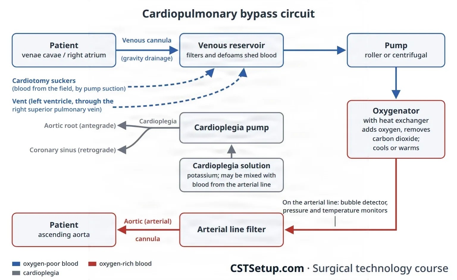
The perfusionist primes the circuit with fluid, clears it of air and runs its safety devices, such as a bubble detector and an arterial line filter; chapter 23 teaches the machine as equipment. At the field, the scrub helps place and remove the cannulas.
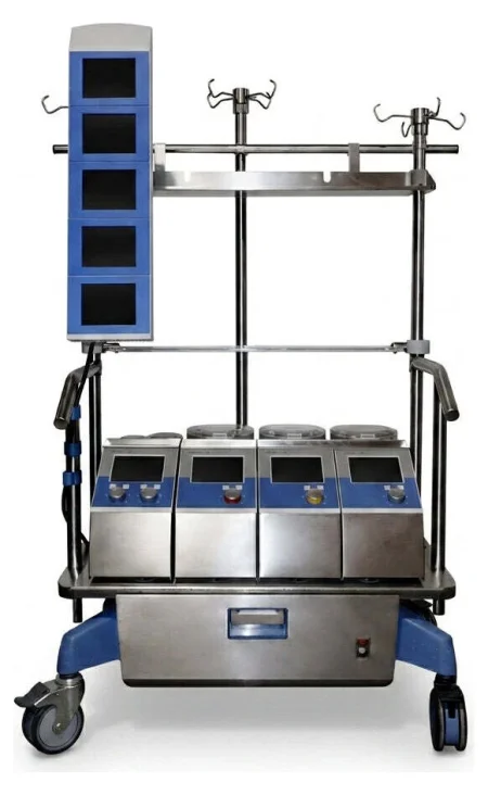
watch the bypass lines, and the heart, for air. Tell the surgeon at once if you see it, because air pumped into the arteries becomes an air embolus.
The heater-cooler that warms and cools the patient has spread Mycobacterium chimaera infection through its tank water carried in the air, so it is cleaned by the maker's instructions, filled only with sterile or filtered water, and its exhaust is pointed away from the sterile field.
Heparin and the ACT
The patient is given heparin before bypass, and the clotting time is checked with the activated clotting time (ACT) before bypass starts and at intervals during it. It is kept above about 480 seconds, the target varying with the test device; one reference gives above 500 and above 600 seconds. If heparin must be given again after protamine, as when bypass has to be restarted, it is confirmed before the pump runs. For a patient with heparin-induced thrombocytopenia (HIT), a planned operation waits until the antibody tests are negative, and an urgent one may use bivalirudin in place of heparin.
Median sternotomy
Most bypass cases open the chest through the sternum.
- The incision runs from the sternal notch to below the xiphoid. The fat and the linea alba are divided with the knife or ESU, the surgeon frees the tissue behind the sternal notch and the xiphoid with a finger, and the xiphoid is split in the midline with curved Mayo scissors.
- The sternal saw divides the sternum. Army-Navy retractors lift the edges while bleeding from the periosteum is coagulated, and bone wax may be pressed on the marrow.
- The sternal retractor is opened, sometimes over moist laps, to show the pericardium.
- The pericardium is lifted with forceps so the heart is not cut, opened with the ESU or scissors, and its edges sewn up to the chest wall with traction sutures; this lifts the heart and makes a well that holds irrigation.
For a repeat sternotomy, scar may hold the heart against the back of the sternum. The surgeon uses the oscillating saw, may expose the groin or axillary vessels first so bypass can start quickly, and the perfusion team is in the room for the opening.
Cannulation
Cannulas go in a set order unless the surgeon says otherwise: the arterial cannula first, so blood can be given at once if needed, then the venous, the retrograde cardioplegia catheter, the vent and the antegrade cardioplegia cannula. TEE may guide and confirm placement. The technique is much the same at every site: a purse-string stitch, a Rummel tourniquet over its ends held with a hemostat, an opening inside the purse-string, the cannula in, and the tourniquet snugged and tied to the cannula with heavy silk.
- The aortic cannula: two purse-strings, with or without pledgets, go in the front of the ascending aorta. The surgeon cuts a small opening between them with a #11 blade (an aortotomy, an incision into the aorta) and covers it with a finger, then inserts the cannula, whose end is clamped so the aorta cannot bleed back through it. It is filled with blood to push out the air before it is joined to the arterial line, under a saline drip or while the perfusionist runs a little prime out of the line.
- The venous cannula: through a purse-string in the right atrial appendage, held with a clamp (such as a Cooley spoon clamp) while the tip of the appendage is cut off. A two-stage cannula drains both the atrium and, from its tip in the inferior vena cava, the lower body; it is enough when the atrium need not be emptied completely, as in many aortic valve replacements. Bicaval cannulation places one cannula in each vena cava; with tapes and tourniquets snugged around both cavae, all the venous blood goes to the pump (total bypass), which is used for mitral valve replacement, septal defects, ventricular aneurysm repair and transplant, and whenever the right side of the heart is opened. In partial bypass some blood still passes through the heart.
- Retrograde cardioplegia catheter: through a purse-string in the right atrium into the coronary sinus, its place confirmed by TEE or by the surgeon's touch; its pressure is monitored.
- The vent: through the right superior pulmonary vein, across the mitral valve into the left ventricle, connected to the pump suction.
- Antegrade cardioplegia cannula: a small needle-tipped cannula through a pledgeted purse-string in the ascending aorta, between the cross-clamp and the heart.
- Femoral cannulation: through the groin, femoral cannulas are placed in the femoral artery and vein over a guidewire with dilators (the Seldinger technique), as in minimally invasive surgery, a dissection or a repeat sternotomy, or when the aorta cannot be used. The axillary artery is another arterial site, often through a short graft sewn onto it, when the aorta is diseased or the brain will be perfused during circulatory arrest.
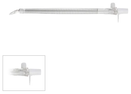
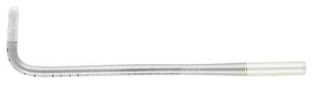
Keep a tubing clamp on each cannula until it is connected, and know which lines bring blood to the patient and which take it away.
Stopping and protecting the heart
Once bypass is running, the surgeon places the aortic cross-clamp across the ascending aorta, between the aortic cannula and the heart. This cuts the heart off from the circulation so cardioplegia can fill the coronary arteries without being washed out.
Cardioplegia is the planned stopping of all the heart's activity. An arrested heart uses far less energy than a beating or fibrillating one, and cardioplegia protects the muscle while its blood supply is cut off. The solution contains potassium (2 to 50 mEq/L), its most important ingredient, which stops the heart in diastole, with buffers against the acid that builds up; it is usually given cold, at 4 to 10°C, and may be given warm. It is given two ways:
- Antegrade, into the aortic root below the cross-clamp. The pressure closes the aortic valve, so the solution has nowhere to go but the coronary arteries. If the aortic valve leaks, the solution fills and stretches the left ventricle instead, so it is given retrograde. It can also go into finished vein grafts; handheld cannulas can put it straight into the coronary openings, but because they can injure the openings they are kept for when retrograde delivery cannot be used.
- Retrograde, through the coronary sinus, backward through the heart's veins; it helps when the coronary arteries are blocked.
When the ECG goes flat, the heart is arrested; the dose is repeated about every 15 to 20 minutes, or when fine fibrillation appears. A last warm dose before the clamp comes off helps the heart recover.
Cooling: the heat exchanger cools the whole body, and cold saline or slush is applied around the heart. Moderate cooling to 28°C halves the body's oxygen use. Slush is kept free of large ice chips, which can injure the phrenic nerves in the pericardium. Ventricular fibrillation is less likely above about 32°C, and cooling harms clotting and wound healing, which may slow the end of bleeding after heparin is reversed. Chapter 13 teaches deliberate hypothermia.
Circulatory arrest: for complex aortic arch surgery, where no clamp can be placed, the patient is cooled to about 18°C and the pump is stopped, so the field is dry, while the brain is protected as the arch section below describes.
Coming off bypass
- Near the end of the repair the perfusionist rewarms the patient, and the surgeon removes the air from the left ventricle and the aorta, using the vent, a needle in the aorta, the table tilted head down, the lungs inflated and TEE to find any air left.
- The cross-clamp comes off. The heart often starts beating on its own; if it fibrillates, it is defibrillated.
- Temporary pacing wires are sewn to the atrium and ventricle.
- With the heart beating and the lungs ventilated, the perfusionist slowly reduces the venous drainage and the arterial flow until the heart carries the circulation, and bypass ends.
- The vent is usually removed first, before bypass ends, then the venous and aortic cannulas, each purse-string tied as its cannula comes out. Femoral cannulas come out the same way, the vein first, and each vessel is closed with fine polypropylene.
- Protamine reverses the heparin; the pump suckers go off as it starts.
- Tubes drain the pericardium, and the pleura if opened. The pericardium is usually left open, so collecting blood cannot cause tamponade.
Problems after bypass: bleeding from cannulation sites, from heparin not fully reversed or from damaged tissue; atelectasis; brief changes in thinking and perception. Blood's contact with the circuit can also set off a body-wide inflammatory response, fluid shifts and showers of tiny emboli.
12Coronary artery surgery
Coronary angioplasty
What and why: percutaneous coronary intervention (PCI) is done in the cardiac catheterization lab. A balloon catheter is passed into the narrowed coronary artery and inflated to open it (percutaneous transluminal coronary angioplasty, PTCA), and a bare-metal or drug-eluting stent is usually placed to hold it open. Clot-dissolving drugs can reopen a freshly blocked artery, and antiplatelet drugs, such as aspirin and clopidogrel, help keep it from narrowing again. PCI is often used during a heart attack and can spare a patient bypass surgery, though coronary disease keeps progressing and some patients need a bypass later. For disease of the left main coronary artery or of all three vessels, bypass surgery is still the standard of care. In a hybrid room, a surgeon may sew a bypass graft and a cardiologist place stents in the same session. Chapter 22 teaches balloons, stents and catheter work. A catheter case through the skin enters no tract, so it fits Class I.
Coronary artery bypass grafting (CABG)
What and why: CABG routes blood around a narrowed coronary artery with a graft, a conduit (a channel for blood) taken from the patient's own body, to relieve angina and lengthen life in patients with disease of several vessels. The internal mammary artery stays attached at its upper end to the subclavian artery, so it brings its own blood; a vein or a free artery graft is sewn from the aorta to the coronary artery beyond the blockage. Chapter 5 teaches the grafts' anatomy.
The conduits:
- The left internal mammary artery (LIMA) to the left anterior descending (LAD) artery is the first choice: it stays open longest. The STS guideline recommends an internal mammary artery whenever the LAD needs a graft.
- A second arterial graft, the right internal mammary or a radial artery, should be considered in suitable patients, though taking both mammary arteries may add to the risk of a sternal infection.
- The radial artery is taken from the arm the patient uses less, after a test such as the Allen test or a Doppler study shows the ulnar artery alone can feed the hand; a drug is used to prevent spasm. More and more often it, too, is taken with an endoscope.
- The great saphenous vein remains the workhorse when several grafts are needed, usually taken through small incisions with an endoscope. It is reversed, or its valves are cut with a valvulotome, so they cannot block the flow, and it is flushed with heparinized blood or saline and kept moist until it is used. Chapter 40 teaches the vein itself and the open harvest for a leg bypass.
- The right gastroepiploic artery, from the stomach, is rarely used, when other conduits are poor.
Endoscopic vein harvesting (EVH) has become the standard way to take the saphenous vein for a bypass in the United States: compared with one long leg incision, it means less pain and far fewer wound infections, collections of blood or fluid, and wound breakdowns. An emergency may still call for the faster open harvest. Many centers map the vein with Doppler ultrasound before surgery, to find it and check its size.
- With the knee bent and supported, a cut of about 2 cm is made over the vein on the inner leg near the knee, and the vein is found under direct vision.
- A sealing port goes into the incision, and the endoscope is passed along the vein inside its cannula while carbon dioxide holds the tunnel open.
- The side branches are sealed and cut with the kit's cautery device, or with clips and bipolar energy, and small cuts at the groin and ankle let the vein's ends be divided; the vein is drawn out through the knee incision.
- The tunnel is checked for bleeding, a drain is placed if needed, the incisions are closed and the leg is wrapped firmly.
- On the back table the vein is checked for tears and loose branches, which are clipped or repaired, and it is kept in heparinized saline until it is sewn in.
The harvester can injure the saphenous nerve beside the vein, leaving pain or numbness on the inner leg, and the carbon dioxide can spread under the skin or into the abdomen, or cause an air embolism.
Position, prep and drape: supine; a prep from the jaw to the toes, the legs all the way around, with a urinary catheter.
Setup: a major cardiac set, a coronary artery set, a vascular set, vessel loops, clips and Rummel tourniquets; an IMA retractor, coronary dilators, an aortic punch and fine polypropylene sutures.
Key steps (on bypass):
- The surgeon opens the sternum while the assistant takes the vein or radial artery.
- The IMA takedown: a retractor lifts one side of the sternum, and the artery is freed from the chest wall with fine forceps and the ESU or ultrasonic scalpel, its small branches clipped. Heparin is given before it is cut at its lower end, the artery is checked for good flow, papaverine is put on or into it, and a small bulldog closes its tip; it is wrapped in a papaverine-soaked sponge and tucked into the pleural space until needed.
- Bypass starts, usually with light cooling; the aorta is cross-clamped and cardioplegia is given.
- Distal anastomoses: the surgeon clears fat from the coronary artery with a #64 Beaver or #15 blade, opens it with a #11 or Beaver blade and extends the opening with Potts scissors. A dilator may size it; the graft end is cut on a slant and sewn on with 7-0 polypropylene, then filled with cardioplegia to check for leaks and its length.
- Proximal anastomoses: a #11 blade and the aortic punch make small holes in the aorta, and the vein grafts are sewn to them, either during the cross-clamp or with a partial occlusion clamp on the aorta after the cross-clamp comes off. Epiaortic ultrasound may find a spot free of plaque first.
- A fine needle, 25 or 27 gauge, lets trapped air out of each vein graft, and every join is checked for leaks.
- The aortic ends of the vein grafts may be marked with radiopaque rings or clips, so they can be found at a later catheterization.
- Bypass ends, pacing wires and chest tubes are placed and the chest is closed.
After surgery: bleeding, arrhythmias (atrial fibrillation is common after CABG), stroke, infection and ischemia are the main complications, and a graft that clots can cause an MI.
Wound class: I; no tract is entered in the chest or the leg.
Off-pump and minimally invasive bypass
Off-pump coronary artery bypass (OPCAB), beating-heart surgery, avoids bypass, cardiac arrest and cooling. A suction stabilizer, attached to the sternal retractor, holds still the small patch of heart around the artery being grafted, and a suction cup on the apex lifts the heart to reach the arteries on its side and back. Blood from the opened artery is cleared with a mist of humidified carbon dioxide from a blower, elastic coronary tourniquets or a small Silastic shunt placed inside the artery. Bypass stands by, with the perfusionist assigned and the circuit ready, in case the heart fibrillates or the patient becomes unstable. Off-pump techniques are not used when a chamber of the heart must be opened, because air in the open chamber would travel to the brain. Long-term studies have found some off-pump disadvantages, such as less complete grafting and grafts that stay open less well.
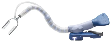
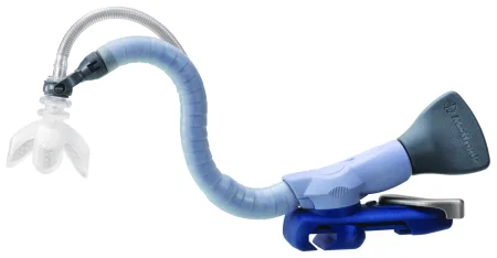
Minimally invasive direct coronary artery bypass (MIDCAB) joins the LIMA to the LAD on the beating heart through a short left anterior thoracotomy, about 10 cm, in the fourth rib space, for disease of one vessel. Some surgeons have the legs prepped too, in case the LIMA cannot be used.
Wound class (OPCAB and MIDCAB): I, as for CABG; no tract is entered.
13Valve surgery
Valve prostheses
- Mechanical valves, most with a tilting disk design, last a long time but need lifelong anticoagulation, and a clot can suddenly block one. Protect them from scratches.
- Biological valves (bioprostheses) are made from pig valves or from cow or horse pericardium. Most patients need no long-term anticoagulation, but a tissue valve can wear out and need replacing.
- Allografts (homografts), human donor valves, resist infection and need no anticoagulation; they arrive frozen and are thawed by the supplier's protocol.
- Valved conduits join a valve to a tube graft, to replace the aortic valve and the ascending aorta together.
- Annuloplasty rings and bands are sewn around a valve to repair it rather than replace it.
Valves are very expensive. Handle them as little as possible, and verify the type, size and serial number with the circulator, who records them. Each prosthesis has its own sizers, holders and handles. Biological valves stored in glutaraldehyde are rinsed in saline baths before they are sewn in; the number of baths, the volume and the time differ between makers, so follow the instructions for use every time. Keep a biological valve moist with saline from then on, because dry leaflets are damaged, and never soak it in antibiotic solution. Check that a valve's temperature indicator shows it was stored correctly.
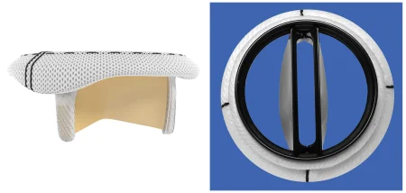
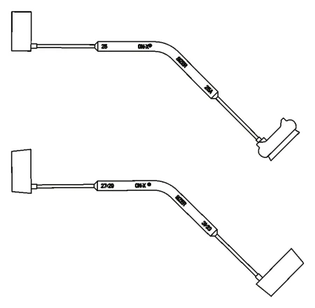
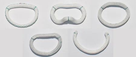
The patient's choice matters, including any social, cultural or religious objection to animal tissue. After any valve operation the risks include clots and stroke, bleeding from the anticoagulants, infection of the prosthesis (prosthetic valve endocarditis), a leak around the valve, a valve too small for the patient's needs and failure of the prosthesis.
Aortic valve replacement (AVR)
What and why: the aortic valve is replaced for stenosis, when calcified, stiff leaflets narrow the opening to a slit, or for insufficiency, when a leaking valve lets blood fall back into the left ventricle; either way the ventricle overworks and can fail. A partial sternotomy may be used for an aortic valve alone.
Setup: an open-heart set, aortic valve instruments, the valves, sizers and holders the surgeon prefers, a retrograde cardioplegia catheter and vents, and basins of saline to rinse a tissue valve.
Key steps:
- Bypass with a two-stage venous cannula; a vent through the right superior pulmonary vein and a retrograde catheter into the coronary sinus.
- The aorta is cross-clamped. With stenosis, the first dose of cardioplegia can go into the aortic root; with a leaking valve it goes retrograde, because antegrade solution would fill the ventricle.
- The aorta is opened and the diseased cusps are cut out; calcium is removed with rongeurs. Narrow packing in the ventricle catches loose bits, and you wipe each instrument with a moist sponge, so no fragment is left to embolize.
- The annulus is measured with the valve's own sizers, and the circulator opens the chosen valve.
- Interrupted, often pledgeted, sutures are placed through the annulus and then through the valve's sewing ring, in three groups, one for each cusp, and held in order.
- The valve is lowered into place and the sutures tied; the aorta is closed.
- Air is cleared from the left heart through the vent, by tilting the table head down and inflating the lungs, and the heart is not allowed to eject until the surgeon, helped by TEE, is satisfied that no air is left. The cross-clamp comes off, and the heart is defibrillated if needed.
- Pacing wires and chest tubes are placed and the chest is closed.
After surgery: bleeding, arrhythmias, stroke, infection, ischemia and failure of the valve.
Mitral valve repair and replacement
What and why: a stenotic mitral valve is opened, or a leaking one made to close. Repair is preferred whenever possible, because it avoids a prosthesis and lifelong anticoagulation. Instruments and valves for replacement are always ready, since the surgeon decides only after looking at the valve. Bicaval cannulation decompresses the heart and improves the view, and TEE checks the repair once the heart beats again.
Repairs:
- Commissurotomy: fused leaflets are cut or spread apart, sometimes with a dilator, for stenosis.
- Annuloplasty: a complete or partial (C-shaped) ring, sized with its obturator, is sewn into a stretched annulus to make it smaller.
- Leaflet and chord repair: a floppy section of the posterior leaflet is cut out and the edges sewn, and torn or stretched chordae are replaced with PTFE suture.
- Edge-to-edge (Alfieri) repair: a stitch, or a clip placed through a catheter, joins the middle of the two leaflets.
The repair is tested by filling the ventricle with saline from a bulb syringe and watching the leaflets close.
Replacement: the left atrium is opened along the groove between the atria and held open with an atrial retractor. The anterior leaflet is cut out, but the posterior leaflet and its chordae are kept when possible to preserve the shape and pumping of the ventricle. Fifteen to 20 or more pledgeted sutures go through the annulus and the valve's sewing ring, kept moist and in order; the valve is seated and tied, the atrium closed, and air removed with the table head down and the lungs inflated.
Mitral surgery can also be done through a right mini-thoracotomy or with the robot, usually with femoral cannulation, and the aorta is closed off with a cross-clamp or a balloon inside it.
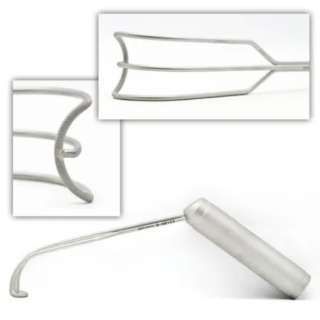
The tricuspid valve is most often operated on at the same time as a mitral valve and is repaired, for example with a ring, or replaced with a mitral prosthesis, its stitches kept clear of the conduction tissue near the AV node.
Two valves: when the aortic and mitral valves are both replaced, the mitral goes in first, because the firm ring of an aortic prosthesis already in place could injure the heart and make the mitral harder to insert; when a CABG is added to an aortic valve, the distal grafts are sewn before the new valve goes in and the proximal grafts last.
Transcatheter aortic valve replacement (TAVR)
What and why: a tissue valve compressed onto a catheter is placed inside the diseased aortic valve without opening the chest, for severe aortic stenosis with symptoms in patients at high risk for open surgery, and more and more in lower-risk patients. It is done in a hybrid room or the catheterization lab, usually through the femoral artery; the axillary artery or the apex of the heart is used when the leg arteries are too narrow or diseased.
Setup: endovascular valve and femoral access instruments, the valve and its deployment system, guidewires and catheters with the catheter lubricants their makers name, contrast media, lead aprons and fluoroscopy. The valve is rinsed or kept moist by its maker's instructions.
Key steps:
- A sheath goes into the femoral artery over a guidewire, and the catheter is threaded up the aorta to the valve.
- Landmarks are confirmed on fluoroscopy, and the valve is brought into the aortic annulus.
- A self-expanding valve is released; a balloon-expanded one is opened by inflating its balloon.
- The surgeon looks for leaks around the valve. A self-expanding valve can be adjusted; with a balloon-expanded valve, a second one is placed inside the first.
- The access is closed.
Be ready to convert from the catheter method to open surgery in every case; injury to the heart or to the access vessel is one of the risks of a valve placed through a catheter.
Wound class (valve surgery, TAVR included): I, no tract is entered.
14Aortic aneurysm and dissection
The aortic arch
What and why: an aneurysm or a dissection of the arch can cut off blood to the brain and arms, because the arch gives off the brachiocephalic, left carotid and left subclavian arteries; in the ascending aorta, one may burst or keep the aortic valve from closing.
Position and setup: supine for a median sternotomy, or a modified thoracotomy position for the far end of the arch. Aneurysm instruments and woven or collagen-sealed grafts in a range of sizes, with valve and coronary instruments, valves and valved conduits ready. Have a basin for clot and debris. Since the weakened aorta may not take a cannula, the femoral or axillary artery is often used for arterial inflow, and the femoral artery and vein may be isolated before the chest is opened in case the aneurysm ruptures.
Key steps:
- The right atrium and the right axillary or a femoral artery are cannulated, and the patient is cooled for circulatory arrest; the head may be packed in ice bags, padded against frostbite.
- At the planned temperature the surgeon clamps the arch vessels one by one or stops the circulation, and the brain is perfused through an arch branch or backward through the superior vena cava.
- The tissue holding all three arch vessels is cut out as an "island," and a graft, straight or with side branches, is sewn first to the descending aorta.
- An opening cut in the graft is sewn to the island of arch vessels.
- The graft is de-aired and bypass restarts, perfusing the head and the lower body again; the graft is then joined to the ascending aorta.
- Glue may reinforce the suture lines, the patient is rewarmed and weaned from bypass, and the chest is closed.
After surgery: bleeding, stroke, infection and death are the risks of aortic surgery, and a join that keeps bleeding may need repair.
Dissection, the aortic root and stent grafts
A Type A dissection, one that involves the ascending aorta, is an emergency because the false channel can rupture, cut off the aorta's branches or keep the aortic valve from closing. It is repaired at once on bypass, with the arterial cannula often in the femoral or axillary artery, by replacing the torn aorta with a woven graft, or with a valved conduit when the aortic root is involved.
When the root is involved, the Bentall procedure replaces the aortic root, the valve and the ascending aorta with a valved conduit, and the coronary arteries are sewn into openings that the electrocautery makes in the graft, sealing its cut edges at the same time.
Most aneurysms and dissections of the descending aorta are now treated by endovascular repair (TEVAR): a stent graft goes in through the femoral artery under fluoroscopy, its guidewires, which can be more than 6 feet long, are anchored on the field with wet sterile towels, and the open set stays ready in case of bleeding; chapter 40 teaches stent grafts.
Left ventricular aneurysm
What and why: the thin scar of an old MI is removed to reduce the risk of rupture and of clots breaking off.
Setup: the major cardiac set, with a Dacron patch and Teflon felt.
Key steps: through a median sternotomy, on bypass with the aorta cross-clamped, the surgeon opens the aneurysm and gently removes any clot; keep the instruments clean so no clot gets into the circulation. Most surgeons now sew a patch of Dacron, Teflon or pericardium inside, on the border of healthy muscle (endoaneurysmorrhaphy), because it keeps the ventricle's shape better. The ventricle is vented and de-aired before the last stitch is tied.
cutting the scar out and closing the ventricle by sewing its edges together between strips of Teflon felt, which one reference still describes.
After surgery: the patient is watched closely in the intensive care unit for a low cardiac output and ventricular arrhythmias; failure of the left ventricle is the most common cause of death in the hospital.
Wound class (aorta and ventricle): I.
15Surgery for rhythm problems
Many, but not all, pacemakers and defibrillators now go in in the electrophysiology (EP) lab or the catheterization lab, and ablation for atrial fibrillation is normally done in the EP lab. Chapter 20 teaches energy safety around implanted devices.
Temporary pacing wires
At the end of heart surgery, temporary wires are sewn to the surface of the right atrium, the right ventricle or both, before bypass ends, while the heart is easy to reach and the pump still protects the circulation if handling the heart causes an arrhythmia. The needle at the heart end is cut off after it passes through the muscle, the wire is sewn in place, and the needle at the other end brings it out through the skin, where it is stitched down; an alligator cable connects it to the external pacemaker, so the anesthesia provider can pace the heart. The wires stay in after surgery for a slow or fast rhythm.
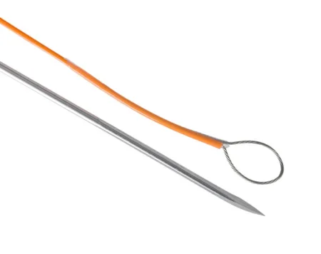
Permanent pacemaker
What and why: a pulse generator and its leads start the contraction of the atrium, the ventricle or both, most often for complete heart block and slow rhythms; a pacemaker is a device that helps control abnormal heart rhythms.
Before surgery: local anesthesia with monitored anesthesia care; fluoroscopy, a defibrillator in the room and a pacing system analyzer, often brought by the maker's representative. The table is tilted head down to fill the vein and to prevent an air embolism.
Key steps (transvenous):
- A needle enters the subclavian, axillary or internal jugular vein, and a guidewire is passed into the right atrium under fluoroscopy.
- A pocket for the generator is made under the skin below the collarbone.
- A sheath and dilator go over the wire, and the lead, stiffened with a stylet, is advanced into the right ventricle and fixed at its apex, by a small screw or by lodging in the muscle ridges.
- The lead is tested with the analyzer through alligator cables, then sewn to the muscle; a second lead goes into the right atrium for a dual-chamber pacemaker.
- The pocket is irrigated with an antimicrobial solution, the generator is connected and sewn into the pocket, and the incision is closed in layers.
After surgery: the complications include bleeding into the pocket (a hematoma), which raises the risk of infection, infection of the pocket or the leads, a lead that moves or breaks, and, rarely, a lead that punctures the heart.
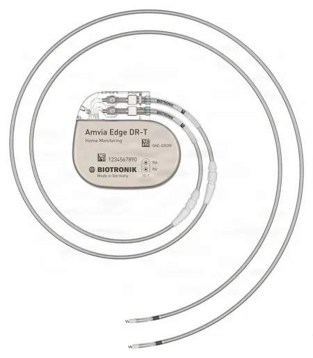

Other options: a leadless pacemaker, placed through a catheter into the right ventricle, needs no leads or pocket. Epicardial leads are fixed onto the heart through a subxiphoid incision, a small thoracotomy or a sternotomy. Cardiac resynchronization therapy adds a lead through the coronary sinus to pace the left ventricle too, for heart failure.
Wound class: I.
Implantable cardioverter defibrillator (ICD)
An ICD watches the heart's rhythm and shocks it when it senses ventricular tachycardia or fibrillation, while a pacemaker responds when the heart fails to beat; ICDs can pace as well. The operation is much like a transvenous pacemaker: the leads go in through a vein under fluoroscopy, the generator goes in a pocket in the chest wall, and the device is tested; EP studies before and after show the rhythm and check the device.
Ablation and the Maze procedure
Catheter ablation: in the EP lab, the electrophysiologist threads a catheter from the femoral vein or artery into the heart, provokes the arrhythmia to find where it starts, and destroys that spot with radiofrequency energy or freezing (cryoablation). Radiofrequency ablation destroys tissue with the heat of high-frequency energy.
The Maze procedure treats atrial fibrillation in the OR. Lines of scar are made in the atria; electrical signals cannot cross scar, so they are forced along one path, the "maze," from the SA node to the AV node. Today the lines are made with cryothermal, radiofrequency or ultrasonic energy rather than cut and sewn, especially around the pulmonary veins.
- Through a sternotomy: on total bypass with the heart arrested, the lesions are made through an opening in the right atrium and then one in the left.
- Through a right mini-thoracotomy: the patient tilted slightly left with a roll under the right chest, femoral cannulation, and the right atrial lesions made before bypass starts, the left ones after the heart is arrested.
After surgery the rhythm is watched, and it can take up to 3 months for the heart to beat normally.
Wound class (ICD, ablation and Maze): I.
16Congenital heart surgery
Children's heart surgery uses the open-heart setup scaled to the child's size, with patch material ready. Chapter 13 teaches the child under anesthesia, and chapter 18 the child and family as people.
Patent ductus arteriosus (PDA)
What and why: when the ductus stays open after birth, blood shunts from the aorta into the pulmonary artery, overloading the lungs and the heart, which enlarges and can fail. It is common in premature babies. A ductus is not closed when it is keeping the child alive, as with some other defects such as severe pulmonary stenosis.
Before surgery: for a newborn, the operation may be done at the bedside in the intensive care nursery, since the operation is short and the baby need not be moved; older children come to the OR. No bypass is needed. A thoracoscopic closure with clips is an option, with the open instruments ready.
Key steps (open):
- Right side down, a left posterolateral thoracotomy through the third or fourth rib space, with a Finochietto spreader; the lung is held back with a moist pack.
- The pleura over the ductus is opened between the phrenic and vagus nerves, and the recurrent laryngeal nerve is found and protected.
- The ductus is tied twice, close to the aorta and close to the pulmonary artery, or clamped with ductus clamps, divided and both ends sewn closed; in a tiny baby it may simply be tied or clipped.
- A small chest tube is placed and the chest is closed.
After surgery: the complications include infection, bleeding, injury to the recurrent laryngeal nerve with a paralyzed vocal cord, injury to the vagus nerve or the thoracic duct, and a ductus that is not fully closed.
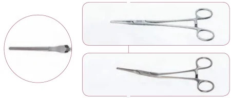
Coarctation of the aorta
What and why: a narrowing of the aorta, usually near where the ductus joined it, blocks flow to the lower body; the heart enlarges from pumping against it, and the lower body may be underdeveloped.
Setup: a cardiac set, with coarctation clamps, bulldogs and moist umbilical tape.
Key steps:
- Lateral position and a posterolateral thoracotomy; a moist lap protects the retracted lung.
- The mediastinal pleura is opened, a moist umbilical tape is passed around the aorta, and the ductus or its remnant (the ligamentum arteriosum) is tied and divided.
- The aorta is clamped above and below the narrowing, with bulldog clamps on vessels between the clamps.
- The narrowed segment is cut out and the ends are joined; interrupted sutures in a child allow growth. A tube graft, or a flap of the left subclavian artery, is used when the ends will not meet.
- The clamps come off, restoring flow to the lower body, and the chest is closed over a chest tube.
After surgery, the cross-clamp can starve the spinal cord of blood and cause paraplegia, the most devastating complication, and adults often have high blood pressure for the first few days.
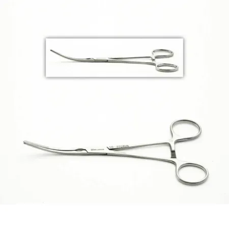
Atrial and ventricular septal defects
What and why: an atrial septal defect (ASD) is a hole between the atria, so blood shunts from the left atrium to the right. Children often have no symptoms, but over years the right heart and the lung vessels are overloaded, with pulmonary hypertension, heart failure and arrhythmias, and the shunt can reverse. Some septal defects can be closed with a device in the catheterization lab.
Key steps (ASD):
- A median sternotomy (or a right anterolateral thoracotomy) and bypass, usually with bicaval cannulation.
- The right atrium is opened and the defect examined.
- It is closed with a running stitch or a patch of pericardium or synthetic material; the left atrium is filled with blood to push out air before the last stitches are tied.
- Bypass ends and the chest is closed.
After surgery: the child is watched for arrhythmias; the complications include infection, bleeding, failure of a synthetic patch, a leaking mitral valve and, rarely, heart block.
A ventricular septal defect (VSD), a hole between the ventricles, is one of the most common heart defects, and a large one floods the lungs at high pressure, so it is closed to prevent pulmonary hypertension. It is often reached through the right atrium and the tricuspid valve and closed with a patch, the stitches placed with great care near the conduction system, because damage causes heart block.
Tetralogy of Fallot
What and why: four defects together: a VSD, narrowing below or at the pulmonary valve, an aorta that sits over the septum and a thickened right ventricle. Oxygen-poor blood shunts from right to left, so the child is cyanotic, with spells of severe cyanosis ("tet spells"), clubbed fingers and too many red cells. A complete repair is preferred; when it must wait, a shunt from a systemic artery to the pulmonary artery improves the oxygen until the child is bigger.
Key steps:
- A median sternotomy, bypass with cooling and cardioplegia.
- The right ventricle is opened over its outflow tract, and the thick muscle blocking it is cut away; a fused pulmonary valve is opened.
- The VSD is closed with a patch, the stitches kept clear of the conduction system.
- If the outflow is still too narrow, a patch widens it, sometimes across the valve ring onto the pulmonary artery.
- The pressures in the pulmonary artery and right ventricle are measured to judge the repair, and bypass ends.
After surgery: for the first 24 hours, bleeding in the chest is watched closely, because the long-standing extra red cells raise its risk, along with blood collecting around the heart (tamponade) and the cardiac output; the complications include infection, bleeding, a low cardiac output and, rarely, heart block.
Other repairs include a valvulotomy, which opens a narrowed pulmonary valve on bypass, and, for babies with only one working ventricle, as in hypoplastic left heart syndrome, a series of three operations ending with the Fontan procedure, which sends the blood from the venae cavae straight to the lungs.
Wound class (congenital heart repairs): I.
17Heart failure: support devices and transplant
When the heart is failing, devices can support it for a short time, a long time or for good, and a transplant replaces it when they cannot reverse the decline.
Intra-aortic balloon pump (IABP)
What and why: a balloon on a catheter sits in the descending thoracic aorta and works by counterpulsation. A console fills it with helium as the ventricle relaxes, pushing blood into the coronary arteries and the organs, and empties it just as the ventricle contracts, lowering the pressure the heart must pump against. It reduces the heart's work after an MI and helps patients who cannot come off bypass.
Key steps:
- The balloon goes in through a femoral artery, under local anesthesia or at the end of heart surgery.
- A needle punctures the artery, a guidewire is passed and the needle removed; dilators widen the track and a sheath goes in.
- The air is removed from the balloon with its one-way valve and a syringe.
- The balloon catheter is advanced over the wire to just below the left subclavian artery; the surgeon marks the length with a silk tie.
- Its connections are passed off the field and joined to the console.
After: complications include ischemia of the leg and compartment syndrome, emboli, dissection of the aorta or iliac artery, bleeding and failure of the balloon.
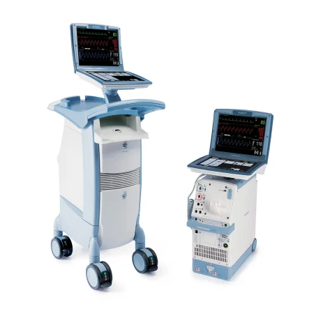
Ventricular assist device (VAD)
What and why: a pump that takes blood from a failing ventricle and sends it on, as a bridge to a transplant, as permanent destination therapy for a patient who cannot have one, or to help a patient off bypass. Devices support the left ventricle (LVAD), the right (RVAD) or both (BiVAD). An LVAD has the pump, an inflow cannula in the left ventricle's apex, an outflow graft to the aorta and a driveline that leaves the body through the skin to its power source.
Key steps (LVAD):
- A median sternotomy, and bypass with the heart still beating.
- The atrial septum is checked for a hole, which is closed if found, so blood cannot shunt once the pump runs.
- The apex of the left ventricle is cored out with the device's coring knife, and pledgeted sutures, sometimes with a felt strip, are placed around the hole.
- The inflow cuff is sewn into the apex and the pump attached.
- The outflow graft is measured, a side-biting clamp is placed on the ascending aorta and the graft is sewn end to side onto it.
- The driveline is tunneled under the skin and out through the abdominal wall.
- The pump is started as bypass is withdrawn.
Bleeding, clots, infection and device failure are the main complications, and a total artificial heart is used for some patients whose two sides both fail.
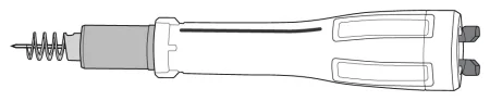
Extracorporeal membrane oxygenation (ECMO)
What and why: a pump and an oxygenator outside the body that take over the lungs' work, or the heart's and lungs', for a time, while the organs recover. Venovenous (VV) ECMO drains blood from a vein and returns it to a vein, supporting the lungs only; venoarterial (VA) ECMO returns it to an artery, supporting the heart and the lungs, for cardiogenic shock or cardiac arrest.
Cannulation:
- Peripheral VA, in an emergency or for cardiogenic shock: a drainage cannula in a femoral vein and a return cannula in a femoral artery. A small extra cannula is often placed to carry blood down the leg beyond the arterial cannula, because without it the leg can lose its blood supply.
- Central VA, after heart surgery: the bypass cannulas in the right atrium and aorta are moved over to the ECMO circuit; a patient who cannot be weaned from bypass can be switched to ECMO in the OR.
- In children, VA ECMO usually uses the right common carotid artery and the internal jugular vein in the neck.
Setup: ECMO often starts in the ICU or the emergency department. Plan room at the bedside for the ECMO pump, a sterile table, the ESU, a headlight and a defibrillator with external and internal paddles; everyone wears surgical attire and traffic is limited; keep the instruments sterile until an X-ray confirms the cannulas are in the right place. A continuous heparin infusion prevents clots, and bleeding and clots are the most common complications.
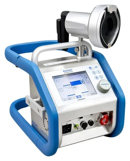
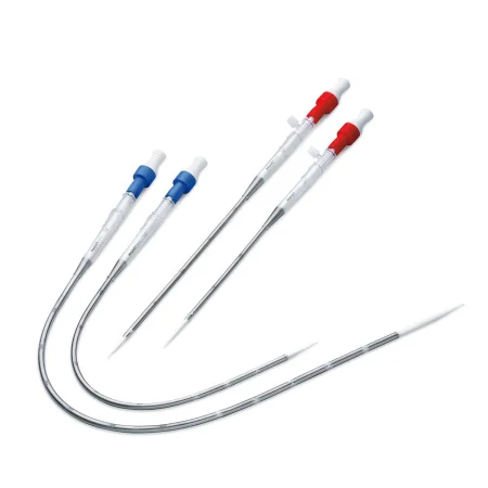
Heart transplant
What and why: a donor heart replaces a heart with end-stage disease, such as coronary artery disease, a congenital defect, valve disease or rejection of an earlier transplant. Orthotopic transplant, replacing the heart in its own place, is the usual operation. The donor and the recipient each need their own instrument setup, and the donor heart arrives cooled in saline in a transport container. Chapter 18 teaches organ donation.
Key steps (recipient):
- Supine, prepped from the chin to the knees; a median sternotomy and heparin.
- Bicaval cannulation with tapes around each vena cava; the patient is cooled to about 32 to 34°C, then the cross-clamp goes on the aorta and the tapes are snugged.
- The aorta and pulmonary artery are divided above their valves, and the atria are cut so the back walls of the recipient's atria remain; the heart is removed.
- On the back table, in a basin, the surgeon trims the donor heart.
- The donor heart is placed in the pericardium, and the left atria are joined first; then the venae cavae (the bicaval technique, which causes fewer rhythm and tricuspid problems than joining the right atria), the pulmonary artery and the aorta.
- Air is removed, the cross-clamp comes off, and a single shock usually starts the heart; pacing wires and chest tubes are placed and the chest is closed.
A combined heart-lung transplant also spares the donor's SA node and the recipient's phrenic, vagus and recurrent laryngeal nerves. Afterward the patient takes immunosuppressive drugs for life, with regular biopsies of the heart muscle to look for rejection; rejection and infection are the main complications.
Wound class (support devices and transplant): I.
18Closing the chest and after surgery
Closing a sternotomy
- Before the chest is closed, the pacing wires and usually two chest tubes are in place.
- The surgeon places six to eight stainless steel wires through the sternal edges with a heavy sternal needle holder. As each wire is passed, hold its free ends so they stay under control and off anything unsterile.
- The holes are checked for bleeding, and the wires are tightened and twisted to bring the halves of the sternum together, cut with wire cutters, and their twisted ends buried in the periosteum. Some surgeons use small metal crimpers instead of twisting; a frail sternum may get extra wires running along each edge, bands or plates.
- The fascia over the sternum, the linea alba, the fat and the skin are closed. If the skin is stapled, a staple remover goes with the patient.
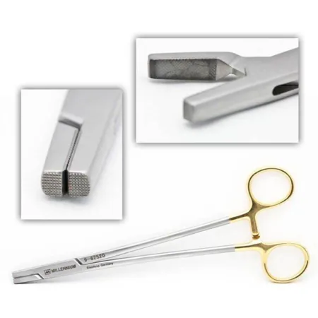
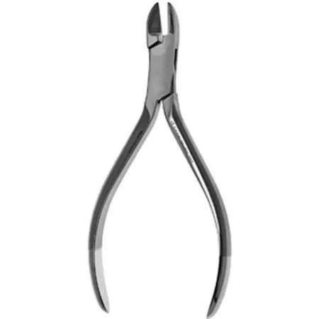
Before the patient leaves, the circulator phones a handoff report to the intensive care unit, covering, among other things, the grafts used, the cross-clamp and pump times, the blood loss, the blood and drugs given and whether the heart is being paced; chapter 16 teaches handoffs.
After heart surgery
After heart surgery, patients go to a cardiovascular intensive care unit, where the blood pressure, the heart rhythm, the chest tube drainage and the temperature are watched closely. The breathing tube may come out within a few hours or stay until the patient can breathe alone. Many patients go home after about 3 to 7 days, and a sternotomy takes about 4 to 12 weeks to recover from, compared with 1 to 2 weeks after a minimally invasive operation.
- Bleeding and tamponade: chest tube drainage of more than 100 mL an hour is investigated, and the surgeon is told before the patient leaves the room; the chest may have to be reopened to stop bleeding. Sudden bleeding that causes tamponade needs a fast, organized response, through the chest tubes or by reopening the chest. This is one reason the sterile setup stays intact until the patient has left the room.
- Arrhythmias: atrial fibrillation is common after bypass surgery, and beta-blockers or amiodarone help prevent it; the pacing wires treat slow rhythms.
- Sternal wound infection (mediastinitis, an organ or space SSI from chapter 11) can mean a return to the OR to clean the wound and cover it with a muscle flap, an operation that is Class IV because the infection is already present. Its risks include diabetes, obesity, COPD, smoking, use of both mammary arteries, a balloon pump, a long operation and a return for bleeding; blood glucose is kept below 200 mg/dL.
- Other complications: stroke, kidney failure, a long time on the ventilator, pneumonia, clots in the legs and the lungs, infection and death.
- Teaching: a patient with a mechanical valve needs to understand lifelong anticoagulation and the signs of bleeding to report.
After lung surgery
The priorities are breathing and pain control. The patient breathes deeply, coughs, uses the incentive spirometer every 1 to 2 hours while awake, and gets up on the day of surgery; some go home on oxygen. An enhanced recovery (ERAS) pathway moves the chest tube to water seal after a short time on suction. An air leak or a lung infection can delay going home.
Lo que te llevas de este capítulo
- Chest surgery is planned around negative pressure: an opened chest collapses the lung, so it is closed over chest tubes and a drainage unit kept upright and below the chest. If a tube comes out, petroleum gauze covers the hole at once.
- Most lung operations are now VATS, with the patient lateral on a double-lumen tube and the open thoracotomy set ready. The bronchus is opened under suction, lung staple lines are tested for air leaks under warm saline, and every lymph node is labeled by its station.
- A heart field runs on two back tables and three Mayos, a separate setup and light for the leg, clean instruments free of calcium and clot, cold and warm solutions, and every drug labeled, heparin and papaverine above all.
- Bypass drains venous blood to the heart-lung machine, which pumps it through the oxygenator and back into the aorta. Heparin and an adequate ACT come first; the cannulas go in arterial first; the cross-clamp isolates the heart, and potassium cardioplegia, antegrade or retrograde, stops it in diastole. Watch the lines for air.
- Coming off bypass: de-air, remove the clamp, defibrillate if needed, place pacing wires, wean, decannulate, then protamine, with the pump suckers turned off as it starts.
- CABG joins the aorta to a coronary artery beyond the blockage; the LIMA to the LAD is the first choice, and OPCAB works on the beating heart with stabilizers, bypass on standby. Valves are handled little, verified, and, if biological, rinsed by the maker's instructions and kept moist.
- Arch surgery cools the patient to about 18°C, stops the circulation and perfuses the brain; rhythm devices go in through a vein under fluoroscopy and are tested with an analyzer; congenital repairs close shunts and open narrowings, guarding the conduction system and the nerves.
- The IABP inflates in diastole and deflates before systole; a VAD bridges to transplant or replaces it; VA ECMO supports heart and lungs, VV ECMO the lungs. After heart surgery, watch for bleeding over 100 mL an hour and tamponade, and keep the setup sterile until the patient leaves.
Ponte a prueba6 preguntas rápidas
Gratis para todos. Elige una respuesta para cada una y luego revísalas todas juntas.
En el quirófanoDónde vas a usar esto
Todo el banco de preguntas del capítulo en series de 10, con la respuesta explicada después de cada una.
Parte del programa para educadoresLas palabras del capítulo en tarjetas de estudio: las que fallas vuelven antes.
Parte del programa para educadoresLo asigna tu docente. Cada grupo saca sus propias 25 preguntas del banco del capítulo.
Parte del programa para educadores
Sugerir una corrección
¿Algo está mal, confuso o desactualizado en este capítulo? Avísanos y lo corregimos para todos. Tu correo solo se usa para responderte.