Peripheral vascular surgery
Arteries, veins and access for dialysis: the open and endovascular cases of vascular surgery, the clamps, wires and grafts they run on, and how the scrub keeps blood flowing past every repair.
1What vascular surgery covers
Peripheral vascular surgery is the specialty of the arteries, veins and lymph vessels outside the area of the heart. It treats disease, injury and malformations a patient is born with. Vascular surgeons also build the access that dialysis needles use, and they repair arteries torn by gunshots, stab wounds and crashes.
The Core Curriculum sorts the specialty's cases three ways: operations on a blood vessel, on an artery and on a vein. Each can be done open or by a minimally invasive, endovascular approach, either through the skin (percutaneous) or with a scope. Three words come up again and again:
- Open: the surgeon cuts down to the vessel, controls it, clamps it, opens it and repairs it under direct view.
- Endovascular: the work happens inside the vessel. A needle or a small cutdown gives access, and wires, sheaths, catheters, balloons and stents travel through the vessel under live x-ray. Percutaneous means through the skin: no incision is made, and the devices go in through a puncture.
- Hybrid: an endovascular repair done in a hybrid operating room, a room built for both endovascular and open surgery, with fixed imaging and everything needed to open if the repair goes wrong.
Many operations that were once done open are now done from inside the vessel, with far less trauma and a faster recovery. Endovascular repair of an abdominal aortic aneurysm, for example, is now done more often than the open repair. Vascular cases also share a great deal: the same clamps, the same steps for opening and closing an artery, and the same drugs on the field. The Core Curriculum calls cases like these co-related procedures, so learn the pattern once and each new case is mostly a new middle. Most cases below link to their setup card, which holds the full instrument list and every step.
Vascular surgery asks a lot of the scrub. Some steps stop the blood flow to an organ or a limb, and during those minutes you watch the field closely and have the next item ready, from a well-organized instrument table. Surgeons often name exactly which vascular instruments they want on the setup, so the right clamp is there at the critical moment.
Other chapters hold the pieces around this one:
- The arteries and veins by name, the systemic and pulmonary circulations, the pulse points and blood pressure: chapter 5.
- Heparin, protamine, contrast and the other drugs as drugs: chapter 12.
- Interventional radiology, guidewires and sheaths as IR uses them, radiation safety and the hybrid OR as a room: chapter 22.
- Heart surgery, the thoracic aorta and coronary artery disease, the cardiothoracic supplies (such as the balloon pump), and endoscopic vein harvesting: chapter 41.
- Amputation technique: chapter 39.
- Hemostatic agents and how they are prepared: chapter 29.
2Anatomy that matters
The vessel wall
Chapter 5 teaches the three layers of a vessel wall: the intima inside, the muscular media and the tough adventitia outside. Two of them matter most here. Fatty, calcified plaque builds up on the intima, the smooth lining that normally keeps platelets from clumping. An endarterectomy lifts the plaque out along a cleavage plane inside the wall, usually in the media, and leaves the smooth outer wall behind.
The space inside a vessel is its lumen. A vein is thinner and more fragile than an artery, and venous bleeding can be hard to control. Veins have one-way valves, so a vein used as an arterial graft must be reversed, or its valves cut, before arterial blood can flow through it.
A fork in an artery or a graft is a bifurcation, and plaque is thickest there. A narrowing, a curve or a fork makes the blood swirl, and that turbulence makes a bruit, a low-pitched blowing sound heard through a stethoscope, and an abnormal Doppler signal. The pressure, flow and resistance of the blood in the system are its hemodynamics.
The vessels you will work on
Chapter 5 names every major artery and vein and where each runs. This is where each one meets the operations in this chapter.
| Vessel | The surgical point | Operations |
|---|---|---|
| Abdominal aorta | Aneurysms form mostly below the renal arteries, above its fork into the common iliac arteries | Aneurysm repair, open and endovascular; aortofemoral bypass |
| Iliac arteries | A common site of atherosclerosis; an EVAR's stent graft seals in them at its lower end | EVAR limbs, angioplasty and stents |
| Femoral arteries | The common femoral, in the groin, is the access point used most for endovascular work | Embolectomy, bypass, EVAR access |
| Popliteal and leg arteries | A leg bypass runs from the femoral artery to the popliteal artery at the knee, or to a tibial artery below it | Femoropopliteal and tibial bypass; pulse checks |
| Carotid arteries | Disease sits mostly at the fork where the common carotid divides into the internal carotid, which feeds the brain, and the external carotid | Carotid endarterectomy and stenting |
| Arm vessels | Dialysis access joins the radial or brachial artery to the cephalic or basilic vein; the basilic runs deep, under the fascia, in the upper arm | Dialysis fistulas and grafts |
| Leg veins | The great saphenous vein, under the skin of the inner leg, is the vein varicose disease affects most and the usual vein graft; it joins the femoral vein in the groin | Vein stripping; vein grafts; venous thrombectomy |
| Venae cavae and neck veins | A filter goes in the inferior vena cava below the renal veins; central lines and ports enter the internal jugular or subclavian vein, the tip in the superior vena cava | Vena cava filter; central lines and ports |
When an artery narrows slowly, the body grows a network of side branches, collateral vessels, that carry some blood around the blockage. The portal circulation of the liver (chapters 5 and 7) and the lymph system (chapter 5) are taught with their organs.
3Diseases of the arteries and veins
Hardening and narrowing of the arteries
- Arteriosclerosis: thickening, hardening and loss of elasticity of the artery wall. It comes partly with age.
- Atherosclerosis: the most common form of arteriosclerosis and the disease that most often brings a patient to vascular surgery. Fatty plaque and calcium build up on the intima and narrow the lumen, a narrowing called stenosis, so less blood gets through.
Plaque begins with damage to the intima. Platelets gather there, inflammation follows, and fat and scar tissue build a deposit called an atheroma, which hardens with calcium into plaque. Plaque may be soft and rubbery, hard and calcified, or both. Smoking, high cholesterol, high blood pressure and diabetes are the firmly established risk factors; age, male sex, an inactive life and family history add to the risk.
Atherosclerosis harms in two ways. It slowly starves tissue of blood, and pieces of plaque can break off and travel downstream, which in the carotid artery means a stroke. Ischemia is too little blood flow to a tissue, usually from a blocked vessel. An infarction is tissue that dies when its blood supply stops. Peripheral artery disease (PAD) is the narrowing or blockage of the arteries that carry blood out from the heart, most often in the legs, and atherosclerosis is its most common cause.
From claudication to gangrene
Blocked leg arteries cause trouble in stages.
- Intermittent claudication, the first and most common symptom: cramping pain in a working muscle below the blockage, such as the calf, that comes on with walking, at about the same distance each time, and eases with rest. Blood flow is enough at rest but not for exercise. Surgery is not usually done for claudication unless it is disabling.
- Rest pain: as the disease worsens, a constant burning pain in the lower leg, foot or toes comes on even at rest, often worse at night. Ischemia at this stage is called critical.
- Ulcers and gangrene: without treatment, sores that will not heal and gangrene, the death and decay of tissue whose blood supply has failed, follow. Dry gangrene can develop in a severe blockage left untreated.
Patients with diabetes are at special risk, because nerve damage in the feet (peripheral neuropathy) can keep them from feeling the tenderness and early signs of an infection, and a foot infection can grow severe enough to threaten the leg.
Thrombus and embolus
A thrombus is a clot that stays where it formed, attached to the vessel wall. An embolus moves through the circulation until it reaches a vessel too small to pass, where it lodges and blocks the flow. An embolus may be a blood clot, a piece of plaque, air, fat, a foreign body or tumor cells. Chapter 11 teaches thrombosis and embolism as disease processes.
Emboli in the arteries most often come from the heart: a clot forms in a heart that beats irregularly in atrial fibrillation, or on the wall of a heart damaged by a heart attack. When one lodges in a limb artery, the limb shows the signs of acute arterial blockage, often taught as the six Ps: pain, sudden and severe; pulselessness; paresthesia (numbness and tingling); paralysis; pallor; and poikilothermia, a limb gone cold.
Heparin is started at once so the clot cannot grow. A threatened limb then needs the clot removed, by surgery (embolectomy) or by a drug that dissolves it (a thrombolytic). Once the muscle has become rigid, the limb cannot be saved, and amputation is done to save the patient.
Aneurysms
An aneurysm is a weakened section of artery that has ballooned to more than half again its normal diameter. As it grows, it can burst. There are three kinds:
- True aneurysm: all three layers of the wall are stretched out together.
- False aneurysm (pseudoaneurysm): a hole goes through every layer of the wall, and the blood that escapes is held in by the tissue around the vessel. It follows trauma, infection or a suture line that comes apart, and it can form at a puncture site after a catheter procedure.
- Dissecting aneurysm: a tear in the intima lets blood force its way between the layers and split the wall.
By shape, a fusiform aneurysm bulges evenly all the way around and is the most common; a saccular aneurysm bulges out from one side. Atherosclerotic damage to the wall is the most common cause, with genes, age and inflammation playing a part; the Core Curriculum's full list of causes is in the EVAR section.
The abdominal aortic aneurysm (AAA) is the most common. It forms mostly below the renal arteries and above the aortic bifurcation, and often reaches the iliac arteries. True aneurysms also form in the thoracic aorta and the iliac, femoral and popliteal arteries. AAAs affect more men than women, mostly older adults, and they run in families.
Most AAAs cause few or no symptoms and are found on a routine exam or scan. Severe back pain with low blood pressure and shock usually means a rupture, a true emergency. Repair is planned once an aneurysm without symptoms reaches about 5.5 cm across in a man or 5.0 cm in a woman, or grows fast. Trials found that the risk of rupture does not outweigh the risk of repair until an aneurysm reaches about 5.5 cm. One that causes symptoms or has ruptured is repaired urgently. The patients often have coronary artery disease, high cholesterol, high blood pressure or a smoking history as well, and a heart attack is the leading cause of death after AAA repair. Aneurysms in the chest and heart are chapter 41's, and those in the brain chapter 42's. Coronary artery disease, which the Core Curriculum lists with the peripheral vascular diseases, is chapter 41's as well.
Carotid disease and stroke
Plaque in the carotid artery builds up mostly at the bifurcation, where the common carotid divides. It narrows the flow to the brain, and small pieces can break off and lodge in the brain's vessels. The warning signs are:
- A transient ischemic attack (TIA): a short episode of lost brain function, such as weakness or trouble speaking, that clears within 24 hours.
- Amaurosis fugax: brief loss of part or all of the vision in one eye, often described as a curtain coming down, because the first branch of the internal carotid feeds the eye.
Either can lead on to a full stroke (a cerebrovascular accident). A TIA, or a severe narrowing even without symptoms, is a reason for a carotid endarterectomy. A carotid artery that is completely blocked is generally not opened.
Raynaud phenomenon
Raynaud phenomenon is an exaggerated response of the extremities to cold or emotional stress: the fingers typically turn white, then blue, then red as the blood returns. The common primary form, which the Core Curriculum calls Raynaud's disease, is a harmless, reversible spasm; the secondary form, which comes with another disease, can lead to ulcers of the fingers or toes, scarring or gangrene.
Intimal hyperplasia
Intimal hyperplasia is an overgrowth of the intima that thickens it, in an artery or a vein, after a long-term central line, a graft or another treatment irritates the vessel. In an artery it narrows the lumen much as atherosclerosis does; in a vein it slows the return of blood and causes swelling and tissue breakdown.
Diseases of the veins
- Varicose veins: a varicose vein is an enlarged, swollen vein, seen and felt under the skin, whose wall has weakened and whose valves no longer close. In primary varicose veins, the more common kind, the superficial saphenous veins are affected and the deep veins are healthy; secondary varicose veins follow failure of the deep venous system. The great saphenous vein is affected most often. The risk is higher in women, with age and in pregnancy.
- Venous stasis is the pooling of blood in the veins from inactivity or disease. The pressure in the veins rises, fluid leaks out of the capillaries into the tissues, and the leg swells (edema); venous stasis ulcers can follow.
- Chronic venous insufficiency: over time the vein walls or valves stop returning blood well; it is seldom treated with surgery, and graduated compression stockings are the usual treatment.
- Thrombophlebitis: painful clots and inflammation in the superficial veins of the legs, often from venous stasis after inactivity.
- Deep vein thrombosis (DVT): a clot, or several, in the deep veins of a leg. Three things set it off, known as the Virchow triad: injury to the vessel lining, pooling of blood, and blood that clots too easily. Its danger is that a piece breaks off and travels to the lungs as a pulmonary embolism, which can kill; most pulmonary emboli start as a DVT in the legs. A DVT is treated with heparin; when heparin cannot be used or does not work, a vena cava filter may be placed. Chapter 11 teaches how compression devices and drugs prevent it.
4Diagnostic tests
Diagnosis starts with the patient's history, the physical exam and routine blood tests; more specific studies follow from what they show.
- Pulses and skin: the color, warmth and pulses of the limbs, and any ulcers, wasted muscle or dead tissue, are checked and recorded so there is a baseline to compare with during and after surgery. Chapter 5 teaches where to feel each pulse.
- Ankle-brachial index (ABI): the simplest test for blocked leg arteries. Blood pressures are taken at both arms and both ankles, and the higher ankle pressure on each side is divided by the higher arm pressure; the lower the index, the worse the disease.
- Segmental pressures and plethysmography: blood pressure cuffs placed at several levels on the leg show where the flow drops off, either as pressures read with a Doppler or as the changing volume of the limb with each beat (plethysmography).
- Doppler: a probe aimed at a vessel turns the moving blood into sound, a swishing signal, not a pulse. A normal arterial signal has two or three phases; a single low-pitched phase suggests a narrowing or a blockage. Gel carries the signal through the skin.
- Duplex ultrasound (Doppler duplex ultrasonography): Doppler combined with a live ultrasound picture of the vessel. It shows narrowing, clots and swirling flow, and it is the test of choice for venous insufficiency.
- Angiography (arteriography for the arteries): contrast is injected into the vessels and x-ray images follow it through them, showing exactly where an artery is narrowed or blocked, which the surgeon needs to plan a bypass. A venogram does the same for the veins, to find a DVT or test the valves.
- CT angiography and MR angiography: CT angiography has largely taken the place of catheter angiography for diagnosis, and MRI gives detailed 3-D pictures of the carotids, the aorta and the leg arteries.
- Intravascular ultrasound (IVUS): a tiny ultrasound probe on a catheter inside the vessel maps the lumen, the wall and its plaque in a full 360-degree image.
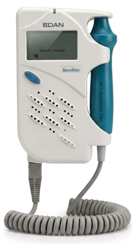
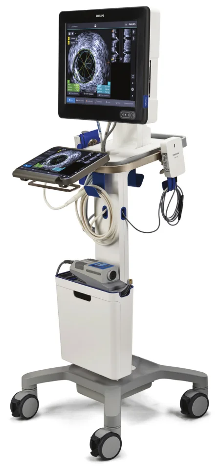
Before a study with contrast during surgery, the team reviews the patient's allergies and clotting status, with the kidney checks chapter 22 teaches.
5The patient and the room
Before the patient arrives
Vascular disease is usually a disease of the whole body, so the patient's heart, kidney and lung disease is reviewed before surgery. Many fear a stroke or the loss of a leg, so answering their questions is part of the team's care.
The circulator confirms that blood ordered from the blood bank is ready, and whether a cell saver will be used to return the patient's own shed blood. Chapter 12 teaches the cell saver. Because angiography or fluoroscopy may be needed in almost any vascular case, the patient lies on a bed that x-rays can pass through, with contrast, catheters, sterile x-ray covers and lead aprons in the room. Chapter 22 teaches radiation safety.
Vascular cases are often long, so the team works to keep the patient warm: as little skin uncovered as possible, a forced-air warmer, warmed IV fluids, and irrigation at body temperature, 37°C and no warmer, as chapter 29 teaches. A prophylactic antibiotic is given within 1 hour before the incision, because an infected graft can cost a patient a leg, or a life.
Anesthesia and monitoring
Chapter 13 teaches the anesthetics and the monitors. Open vascular surgery may be done under general, regional or local anesthesia, and most percutaneous endovascular cases need only short-acting IV sedation with local anesthetic at the access site. What vascular cases add: an arterial line in the radial artery for a long case; a central venous or pulmonary artery catheter as needed, the pulmonary artery catheter common in aortic surgery; an EEG to watch the brain in a carotid case; and a urinary catheter when the case involves the renal arteries or clamps the aorta above them, heavy blood loss is expected, the case is long, or a spinal or epidural is used.
Position, prep and drape
Most vascular cases are supine, padded and with the arms placed as chapter 31 teaches. The patient lies on a pressure-redistributing surface, and a footboard can keep the weight of the drapes off the feet. For a leg case, the thigh may be turned outward and spread a little with the knee bent over a small pillow. The patient's pulses are found, graded and marked on the skin before the prep, so the surgeon can find them fast when a pulse check is called for.
The prep areas are wide:
- Carotid: from the ear and chin on the operative side to below the collarbone.
- Abdominal aorta: from the nipple line to mid-thigh, so the groins are ready if the femoral arteries must be reached.
- Legs: from the umbilicus to the feet, each leg prepped all the way around (circumferentially) and then draped free.
An alcohol-based prep must dry completely before the drapes go on, because its fumes can catch fire. The drapes are impervious, and they leave the surgeon every area the case might need: both groins in an aortic case, the other leg when its saphenous vein may be taken. When both legs are draped, the genitals are covered with a towel and a barrier drape.
6Vascular instruments
Vascular surgery uses general surgery instruments with a vascular set added, plus extra instruments for the region. Every vascular case needs right-angle (Mixter) clamps, tonsil (Schnidt) clamps, and Kelly, Crile and mosquito clamps.
Clamps
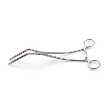
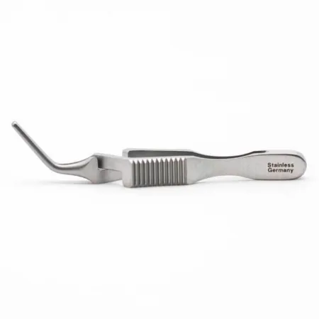
Vascular clamps close a vessel without damaging it. Their jaws carry fine, atraumatic serrations that grip without crushing the wall, even when the clamp is fully closed. They come in hundreds of shapes, made to fit around a vessel as it lies, and the peripheral ones are smaller than the clamps of heart surgery. The two common serration patterns are the DeBakey and the Cooley types.
- Occlusion clamps close the vessel completely. A DeBakey or similar angled clamp crosses the aorta or the iliac arteries.
- Partial occlusion (tangential) clamps, such as the Satinsky, close off only part of a large vessel, so blood keeps flowing past while the surgeon works on the side wall.
- Clamps with inserts, such as the Fogarty, carry disposable plastic inserts that slide onto each jaw and protect the vessel; the Core Curriculum lists them as vascular clamp inserts. Clamps with rubber, plastic or fluid-filled jaws protect a vein graft.
- Bulldogs are small, spring-loaded clamps with atraumatic serrations that close off a small vessel or a side branch while the surgeon works.
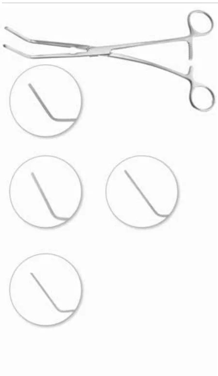
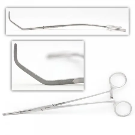
Scissors, forceps, retractors and suction
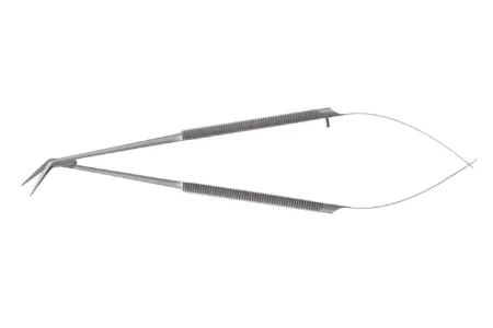
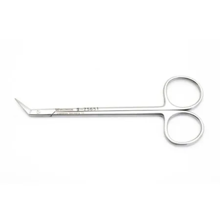
- Scissors: Potts scissors, with blades angled to the side, open and lengthen the cut in an artery; De Martel scissors are straight, and fine Metzenbaums dissect. Vascular scissors are very delicate. Never use them on suture or graft material, which dulls them and knocks the blades out of line.
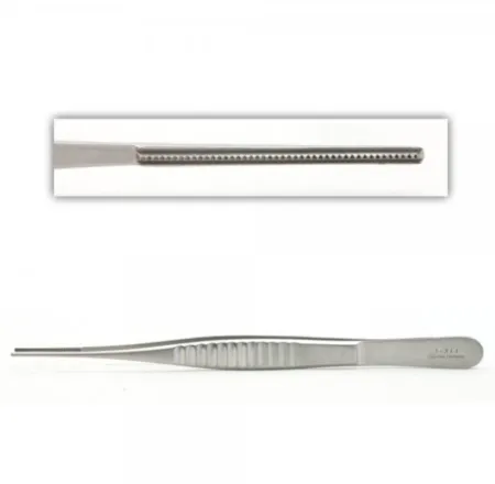
- Forceps: vascular forceps, such as the DeBakey, have very fine serrations at the tips that hold a vessel without injuring it. Pass them whenever a vessel or other delicate tissue will be handled; plain forceps do not grip it well enough, and toothed forceps are never used on a vessel, because they can puncture its wall.
- Retractors: shallow, blunt retractors for the superficial cases: a blunt Weitlaner, the Senn, vein retractors, small Richardsons. Take great care with any sharp retractor near a vessel. Deep cases use the Deaver, Richardson and Army-Navy.
- Suction: a dry field is critical, and suction is used more than sponges. Small Frazier tips are used near vessels, with the suction pressure turned down for the vessel itself. They clog easily, so keep two ready, one on the field and one in reserve, and clear a clogged tip with its stylet or by suctioning a little water through it. The aorta needs larger tips, sometimes two suctions.
Tunnelers
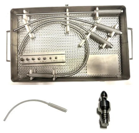
A tunneler is a long blunt-tipped metal shaft that is pushed under the skin or between tissue layers from one incision to another, making a path for a graft. The graft is then pulled through, by the tunneler itself or by a long clamp passed through the tunnel.
7Vascular supplies
Vessel loops, tapes and the Rummel tourniquet
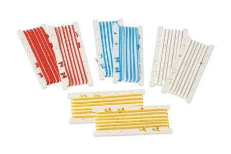
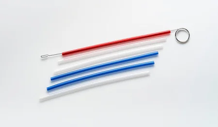
Before a vessel is opened, the surgeon frees it and passes a vessel loop around it with a right-angle clamp, to lift it, pull it aside or stop its flow. The ends are clamped together with a hemostat. Most loops are thin colored Silastic; chapter 25 gives the color code, usually red for arteries and blue for veins. Umbilical tapes, flat cotton tapes used wet, do the same job. Load the loops on tonsil clamps on the back table so they are ready to pass. Vessel loops and tapes used in the wound are counted with the miscellaneous items.
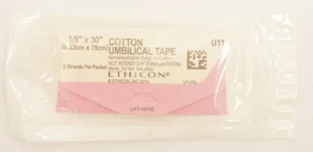
A Rummel tourniquet is an umbilical tape passed around a vessel, with its ends threaded through a short length of tubing. The tubing is pushed down snug against the vessel and secured, so it closes the vessel for a time, as a temporary tourniquet. To get one ready, feed one end of the tape into the tubing. The surgeon carries the tape around the vessel on a right-angle clamp, and a hemostat holds its ends.
Sutures, boots and pledgets
Vascular sutures are synthetic nonabsorbable monofilaments, most often polypropylene, because a monofilament is nonreactive, which keeps clot from forming on it inside the vessel. Sizes run from about 3-0 for the aorta to 6-0 or 7-0 for small arteries and a dialysis fistula, with finer still in microsurgery. The needles are swaged on, usually with a 3/8 or 1/2 curve, and the suture may be single-armed or double-armed, a needle at each end.
- Loading: vascular needles are delicate, so use fine-tipped needle holders and draw the suture gently out of the package.
- Double-armed sutures: mount one needle on the needle holder and put the other on a small hemostat whose jaws wear jaw covers: short pieces of Silastic tubing, also called suture boots or booties, that cushion the grip so the suture is not damaged.
- Never pull a needle holder away from the field until you are sure it is free of the anastomosis. The two ends of a double-armed suture work together, and pulling one can drag the other out of the tissue.
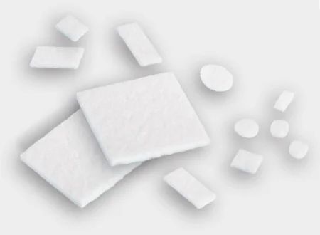
Pledgets are small squares of Teflon (PTFE) felt, or leftover pieces of graft, threaded onto a suture as a buttress, to keep the stitch from cutting through a fragile vessel wall or to seal a leaking anastomosis. The pledget stays on the suture line.
Grafts and patches
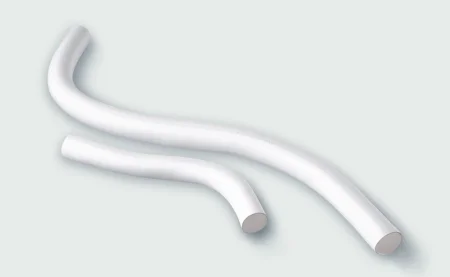
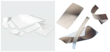
A vascular graft replaces a section of vessel, carries blood around a blockage (a bypass), patches a vessel, or makes dialysis access. Grafts come straight, tapered, bifurcated (Y-shaped) for the aorta and both iliac or femoral arteries, or as patches cut to size. Their length is measured in centimeters and their outside diameter in millimeters.
- Polyester (Dacron): woven polyester is strong and leaks little through its fabric but is stiffer, so it is kept for large arteries such as the aorta; knitted polyester is easier to handle. Many polyester grafts now come coated with albumin, collagen or gelatin, so they need no preclotting.
- PTFE (expanded polytetrafluoroethylene): sometimes with rings on the outside to keep it from being squeezed or kinked. It can cross the knee without kinking. It does not stretch, so its needle holes can bleed.
- Biologic grafts: the patient's own vein (an autograft, usually the great saphenous vein), a banked human umbilical cord, a bovine (cow) carotid artery, or a cryopreserved donor saphenous vein.
The patient's own vein is the best choice below the groin, because its living lining resists clotting; synthetic grafts are used above the knee, to spare the veins, or for a patient who cannot have a vein taken. Some synthetic grafts carry an antibacterial substance to lower the risk of infection.
a knitted polyester graft is porous, so the scrub once preclotted it, soaking it in a little of the patient's own blood drawn before the heparin, until fibrin sealed the fabric. Coated polyester grafts and PTFE need no preclotting.
A graft is an implant, and chapter 29's rules apply. Grafts are very expensive: keep each one in its package until the surgeon is ready and has asked for the size aloud, then confirm it with the circulator before you open it. Its size, lot and identification number go on the patient's record. A synthetic graft needs only trimming; a bovine graft is soaked in saline, and a cryopreserved vein rinsed of its preservative, exactly as the maker's instructions say. Keep a graft from touching the patient's skin, where it could pick up bacteria. A harvested vein is kept moist until it is used.
Other vascular supplies
- Embolectomy catheters: thin catheters with a small balloon behind the tip, passed beyond a clot, inflated and drawn back to pull the clot out.
- Irrigation tips and syringes: a blunt heparin flushing needle or a soft catheter on a 20 or 30 mL syringe of heparinized saline.
- Shunts: flexible tubes that carry blood past an opened artery, as in a carotid endarterectomy.
- Valvulotome: a cutting instrument passed through a vein to cut its valves, so arterial blood can flow through the vein.
- Vein stripper: a long, very flexible cable with olive (acorn) tips that pulls a vein out through its own lumen.
- Aortic punch: punches a round hole in an artery for a graft to be sewn to.
- Sterile Doppler probe: sterilized by its maker's instructions or put in a sterile cover, to check flow on the field. The probes are heat sensitive, and the tip is easily damaged if it is dropped or crushed.
- Endovascular supplies: guidewires, sheaths, catheters, balloons and stents, taught below and in chapter 22.
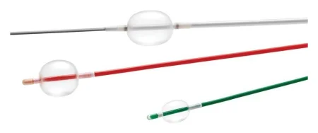
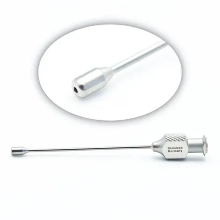
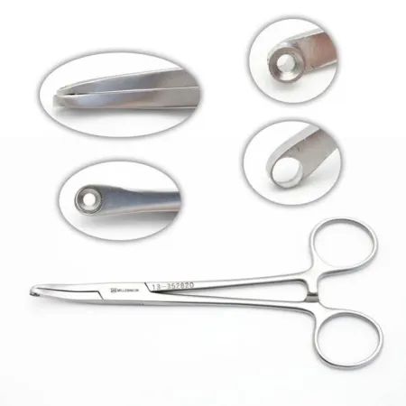
8Drugs on the vascular field
Several high-alert drugs share the vascular field, and most of them are clear liquids, so every cup and syringe is labeled the moment it arrives, as chapter 12 teaches. Keep track of how much of each drug and of the contrast is used, because the circulator records every drug and solution and the amount given.
- Heparin, systemic: given into a vein by the anesthesia provider shortly before an artery is clamped, so no clot forms while the flow is stopped; it acts within minutes. The activated clotting time (ACT), a quick test of how long the blood takes to clot, shows whether more is needed.
- Heparinized saline: heparin mixed into saline to the surgeon's order, for flushing the open vessel, a graft, a shunt and every endovascular device. It keeps clot from forming at the repair and lowers the risk of an embolus. Draw it up in a 20 or 30 mL syringe with a tapered irrigation tip.
- Protamine sulfate: reverses heparin at the end of the case, given slowly into the vein by the anesthesia provider, because a fast dose can drop the blood pressure and the heart rate. It is made from fish, so ask about fish allergy and about insulin that contains protamine.
- Papaverine: relaxes the smooth muscle of the vessel wall, which widens the vessel and eases spasm. It is often added to heparinized saline to distend a vein graft gently.
- Drugs for vasospasm: when a vessel clamps down in spasm, the surgeon may lay on it a gauze sponge soaked in papaverine, lidocaine or verapamil.
- Dextran 40: an antithrombotic, given into a vein after a bypass or endarterectomy, or diluted as an irrigation on the field.
- Topical hemostatic agents: collagen or fibrin products, a gelatin sponge or topical thrombin, laid on an oozing anastomosis. Chapter 29 teaches preparing them.
- Contrast: for the angiograms. Track the total amount and strength used.
Chapter 12 teaches each drug, its doses and its cautions, including the strengths of heparin that look alike and how much protamine neutralizes it.
Never inject topical thrombin, or put it in a syringe: in a blood vessel it can cause massive clotting and death. Heparinized saline, contrast, saline and papaverine share the field, and most field drugs are clear, so if a cup or syringe is not labeled, it does not go on the field.
9Opening and joining vessels
Most open vascular operations follow the same order. Learn it once, and you can follow the open cases in this chapter.
- Exposure: the surgeon dissects down to the vessel and frees it from the tissue around it.
- Control: vessel loops or umbilical tapes go around the vessel above and below the area to be opened, and around its branches.
- Heparin: the anesthesia provider gives systemic heparin just before the clamps go on; it acts within minutes.
- Clamping: the surgeon chooses a clamp that fits the vessel where it lies. The circulator records the time each clamp goes on and comes off, because that is how long the tissue beyond it is without blood.
- Arteriotomy: an incision into the artery, usually with a #11 blade, lengthened with Potts scissors.
- The repair: the plaque or clot is removed, or a graft is sewn on.
- Flushing: before the last stitches are tied, each clamp is opened briefly, one at a time, so blood flushes air, clot and loose debris out through the gap. Heparinized saline may be injected too.
- Restoring flow: the clamps come off in the order the surgeon chooses, so anything still loose travels where it does the least harm.
- Checking: the surgeon looks for leaks and closes them with extra stitches, pledgets or a hemostatic agent, and checks the flow beyond the repair with a sterile Doppler, the pulses or an angiogram.
When the clamps are on, the clock is running. Have the next suture loaded, the heparinized saline ready and the Doppler on the field, so the surgeon never waits while the tissue beyond the clamp has no blood.
Endarterectomy
An endarterectomy removes plaque from inside an artery. The surgeon finds the plane between the plaque and the wall and lifts the plaque away with a Penfield or Freer elevator, using gentle traction; the plaque may be cut across over a right-angle clamp at its ends. Loose plaque can break up and travel as an embolus, especially when the clamps come off. Hand the plaque off as a specimen, in a small basin.
Anastomosis
An anastomosis is the join between two vessels, or between a vessel and a graft. It can be made three ways:
- End to end: two cut ends joined, as when a damaged piece of vessel is replaced.
- End to side: the cut end of one vessel or graft sewn over a slit in the side of another. It is the join a bypass graft usually makes at each end, and the usual join in a dialysis fistula. The end of the graft is cut at a slant (beveled) so it meets the artery at a gentle angle.
- Side to side: two vessels lying next to each other, each slit and the edges sewn together.
The surgeon usually sews with a double-armed suture, often anchoring a stitch at each end first and then running a continuous stitch around. An arteriotomy can also be closed with a patch, a piece of vein or synthetic material sewn into the opening, so that closing a small artery does not narrow it.
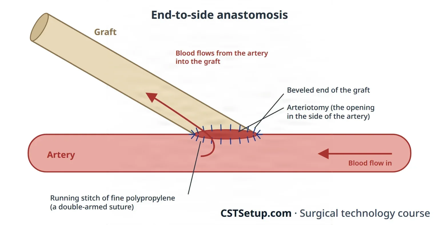
Tunneling a graft
A bypass graft often runs between two incisions some distance apart. The surgeon makes a tunnel between them with the fingers or a tunneler, and the graft is pulled through without kinks or twists.
10Endovascular supplies and how they are used
Chapter 22 teaches the endovascular supplies as interventional radiology uses them: the way into the vessel, the guidewire's job and lengths, the access sheath, the five kinds of catheter, the balloon and its inflation device, and closing the site when the sheath comes out. This section adds what a vascular case asks of the scrub.
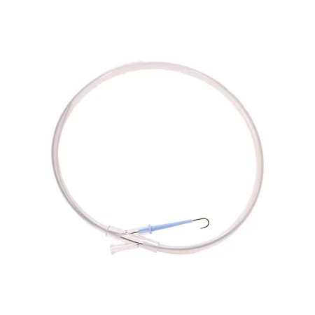
Guidewires
- Diameter: usually 0.014, 0.018 or 0.035 inch. The channel of each catheter, balloon and stent delivery system is sized for one wire diameter, so check that they match.
- Material: stainless steel, or nitinol, a more flexible metal alloy.
- Tip: stiff and rigid, J-curved, or floppy and flexible to protect a delicate vessel wall; a stiff wire carries a large device such as a stent graft.
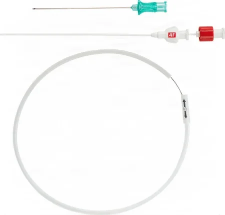
- A smaller first stick: a micropuncture set starts with a 21-gauge needle and a thin 0.018-inch wire, then changes up to the 0.035-inch wire.
Keep every wire wet and free of clot and debris. Wipe it with a moist gauze sponge before and after each pass into the patient, and coil it back quickly and safely after each use; long wires slide off a field easily, and holders are made to keep them on it.
Sheaths and the French scale
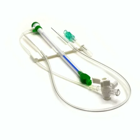
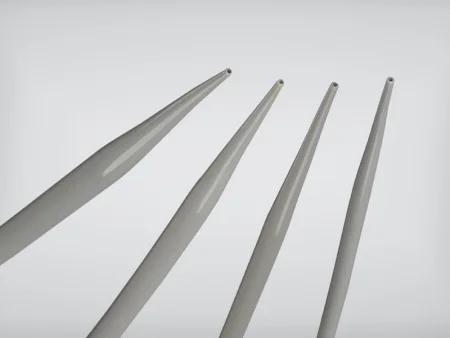
Sheaths and catheters are sized on the French (Fr) scale: each French unit is about a third of a millimeter of diameter, so a 24 Fr sheath is about 8 mm across. Sheaths run from 4 to 24 Fr, chosen for the vessel and the devices that must pass. When a larger sheath is needed, a wire holds the access while the small sheath comes out and the larger one goes in over the same wire; dilators are part of the setup. The sheath stays in until the end of the procedure.
Balloons and the inflation device
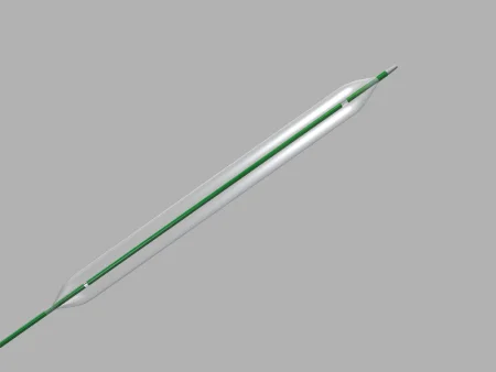
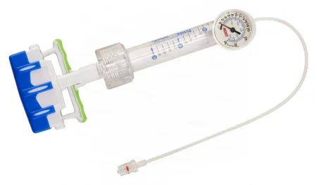
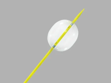
An angioplasty balloon stretches a narrowed artery or vein open; a stent expansion balloon presses a stent firmly against the vessel wall. Balloons are inflated and held for several seconds to several minutes.
The inflation device fills the balloon to an exact pressure, measured in atmospheres (atm). You load it with 10 to 20 mL of the contrast solution, so the balloon shows on fluoroscopy, connect it to the balloon catheter, and clear every bit of air from the device, its tubing and the balloon: trapped air blurs the image and could become an air embolus if the balloon burst.
Stents and stent grafts
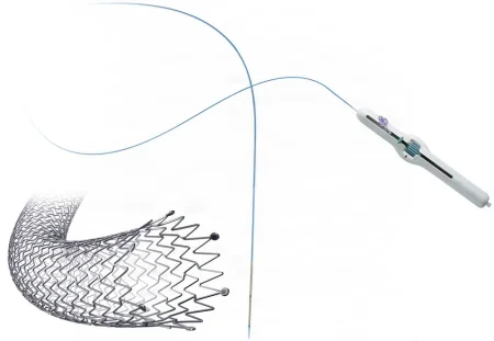
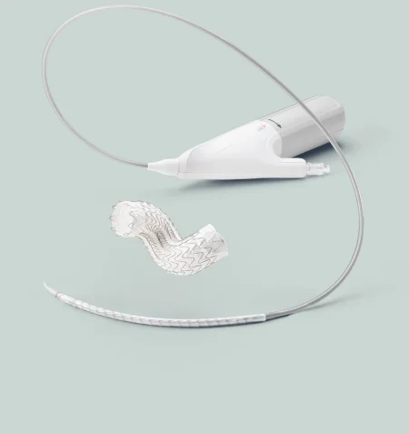
A stent is a tube of metal mesh that fits against the inside of a vessel and holds it open. In an artery it walls the plaque off from the lumen and lets blood flow freely without platelets clumping. Stents are made of stainless steel, titanium or nitinol, and they stay in for life.
- Balloon-expandable stent: the mesh is crimped onto a balloon, and inflating the balloon pushes it out against the wall.
- Self-expanding stent: it opens out against the wall by itself as it is deployed, with no balloon needed to expand it.
- Stent graft (covered stent): a metal frame covered with ePTFE or woven polyester. It lines a weakened artery from the inside, gives the blood a smooth path, and walls the aneurysm sac off from the circulation. It opens partly on its own, then a balloon presses it snug against the wall so it does not leak. Stent grafts come tubular or bifurcated, and self-expanding or balloon-expanded.
Every stent and stent graft is an implant. Confirm its exact type, length and diameter with the surgeon and the circulator before you open it, and make sure its lot and serial numbers reach the record, as chapter 29 teaches.
Other endovascular tools
- Snare: a device passed into the vessel to catch and pull on a wire or a device inside it, such as the hook of a vena cava filter.
- Embolization: blocking a vessel from inside on purpose, with platinum or titanium coils, fast-setting glue or tiny plastic particles. It is used to shrink an abnormal tangle of vessels before surgery, and to close a side branch that keeps filling an aneurysm after an endovascular repair.
- Intravascular ultrasound: the IVUS probe rides into the vessel on a catheter to measure it from inside.
- Closure devices: at the end, an arterial puncture can be closed with a device instead of a stitch, such as one that pulls a purse-string suture tight around the hole, or a collagen plug; the Core Curriculum lists vascular closure devices and plugs.
- Atherectomy: newer devices shave or laser plaque out of the artery from inside.
11Angiography, angioplasty and angioscopy
Angiography in the OR
What and why: an angiogram injects contrast into a selected artery and its branches to show exactly where a vessel is narrowed, blocked or malformed. Done before surgery it maps the disease; done during surgery it guides an angioplasty or a stent, places a stent graft, and checks a new bypass, looking above and below the repair for blockages or emboli.
Setup: two or more 30 or 60 mL syringes, an arterial needle or angiocatheter, one or more IV extension tubes, a three-way stopcock, contrast as the surgeon orders, IV saline and heparin as ordered. Position, prep and drape depend on the vessel.
Steps:
- The circulator passes you the contrast. Draw it into two syringes and fill a third with IV saline to flush the contrast out of the artery afterward. Label all three.
- Connect the needle to the extension tubing and the tubing to the stopcock and the contrast syringe. Remove every air bubble, so no air goes into the artery; a hemostat across the tubing keeps air from backing in, and the stopcock stays closed until the injection.
- Move metal instruments out of the area to be imaged. The C-arm is draped just before it is needed and parked a little away from the field. Everyone in the room wears a lead apron, put on before scrubbing, as chapter 22 teaches.
- The surgeon injects through a needle placed between the stitches of a closed arteriotomy, through a catheter tied into the vessel, or through the stopcock of a sheath already in place, and images are recorded during the injection.
- The contrast is flushed out with saline, and injections are repeated as needed.
Wound class and after: a percutaneous angiogram enters no tract, so by the CDC's definitions it is Class I; done during an operation, it takes that operation's class. Afterward the puncture site is watched for bleeding and a hematoma, and the patient for a reaction to the contrast, which can come late, as chapter 22 teaches.
Angioplasty and stenting
What and why: angioplasty opens a narrowed or blocked artery from inside, with a balloon. The balloon cracks the plaque, and the artery heals with a new lining. A stent may follow to hold the artery open, especially when the balloon result is not good enough. Percutaneous transluminal angioplasty (PTA) is used routinely for narrowings and short blockages of the iliac arteries. Angioplasty works well at first, but the artery can narrow again (restenosis).
Diagnosis: detailed angiographic mapping locates the narrowing and measures it before the balloon is chosen; have the images in the room.
Anesthesia and position: local anesthetic with monitored sedation, often as same-day surgery; supine, prepped from the groin to the knee.
Setup: an angioplasty tray with an arterial introducer needle and a sheath introducer; a guiding catheter, guidewires, the balloon catheter and stents; heparin and contrast; the C-arm and its drape.
Key steps:
- Under fluoroscopy, a needle enters the femoral artery, often with the flow (antegrade), the best approach for most narrowings in the legs; a guidewire goes in and a small sheath, 4 to 8 Fr, goes over it.
- Contrast shows the narrowing, and its place is marked on the images. Be ready with heparin for the surgeon to give through the catheter.
- The wire is passed through the narrowing, the most delicate part of the case.
- The balloon goes over the wire to the narrowing and is inflated with the inflation device to a set pressure for a set time, watched on fluoroscopy.
- A stent may be placed over the wire and expanded against the wall, and the final images are recorded.
Wound class and after surgery: Class I, clean. The complications include a hematoma at the puncture site, which may need surgical repair, a reaction to the contrast, a surgical site infection and a heart attack.
Carotid artery stenting is the endovascular alternative to carotid endarterectomy; one large database study found the two about equally safe overall, with slightly more strokes after stenting and slightly more heart attacks after endarterectomy.
Angioscopy
What and why: angioscopy looks inside a vessel with a thin, flexible fiberoptic scope, about 0.5 to 3.0 mm across, on a video camera and monitor. It lets the surgeon see the inside of the vessel, as when the valves of a vein are cut under direct view for an in situ bypass, or after a clot is pulled out. Its main challenge is clearing the blood, because even a little blood blurs the view, so the segment is flushed with pressurized irrigation.
Setup: the angioscope (confirm its size with the surgeon, and pick one smaller than the vessel, so it does not rub the wall into spasm), a light cord, the video system, a vein irrigating catheter, and a 1,000 mL bag of Ringer's solution in a pressure cuff inflated to 200 to 300 mm Hg, or an irrigation pump; a Doppler with a sterile probe.
Anesthesia and position: general anesthesia, supine. The prep depends on where the scope goes in, but it is still wide.
Key steps: the vessel is exposed through a cutdown and a segment is isolated between two vascular clamps. A small arteriotomy or venotomy is made with a #11 or #15 blade, the blood is flushed out, and the irrigating catheter is placed. The scope goes in down the center of the vessel, the far clamp comes off, and you may help advance the irrigating catheter beside the scope. At the end both come out, the vessel is closed, the last clamp is removed and flow is checked with the Doppler.
Wound class and complications: Class I. Complications include infection, vasospasm, injury to the intima from the scope or the force of the irrigation, and a loosened piece of plaque that travels as an embolus.
12Carotid endarterectomy
What and why: a carotid endarterectomy (CEA) removes atherosclerotic plaque from the carotid artery, through an incision in the artery, at the bifurcation where the common carotid divides. It restores the flow of blood to the brain and lowers the risk that plaque or clot will break off and cause a stroke. Most patients go home the day after surgery.
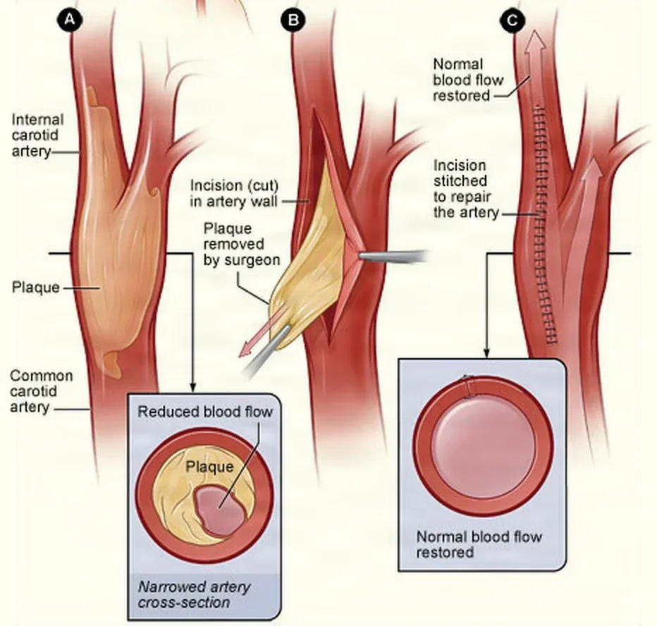
Diagnosis: duplex ultrasound and arteriography show where the plaque is and how much it narrows the artery, and a CT scan or MRI checks the brain for an earlier stroke. Have the patient's images in the room.
Anesthesia and watching the brain: general or regional anesthesia, such as a cervical plexus block. While the carotid is clamped, the brain must get its blood through the other arteries joined at its base (the circle of Willis, chapter 5), and in some patients that is not enough, so the team watches for signs of trouble:
- An awake patient under a regional block is asked to squeeze a hand or to speak.
- Under general anesthesia, an EEG shows the brain's electrical activity slowing.
- The back pressure in the carotid above the clamp (the stump pressure) can also guide the decision.
If the brain shows signs of too little blood flow, the surgeon places a temporary shunt, a tube that carries blood around the open artery. Placing it takes extra dissection and can knock debris loose, so not every surgeon uses one, but a shunt must be ready at the start of every case. The time the clamps are on is kept as short as possible.
Position, prep and drape: supine, the head on a head support or donut, turned away from the operative side, with a small roll between the shoulder blades so the neck is slightly extended; the arms are at the sides. The prep runs from the ear and chin to below the collarbone on the operative side, and the draping is like a thyroidectomy's. If a vein patch is likely, a leg is prepped and draped as well.
Setup: a vascular set with the surgeon's choice of right-angle vascular clamps, right-angle clamps, Freer and Penfield elevators, vessel loops, Rummel tourniquets, umbilical tapes, jaw covers, and topical hemostatic agents. On hold: a carotid shunt and the clamps that hold it, such as the Javid clamp, contrast and the C-arm, and the Doppler. Small bulldog clamps close side branches, such as the superior thyroid artery off the external carotid. For irrigation inside the artery, a 20 mL syringe with a heparin needle or an angiocatheter cannula. Retractors include a blunt Weitlaner, blunt rakes and Army-Navy retractors.
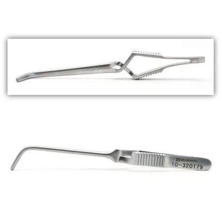
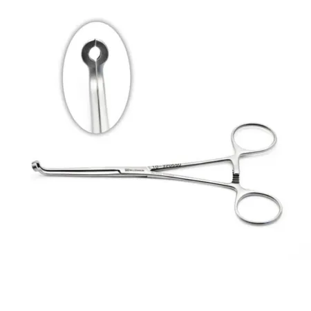
Medications: systemic heparin; heparinized saline for irrigation; 1% lidocaine, which the surgeon may inject around the carotid sinus to prevent the slow heart rate and low blood pressure that handling it can cause (chapter 5 explains the reflex); topical hemostatic agents.
Key steps:
- The incision runs along the front border of the sternocleidomastoid muscle, and a Weitlaner holds it open.
- The surgeon dissects with vascular forceps, the ESU, Metzenbaum scissors and sponge dissectors down to the carotid arteries, moving the internal jugular vein aside.
- Vessel loops go around the common, internal and external carotid arteries, their ends clamped with small hemostats; the bifurcation itself is not fully freed.
- Heparin is given. Have ready a #11 blade, Potts and De Martel scissors, Penfield and Freer elevators, straight hemostats and an atraumatic suction tip.
- The three arteries are clamped, and the surgeon tells the anesthesia provider and the circulator. The clamp time is recorded and the EEG watched. The order of clamping varies with the surgeon and the reference.
- The arteriotomy starts in the common carotid and is extended up into the internal carotid with Potts scissors. The plaque shows as a thick, yellow, rubbery layer stuck to the lining.
- If a shunt is used, it goes in now, one end in the common carotid and one in the internal carotid, held by a shunt clamp or by vessel loops; flush it with heparinized saline before you pass it.
- The surgeon lifts the plaque away with an elevator, finishes it cleanly in the internal carotid, and flushes the artery with heparinized saline to wash away loose strands. Keep a small basin ready for the plaque.
- The arteriotomy is closed with 6-0 or 7-0 polypropylene, directly or with a patch of PTFE, polyester or vein.
- Just before the closure is finished, any shunt comes out, and each clamp is opened briefly so blood flushes out air and debris.
- The clamps come off the external carotid first, then the common carotid, and the internal carotid last, so any debris or air still loose is carried up the external carotid to the face instead of to the brain. One reference releases the common carotid first; all agree the internal carotid is released last. The time flow returns is noted.
- The surgeon checks for leaks and the flow with the Doppler, places a drain through a separate stab wound, and closes the wound.
Specimen: the plaque.
Counts: initial and closing, with the vessel loops and tapes included.
Dressing: gauze, or a clear film the patient can shower with.
Wound class: I, clean.
After surgery: the patient is watched closely for any new neurologic deficit, bleeding and breathing problems. Stroke is one of the most feared complications. Injury to a cranial nerve is the most common, most often the hypoglossal nerve, which makes the tongue point to the operated side, and most of these injuries recover. Bleeding, infection and too much blood flow to the brain once the artery is open (hyperperfusion) can also occur. Patients are taught that numbness around the incision is normal, and to report any new TIA symptoms, confusion or trouble speaking.
13Open abdominal aortic aneurysm repair
What and why: the open repair replaces the aneurysm with a graft, a straight tube from aorta to aorta or a bifurcated graft from the aorta to both iliac arteries, which restores the flow to the legs and pelvis. It can be planned (elective) or an emergency. The sac is not removed: it is opened, emptied, and closed again around the graft.
Diagnosis: ultrasound and CT angiography measure the aneurysm, and the heart is checked carefully first, because heart disease is common in these patients.
Anesthesia and position: general anesthesia, with a central venous line and an arterial line; supine. The pedal pulses are marked, and a urinary catheter goes in before the prep.
Prep and drape: from the nipple line to mid-thigh, draped for a laparotomy that leaves the groins open in case the femoral arteries are needed.
Setup: a major laparotomy set with long instruments, Deaver retractors, a self-retaining retractor that mounts on the table (a Bookwalter or an Omni-Tract), a major vascular set, vessel loops, umbilical tapes, jaw covers, a long Penrose drain for retraction, the graft, IV saline and heparin, topical thrombin and other hemostatic agents, and a cell saver. Have extra aortic clamps, suction and mounted sutures ready, and a basin to collect the clot and plaque.
Medications: systemic heparin before the clamps, heparinized saline, topical thrombin and other hemostatic agents, and protamine at the end if the heparin must be reversed.
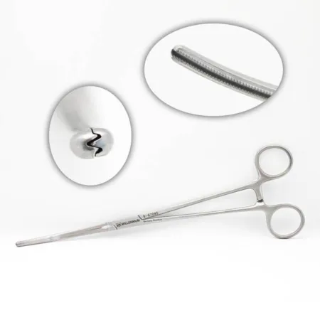
Key steps:
- A long midline incision from the xiphoid to the pubis. A self-retaining retractor goes in, and the small bowel is moved to the right, in moist laps or a bowel bag.
- The tissue behind the peritoneum (the retroperitoneum) over the aneurysm is opened with long Metzenbaum scissors and the ESU; a long ESU tip may be needed.
- The aorta is freed above the aneurysm (its neck), avoiding the renal arteries and the ureters, and the iliac arteries below it, mainly by blunt dissection; the surgeon checks the iliacs for aneurysms, clot and calcium. Have sponge sticks and small sponge dissectors ready. An injured vena cava is repaired at once with 4-0 or 5-0 vascular suture.
- Heparin is given. The iliac arteries are clamped with angled vascular clamps, and the aorta is cross-clamped above the aneurysm with the surgeon's choice of aortic clamp.
- The aneurysm is opened lengthwise with the ESU or a blade, with short cuts across each end. Clot and plaque come out piece by piece into your basin, and the lumbar arteries that bleed back into the sac from behind are oversewn with polypropylene. The sac itself is left in place.
- Pass the graft the surgeon names in a basin with straight Mayo scissors to trim it; a polyester graft coated with collagen, albumin or gelatin may be used. The upper (proximal) end is sewn to the aorta with a double-armed 3-0 polypropylene suture, sometimes reinforced with a strip of felt.
- The vessels below are opened to check that they bleed back, and flushed with heparinized saline. The lower end, or each limb of a bifurcated graft, is sewn on with a smaller suture.
- When the first side is done, blood is let into that limb while the other limb stays clamped; then the second side is finished.
- The suture lines are tested with saline or by easing the clamps; leaks get figure-of-eight stitches, pledgets or a hemostatic agent. The pedal pulses are checked with the Doppler.
- The aneurysm wall is closed over the graft with running polypropylene, and the retroperitoneum is closed over it. The abdomen is closed, and the skin is stapled.
be ready for the moment the aortic clamp comes off. Blood rushes into the widened vessels of the pelvis and legs, and the blood pressure can drop suddenly and severely (declamping shock). If heavy bleeding is likely even with the clamps on, a Foley catheter with a 30 mL balloon can be passed into the aorta and inflated to block it from inside.
Ruptured aneurysm: the first goal is to stop the bleeding by blocking the aorta above the rupture, by a clamp or by a balloon passed up from the groin. A ruptured aneurysm may also be repaired from inside, and the room is made ready for either repair.
Counts: as for any laparotomy, chapter 28's rules, with the vessel loops and tapes counted.
Wound class: I, clean.
After surgery: the patient goes to the PACU, the ICU or a cardiac unit, where the heart, lungs and kidneys are watched, and the legs are checked every hour. The risks are heavy bleeding, low blood pressure, injury to a ureter, kidney failure, a spinal cord that loses its blood supply, and death. A graft infection is rare but very serious. Patients with a graft may need antibiotics before future dental or endoscopic procedures.
14Endovascular abdominal aortic aneurysm repair (EVAR)
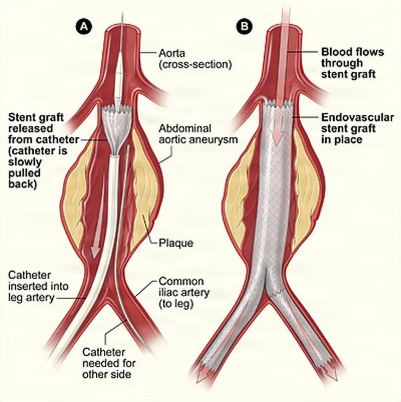
This is the Core Curriculum's model case for vascular surgery, so it is taught here from start to finish. In an EVAR the surgeon reaches the aneurysm from the femoral arteries and lines it from inside with a stent graft. The graft is held to the healthy aorta below the renal arteries and to the iliac arteries by self-expanding or balloon-expanded stents with hooks, not by sutures, so blood flows through the graft and the aneurysm sac is shut out of the circulation. No large abdominal incision is made, so the hospital stay is short, often 1 to 2 days, recovery is quick, and death and complications are fewer than after the open repair. It can also be used for a patient at high risk from open surgery.
Pathophysiology
The Core Curriculum's outline for the case reviews what you met earlier in this chapter:
- The aneurysm: caused by arteriosclerosis, atherosclerosis, a defect present from birth, infection, inflammation or trauma; it lies between the renal arteries and the aortic bifurcation; it is fusiform or saccular in shape; and it is a true aneurysm or a dissecting one.
- Atherosclerosis: atheroma and plaque in the wall, and thrombi, the clot that lines the sac.
- Turbulent blood flow in the widened sac.
- Other disease the patient brings: coronary artery disease, high cholesterol, high blood pressure and smoking.
Diagnosis
A Doppler exam, then imaging: angiography, an aortogram (an angiogram of the aorta), arteriography, CT scan and CT angiography, fluoroscopy, MRI and ultrasound. The images decide whether an EVAR is possible. The graft needs healthy, undiseased aorta and iliac artery to seal against at each end, called the landing zones: a neck of at least 1 to 2 cm below the renal arteries, iliac arteries for the lower end, and femoral and iliac arteries wide and straight enough for the large delivery system to pass. The size and the place of the aneurysm can rule an EVAR out. So can the need for imaging for the rest of the patient's life.
Preparing the patient
- Anesthesia: epidural, general or spinal. If an epidural is used, mechanical ventilation must be ready in case the case converts to an open repair.
- Position: supine, on a radiolucent bed, so the whole aorta and both groins can be imaged.
- Monitoring: the depth of anesthesia (BIS) and exhaled carbon dioxide (capnography) under general anesthesia; a Doppler probe for the pulses; DVT prophylaxis; and the hemodynamic signs: blood pressure, heart rate, oxygen saturation, breathing rate, temperature and urine output. A central venous line and an arterial line go in, and the circulator places a urinary catheter.
- Skin prep: the abdomen, and the legs all the way around.
- Draping: a sheet below the legs, the genitals isolated, folded towels to square off the groins, incise drapes, and fenestrated or plain sheets, draped so the case can become an open repair at once.
- Verification: the time out. Blood ordered from the blood bank is confirmed before the procedure begins.
The room, instruments and supplies
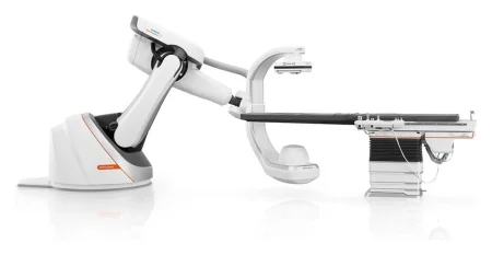
EVAR is done in a hybrid OR, which gives the team fixed and rotating angiography and fluoroscopy beside a full surgical setup. Chapter 22 teaches the hybrid room and radiation safety.
Equipment: extra tables (a back table, a Mayo stand and a ring stand); a cell salvage system; the Doppler; an ESU; fluoroscopy, with its control room and imaging monitors; a headlight; lead aprons or shields; positioning aids (armboards and safety straps); and suction.
Instruments: assorted vascular clamps and a minor vascular tray, with a major vascular tray and a self-retaining abdominal retractor on hold.
Basic supplies: a back table or custom pack, a basin set, #10 blades, an ESU pencil with its holder and tips, light handle covers, suction tubing, drapes, gowns, gloves and sponges.
Specialty supplies:
- Arteriogram supplies: an angiocatheter, an arterial (butterfly) needle, IV extension tubing, syringes and three-way stopcocks
- Balloons: angioplasty balloons with their inflation device (the Core Curriculum calls it an insufflation device), and stent expansion balloons
- Catheters: balloon, directional, exchange, pigtail and sizing
- Guidewires: exchange, introducing, steerable and non-steerable, with flexible, J-curved, rigid or stiff tips
- Needles: a Seldinger needle and an 18-gauge spinal needle
- Sheaths, each with its introducer (dilator), port and stopcock
- The stent graft, tubular or bifurcated, balloon-expanded or self-expanding
- A snare, hemoclips, a Rummel tourniquet, vessel loops and suture boots
- Pressure bags with high-pressure tubing, and cell salvage suction tubing
- Frazier and Yankauer suction tips, 30 and 60 mL syringes, and vascular closure devices or plugs
- Sutures for hemostasis, the femoral arteries, the fascia, the subcutaneous layer and the skin
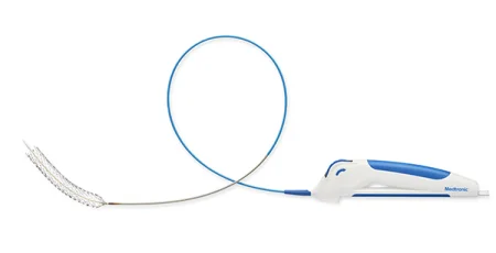
Most stent grafts are modular: the main body comes separate from the limbs that make the two legs of the Y. Each part comes loaded in its own delivery system, and the controls at its handle release and open it in steps. Exactly how depends on the maker, so the maker's instructions for that system are followed, but the patient's preparation is the same for all of them.
Medications: contrast; heparin; injectable saline; irrigation fluid; papaverine; topical hemostatic agents. Label each one, and keep a running total of the contrast and heparin.
The operation
The Core Curriculum's incisions are oblique groin incisions on both sides, from the inguinal ligament to below the femoral bifurcation; the right femoral artery is opened (an arteriotomy) and the left is punctured with an introducer, and a vessel may be entered against the flow (retrograde) or with it (antegrade). Access through the skin, closed with suture closure devices, is growing; because about 5 to 10 percent of closures of larger arteriotomies fail, a surgical cutdown to close the artery must be ready at once.
- Bilateral oblique groin incisions are made at the inguinal ligament, extending to below the femoral bifurcation, with a knife and toothed tissue forceps.
- A Halsted mosquito or a Kelly or Crile clamp spreads the tissue bluntly down to the femoral arteries, and Weitlaner retractors are placed.
- The common, superficial and deep femoral arteries are freed and controlled with vessel loops, a Rummel tourniquet and mosquito clamps.
- The anesthesia provider gives heparin.
- A DeBakey or similar vascular clamp goes across the right femoral artery.
- An arteriotomy is made in the right femoral artery with a #11 blade and lengthened with Potts-Smith scissors.
- You flush the sheath with heparinized saline. It goes into the right femoral artery and is advanced up into the right external iliac artery.
- On the left, the surgeon punctures the femoral artery with an introducer needle and advances a second sheath up into the left external iliac artery.
- An angiocatheter goes in and an arteriogram is taken, marking the renal arteries and the aortic bifurcation on the screen.
- A sizing catheter measures the aorta so the right size and length of graft can be chosen. Confirm the graft with the surgeon before you open it.
- The graft, on its delivery system, goes into the right femoral artery over a stiff guidewire and is positioned just below the renal arteries under fluoroscopy.
- A measuring catheter checks the position of the graft segment.
- A stent expansion balloon is passed up to the graft and inflated with the inflation device, to secure the graft's attachment.
- The limbs of the graft are positioned in the iliac arteries. The main body has a short limb with an opening on the far side; a second guidewire is passed up into it from the other groin, and the separate limb is slid in and opened so it overlaps snugly inside the main body.
- The graft is fully deployed, and its hooks grip the aortic and iliac walls.
- The stent expansion balloon is inflated again to press each attachment snug; for a stent graft this is a large, soft (compliant) molding balloon, pressed at each seal and where the parts overlap.
- A completion arteriogram confirms the position and that no blood still reaches the sac, and IV saline is injected to flush out the contrast. The graft is ideally placed so it covers the inferior mesenteric artery, which then cannot keep filling the sac, while the flow to the internal iliac arteries is kept.
- The guidewires, catheters and sheaths come out, and the arteries are irrigated with heparinized saline.
- Each femoral arteriotomy or puncture is closed, with a double-armed nonabsorbable suture loaded on a Castroviejo needle holder, or with a collagen plug or another vascular closure device.
- The fascia is closed with a continuous absorbable suture of the surgeon's choice.
- The subcutaneous layer may be closed with a continuous absorbable suture.
- The skin gets a subcuticular absorbable stitch or staples.
In an EVAR everything stays sterile until the patient has left the room, because a failed endovascular repair, a burst artery or a rupture means an open repair at once. Wipe every guidewire, catheter and sheath before and after each pass, and keep the open setup ready.
Ruptured aneurysm: when an aneurysm has burst, an aortic occlusion balloon may be passed up from the groin on a sheath pushed fully up into the aortic neck, and gently inflated to block the aorta above the aneurysm, holding back the bleeding while the stent graft is readied.
Dressings: nonadherent pads, 4x4 gauze and clear film over the groins.
Counts: initial, at fascia closure and at skin closure.
Wound class: I, clean.
After surgery
The patient goes to PACU, or to the ICU. The outlook is guarded when complications occur; otherwise medicines continue, the patient returns to normal activities, and illness and death are lower than after an open repair. The pulses in the legs are checked often in the first hours, and the patient is watched for belly or back pain and for the feeling and movement of the legs. Many walk within hours and go home on the second day. Imaging follows for life, because a stent graft can later leak or move, and some need another procedure.
The Core Curriculum lists the complications: death; an endoleak, and enlargement of the aneurysm sac; migration (movement) or occlusion of the graft; hemorrhage; infection of the graft, and surgical site infection; thrombosis of a limb of the graft, and other thrombus formation; pelvic ischemia; a pseudoaneurysm; and rupture.
An endoleak is blood still flowing into the aneurysm sac after the stent graft is placed. There are five types:
- Type I: blood leaks around the end of the graft, at the top (IA) or the bottom (IB) seal. It carries a high risk of rupture and is fixed at once, often by extending the graft or adding a bare stent at the neck.
- Type II: a side branch, such as the inferior mesenteric or a lumbar artery, keeps filling the sac backward. Its risk of rupture is small, and many close on their own within 6 to 12 months; it is treated, with coils or glue, only if the sac keeps growing.
The other three are a hole in the graft fabric or a gap where two parts overlap (Type III), blood seeping through porous fabric, seen as a faint blush on the final angiogram (Type IV), and a sac that keeps growing with no leak that can be seen (Type V, endotension).
If the bleeding cannot be stopped from inside, the graft cannot be placed, or the repair fails, the case converts to an open repair, and the team must be ready for it from the first minute.
15Bypass operations
A bypass carries blood around a blocked artery, through a graft sewn to healthy artery above the blockage (the inflow) and below it (the outflow). The steps are the same ones you learned for opening and joining vessels: exposure, control, heparin, clamps, arteriotomy, anastomosis, flushing and a flow check. What changes from case to case is where the graft runs and what it is made of.
Aortofemoral (aortobifemoral) bypass
What and why: the iliac arteries are a common site of atherosclerosis. An aortobifemoral bypass treats blockage of the aorta and iliac arteries (aortoiliac occlusive disease) with a bifurcated graft from the aorta to both femoral arteries, passing over the iliacs. It stays open better than an endarterectomy or an aortoiliac bypass, and it works even when the arteries are heavily calcified.
Diagnosis: arteriography or CT angiography shows where the aorta and iliac arteries are blocked and which arteries below are open.
Position, prep and drape: supine; a urinary catheter before the prep. The prep runs from the chest to the mid-thighs or below, both legs are draped all the way around, and the genitals are covered with a towel and a barrier drape. Confirm that blood has been ordered and is available, and have the patient's imaging in the room.
Setup: a major laparotomy set, a major vascular set, vessel loops, umbilical tapes, jaw covers, vessel clips, pledgets, topical hemostatic agents and thrombin, IV saline and heparin, and the surgeon's choice of graft, with long Penrose drains or straight Robinson catheters for the tunnels, and the surgeon's tunneling device. Have several graft sizes in the room, unopened, for the surgeon to choose from.
Key steps:
- A long midline laparotomy. The aorta is exposed up to the renal vein, with the duodenum moved aside, and down to its lower end.
- Both groins are opened, and the common, superficial and deep femoral arteries are freed and looped. The groin wounds may be covered with sterile towels while the abdomen is worked on.
- A tunnel is made on each side from the groin up to the aortic bifurcation, by finger, with a long clamp or with a tunneler. A Penrose drain or Robinson catheter may be drawn through each tunnel, its ends clamped with hemostats so it cannot slip back.
- Heparin is given, and the aorta is clamped just below the left renal vein. The aorta is divided, its lower stump oversewn, and the graft sewn to the upper end, end to end, with a continuous polypropylene suture. Pledgets may reinforce a fragile aorta.
- Each limb of the graft is drawn down through its tunnel to the groin. The common femoral artery is clamped, with bulldogs on its branches, and opened; the limb is cut to length, beveled and sewn end to side with a double-armed 5-0 or 6-0 polypropylene.
- Just before each join is finished, the clamps are let off briefly to flush clot and air out of the graft, and the suture line is flushed with heparinized saline; a small basin catches the blood.
- The surgeon closes the peritoneum and fascia at the back of the abdomen over the graft, then the abdomen and both groins, layer by layer.
Wound class: I, clean.
After surgery: the ICU or surgical unit, with Doppler checks of the circulation in the legs through the first 48 hours.
Femoropopliteal bypass
What and why: a femoropopliteal bypass carries blood around a blocked stretch of the femoral artery, from the femoral artery in the groin to the popliteal artery at the knee. The graft is the patient's own great saphenous vein or a synthetic tube. When the popliteal artery is diseased too, the lower end goes to a tibial artery below the knee (a femorotibial bypass), which may need microvascular instruments.
Diagnosis: arteriography maps the blocked femoral artery and the arteries below the knee, and a duplex ultrasound scan can check before surgery that the saphenous vein is good enough to use.
Position, prep and drape: supine, the hip turned out and spread a little, with the knee bent. The whole groin and leg are prepped and draped, with the other leg prepped when its vein may be needed. The incision lines and the pedal pulses are marked on the skin.
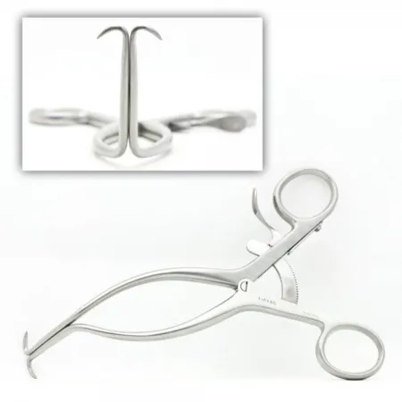
Setup: a basic minor set and a vascular set, Gelpi and Weitlaner retractors, a tunneler, umbilical tapes or vessel loops, and supplies for an angiogram during the operation.
Key steps (reversed vein):
- An incision along the inner thigh below the groin exposes the common femoral artery, and moist umbilical tapes or vessel loops go around the common, superficial and deep femoral arteries.
- A second incision on the inner side of the knee, above or below it, exposes the popliteal artery, which is looped; an angiogram may check that it and the arteries below are open.
- The saphenous vein is exposed through one long incision or several short ones, its side branches are tied with fine silk or clipped, and the length needed is removed. Because of its valves, the vein is reversed, so the end that was at the groin is sewn to the popliteal artery. Keep the vein moist in the solution the surgeon orders, which may be heparinized saline with papaverine.
- The graft is passed through a tunnel from the knee to the groin, without kinks or twists.
- Heparin is given, the femoral artery is clamped and opened with a #11 blade and Potts scissors, and the upper end of the graft is sewn on with fine vascular suture. Keep heparinized saline ready to irrigate both sites.
- The knee is bent, the popliteal artery is clamped and opened, and the lower end is sewn on. Before the last stitches, the femoral clamp is opened for a moment to flush out air and debris.
- All the clamps come off, the joins are checked for leaks, and the flow is checked by angiogram, Doppler or IVUS before the wounds are closed.
Taking the vein through small incisions, with an endoscopic vein harvesting system, is chapter 41's.
In situ bypass: in situ means in its natural place. The saphenous vein stays in its bed, its narrow lower end sewn to the popliteal artery and its wide upper end to the femoral artery. Its valves are cut with a valvulotome passed through it, and its side branches are tied or clipped so they cannot act as small fistulas.
Femorofemoral and axillofemoral bypass: when an aortic operation is undesirable, an extra-anatomic bypass, run outside the normal path of the arteries, carries blood from one good iliac and femoral artery through a tunnel under the skin across the pubis to the other femoral artery (femorofemoral), or from the axillary artery down to the same side's femoral artery (axillofemoral).
Wound class: I, clean.
After surgery: the pedal pulses are checked often; a pulse that fades or disappears can mean the graft has blocked. Swelling of the operated leg is common afterward, and some numbness of the lower leg may follow; blockage of the graft and infection are the main complications. Patients are taught foot care, to stop smoking and to report any change in the color, warmth or feeling of the leg.
16Embolectomy and thrombectomy
What and why: an embolectomy removes an embolus that has lodged in an artery; a thrombectomy removes a thrombus, a clot that formed where it sits. Both open the vessel and pull the clot out, usually with a balloon embolectomy catheter, to restore the flow before the tissue beyond dies, and both can save a limb or a life. About 80 percent of emboli that travel to the peripheral arteries lodge in a leg, about half of all of them where the common femoral artery divides, which is why a leg clot is usually removed through the groin. The usual causes of the clot are atherosclerosis, a heart that throws emboli, surgery on large vessels, and trauma such as a hip fracture.
Diagnosis: angiograms, duplex Doppler ultrasound and MRI show where the clot is and guide where the incision goes.
Anesthesia and position: general or regional anesthesia; supine, prepped and draped for the clot's location, most often a leg.
Setup: a laparotomy or general set, a vascular set, vessel irrigation tips, Frazier suction, 30 and 60 mL syringes and heparinized saline, irrigation catheters, vessel loops and topical hemostatic agents. Embolectomy catheters in several sizes are in the room but are opened only when they are needed.
Key steps (femoral embolectomy):
- A groin incision exposes the common femoral artery and its branches, and vessel loops go around them.
- Heparin is given, the vessels are clamped, and an arteriotomy is made with a #11 blade and lengthened with Potts-Smith scissors; have suction ready at the arteriotomy.
- You prepare the embolectomy catheter the surgeon names. The surgeon passes it beyond the clot with the balloon down, inflates the balloon, and slowly pulls the catheter back, drawing the clot out of the artery. Passes are made toward the heart and down each branch, and the surgeon checks that each branch bleeds back well.
- Have a vascular clamp ready to hand the moment the catheter and clot come out, so the bleeding is controlled.
- The artery is flushed with heparinized saline through a small irrigating catheter. An angiogram, angioscopy or Doppler checks the result.
- The arteriotomy is closed with 6-0 or 7-0 nonabsorbable vascular suture, and the wound is closed.
Specimen: the clot is kept as a specimen.
After surgery: once blood flows again into muscle that has been without it, the muscle can swell inside its fascia (ischemia-reperfusion injury), causing compartment syndrome. The surgeon may open the fascia at the same operation (a fasciotomy), and afterward the limb is checked often for the signs chapter 11 teaches. Dying muscle also releases myoglobin and potassium into the blood, which are watched.
Wound class: I, clean, when no infection or dead tissue is present.
Venous thrombectomy
A deep vein thrombosis in the iliac and femoral veins may be dissolved through a catheter, or removed in an open venous thrombectomy: a balloon catheter is passed up the femoral vein under fluoroscopy to pull the clot out, and any narrowing left in the iliac vein is opened with a balloon and, if needed, a stent; like the other planned vascular operations, it is Class I.
17Access for hemodialysis
Chapter 34 teaches kidney failure and hemodialysis, in which a machine filters the blood. Dialysis needs strong blood flow through large needles, often 15 to 17 gauge, placed again and again. An ordinary vein cannot take that, so the surgeon builds an access. For urgent dialysis, a double-lumen dialysis catheter can be placed in a large vein and used at once.
Arteriovenous fistula
An arteriovenous fistula (AV fistula) is a direct connection between an artery and a vein made by the surgeon. The vein, filled with arterial blood under pressure, grows wide and thick-walled over the following weeks (it matures), until it can take the dialysis needles. It is the standard access: it uses the patient's own vessels, and it is preferred over an external shunt, which carries a high risk of clotting and infection. A working fistula has a buzz you can feel, the thrill, and a continuous rushing sound you can hear, the bruit.
Of the four ways to join the artery and vein, sewing the end of the vein to the side of the artery is used more and more, and side-to-side joins less. Surgeons start as far out on the arm as they can and move up the arm with each new fistula. The classic first fistula, the Brescia-Cimino fistula, joins the radial artery and the cephalic vein at the wrist; later ones join the brachial artery to the cephalic or basilic vein in the upper arm. The basilic vein lies deep, under the fascia of the upper arm, so for a basilic fistula it is also moved up near the skin where needles can reach it (a transposition).
Before surgery: the fistula may be made ahead of need, when the kidney function falls far enough that dialysis is near, so it has time to mature. The veins and arteries of the arm are checked first, often in the vascular lab with ultrasound, because the veins vary from one person to the next.
Position, prep and drape: supine, the arm (often the nondominant one) out on an armboard. The prep may include the hand, arm, chest and axilla, and the arm is draped free. Local anesthesia is common.
Setup: a minor plastic set and a peripheral vascular set, vessel loops, mosquito clamps with jaw covers, a small self-retaining retractor, and loupes for the surgeon.
Key steps (radiocephalic fistula):
- An incision with a #15 blade over the radial artery and the cephalic vein in the forearm.
- The vein and the artery are freed, with vessel loops around them and small bulldogs or vascular clamps on each side of the planned join.
- An arteriotomy is made in the radial artery with a #11 blade and lengthened with Potts-Smith scissors, and the surgeon may widen the artery with vascular dilators: have them lined up from smallest to largest, and say the size of each one as you hand it.
- The vein is clamped, divided and tied off at its far end, and the near end is opened (a venotomy).
- The vein is sewn end to side onto the artery with 6-0 or 7-0 polypropylene; the surgeon may inject a little heparin into the join just before the last stitches.
- The clamps come off, and the surgeon gently feels the join to confirm blood flow. The wound is closed in layers and dressed with a nonadherent pad and dry gauze.
Arteriovenous graft
When the patient's own veins will not do, an arteriovenous graft bridges an artery and a vein with a tube, usually PTFE, laid in a tunnel under the skin; the Core Curriculum's "shunt" includes it. A bovine artery or a vein graft can be used too. A graft does not need to mature and can be used at once, though waiting about 2 weeks lets its joins heal first. A loop (U-shaped) graft joins an artery and a vein that lie close together; the usual sites run from the brachial artery to a vein at the elbow or to the axillary vein. A tunneler lays the graft in the fat just under the skin.
Once the arm sites are used up, most surgeons place the graft in the thigh, looped from the femoral artery to the great saphenous or femoral vein, where it stays open about as well but becomes infected more often.
Wound class (a fistula or a graft): I, clean.
After surgery: a fistula or graft can narrow, most often where it joins the vein, and then clot; that is its most common problem, and it is usually treated by another procedure, such as a patch angioplasty or removal of the clot. Bleeding, infection, an aneurysm or pseudoaneurysm, and heart failure from too much flow can also occur. In steal syndrome, the access draws so much blood away from the hand beyond it that the hand becomes ischemic; the access may have to be tied off or revised. Patients guard their access closely, as chapter 34 teaches.
18Venous access
Some patients need a large vein for weeks, months or years: for antibiotics, chemotherapy and hemodialysis, the uses the Core Curriculum names, and for IV nutrition and blood draws. A central venous catheter (central line) is a soft tube whose tip lies in the superior vena cava near the right atrium, entered through the skin into the subclavian, internal jugular or, less often, the femoral vein; its outer end may have two or three lumens.
- Tunneled catheter: part of the catheter runs in a tunnel under the skin between the vein and an exit site on the skin. Common ones are the Hickman, the Broviac and the Groshong. They are for long-term therapy, with one lumen or several.
- Non-tunneled catheter: for short- or medium-term therapy.
- Peripherally inserted central catheter (PICC): a thin tube put into a vein of the upper arm and guided up to the superior vena cava, for weeks or months, such as a course of antibiotics.
- Port: a reservoir implanted under the skin, usually of the upper chest, joined to a catheter in a central vein. Its self-sealing silicone dome is entered through the skin with a special noncoring needle, which does not cut out a core of the dome. A port can stay for months to years, and when it is not in use the patient lives normally, with nothing outside the skin.
Central lines are placed with strict aseptic technique in interventional radiology or the OR, guided by fluoroscopy or ultrasound. Every central line carries a risk of bloodstream infection; among the long-term devices, a fully implanted port carries the lowest.
Before surgery: electrolytes and clotting studies are checked, any earlier central lines are asked about to help choose the site, and ultrasound can find the vein. A single preoperative dose of antibiotic is given.
Anesthesia, position, prep and drape: usually moderate sedation or local with monitored anesthesia care, as an outpatient. Supine, the arms tucked, often in slight Trendelenburg to fill and widen the vein, with the head turned away. Hair is clipped, and a wide chlorhexidine prep covers the side of the neck and the chest, under the full sterile barrier: a drape that exposes the area over the collarbone and breastbone up to the chin.
Setup: the catheter or port kit, with its introducer needle, guidewire, dilator, peel-away sheath and tunneler; a minor instrument set; contrast, the C-arm and its drape; heparinized saline.
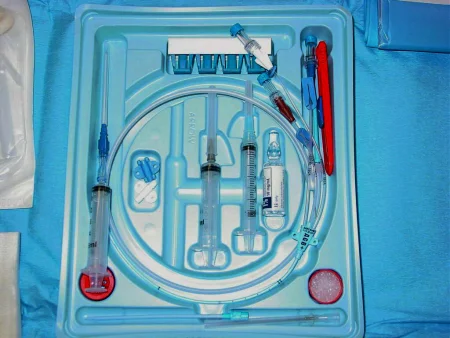
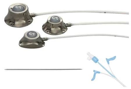
Key steps (port in the internal jugular vein):
- The vein is checked with ultrasound, then entered through a tiny skin nick with a small needle under live ultrasound, by the modified Seldinger technique (chapter 22), and a guidewire is passed; a micropuncture set may be used first.
- A small dilator makes a track over the wire.
- A 3 to 4 cm incision two fingerbreadths below the collarbone opens a pocket on the fascia of the pectoralis muscle, and a tunnel is made under the skin from the pocket to the neck.
- The dilator is swapped over the wire for an introducer with a peel-away sheath. The catheter goes in through the sheath and is placed with its tip in the right atrium under fluoroscopy; the sheath is split and peeled away. Hand the catheter coiled and tip first, and help steady it while it goes in.
- The catheter is cut to length and pushed onto the port's hub, and a slide-on boot locks it there.
- The port is drawn back and flushed to check that it runs freely, its position is checked on fluoroscopy, and it is stitched to the pectoralis fascia with nonabsorbable monofilament.
- The pocket and the skin are closed with absorbable sutures, and the port is filled with a dilute heparin solution.
A tunneled catheter goes in the same way, but its outer end comes out through an exit site, and a small cuff around it, left in the tunnel, grows into the tissue and holds it. It is stitched to the skin for about 2 weeks while the cuff takes hold.
Medications: heparin in low strengths flushes and fills a port or catheter; count it as heparin given into a vein. Have the right strength and label it clearly, because heparin vials that look alike hold very different doses.
Wound class: I, clean.
After surgery: a pneumothorax, a lung collapsed by a needle that strayed into it, is ruled out with a chest x-ray, which may be taken in the OR. Other complications are puncture of the subclavian artery and a catheter tip in the wrong place, such as up the jugular vein.
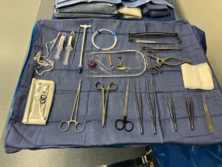
19Vena cava filter
What and why: a vena cava filter is a small metal device, shaped like an umbrella without its fabric, placed in the inferior vena cava to catch clots on their way from the leg veins to the lungs. It is made of titanium, stainless steel or nitinol, opens out against the vein wall, and may be permanent or temporary. It is used when:
- a patient with a venous clot cannot take anticoagulants, as after recent surgery or a bleeding stroke
- a patient survived a massive pulmonary embolism and another would be fatal
- clots keep forming in spite of anticoagulants
- the risk of a pulmonary embolism is high
It is placed through the skin, under fluoroscopy, in the catheter lab or the OR, with local anesthesia or moderate sedation. Several designs exist, and a kit made for the chosen route, femoral or jugular, must be opened.
Position and prep: supine on a bed that x-rays pass through, imaged at the level of the renal veins. The right femoral vein is the usual route, because the left femoral vein's path makes threading harder; for a jugular approach the head turns to the left. The circulator makes sure the local anesthetic, the contrast and heparinized saline for flushing the filter's kit are in the room.
Key steps:
- The groin is prepped, draped and numbed, and the vein is entered with a needle, often 18-gauge.
- A guidewire is passed under fluoroscopy up past the renal veins.
- Every lumen of the device is flushed with heparinized saline, and the sheath or dilator goes over the wire.
- The introducer catheter, carrying the filter preloaded in a capsule, goes up to the planned spot below the renal veins, and the filter is pushed out, where it opens; its place is checked on fluoroscopy.
- The introducer and wire come out, and pressure is held on the site until it stops bleeding, about 5 to 15 minutes in the references; a pressure dressing goes on.
The patient lies flat for some hours afterward so the site does not bleed, keeping the leg straight after a femoral insertion. A retrievable filter has a small hook at its top: through a sheath in a jugular or subclavian vein above it, the surgeon snares the hook and draws the filter up, and it folds closed into the sheath like an umbrella; an angiogram then checks the vena cava.
Wound class and complications: Class I. Bleeding at the site, a pulmonary embolism, a filter that moves or becomes infected, clot in the vena cava, and a filter that pierces the vein wall, causing bleeding behind the peritoneum.
20Vein ligation and stripping
What and why: the operation ties off (ligates) the great or small saphenous vein where it joins the deep vein and removes the diseased length of it, with its varicose branches, to relieve the pain, swelling and tiredness of the leg and to prevent ulcers.
Diagnosis and anesthesia: duplex ultrasound shows which veins and valves have failed. Stripping is usually done under general anesthesia.
Before surgery: before the patient comes to the OR, the surgeon has the patient stand and marks the varicose veins on the skin with a marker that will not wash off in the prep, so every varicose vein can be seen and removed.
Position, prep and drape: supine with the legs slightly apart, often tilted head down during surgery (Trendelenburg) to drain the veins; the small saphenous vein, behind the calf, may need the prone position. The leg is draped so the knee can be bent and lifted.
Setup: a minor set, Weitlaner retractors, #11 blades, skin hooks, mosquito clamps, vein strippers with several sizes of olive (acorn) tip, and elastic bandages. Confirm the size of acorn with the surgeon, and keep the long, very flexible stripper from sliding off the field.
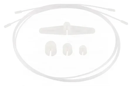
Key steps:
- An incision in the upper thigh, parallel to the groin crease, and a Weitlaner.
- The saphenous vein is found and its branches are tied twice with silk and divided. The vein is traced up to where it enters the femoral vein (the saphenofemoral junction) and tied there.
- A second incision near the ankle exposes the lower end of the vein, which is tied and divided.
- The stripper is threaded up the inside of the vein from the ankle to the groin. The acorn is screwed on, the vein is tied tightly to the stripper just below it, and the stripper is pulled out, turning the vein inside out as it comes. As the surgeon pulls, the assistant or the scrub presses along the leg.
- Branches are removed through many tiny incisions, 2 to 3 mm long, a technique called stab avulsion.
- The groin is closed in layers, the small incisions with sutures or staples, and the leg is wrapped firmly in elastic bandages.
Medications: none are routinely needed on the field.
Specimen: you remove the inverted vein from the stripper as the specimen.
Endovenous ablation: a newer outpatient alternative seals the vein from inside instead of removing it, with laser energy through a fiber-optic catheter placed under ultrasound, under the laser safety rules of chapter 20.
Wound class: I, clean. Most patients go home the same day; bleeding, a hematoma, infection and recurrence are the main complications.
After surgery: the patient walks, avoids long sitting and standing, keeps the legs raised when sitting, and wears the bandages or stockings as told.
21Vascular trauma
Chapter 11 teaches trauma care and how penetrating and blunt injuries harm the body; this is the vascular part. An injured major artery can cost a limb or a life, so its repair is urgent. In the United States most peripheral vascular injuries come from penetrating violence, gunshots and stab wounds.
- Penetrating trauma: a bullet's track and its breaking up can injure vessels far from the entry wound, and shotgun pellets scatter widely. A knife cuts a vessel partly or completely.
- Blunt trauma: crushing, as in a vehicle or industrial accident, or a broken long bone pressing on the vessels, blocks the flow to the limb. A sudden stop at high speed can split the layers of the aorta (a dissection).
The hard signs of an artery injury are absent pulses, active bleeding, a limb that is plainly ischemic, a hematoma that keeps growing, and a thrill or bruit over the wound; a patient with them usually goes straight to the OR. In the emergency department a Foley catheter may be passed into the wound and inflated as a tamponade to hold back the bleeding.
Setup: a vascular setup with general surgery instruments and every major vascular supply: the drugs, solutions, hemostatic agents, vessel loops and Fogarty catheters to clear clot from damaged vessels, with orthopedic and amputation instruments available. Get the sutures ready as soon as you know which vessel will be repaired, both single-armed and double-armed. Unless the surgeon names others, start with 2-0 and 0 silk ties on right-angle passers. Chapter 25 teaches loading needle holders ahead in trauma, with the needle tips left in their packet.
The repair: the surgeon exposes the injury, controls the bleeding with clamps or pressure, then repairs the vessel, directly, with a patch, or by bypassing the damaged part; blood and fluids are replaced, and bone or metal fragments that threaten more injury are dealt with. In a patient too unstable for a full repair, a temporary shunt, a tube placed inside the injured artery, restores blood flow to the limb until the patient can return for the definitive repair. This is vascular damage control. A severely mangled limb, or an injury to a major nerve and vessel bundle together, may lead to amputation instead, which chapter 39 teaches. Compartment syndrome threatens after the flow returns, and a fasciotomy may be done (chapter 11).
Wound class: a fresh traumatic wound is Class III, contaminated (chapter 11).
22Microvascular surgery
Microsurgery is any operation done under magnification, through loupes or the operating microscope; microvascular work joins vessels about 1 mm across, in replantation, plastic and reconstructive surgery, trauma, urology and neurosurgery. Chapter 38 teaches the scrub's part (the spring-handled microinstruments, the microvascular clamps marked A or V, sutures of 8-0 to 12-0, gentle irrigation and suction, and tissue kept moist and away from the table's edge), and chapter 23 the operating microscope.
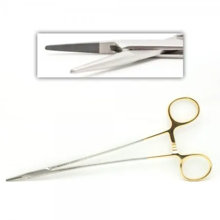
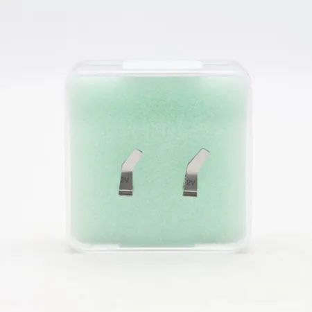
23Wound classes and care after vascular surgery
Almost every planned vascular operation is Class I, clean, because no tract is entered. A fresh traumatic wound is Class III, and an infection already present, such as an infected graft or infected gangrene, is Class IV. The scrub's part in preventing infection matters especially here, because an infected graft can mean losing a limb or a life: the antibiotic goes in on time, the graft stays off the skin, and sterile technique holds.
Everything on the field, the back table and the Mayo stand included, stays sterile until the patient has left the room. Bleeding from an anastomosis may need another stitch, and a new neurologic deficit in the OR after a carotid endarterectomy means the incision is opened again to check the repair.
Watch for these after vascular surgery, and report them at once:
- Bleeding: at an anastomosis, a groin puncture or a sheath site; a growing hematoma.
- A blocked graft or vessel: a pulse that fades or disappears, or a limb that turns pale, cold, numb or painful.
- Compartment syndrome after flow returns to an ischemic limb: swelling and severe pain, with the other signs chapter 11 teaches.
- Stroke and nerve injury after carotid surgery: a new weakness, confusion or trouble speaking, or a tongue that points to one side.
- Infection of the wound or the graft.
- Kidney injury, after contrast or aortic surgery.
Patients go home with teaching tailored to the operation: wound care, the signs of graft failure and infection, foot care after a leg bypass, how to take an anticoagulant safely, and above all to stop smoking, which harms both the vessels and wound healing.
Lo que te llevas de este capítulo
- Vascular surgery treats the arteries and veins outside the heart, open, endovascular (inside the vessel, often through the skin) or both in a hybrid room. Atherosclerosis is the disease behind most of it: plaque narrows arteries, causing claudication, rest pain and gangrene in the legs and TIAs and strokes from the carotids, and it weakens the aorta into an aneurysm.
- A thrombus stays where it formed; an embolus travels until it lodges. A cold, pale, pulseless limb is an emergency, and an embolectomy pulls the clot out with a balloon catheter.
- Vascular instruments never crush: atraumatic clamps and bulldogs, Potts scissors that never cut suture or graft, vascular forceps and never toothed ones on a vessel, two small suction tips, vessel loops, umbilical tapes and Rummel tourniquets.
- Every open repair runs the same way: expose, loop, heparin, clamp (and record the time), arteriotomy, repair, flush before the last stitch, release the clamps in order, check the flow with a Doppler. In a carotid endarterectomy the internal carotid clamp comes off last.
- Double-armed polypropylene sutures go out with the second needle on a booted hemostat, and no needle holder is pulled away until it is free. A graft or stent is an implant: opened only when the surgeon names the size, confirmed, and recorded.
- Heparin is given before clamping and heparinized saline flushes every vessel and device; protamine reverses heparin; papaverine eases spasm; topical thrombin is never injected. Label every clear liquid and total the heparin and contrast.
- Endovascular work: wires (0.014, 0.018 or 0.035 inch, steel or nitinol) kept wet and wiped; sheaths of 4 to 24 Fr flushed and left in to the end; balloons filled with contrast with no air; stents that stay for life. In an EVAR the stent graft seals below the renal arteries and in the iliacs, endoleaks are checked on the final angiogram, it is Class I, and everything stays sterile until the patient leaves, ready to open.
- Dialysis access: a fistula joins artery to vein and must mature, a graft bridges them and can be used early, and a working access has a thrill and a bruit. Ports and central lines go in under strict asepsis, a vena cava filter catches clots below the renal veins, and a stripped vein comes out inside out.
Ponte a prueba5 preguntas rápidas
Gratis para todos. Elige una respuesta para cada una y luego revísalas todas juntas.
En el quirófanoDónde vas a usar esto
Todo el banco de preguntas del capítulo en series de 10, con la respuesta explicada después de cada una.
Parte del programa para educadoresLas palabras del capítulo en tarjetas de estudio: las que fallas vuelven antes.
Parte del programa para educadoresLo asigna tu docente. Cada grupo saca sus propias 25 preguntas del banco del capítulo.
Parte del programa para educadores
Sugerir una corrección
¿Algo está mal, confuso o desactualizado en este capítulo? Avísanos y lo corregimos para todos. Tu correo solo se usa para responderte.