Chest and heart surgery
The lungs, the heart and the machine that stands in for both: how chest and heart cases are opened, set up and closed, and how the scrub keeps a crowded, fast-moving field safe.
1What chest and heart surgery covers
Cardiothoracic surgery treats the organs inside the chest. thoracicthuh-RAS-ikRelating to the chest, or to the middle part of the spine.thorac/o chest-ic relating toFull entry surgery works on the lungs, the bronchusBRONG-kusOne of the large air tubes that branch from the windpipe into each lung.bronch/o airwayFull entry and the smaller airways, the pleuralPLOOR-ulHaving to do with the pleura, the thin two-layer membrane that covers the lungs and lines the chest wall.pleur/o pleura-al relating toFull entry , the chest wall and the mediastinum; cardiac surgery works on the heart and the great vessels, for disease present at birth and disease acquired later in life. A thoracic surgeon may also operate on the esophagusih-SOF-uh-gusThe muscular tube that carries food and liquid from the throat to the stomach.esophag/o esophagusFull entry and the thymusTHY-musA small gland in the upper chest, behind the breastbone, where T cells (a kind of white blood cell) mature.Full entry , or a general surgeon may, helped by a thoracic surgeon.
Heart surgery builds on what you learn in general, vascular and thoracic surgery. Much of what makes it complex is the number of different devices and how they fit together, which you learn by practice, study, mentors and in-service training.
The team:
- The surgeon and assistants, one of whom often takes the leg vein.
- The anesthesia provideran-us-THEE-zhuh pruh-VY-derThe licensed professional who gives anesthesia and watches over the client's breathing and vital functions during a procedure.Full entry , who places a double-lumenLOO-menThe open space inside a tube, such as a blood vessel, the intestine or a catheter.Full entry breathing tube so one lung can be collapsed, runs the monitoring lines, and often watches the heart with a transesophageal echocardiogramek-oh-KAR-dee-oh-gramAn ultrasound picture of the heart that shows its chambers, valves, walls and how well it pumps.echo soundcardi/o heart-gram recordFull entry (TEE) during the case.
- The perfusionist, who sets up and runs the heart-lung machine and is responsible for its safe operation, oxygenationok-sih-juh-NAY-shunThe process of getting oxygen into the blood and delivering it to the body's tissues.Full entry the blood outside the body while the heart is stopped, and who by AmSECT's standards is board certified or equally qualified.
- The scrub and the circulator, who keep two back tables and several Mayo standMAY-oh standA small tray on an adjustable stand placed over or beside the patient during surgery to hold the instruments used most often.Full entry in order in a room crowded with monitors, the bypass machine and solution basins.
Heart rooms are large, with ceiling booms that carry equipment and gases off the floor, and some are hybrid rooms that add a catheterization lab's imaging; chapter 17 teaches room design and chapter 22 the hybrid room.
As in every procedure chapter, operations that share instruments, positions and steps are taught together as co-related procedures, and any approach other than open counts as minimally invasive surgery (MIS). The Core Curriculum lists four approaches for the chest (open, endoscopicen-doh-SKOP-ikDone with an endoscope, a thin tube with a light and camera passed into the body.endo- within-scopic viewingFull entry , robot-assisted and video-assisted thoracic surgery) and, for the heart, sternotomy or thoracotomythor-uh-KOT-uh-meeA surgical cut through the chest wall to reach the lungs, heart or other organs in the chest.thorac/o chest-tomy cutting intoFull entry , robot-assisted and percutaneous (through the skin).
Other chapters hold the pieces around this one: the heart and vessels (chapter 5), the lungs (6), the thymus as a gland (9), the drugs (12), the monitors (13), scopes and the robot (21), the heart-lung machine, cell saver and slush machine (23), drains (25), hemostatichee-moh-STAT-ikStopping bleeding; also, a product or agent used to stop bleeding.hem/o blood-stasis stopping, standing still-ic relating toFull entry at the field (29), positioning (31), and the saphenous vein's open harvest and all peripheral vascular work (40); the endoscopic vein harvest for a heart bypass is taught here, and airway endoscopyen-DOS-kuh-peeA procedure that uses a thin, flexible tube with a light and camera to look inside the body.endo- within-scopy looking with an instrumentFull entry by ENT surgeons in chapter 36.
2Anatomy that matters
The chest wall and mediastinum
The sternum has three parts: the manubrium at the top, the body (gladiolus) and the xiphoid processZY-foyd PROS-esThe small, pointed tip at the lower end of the breastbone.Full entry at the bottom. Twelve pairs of ribs form the sides, and chapter 3 teaches how they join the sternum and the spine.
intercostalin-ter-KOS-tulBetween the ribs.inter- betweencost/o rib-al relating toFull entry muscles fill the spaces between the ribs, and an intercostal artery, vein and nerve run with each one. The nerve lies in the groove on the lower edge of its rib, so surgeons take care not to injure it, and when it must be disturbed they may inject a local anestheticLOH-kul an-es-THET-ikA drug that blocks feeling in one part of the body without making the person unconscious.Full entry around it to ease pain afterward. Inside the front chest wall run the internal mammary (internal thoracicthuh-RAS-ikRelating to the chest, or to the middle part of the spine.thorac/o chest-ic relating toFull entry ) arteries, which chapter 5 teaches as coronary graftGRAFTTissue or a man-made material moved into place in surgery to replace or repair a damaged part.Full entry .
The mediastinum is the space between the two lungs. It holds the heart, the great vessels, the tracheaTRAY-kee-uhThe windpipe: the tube that carries air from the throat to the lungs.Full entry and main bronchusBRONG-kusOne of the large air tubes that branch from the windpipe into each lung.bronch/o airwayFull entry , the esophagusih-SOF-uh-gusThe muscular tube that carries food and liquid from the throat to the stomach.esophag/o esophagusFull entry and the thymusTHY-musA small gland in the upper chest, behind the breastbone, where T cells (a kind of white blood cell) mature.Full entry . It is divided into compartments: the anterior mediastinum, behind the sternum, holds the thymus, lymph nodelimf nohdA small, bean-shaped organ of the immune system that filters lymph fluid and holds white blood cells.Full entry and fat; the middle holds the heart, the pericardiumpair-ih-KAR-dee-umThe thin, two-layered sac that surrounds the heart.peri- aroundcardi/o heart-um a structureFull entry and the roots of the great vessels; and the posterior lies between them and the spine. The thymus is large in childhood and shrinks in adults, its tissue slowly replaced by fat; chapters 9 and 10 teach its role.
The diaphragmDY-uh-framThe dome-shaped sheet of muscle under the lungs that is the main muscle of breathing; also the flat side of a stethoscope.Full entry closes the chest off from the abdomen, and the phrenic nerves, which run along the sides of the pericardium, make it contract.
The lungs and breathing
There are three lobes in the right lung and two in the left. Each lung divides into about 10 segments, each with its own bronchus and its own branches of the pulmonary arteryPUL-muh-nair-ee AR-ter-eeThe large blood vessel that carries blood low in oxygen from the right side of the heart to the lungs.Full entry and vein, which is what lets a surgeon remove one segment alone. The bronchus, the pulmonary vessels, the nerves and the lymphLIMFThe clear fluid that flows through the lymphatic vessels and carries white blood cells.Full entry vessels enter each lung at its hilum, on the side facing the mediastinum; the apex of each lung rises just above the collarbone, and its base rests on the diaphragm.
The pleuralPLOOR-ulHaving to do with the pleura, the thin two-layer membrane that covers the lungs and lines the chest wall.pleur/o pleura-al relating toFull entry is a double membrane. The parietalpuh-RY-uh-tulRelating to the wall of a body cavity; also names a lobe of the brain and two bones of the skull.pariet/o wall-al relating toFull entry pleura lines the chest wall and the visceralVIS-er-ulHaving to do with the internal organs, especially those in the chest and belly.viscer/o internal organs-al relating toFull entry pleura covers the lung, with a thin film of fluid, about 50 mL, between them. The space between them holds a negative pressureNEG-uh-tiv PRESH-erPressure that is lower than the pressure around it, so air or fluid is drawn in.Full entry . If the chest wall or pleura is opened, air rushes in and the lung collapses, which is why an opened chest is closed with a drainage system that restores the pressure. Chapter 6 teaches the respiratory system in full.
Three words describe the work of breathing:
- ventilationven-tuh-LAY-shunAir moving in and out of the lungs as a person breathes.Full entry : moving air into and out of the lungs. The diaphragm contracts and the ribs rise during inspiration (breathing in), and air flows out passively during expirationek-spuh-RAY-shunBreathing out; letting air flow out of the lungs. Also the date after which a product should not be used.ex- outspir/o breathe-ation processFull entry (breathing out).
- diffusiondih-FYOO-zhunThe movement of particles from an area where they are more concentrated to one where they are less concentrated.dif- apartfus pour-ion processFull entry : oxygen crossing from the air sacs (alveolusal-VEE-uh-lus (plural alveoli, al-VEE-uh-ly)One of the tiny air sacs at the ends of the smallest airways in the lungs.Full entry ) into the blood.
- perfusionper-FYOO-zhunThe flow of blood through the body's tissues, bringing them oxygen and nutrients.per- throughfus pour-ion processFull entry : oxygen delivered to and taken up by the tissues.
The heart
Chapter 5 teaches the heart, its valves, the coronary arteryKOR-uh-nair-ee AR-ter-eeOne of the blood vessels on the surface of the heart that bring oxygen-rich blood to the heart muscle.Full entry and the great vessels in full; here are the terms and landmarks heart surgery uses. The wall has three layers, the epicardium, the myocardiummy-oh-KAR-dee-umThe thick middle layer of the heart wall, made of heart muscle that contracts to pump blood.my/o musclecardi/o heart-um structureFull entry (the muscle) and the endocardium, and the apex is the lower left tip of the left ventricleLEFT VEN-trih-kulThe heart's lower left chamber, which pumps oxygen-rich blood into the aorta and out to the body.Full entry . The tricuspid and mitral valveMY-trul valvThe heart valve between the left atrium and the left ventricle.Full entry are tied to papillary muscles by the chordae tendineae, and the pulmonary and aortic valveay-OR-tik valvThe heart valve between the left ventricle and the aorta, which opens to let blood out to the body.Full entry guard the exits. One full heartbeat is the cardiac cycleKAR-dee-ak SY-kulThe series of events in one heartbeat, from the start of one beat to the start of the next.cardi/o heart-ac relating toFull entry : the ventricleVEN-trih-kulOne of the two lower chambers of the heart, or one of the fluid-filled spaces inside the brain.Full entry contract in systoleSIS-tuh-leeThe part of each heartbeat when the heart muscle squeezes and pumps blood out.Full entry and relax and fill in diastoledy-AS-tuh-leeThe part of each heartbeat when the heart muscle relaxes and the chambers fill with blood.Full entry .
The beat starts in the conductionkun-DUK-shunThe passing of something, such as an electrical signal or heat, from one place to another through a material.Full entry system. A node is a group of specialized cells that make and pass on the electrical signal: it begins in the sinoatrial (SA) node, the heart's pacemakerPAYS-may-kerA small device that sends electrical pulses to the heart to keep it beating at a set rate; also the heart's own natural rhythm center.Full entry , travels to the atrioventricular (AV) node and down the bundle of HisBUN-dul uv HISSA band of special heart fibers that carries the electrical signal from the AV node down to the ventricles.Full entry , the bundle branches and the Purkinje fiberspur-KIN-jee FY-berzSpecial fibers in the walls of the heart's ventricles that spread the electrical signal so the ventricles contract.Full entry . In valve and septumSEP-tumA wall of tissue that divides two spaces or cavities, such as the two sides of the heart or nose.sept/o wall, partitionFull entry surgery the surgeon keeps stitches away from the conduction tissue near the AV nodeay-VEE nohdAtrioventricular node: a small group of cells between the upper and lower chambers of the heart that passes on the heartbeat signal.atri/o atriumventricul/o ventricle-ar relating toFull entry , because injuring it blocks the signal.
The coronary arteries, the first branches of the aorta, feed the heart muscle. On the left, the recurrent larynxLAIR-inksThe voice box: the part of the airway in the neck, above the windpipe, that holds the vocal cords.laryng/o larynxFull entry nerve leaves the vagus nerveVAY-gus nervThe 10th cranial nerve, which runs from the brain stem to the chest and belly and slows the heart and helps digestion.Full entry and curves around the ductus arteriosusDUK-tus ar-teer-ee-OH-susA blood vessel in the fetus that connects the pulmonary artery to the aorta, so blood can skip the lungs.Full entry , or its remnant, beside the aortic arch, and the phrenic and vagus nerves cross the field, so surgeons protect them in lung, ductus and transplant operations.
3Tests before surgery
The lungs
- Imaging: chest X-rays show fluid or air in the pleural spacePLOOR-ul SPAYSThe thin space between the two layers of the pleura, the membrane that covers the lungs and lines the chest wall.Full entry , tumors and deformities; computed tomographykum-PYOO-tid toh-MOG-ruh-feeAn imaging test that uses x-rays and a computer to make cross-section pictures of the inside of the body.tom/o cut, slice-graphy process of recordingFull entry and magnetic resonance imagingmag-NET-ik REZ-uh-nunts IM-uh-jingAn imaging test that uses a strong magnet and radio waves to make detailed pictures of the inside of the body.Full entry study a mass more closely; positron emission tomographyPOZ-ih-tron ih-MISH-un toh-MOG-ruh-feeAn imaging test that uses a small amount of a radioactive tracer to show how active the body's tissues are.tom/o cut, slice-graphy recordingFull entry helps tell a cancerous noduleNOD-yoolA small, firm, rounded lump of tissue that can be felt or seen, in or under the skin or in an organ.Full entry or lymph nodelimf nohdA small, bean-shaped organ of the immune system that filters lymph fluid and holds white blood cells.Full entry from a harmless one; pulmonary angiographyan-jee-OG-ruh-feeAn imaging test that shows blood vessels by injecting a contrast dye and taking x-ray, CT or MRI pictures.angi/o vessel-graphy process of recordingFull entry looks for a pulmonary embolismPUL-muh-nair-ee EM-buh-liz-umA blockage of an artery in the lungs, usually by a blood clot that has traveled there from elsewhere in the body.pulmon/o lung-ary relating toembol/o plug-ism conditionFull entry when CT cannot settle it. The chest X-ray should be in the room during surgery.
- pulmonary function testsPUL-muh-nair-ee FUNK-shun testsBreathing tests that measure how much air the lungs hold and how fast air moves in and out.pulmon/o lung-ary relating toFull entry (PFTs): breathing tests that measure how well the lungs work, such as the tidal volumeTY-dul VOL-yoomThe amount of air moved in or out of the lungs with one normal breath.Full entry , the vital capacity, the total lung capacity and the forced expirationek-spuh-RAY-shunBreathing out; letting air flow out of the lungs. Also the date after which a product should not be used.ex- outspir/o breathe-ation processFull entry volume in 1 second (FEV1). An FEV1 below 1 liter marks a patient at extremely high risk of lung complications after surgery.
- arterial blood gasar-TEER-ee-ul BLUD GASA blood test from an artery that measures oxygen, carbon dioxide, bicarbonate and the blood's pH.Full entry (ABGs): oxygen, carbon dioxideKAR-bun dy-OK-sideA gas made by the body's cells as waste, carried in the blood to the lungs and breathed out.Full entry and pH in arterial blood, among the most important blood tests for the lungs. A complete blood count, cultures of sputumSPYOO-tumMucus and other material coughed up from the lungs and lower airways.Full entry and tumor markerMAR-kerSomething that can be measured, such as a substance in the blood, that shows a disease or body process is present.Full entry are also used.
- ventilationven-tuh-LAY-shunAir moving in and out of the lungs as a person breathes.Full entry and perfusionper-FYOO-zhunThe flow of blood through the body's tissues, bringing them oxygen and nutrients.per- throughfus pour-ion processFull entry studies show how much each part of the lung breathes and receives blood, which helps the team keep gas exchangeGAS eks-CHAYNJThe movement of oxygen from the air sacs of the lungs into the blood and of carbon dioxide from the blood into the air sacs.Full entry working during the operation.
- endoscopyen-DOS-kuh-peeA procedure that uses a thin, flexible tube with a light and camera to look inside the body.endo- within-scopy looking with an instrumentFull entry for tissue: bronchoscopybrong-KOS-kuh-peeA procedure in which a thin, lighted tube with a camera is passed through the nose or mouth to look inside the airways.bronch/o airway (bronchus)-scopy looking with an instrumentFull entry , mediastinoscopy and thoracoscopy, each with biopsies, are the three procedures that confirm a diagnosis. They are taught below.
The heart
- Noninvasive tests: the resting and exercise electrocardiogramih-lek-troh-KAR-dee-oh-gramA test that records the heart's electrical activity as waves on paper or a screen.electr/o electricitycardi/o heart-gram recordFull entry (stress test), chest X-ray, echocardiogramek-oh-KAR-dee-oh-gramAn ultrasound picture of the heart that shows its chambers, valves, walls and how well it pumps.echo soundcardi/o heart-gram recordFull entry from the chest wall (TTE) or the esophagusih-SOF-uh-gusThe muscular tube that carries food and liquid from the throat to the stomach.esophag/o esophagusFull entry (TEE), nuclear scans, CT, PET and MRI or MR angiography.
- cardiac catheterizationKAR-dee-ak kath-uh-ter-ih-ZAY-shunA procedure in which a thin tube is threaded through a blood vessel into the heart to take pictures, measure pressures or open blocked arteries.Full entry : a catheter passed from a peripheral vessel into the heart. Left heart catheterization, through the femoralFEM-er-ulRelating to the thigh or the femur, the thigh bone.Full entry , radial or brachial artery, studies the coronary arteryKOR-uh-nair-ee AR-ter-eeOne of the blood vessels on the surface of the heart that bring oxygen-rich blood to the heart muscle.Full entry , the aortic and mitral valveMY-trul valvThe heart valve between the left atrium and the left ventricle.Full entry and the left ventricleLEFT VEN-trih-kulThe heart's lower left chamber, which pumps oxygen-rich blood into the aorta and out to the body.Full entry ; right heart catheterization, through a femoral, subclavian or internal jugular veinin-TUR-nul JUG-yuh-ler vaynA large vein deep on each side of the neck that carries blood from the brain, face and neck back toward the heart.Full entry , studies the right heart and the pulmonary arteryPUL-muh-nair-ee AR-ter-eeThe large blood vessel that carries blood low in oxygen from the right side of the heart to the lungs.Full entry . contrastKON-trastA dye or other substance given before an imaging test so body parts show up more clearly.Full entry injected into the coronary openings outlines blockages, and a ventriculogram shows how well the ventricleVEN-trih-kulOne of the two lower chambers of the heart, or one of the fluid-filled spaces inside the brain.Full entry pump.
- ejection fractionih-JEK-shun FRAK-shunThe percentage of blood in the left ventricle that is pumped out with each heartbeat.Full entry : the share of the blood in the ventricle at the end of filling that it pumps out with each beat, a key measure of heart function.
- Electrophysiology (EP) studies map the heart's electrical pathways.
- Blood tests: a full clottingKLOT-ingThe process by which blood thickens into a gel to plug a damaged vessel and stop bleeding; also called coagulation.Full entry profile, because aspirin and some other drugs weaken the plateletPLAYT-litA tiny cell fragment in the blood that sticks together at a wound to help form a clot.Full entry ; troponinTROH-puh-ninA protein found in heart muscle cells; its level in the blood is a lab test for heart muscle damage.Full entry and other markers of myocardiummy-oh-KAR-dee-umThe thick middle layer of the heart wall, made of heart muscle that contracts to pump blood.my/o musclecardi/o heart-um structureFull entry damage; liver and kidney function; blood glucose. Blood is typed, tested for cold antibodyAN-tih-bod-eeA Y-shaped protein made by the immune system that sticks to one specific germ or foreign substance and marks it for destruction.anti- againstbody Full entry that could clump when the patient is cooled, and kept in a monitored blood refrigerator, with packed red cells ready at once.
4Diseases of the lungs, pleura and chest wall
- Lung cancer is a common reason for a thoracotomythor-uh-KOT-uh-meeA surgical cut through the chest wall to reach the lungs, heart or other organs in the chest.thorac/o chest-tomy cutting intoFull entry . Its types include squamous cell carcinomaSKWAY-mus SEL kar-sih-NOH-muhA cancer that starts in squamous cells, the thin, flat cells of the skin's outer layer and the linings of some organs.Full entry and small cell carcinomakar-sih-NOH-muhA cancer that starts in the cells that line the skin or the inside and outside surfaces of organs.carcin/o cancer-oma tumorFull entry , and it is staged by the TNMtee-en-em (said as letters)Tumor, Node, Metastasis: a system for describing how far a cancer has grown and spread.Full entry system: the tumor's size and spread, the lymph nodelimf nohdA small, bean-shaped organ of the immune system that filters lymph fluid and holds white blood cells.Full entry involved and any metastasismuh-TAS-tuh-sisThe spread of cancer cells from where the cancer started to other parts of the body.meta- change, beyondstasis standing, placeFull entry to distant organs. Lymph nodes are numbered by station for stagingSTAY-jingWorking out how large a cancer is and how far it has spread in the body.Full entry , so every node specimen must be labeled with its exact station. Cancer that spreads to the lung from elsewhere is also removed.
- COPDsee-oh-pee-dee (said as letters)Chronic obstructive pulmonary disease: a long-term lung disease that makes it hard to move air out of the lungs.chronic long-lastingobstructive blockingpulmon/o lung-ary relating toFull entry (COPD) is lasting inflammation of the airways, caused by cigarette smoke, with limited airflow, mucus and scarring. In emphysemaem-fuh-SEE-muhA long-term lung disease in which the walls of the tiny air sacs are damaged and lose their stretch.Full entry the lung loses its elasticity, the air sacs are destroyed and air is trapped, so gas exchangeGAS eks-CHAYNJThe movement of oxygen from the air sacs of the lungs into the blood and of carbon dioxide from the blood into the air sacs.Full entry fails and the patient has dyspneaDISP-nee-uhDifficult or labored breathing, or the feeling of not getting enough air.dys- difficult-pnea breathingFull entry and hypoxiahy-POK-see-uhA lower than normal level of oxygen in the body's tissues.hypo- belowox/o oxygen-ia conditionFull entry ; overstretched areas called blebs form in the worst tissue. In chronic bronchitisKRON-ik brong-KY-tisA long-term inflammation of the airways (bronchi) in the lungs, and one of the main forms of COPD.bronch/o bronchus, airway-itis inflammationFull entry the lining is inflamed and thickened, with mucus and pus blocking the airways.
- Bronchiectasis is lasting widening of the bronchusBRONG-kusOne of the large air tubes that branch from the windpipe into each lung.bronch/o airwayFull entry , usually after infection.
- pleural effusionPLOOR-ul eh-FYOO-zhunA buildup of extra fluid in the pleural space, between the lining of the lung and the chest wall.Full entry is extra fluid in the pleural spacePLOOR-ul SPAYSThe thin space between the two layers of the pleura, the membrane that covers the lungs and lines the chest wall.Full entry , such as serousSEER-usThin, watery and clear to pale yellow, like the fluid part of blood.Full entry fluid, pus or blood; it is a sign of another disease or an injury. A cancer can cause one (a malignant effusionih-FYOO-zhunFluid that has leaked out and collected in a body space, such as around the lungs, the heart or in a joint.Full entry ).
- Empyema is pus in the pleural space, after pneumonianoo-MOHN-yuhAn infection of one or both lungs in which the air sacs fill with fluid or pus.pneum/o lung, air-ia conditionFull entry , a chest injury or a chest operation. It must be drained, or scar tissue forms and divides the space into pockets.
- Pleuritis is inflammation of the pleuralPLOOR-ulHaving to do with the pleura, the thin two-layer membrane that covers the lungs and lines the chest wall.pleur/o pleura-al relating toFull entry .
- pneumothoraxnoo-moh-THOR-aksAir in the space between a lung and the chest wall, which lets part or all of that lung collapse.pneum/o air, lungthorac/o chestFull entry is air in the pleural space, which keeps the lung from expanding, and hemothorax is blood there, usually from an injury; both are taught with chest injuries below.
- atelectasisat-uh-LEK-tuh-sisThe collapse of part or all of a lung, in which the tiny air sacs close and hold no air.atel- incomplete-ectasis expansionFull entry is collapse of all or part of a lung.
- aspirationas-puh-RAY-shunBreathing food, liquid, saliva or stomach contents into the airway and lungs; also, drawing fluid out with suction or a needle.Full entry is breathing an object, particle or liquid into the airway. An inhaled object most often goes to the right lung, because the right bronchus is straighter.
- pulmonary embolismPUL-muh-nair-ee EM-buh-liz-umA blockage of an artery in the lungs, usually by a blood clot that has traveled there from elsewhere in the body.pulmon/o lung-ary relating toembol/o plug-ism conditionFull entry is a clot, usually from a deep vein of the leg, that lodges in the lung's arteries and can kill lung tissue and the patient.
- cystic fibrosisSIS-tik fy-BROH-sisAn inherited disease in which the body makes thick, sticky mucus that clogs the lungs, pancreas and other organs.cyst/o sacfibr/o fiber-osis abnormal conditionFull entry is a genetic disease that thickens the secretionsih-KREE-shunA substance a gland or cell makes and releases, such as mucus, saliva, sweat or a hormone.Full entry of the lungs, pancreasPAN-kree-usA long, flat gland behind the stomach that makes digestive enzymes and the hormones insulin and glucagon.pan- allcreas fleshFull entry , liver and intestines; it is one reason for a double lung transplant.
- Infections of the lungs include bronchitisbrong-KY-tisInflammation of the bronchi, the large airways that carry air into the lungs.bronch/o bronchus, airway-itis inflammationFull entry , pneumonia, tuberculosistoo-ber-kyoo-LOH-sisAn infection caused by the bacterium Mycobacterium tuberculosis, which most often attacks the lungs.tubercul/o small swelling-osis abnormal conditionFull entry and Legionnaires' disease; chapter 10 teaches their germs.
- Mesothelioma, a cancer of the pleura, is treated by removing the pleura (pleurectomy).
- Thymoma is a tumor of the thymusTHY-musA small gland in the upper chest, behind the breastbone, where T cells (a kind of white blood cell) mature.Full entry , the most common tumor of the anterior mediastinum, and it is often linked with myasthenia gravismy-us-THEE-nee-uh GRAV-isA long-term autoimmune disease in which antibodies block the signals passing from nerves to muscles.my/o muscle-asthenia weaknessgravis seriousFull entry , an autoimmune diseaseaw-toh-ih-MYOON dih-ZEEZA disease in which the immune system attacks the body's own healthy tissue.auto- selfimmune protectedFull entry in which antibodyAN-tih-bod-eeA Y-shaped protein made by the immune system that sticks to one specific germ or foreign substance and marks it for destruction.anti- againstbody Full entry block the muscles' acetylcholineuh-see-tul-KOH-leenA chemical messenger that nerves release to signal muscles, glands and other nerves.Full entry receptorrih-SEP-terA structure on or in a cell that a specific chemical, such as a hormone or drug, attaches to in order to act.Full entry and the muscles tire.
- Chest wall deformities: in pectus excavatum (funnel chest) the sternum is sunken; in pectus carinatum (pigeon chest) it sticks out.
The signs to know: dyspnea is difficult breathing, hypoxia a lower than normal oxygen level, cyanosissy-uh-NOH-sisA blue or gray color of the skin, lips or nail beds.cyan/o blue-osis abnormal conditionFull entry a bluish tint of the skin from poor oxygenationok-sih-juh-NAY-shunThe process of getting oxygen into the blood and delivering it to the body's tissues.Full entry , and hemoptysishee-MOP-tih-sisCoughing up blood or blood-streaked mucus from the lungs or airways.hem/o blood-ptysis spittingFull entry coughing up blood.
5Diseases of the heart and great vessels
Coronary artery disease
arteriosclerosisar-teer-ee-oh-skluh-ROH-sisThickening and stiffening of the walls of the arteries, so they lose their ability to stretch.arteri/o arteryscler/o hard-osis conditionFull entry is hardening of the arteries with loss of their elasticity. atherosclerosisath-er-oh-skluh-ROH-sisA buildup of fatty deposits (plaque) inside the walls of the arteries.ather/o fatty pastescler/o hard-osis conditionFull entry , its most common form, is the buildup of cholesterol and fatty deposits in the artery lining. When it narrows the coronary arteryKOR-uh-nair-ee AR-ter-eeOne of the blood vessels on the surface of the heart that bring oxygen-rich blood to the heart muscle.Full entry , it is coronary artery diseaseKOR-uh-nair-ee AR-tuh-ree dih-ZEEZNarrowing of the arteries that supply blood to the heart muscle, most often from fatty plaque buildup.Full entry (CAD).
- ischemiais-KEE-mee-uhToo little blood flow to a part of the body, so the tissue does not get enough oxygen.isch- hold back-emia blood conditionFull entry is too little blood supply to an area because its vessels are blocked.
- anginaan-JY-nuhChest pain or pressure that happens when the heart muscle does not get enough blood and oxygen.Full entry is chest pain from ischemia of the myocardiummy-oh-KAR-dee-umThe thick middle layer of the heart wall, made of heart muscle that contracts to pump blood.my/o musclecardi/o heart-um structureFull entry , most often on exertion.
- infarctionin-FARK-shunDeath of an area of tissue (an infarct) because its blood supply has been cut off.Full entry is death of tissue from lost blood flow; in the heart muscle it is a myocardial infarctionmy-oh-KAR-dee-ul in-FARK-shunDeath of part of the heart muscle because its blood supply is blocked; a heart attack.my/o musclecardi/o heart-al having to do withinfarct area of dead tissueFull entry (MI), a heart attack.
Its risk factors include age, male sex and family history, which cannot be changed, and high blood fats, high blood pressure, smoking, obesity, diabetes, stress and inactivity, which can.
Heart failure and heart muscle disease
heart failurehart FAYL-yerA long-term condition in which the heart cannot pump enough blood to meet the body's needs.Full entry (congestive heart failure) means the heart cannot pump enough, from damage or disease of its muscle. When the left side fails, blood backs up into the lungs; when the right side fails, fluid collects in the limbs and the abdominal organs become congested. The New York Heart Association's Classes I to IV grade how far the symptoms limit ordinary activity.
cardiomyopathykar-dee-oh-my-OP-uh-theeA disease of the heart muscle itself that makes it harder for the heart to pump blood.cardi/o heartmy/o muscle-pathy diseaseFull entry is any disease of the heart muscle that weakens the heart's performance. In hypertensive heart disease, years of high blood pressure make the heart muscle thicken and enlarge, and like other cardiomyopathies it can end in heart failure.
Valve disease
A diseased valve either narrows (stenosisstuh-NOH-sisNarrowing of a passage or opening in the body, such as a blood vessel, heart valve or the spinal canal.sten/o narrow-osis conditionFull entry ) or leaks backward (regurgitationrih-gur-jih-TAY-shunA flowing backward: blood leaking back through a heart valve, or stomach contents coming back up into the mouth.Full entry , also called insufficiencyin-suh-FISH-un-seeA state in which an organ or part does not work well enough to meet the body's needs.in- notsufficient enoughFull entry ); chapter 5 explains both.
- mitral valveMY-trul valvThe heart valve between the left atrium and the left ventricle.Full entry stenosis: in low-income countries rheumatic feverroo-MAT-ik FEE-verAn inflammatory disease in which the immune system attacks the heart, joints, skin and brain weeks after a strep throat infection.Full entry is a frequent cause. The left atriumAY-tree-umOne of the two upper chambers of the heart, which receive blood coming back to the heart.atri/o atrium-um noun endingFull entry dilates, the pressure backs up into the lungs (pulmonary hypertensionPUL-muh-nair-ee hy-per-TEN-shunHigh blood pressure in the arteries that carry blood from the heart to the lungs.pulmon/o lung-ary relating tohyper- abovetension pressureFull entry ), atrial fibrillationAY-tree-ul fib-rih-LAY-shunAn irregular heart rhythm in which the upper chambers of the heart quiver instead of beating in an organized way.atri/o atrium (upper heart chamber)fibrill/o small fibers, quivering-ation processFull entry is not unusual, and clots can form in the left atrialAY-tree-ulRelating to an atrium, one of the two upper chambers of the heart.atri/o atrium-al relating toFull entry appendage.
- Mitral regurgitation comes from leaflets that do not close, as when they prolapsePROH-lapsThe slipping or falling of an organ or part from its normal position.Full entry , a chord ruptures or infection perforates a leaflet.
- Aortic stenosis comes from rheumatic fever, a valve born with two cusps instead of three (bicuspid), or calcification, and aortic insufficiency from rheumatic fever, a widened aortic root, infection or trauma; in time aortic stenosis brings fatigue, breathlessness, angina, fainting and heart failure.
- Rheumatic heart disease is the long-term result of rheumatic fever, a Streptococcusstrep-tuh-KOK-usA group of round bacteria that grow in chains, some of which cause infections in people.strept/o twisted chain-coccus round bacteriumFull entry disease, and it usually deforms the valves.
- A murmurMER-merAn extra whooshing or swishing sound heard with a stethoscope over the heart, made by turbulent blood flow.Full entry is a sign of valve disease: the sound of blood flowing roughly through the valve, heard with a stethoscope.
Inflammation
endocarditisen-doh-kar-DY-tisInflammation of the inner lining of the heart, most often on the heart valves.endo- insidecardi/o heart-itis inflammationFull entry , myocarditis and pericarditispair-ih-kar-DY-tisInflammation of the pericardium, the thin sac that surrounds the heart.peri- aroundcardi/o heart-itis inflammationFull entry are inflammation or infection of the heart's lining (and valves), its muscle and the sac around it. A thick, scarred pericardiumpair-ih-KAR-dee-umThe thin, two-layered sac that surrounds the heart.peri- aroundcardi/o heart-um a structureFull entry that squeezes the heart (constrictive pericarditis) is removed by a pericardiectomy.
Rhythm disorders
An arrhythmiauh-RITH-mee-uhA heart rhythm that is not normal: too fast, too slow or irregular.a- withoutrhythm regular beat-ia conditionFull entry (dysrhythmiadis-RITH-mee-uhA heart rhythm that is not normal: too fast, too slow or irregular.dys- abnormalrhythm/o rhythm-ia conditionFull entry ) is an abnormal heartbeat, sometimes from an extra conductionkun-DUK-shunThe passing of something, such as an electrical signal or heat, from one place to another through a material.Full entry pathway that can be ablated, as in Wolff-Parkinson diseasePAR-kin-sun dih-ZEEZA slowly worsening brain disease in which the nerve cells that help control movement die off.Full entry -White syndrome.
- bradycardiabray-dee-KAR-dee-uhA heart rate slower than normal; in an adult, fewer than 60 beats per minute.brady- slowcardi/o heart-ia conditionFull entry is a slow heart rate, usually under 60 beats a minute in an adult; tachycardiatak-ih-KAR-dee-uhA heart rate faster than normal; in an adult, more than 100 beats per minute.tachy- fastcardi/o heart-ia conditionFull entry is a fast one; chapter 5 gives the numbers.
- fibrillationfib-rih-LAY-shunFast, uncoordinated twitching of muscle fibers, most often in the heart, so the muscle quivers instead of contracting together.fibr/o fiber-ation processFull entry is uncoordinated quivering of the heart muscle instead of pumping. Atrial fibrillation (AF), the most common lasting arrhythmia, robs the ventricleVEN-trih-kulOne of the two lower chambers of the heart, or one of the fluid-filled spaces inside the brain.Full entry of the atria's last push of blood and lets blood pool in the atria, where clots can form and travel to the brain or lungs. ventricular fibrillationven-TRIK-yuh-ler fib-ruh-LAY-shunA heart rhythm in which the lower chambers quiver in a fast, chaotic way instead of pumping.ventricul/o ventricle-ar relating tofibrill small fiber, twitching-ation processFull entry is chaotic activity of the ventricles that pumps no blood.
- Heart block is a partial or complete failure of the conduction cells to start or carry the beat. Complete heart block and slow rhythms are the most common reasons for a permanent pacemakerPAYS-may-kerA small device that sends electrical pulses to the heart to keep it beating at a set rate; also the heart's own natural rhythm center.Full entry .
Aneurysm and dissection
An aneurysmAN-yuh-riz-umA weak spot in the wall of an artery that bulges outward like a balloon.aneurysm a widening (Greek)Full entry is a weak spot in an artery wall, or a heart chamber, that balloons out and can rupture. A saccular aneurysm bulges from one side; a fusiform one involves the whole circumference. A thoracoabdominal aortic aneurysm runs from the chest into the abdomen.
In an aortic dissectiondy-SEK-shunA tear in the inner layer of an artery wall that lets blood split the layers apart; also, the cutting apart of tissue in surgery.Full entry , a tear in the inner lining lets blood into the middle layer of the wall, where it forms a false channel that can block the branches or rupture. Two classification systems, DeBakey and Stanford, group dissections by where they start and how far they reach: a Type A (Stanford) dissection involves the ascending aorta, wherever it starts, and a Type B does not. Blood pressure drugs are usually the first treatment until repair.
A left ventricleLEFT VEN-trih-kulThe heart's lower left chamber, which pumps oxygen-rich blood into the aorta and out to the body.Full entry aneurysm most often follows an MI: thin scar replaces part of the muscle and bulges, and it can rupture or throw clots.
Congenital heart disease
A congenitalkun-JEN-ih-tulPresent at birth.con- withgen birth, origin-al relating toFull entry condition is present at birth. rubellaroo-BEL-uhA contagious illness caused by the rubella virus; also called German measles.Full entry early in pregnancy, the mother's diabetes, alcohol and some chromosomeKROH-muh-sohmA thread-like structure in the nucleus of a cell that carries the genes, made of tightly packed DNA.chrom/o color-some body (they take up colored stains easily)Full entry disorders raise the risk; atrial septal defectAY-tree-ul SEP-tul DEE-fektA hole in the wall between the two upper chambers of the heart.atri/o atrium (upper heart chamber)sept/o wall, partition-al relating toFull entry , for example, are common in children with Down syndromeDOWN SIN-drohmA genetic condition in which a person has a third copy of chromosome 21.Full entry .
Heart defects are grouped by whether the child is cyanosissy-uh-NOH-sisA blue or gray color of the skin, lips or nail beds.cyan/o blue-osis abnormal conditionFull entry and how the defect changes blood flow to the lungs:
- Acyanotic defects with too much lung blood flow, such as a patent ductus arteriosusPAY-tent DUK-tus ar-teer-ee-OH-susA heart defect in which the ductus arteriosus, a blood vessel between the aorta and pulmonary artery, stays open after birth.Full entry (PDA) and an atrial or ventricular septal defectven-TRIK-yuh-ler SEP-tul DEE-fektA hole in the wall between the heart's two lower chambers, present from birth.Full entry (ASDay-es-DEE (said as letters)An abbreviation with several meanings, most often autism spectrum disorder or atrial septal defect.Full entry , VSD), let blood cross from the high-pressure left side to the low-pressure right side and overloadOH-ver-lohdMore of something than the body can handle; in nursing, most often too much fluid in the circulation.Full entry the right heart and lungs.
- Obstructive defects, such as pulmonary stenosis and coarctation of the aortakoh-ark-TAY-shun uv thee ay-OR-tuhA heart defect present at birth in which part of the aorta is narrowed.Full entry , make the chamber behind the narrowing work harder.
- Cyanotic defects, such as tetralogy of Fallotteh-TRAL-uh-jee uv fuh-LOHA congenital heart defect made up of four problems in the structure of the heart.tetra- four-logy a set or studyFull entry , send oxygen-poor blood to the body.
The ductus arteriosusDUK-tus ar-teer-ee-OH-susA blood vessel in the fetus that connects the pulmonary artery to the aorta, so blood can skip the lungs.Full entry is a normal vessel in the unborn child that joins the pulmonary arteryPUL-muh-nair-ee AR-ter-eeThe large blood vessel that carries blood low in oxygen from the right side of the heart to the lungs.Full entry to the descending aorta and should close soon after birth. Coarctation is a narrowing of the aorta present from birth. A shuntSHUNTA passage that moves blood or other fluid from one place to another, either made by a surgeon or present in the body.Full entry is a path that bypasses a structure or carries fluid from one place to another, whether made by disease or by the surgeon. Each defect is taught with its repair below.
6Incisions, position, prep and drape
Incisions
| Incision | Where it runs | Position | Used for |
|---|---|---|---|
| Posterolateral thoracotomythor-uh-KOT-uh-meeA surgical cut through the chest wall to reach the lungs, heart or other organs in the chest.thorac/o chest-tomy cutting intoFull entry | A curve from below the nipple along the ribs toward the back, below the tip of the shoulder blade; the rib space chosen depends on the target | Lateral | Lobectomy, pneumonectomy, first rib resectionrih-SEK-shunSurgery to remove part of an organ or tissue.re- back, againsect to cut-ion processFull entry ; the most common chest incision, though used less since VATS |
| Anterolateral thoracotomy | A curve under the pectoral muscle out to the axillary line | Supine with a pad under the operative side, the arm on that side supported | Minimally invasive coronary bypass, trauma to the front of the heart, a repeat operation |
| Lateral thoracotomy | Along the costochondral junction in front to the back edge of the shoulder blade | Lateral | Lung biopsy, first rib resection, lobectomy |
| Median sternotomy | midlineMID-lineAn imaginary line down the center of the body; also, a type of IV catheter placed in a vein of the upper arm.Full entry , from the sternal notch to below the xiphoid, the sternum split with a saw | Supine | Most heart operations, and both lungs at once |
| Mini-sternotomy, mini-thoracotomy | A partial upper or lower sternal split, or a small incision of about 5 cm between the ribs | Supine, a small roll under the side | Minimally invasive and robotic valve, coronary and rhythm surgery |
| Clamshell (transsternal bilateral anterior thoracotomy) | Under both breasts, side to side between the anterior axillary lines, crossing the sternum at the fourth rib space | Supine | Lung transplant; emergency access to the heart when no sternal saw is available; both internal mammary arteries are cut |
| Subxiphoid | Vertical, from over the xiphoid down about 10 cm | Supine | Draining or biopsying the pericardiumpair-ih-KAR-dee-umThe thin, two-layered sac that surrounds the heart.peri- aroundcardi/o heart-um a structureFull entry , attaching pacemakerPAYS-may-kerA small device that sends electrical pulses to the heart to keep it beating at a set rate; also the heart's own natural rhythm center.Full entry electrodes |
| Thoracoabdominal | Low on the left chest, continued down the abdomen | Chest tilted 45 degrees, abdomen supine | Thoracoabdominal aneurysmAN-yuh-riz-umA weak spot in the wall of an artery that bulges outward like a balloon.aneurysm a widening (Greek)Full entry |
Thoracotomy means any incision into the chest cavity, and sternotomy an incision into the sternum. VATS uses ports instead (below).
Position
- Lateral, with the operative side up, for a thoracotomy and VATS, as chapter 31 teaches; for the chest, the arm on the operative side is brought up above the chest on a double armboard or an overhead board, under 90 degrees, so it does not block the incision, and the cervicalSER-vih-kulRelating to the neck, or to the cervix, the lower end of the uterus.cervic/o neck, cervix-al relating toFull entry spine, brachial plexusPLEK-susA network of nerves or blood vessels that branch and join together.Full entry and ulnar nerves are at risk.
- Supine for a sternotomy, with the arms tucked at the sides and the legs slightly frog-legged so the groins and the saphenous veins can be reached; the head is repositioned during long cases.
- Supine with the operative side raised on a roll for an anterolateral or mini-thoracotomy, the arm on that side supported.
- Semilateral for a thoracoabdominal aneurysm, to reach both the chest and the abdominal aorta.
Prep and drape
- Chest (thoracotomy and VATS): from the neck to the iliacIL-ee-akHaving to do with the ilium, the large, wing-shaped upper part of the hip bone.Full entry crest and from one side of the bed to the other; the upper arm may be prepped and held in an overhead cradle. Drape with towels, an incise drape and a fenestrated thoracotomy drape. In the lateral position, lay a magnetic instrument pad on the top drape as a neutral zone and use the drape's side pockets so instruments cannot slide to the floor.
- Heart, with a leg vein harvest: from the jaw to the toes, the chest and abdomen from bedline to bedline, and both legs and feet all the way around. The perineumpair-ih-NEE-umThe area of skin and muscle between the genitals and the anus.perine/o perineumFull entry and the feet are covered, and a towel folded lengthwise across the umbilicus joins the side drapes. Towels laid over the legs after the vein is taken, and over other exposed areas during rewarming, cut heat loss.
- Heart, without a vein harvest: down to below the knees, so a femoral arteryFEM-er-ul AR-ter-eeThe large artery that carries blood to the leg, running through the groin and down the thigh.Full entry can be reached for pressure monitoring, a balloon pump or cannulation.
Prevent prep solution from pooling at the bedline and in skin folds, because it can burnBURNAn injury to the skin or deeper tissues caused by heat, chemicals, electricity, radiation or friction.Full entry the skin, and an alcohol-based prep that pools is a fire risk. For a repeat sternotomy, adhesive defibrillator pads go on the left side of the chest and the right upper back before the drapes, since the heart cannot be reached with internal paddles until it is freed.
7Trays, instruments and supplies
Chest instruments
Open thoracicthuh-RAS-ikRelating to the chest, or to the middle part of the spine.thorac/o chest-ic relating toFull entry surgery uses a general or laparotomylap-uh-ROT-uh-meeSurgery that opens the abdomen through a large cut to reach the organs inside.lapar/o abdomen-tomy cutting intoFull entry set with long instruments, at least 9 inches (22.5 cm) for most adults, plus chest wall, lung and bronchusBRONG-kusOne of the large air tubes that branch from the windpipe into each lung.bronch/o airwayFull entry instruments, staplers and some vascular clamps.
- Rib retractorrih-TRAK-terA surgical instrument that holds back tissue or organs so the surgeon can see and reach the operative site.Full entry (rib spreaders): the Finochietto is a self-retaining frame with a blade on the end of each arm, opened with a ratcheted crank; it is opened slowly, over moist laps that protect the wound edges.
- scapulaSKAP-yuh-luhThe flat, triangle-shaped bone at the back of each shoulder; the shoulder blade.scapul/o shoulder bladeFull entry retractor, such as the Davidson, to lift the shoulder blade.
- Rib instruments: periosteal elevators and rib strippers (such as the Alexander periosteotome and the Matson stripper) to free the periosteum, rib shears (such as the Bethune) to cut a rib, and Sauerbruch rib shears or a rongeurron-ZHURA strong surgical instrument with sharp, cup-shaped jaws used to bite away small pieces of bone.Full entry to trim the cut end.
- Rib approximator (rib contractor), such as the Bailey, to draw the ribs together at closure.
- Lung instruments: atraumatic lung-grasping forcepsFOR-sepsA hand instrument with two blades or arms used to grasp, hold or pull tissue or objects.Full entry , such as the Duval and the Lovelace, and lung retractors, such as the Allison and the Semb.
- Bronchus clamps, such as the Sarot: large right-angled clamps that close off a main bronchus, their jaws stippled to grip its cartilageKAR-tih-lijFirm, smooth, rubbery tissue that cushions joints and gives shape to parts such as the ear, nose and windpipe.chondr/o cartilageFull entry .
- Right-angle (Mixter) clamps to pass around vessels and bronchi, and sponge dissectors.
- Staplers, both open and endoscopicen-doh-SKOP-ikDone with an endoscope, a thin tube with a light and camera passed into the body.endo- within-scopic viewingFull entry , to divide lung tissue and close the bronchus.
- A chest tubechest toobA tube placed through the chest wall into the space around the lung to drain air, blood or fluid.Full entry passer to place the tubes.
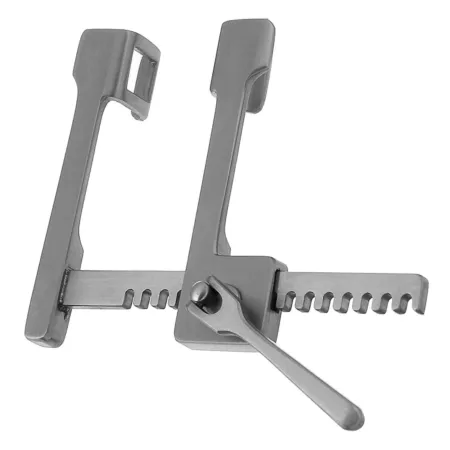
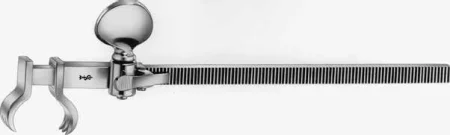
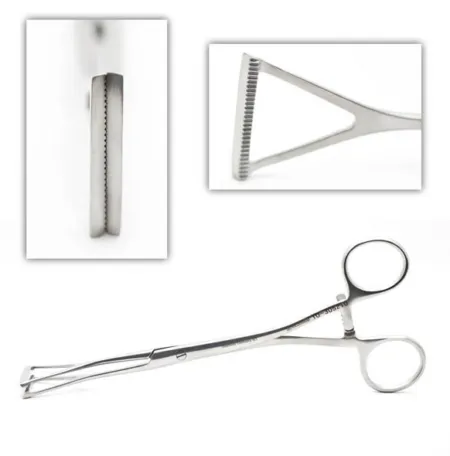
Thoracoscopic (VATS) instruments have two pivot points instead of one, so they open within the small space of a port without pressing on the tissue around it, which keeps them clear of the chest's major nerves.
Heart instruments
A heart case uses a general set with cardiac instruments, thoracic instruments and staplers, and lung instruments if needed; coronary, valve, aneurysmAN-yuh-riz-umA weak spot in the wall of an artery that bulges outward like a balloon.aneurysm a widening (Greek)Full entry and chest wall sets are added for the operation planned.
- The sternal saw splits the sternum. An oscillating saw is used for a repeat sternotomy, because it lets the surgeon cut through gradually and avoid the scar tissue behind the bone. A Lebsche sternal knife and mallet are kept on the setup for when a power saw cannot be used, as in a power failure.
- Sternal retractors, some with attachments that hold the left atriumAY-tree-umOne of the two upper chambers of the heart, which receive blood coming back to the heart.atri/o atrium-um noun endingFull entry open for mitral valveMY-trul valvThe heart valve between the left atrium and the left ventricle.Full entry surgery, and a retractor that lifts one side of the sternum to expose the internal mammary artery.
- Vascular clamps come as cross-clamps, which close a vessel completely, and partial occlusionuh-KLOO-zhunA blockage or closing off of a passage, such as a blood vessel, tube or IV line.Full entry (side-biting) clamps, such as the Satinsky and Lambert-Kay, which close off only part of the wall. Their jaws carry fine rows of teeth or serrationsair-AY-shunA row of small tooth-like ridges, such as the grooves on the jaws of a surgical clamp.Full entry made to hold a vessel without crushing its delicate inner lining, so they must be kept in good condition. Small bulldog clamps close smaller vessels.
- Fine scissors, such as the angled Potts and the Diethrich coronary scissors, to open and extend vessel incisions.
- Fine forceps, such as the DeBakey and the Gerald, and fine needle holders, such as the Ryder and the Castroviejo.
- Coronary dilatorDY-lay-terAn instrument used to stretch or widen an opening, canal or passage in the body.Full entry to size a coronary arteryKOR-uh-nair-ee AR-ter-eeOne of the blood vessels on the surface of the heart that bring oxygen-rich blood to the heart muscle.Full entry , an aortic punch to cut a round hole for a vein graftGRAFTTissue or a man-made material moved into place in surgery to replace or repair a damaged part.Full entry , and sternal wire twisters and cutters.
- Valve instruments: retractors that expose the valve, valve sizers, holders and sutureSOO-cherA stitch, or the thread used to stitch, that holds the edges of a wound or incision together.Full entry holders.
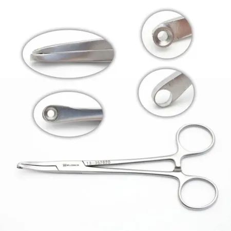
- The Rummel tourniquet: a short length of tubing, bought or cut from a straight catheterizationSTRAYT kath-uh-ter-ih-ZAY-shunPutting a catheter through the urethra into the bladder to drain urine, then taking it out right away.Full entry , slid down over a cannulation stitch or a vessel tape to tighten it, its stylet pulling the strands through and a hemostatHEE-moe-statA clamp with finger rings and a locking ratchet used in surgery to grasp and close off bleeding vessels.hem/o blood-stat stopping, standing stillFull entry holding them.
- Internal defibrillator paddles stay on the field, ready. When the heart fibrillates, the surgeon places one on each side of it and the circulator sets the charge; everyone stands clear of the patient during a shockshokA life-threatening state in which blood flow is too low to bring enough oxygen to the body's tissues.Full entry .
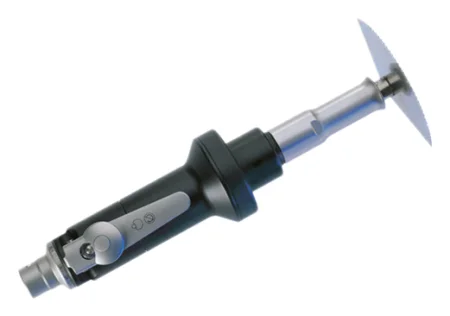
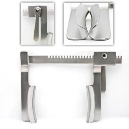
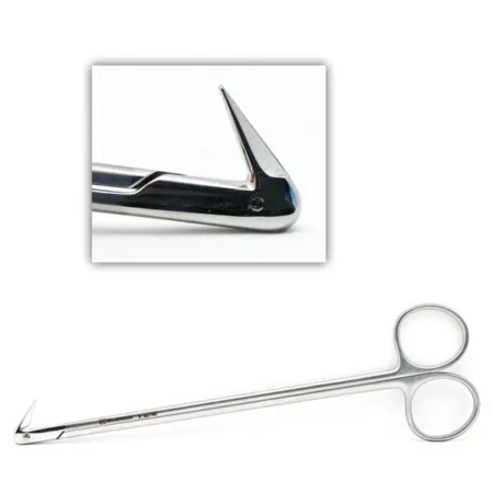
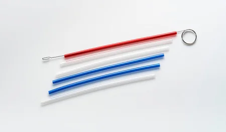
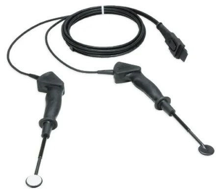
Other equipment in the room includes the heart-lung machine, the cell saver, a defibrillator, an irrigationeer-ih-GAY-shunWashing out a wound, body cavity, tube or eye with a steady flow of liquid.Full entry cooling and warming machine, a warming mattress or forced-air warmer, pacemakerPAYS-may-kerA small device that sends electrical pulses to the heart to keep it beating at a set rate; also the heart's own natural rhythm center.Full entry generators, a headlight, a balloon pump, assist devices and ablationab-LAY-shunA procedure that destroys a small area of tissue, often with heat or cold, to stop it from causing a problem.Full entry sources; chapter 23 teaches the machines.
Supplies, grafts and implants
The Core Curriculum's cardiothoracic supply list includes the aortic punch, the balloon pump, a blower (mister), bulldogs, catheters, defibrillator paddles, the bypass circuit, an off-bypass (stabilizer) system, pacemakers, pledgets, the Rummel tourniquet, shuntSHUNTA passage that moves blood or other fluid from one place to another, either made by a surgeon or present in the body.Full entry , suture boots, a thoracentesisthor-uh-sen-TEE-sisA procedure in which a needle is passed through the chest wall to remove fluid or air from the space around a lung.thorac/o chest-centesis puncture to remove fluidFull entry kit, umbilical tape, valve prosthesispros-THEE-sisAn artificial body part that replaces one that is missing or does not work, such as an artificial leg, joint, eye or heart valve.Full entry , vascular clamp inserts, a vein harvesting system and vessel loops. Its vascular items, the Doppler ultrasoundDOP-ler UL-truh-soundAn ultrasound that uses sound waves to hear or show blood flowing through vessels or the fetal heartbeat.Full entry probe, the valvulotome, the vein stripper and the interventional sheathSHEETHA covering that wraps around a structure; in procedures, a short hollow tube left in a blood vessel to pass catheters through.Full entry , guidewires, stentstentA small tube, often of metal mesh, placed inside a vessel or duct to hold it open.Full entry , tunnelers and ports, are taught in chapter 40.
- Suture boots (rubber shods) slip over hemostat tips so they hold suture without damaging it; small dissecting sponges (peanuts or "pill" sponges), tubing, umbilical tapes and vessel loops identify and lift vessels.
- A thoracentesis kit holds a needle passed through the chest wall into the pleural spacePLOOR-ul SPAYSThe thin space between the two layers of the pleura, the membrane that covers the lungs and lines the chest wall.Full entry , usually to remove fluid.
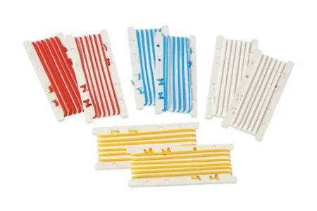
- Sutures: nonabsorbable polypropylene, polyester and similar synthetics, mostly double armed (a needle on each end); size 3-0 is common on large vessels and 5-0 or 6-0 on smaller ones, and finer still on the coronary arteries. Use alternately colored strands and numbered suture holders to keep a valve's many stitches in order.
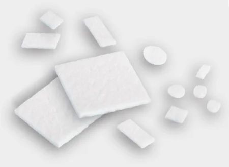
- Pledgets: small pads of Teflon felt on a stitch that reinforceree-in-FORSTo repeat and support teaching or care that has already been started, so the client remembers and follows it.Full entry a suture line. Prepare one by clamping a mosquito across its middle and passing the needle through it on each side of the clamp, or pass the clamp and pledget to the surgeon to load.
- Sternal closure: stainless steel wire, sizes 4 to 7, or cables, bands or plates for a frail sternum.
- Grafts: tubes of Dacron, Teflon or PTFE, straight or branched. Knitted grafts are soft and porous; woven grafts have a tight weave that leaks less blood, so they are used when the patient is fully heparinized. Grafts soaked through with collagenKOL-uh-jenThe main structural protein of the body, which gives strength to skin, bones, tendons, ligaments and scar tissue.coll/a glue-gen producingFull entry need no preclotting, and arch grafts come with side branches for the head vessels. Graft sizers find the size, and only that size is opened.
- Patches of Teflon, PTFE, Dacron, bovine pericardiumpair-ih-KAR-dee-umThe thin, two-layered sac that surrounds the heart.peri- aroundcardi/o heart-um a structureFull entry or the patient's own pericardium close holes and reinforce suture lines.
Every implant (graft, patch, valve, ring or device) is recorded with its type, size, lot and serial number, as the Safe Medical Devices Act requires.
Running a heart field
Expect to keep at least two back tables and three Mayo standMAY-oh standA small tray on an adjustable stand placed over or beside the patient during surgery to hold the instruments used most often.Full entry , each set up the same way every time so you always know where an instrument is and have room to prepare supplies; the room is crowded, so plan the space.
- The saphenous vein is usually taken by a surgical assistant at the same time as the chest is opened, with its own Mayo and its own light: the two ceiling lights are on the chest, so a third light or a headlight is needed for the leg.
- Keep instruments used on the groin or leg apart, and change gown and gloves when moving from the leg to the chest, or after holding the heart and moving to the other side of the table.
- Wipe calcium and debris off instruments with a moist sponge, and keep a small basin for clots and debris, so nothing drops back into the heart and becomes an embolismEM-buh-liz-umThe blocking of a blood vessel by a mass carried there in the blood (an embolus), such as a clot, fat or an air bubble.embol/o plug-ism conditionFull entry .
- Keep the back table and Mayo sterile and stay scrubbed until the patient has left the room, in case the chest must be reopened.
Drugs and solutions
Chapter 12 teaches these as drugs; here is how each is used at the field.
- heparinHEP-uh-rinAn anticoagulant drug, given by injection or IV, that keeps blood clots from forming or growing.Full entry prevents clottingKLOT-ingThe process by which blood thickens into a gel to plug a damaged vessel and stop bleeding; also called coagulation.Full entry in the bypass circuit: 300 to 400 units per kilogram is injected before bypass, through a central venous catheterSEN-trul VEE-nus KATH-uh-terAn IV catheter whose tip sits in a large vein near the heart, used for long-term drugs, fluids, nutrition or blood draws.Full entry or straight into the right atrium. Heparinized saline rinses vessels and grafts.
- protaminePROH-tuh-meenAn IV drug that binds heparin and reverses its blood-thinning effect.Full entry reverses heparin after bypass, given slowly into a vein by the anesthesia provideran-us-THEE-zhuh pruh-VY-derThe licensed professional who gives anesthesia and watches over the client's breathing and vital functions during a procedure.Full entry ; it can cause an allergic reaction. When protamine starts, the pump suckers (cardiotomy suction) are turned off, so that blood that is losing its heparin is not returned into the circuit, where it would clot.
- Papaverine relaxes vessel muscle and prevents spasmSPAZ-umA sudden, involuntary tightening of a muscle or of the muscle in a hollow organ or tube.Full entry in the internal mammary and radial artery grafts. A vein solution, such as a balanced salt solution with heparin and papaverine, distends vein grafts, so the surgeon can check them for twists and leaks.
- Topical antibiotic irrigation, such as bacitracinbas-ih-TRAY-sinAn antibiotic used mostly as an ointment on the skin to prevent infection in minor cuts, scrapes and burns.Full entry in saline. It must never be suctioningSUK-shun-ingRemoving mucus, saliva or other secretions from the mouth, nose or airway with a tube connected to a suction source.Full entry into the cell saver, because in a vein it can harm the nerves and kidneys (chapter 12).
- hemostatichee-moh-STAT-ikStopping bleeding; also, a product or agent used to stop bleeding.hem/o blood-stasis stopping, standing still-ic relating toFull entry : gelatin sponge with thrombinTHROM-binAn enzyme in the blood that turns fibrinogen into fibrin, the threads that hold a blood clot together.thromb/o clot-in substanceFull entry , oxidized cellulose and collagen for oozing; bone sealant or bone wax on the cut sternum and ribs; fibrin sealant to stop air leaking from the lung or a bronchial join; and adhesives such as albuminal-BYOO-minThe main protein in blood plasma, made by the liver, which holds fluid inside the blood vessels and carries drugs and hormones.Full entry with glutaraldehydegloo-tuh-RAL-duh-hideA liquid chemical used for high-level disinfection, or sterilization with longer soaking, of heat-sensitive medical devices.Full entry to seal aortic suture lines. Gelatin works poorly in a fully heparinized patient.
- Cold and warm solutions: cold saline and slush cool the heart during arrest, and warm saline is used before and after it.
- Label every drug and solution, as chapter 29 teaches; heparinized solutions, papaverine, vein solutions and topical hemostatics matter most.
- inotropicin-oh-TROH-pikChanging the force with which the heart muscle contracts; an inotropic drug is one that does this.ino fiber, muscle-tropic acting on, turningFull entry and antiarrhythmican-tee-uh-RITH-mikA drug used to prevent or treat an abnormal heart rhythm.anti- againsta- withoutrhythm a regular beat-ic relating toFull entry are kept on hand, and the team helps give them when they are needed.
Cardioplegia, the solution that stops the heart, is taught with bypass below.
Monitoring
Chapter 13 teaches the monitors, and a heart case uses nearly all of them. Heart and chest cases add sensors on the forehead that measure the oxygen in the brain (cerebralsuh-REE-brul or SAIR-uh-brulHaving to do with the brain, especially the cerebrum, its largest part.cerebr/o brain, cerebrum-al relating toFull entry oximetryok-SIM-uh-treeMeasuring how much of the blood's hemoglobin is carrying oxygen.ox/i oxygen-metry process of measuringFull entry ), TEE that also finds air left in the heart and guides cannulaKAN-yoo-luhA thin tube placed into the body or a body opening to deliver or remove fluid or air.Full entry and balloon pumps, and, with one lung collapsed, frequent blood gases, because blood passing through the airless lung picks up no oxygen.
Counts
An initial instrument count is advised for every procedure in which an instrument could be left behind, as in an opened chest. Count small items that could be lost in the chest, such as peanuts, Kittner dissectors and umbilical tapes, with the sponges; count vessel loops and rubber shods too, and the laps used to stop prep from pooling at the neck. When there are two incisions, such as the chest and the leg, each closure gets its own count. Follow the count times in chapters 28 and 29.
8Chest drainage
After the chest or pleuralPLOOR-ulHaving to do with the pleura, the thin two-layer membrane that covers the lungs and lines the chest wall.pleur/o pleura-al relating toFull entry is opened, closed chest drainage removes air, blood and fluid from the pleural spacePLOOR-ul SPAYSThe thin space between the two layers of the pleura, the membrane that covers the lungs and lines the chest wall.Full entry or the mediastinum and restores the negative pressureNEG-uh-tiv PRESH-erPressure that is lower than the pressure around it, so air or fluid is drawn in.Full entry , so the lung can expand again; chapter 25 teaches the chest tubechest toobA tube placed through the chest wall into the space around the lung to drain air, blood or fluid.Full entry and the three-chamber drainage unit with its water seal. The unit is set up by its maker's instructions. After a lung resectionrih-SEK-shunSurgery to remove part of an organ or tissue.re- back, againsect to cut-ion processFull entry , tubes of 28 to 30 French leave through stab wounds near the eighth or ninth rib space at the anterior axillary line; after heart surgery, tubes drain the mediastinum and the pericardiumpair-ih-KAR-dee-umThe thin, two-layered sac that surrounds the heart.peri- aroundcardi/o heart-um a structureFull entry , and the pleura if it was opened. If a tube comes out, cover the hole at once with the sterile petroleum gauze kept with the patient for this purpose, so air cannot rush in.
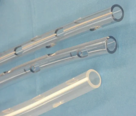
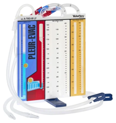
lift the drainage unit to the level of the chest or tip it over, even for a transfer. It stays upright and below the chest at all times, because fluid that runs back can collapse the lung, or compress the heart.
9Lung and chest procedures
A diagnostic endoscopyen-DOS-kuh-peeA procedure that uses a thin, flexible tube with a light and camera to look inside the body.endo- within-scopy looking with an instrumentFull entry often comes first, and thoracicthuh-RAS-ikRelating to the chest, or to the middle part of the spine.thorac/o chest-ic relating toFull entry surgery can then be done open, by VATS or with the robot. Most lung operations, 80 to 90 percent, are now done by VATS, but the open instruments stay ready, often just outside the room, in case the surgeon must convert.
Bronchoscopy and endobronchial ultrasound
What and why: a bronchoscopybrong-KOS-kuh-peeA procedure in which a thin, lighted tube with a camera is passed through the nose or mouth to look inside the airways.bronch/o airway (bronchus)-scopy looking with an instrumentFull entry looks directly at the tracheaTRAY-kee-uhThe windpipe: the tube that carries air from the throat to the lungs.Full entry and bronchusBRONG-kusOne of the large air tubes that branch from the windpipe into each lung.bronch/o airwayFull entry to find a lesionLEE-zhunAn area of tissue that has been damaged or has changed from normal, such as a sore, wound, lump or spot.Full entry and its extent, take biopsies, brushings and washings, and treat: removing a foreign body, opening a narrowing, cutting away a tumor or placing a stentstentA small tube, often of metal mesh, placed inside a vessel or duct to hold it open.Full entry . A rigid scope, under general anesthesiaJEN-er-ul an-es-THEE-zhuhDrug-induced loss of consciousness and feeling over the whole body, so a person feels nothing and is not aware during surgery.an- withoutesthesi/o feeling, sensation-ia conditionFull entry with the patient mechanical ventilationmeh-KAN-ih-kul ven-tuh-LAY-shunBreathing support from a machine, a ventilator, that pushes air into the lungs through a tube or a mask.Full entry through it, takes large instruments, as for a foreign body in a child; a flexible scope reaches the smaller bronchi while the patient breathes around it; both may be used in one session.
Two newer methods help find and stage lung cancer: navigational bronchoscopy steers a guide along a map built from the computed tomographykum-PYOO-tid toh-MOG-ruh-feeAn imaging test that uses x-rays and a computer to make cross-section pictures of the inside of the body.tom/o cut, slice-graphy process of recordingFull entry to a small suspicious area of the lung for biopsy, and endobronchial ultrasound (EBUS) finds lymph nodelimf nohdA small, bean-shaped organ of the immune system that filters lymph fluid and holds white blood cells.Full entry in the mediastinum and the hilum for a needle biopsy.
Key points: chapter 36 teaches the rigid and flexible setups, the Lukens trap and the specimens step by step. When cancer is suspected, the surgeon examines the healthy lung first, so cancer cells are not carried into it. Guide instruments into the channel when asked, and confirm the source of each specimen with the circulator and surgeon. Afterward the patient has nothing by mouth until the gag reflexREE-fleksAn automatic body response to something that stimulates it, which happens without thought, such as the knee jerk.Full entry returns, which may take 2 to 3 hours; the complications are injury to the airway or the lung, a torn blood vessel near the bronchi, and infection.
Wound class: II, the airway is entered through its own opening (as chapter 36 explains).
Mediastinoscopy
What and why: a lighted rigid scope passed through a small incision above the sternal notch, to see and biopsy lymph nodes and tumors around the carina and the upper lobe bronchi. It stages lung cancer before a thoracotomythor-uh-KOT-uh-meeA surgical cut through the chest wall to reach the lungs, heart or other organs in the chest.thorac/o chest-tomy cutting intoFull entry and can biopsy the thymusTHY-musA small gland in the upper chest, behind the breastbone, where T cells (a kind of white blood cell) mature.Full entry . EBUS has largely replaced it.
Position, prep and drape: general anesthesia; supine with the neck extended on a shoulder roll; a thyroid-type prep and drape.
Setup: a minor set, the mediastinoscope with its light cord, suction and aspirating tubes, biopsy forcepsFOR-sepsA hand instrument with two blades or arms used to grasp, hold or pull tissue or objects.Full entry , an electrosurgeryee-LEK-troh-SUR-jer-eeUsing high-frequency electric current through a handheld tool to cut tissue or seal bleeding vessels during surgery.Full entry and a smoke evacuator, and a long aspirating needle. A sterilizationstair-uh-luh-ZAY-shunA process that destroys all microorganisms, including spores, on an item; also a permanent birth control procedure.Full entry scope is preferred to one that is only high-level disinfectiondis-in-FEK-shunCleaning a surface or object with chemicals or heat to kill most germs, though not always all bacterial spores.Full entry . A thoracotomy tray stays in the room in case a biopsy causes heavy bleeding.
Key steps:
- A short incision, about 2 cm, above the sternal notch, down to the fasciaFASH-ee-uhA sheet or band of tough connective tissue that covers and separates muscles and organs.fasci/o bandFull entry in front of the trachea.
- The surgeon tunnels along the front of the trachea with a finger, then passes the scope down the tunnel.
- Each node is first punctured with the needle and syringe to prove it is not a blood vessel; then cup biopsy forceps take tissue. Lift each piece off the forceps onto a saline-moistened Telfa pad or into its container.
- Bleeding is controlled with the ESU, a gelatin sponge or pressure from a small sponge on a carrier, and the incision is closed.
After surgery: many patients go home the same day; bleeding and infection are the complications.
Wound class: I, no tract is entered.
Thoracotomy: opening and closing the chest
Open chest operations generally open and close the same way.
Opening:
- The skin, fat and muscle are cut with the knife and the ESU.
- A scapulaSKAP-yuh-luhThe flat, triangle-shaped bone at the back of each shoulder; the shoulder blade.scapul/o shoulder bladeFull entry retractorrih-TRAK-terA surgical instrument that holds back tissue or organs so the surgeon can see and reach the operative site.Full entry lifts the shoulder blade, and the intercostalin-ter-KOS-tulBetween the ribs.inter- betweencost/o rib-al relating toFull entry muscles are cut to enter the chest.
- Sometimes a rib is removed: the surgeon cuts the periosteum, strips it off with an elevator or rib rasp, cuts the rib with rib shears and trims the sharp ends with a rongeurron-ZHURA strong surgical instrument with sharp, cup-shaped jaws used to bite away small pieces of bone.Full entry .
- Moist laps cover the wound edges and the rib spreader goes in, opened slowly and gently.
Closing:
- chest tubechest toobA tube placed through the chest wall into the space around the lung to drain air, blood or fluid.Full entry are placed and the instruments come out.
- Four to six heavy pericostal sutureSOO-cherA stitch, or the thread used to stitch, that holds the edges of a wound or incision together.Full entry are passed around the ribs above and below, or through holes drilled in them, and tagged with hemostatHEE-moe-statA clamp with finger rings and a locking ratchet used in surgery to grasp and close off bleeding vessels.hem/o blood-stat stopping, standing stillFull entry .
- A rib approximator draws the ribs together while the sutures are tied.
- The muscles, fat and skin are closed in layers, and the tubes are connected to the drainage unit.
A thoracotomy is painful. An epidural anesthesiaep-ih-DOOR-ul an-us-THEE-zhuhPain relief or numbness from an anesthetic drug given through a thin catheter into the space just outside the covering of the spinal cord.epi- upon, overdur/o dura (the tough covering of the spinal cord)-al relating toFull entry , a local anestheticLOH-kul an-es-THET-ikA drug that blocks feeling in one part of the body without making the person unconscious.Full entry catheter in the chest wall or freezing of the intercostal nerves may be used, along with other pain drugs.
VATS and robotic chest surgery
What and why: video-assisted thoracic surgery (VATS) works inside the chest through ports, with a rigid scope and long thoracotomy instruments. It spares the muscles a thoracotomy cuts, has the same goals as the open operation, and needs a full surgical team under general anesthesia. It is used to treat pleuralPLOOR-ulHaving to do with the pleura, the thin two-layer membrane that covers the lungs and lines the chest wall.pleur/o pleura-al relating toFull entry disease, such as blebs and effusionih-FYOO-zhunFluid that has leaked out and collected in a body space, such as around the lungs, the heart or in a joint.Full entry , to biopsy masses, and for wedge resectionrih-SEK-shunSurgery to remove part of an organ or tissue.re- back, againsect to cut-ion processFull entry , lobectomy, lung volume reduction, removal of the pericardiumpair-ih-KAR-dee-umThe thin, two-layered sac that surrounds the heart.peri- aroundcardi/o heart-um a structureFull entry , sympathectomy, stopping bleeding and removing clots. Many authors call it thoracoscopy, though one textbook keeps that word for a smaller look at the pleura under sedationsih-DAY-shunA calm, relaxed or sleepy state brought on by drugs, or the use of drugs to cause it.sed calm, settle-ation processFull entry . Chapter 21 teaches the scopes and ports.
Position, prep and drape: lateral with the operative side up and a chest prep, with a double-lumenLOO-menThe open space inside a tube, such as a blood vessel, the intestine or a catheter.Full entry tube so the lung on the operated side collapses; the surgeon marks the shoulder blade and the port sites on the skin before starting.
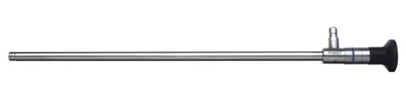
Setup: a 5 or 10 mm thoracoscope, 0 or 30 degree, camera and light source, endoscopicen-doh-SKOP-ikDone with an endoscope, a thin tube with a light and camera passed into the body.endo- within-scopic viewingFull entry graspers, dissectors, scissors, clip appliers and staplers, with open thoracic trays at hand. insufflationin-suh-FLAY-shunBlowing a gas into a body cavity, such as carbon dioxide into the abdomen during laparoscopic surgery.Full entry equipment is in the room; as chapter 21 explains, gas is used only when the anesthesia provideran-us-THEE-zhuh pruh-VY-derThe licensed professional who gives anesthesia and watches over the client's breathing and vital functions during a procedure.Full entry wants help collapsing the lung.
Ports and team: three or four ports are placed in a "baseball diamond" pattern so the instruments do not cross inside the chest, or one larger port (uniportal VATS) carries them all. The first port usually goes in through a cut of 2 to 3 cm somewhere from the fifth to the seventh rib space, for a 10 or 12 mm trocarTROH-karA sharp or bladed instrument inside a tube (cannula), used to pierce a body wall and leave the tube in place as a port.Full entry . In VATS the surgeon and assistant stand at the patient's front, while in an open thoracotomy they stand at the back; the scrub stands at the back at the level of the legs, with the Mayo over the legs.
Wedge resection or lung biopsy (the model VATS case):
- Ports are placed and the scope goes in.
- The lung is held with a grasper and a wedge of tissue, or a small lesion, is cut and sealed with an endoscopic stapler; lymph nodes may be taken too.
- The surgeon fills the chest with warm saline while the lung is inflated and watches the staple line for bubbles of air; leaks are sewn or sealed.
- A chest tube goes in through the lowest port, and the ports are closed.
After surgery: a hospital stay of about 4 to 7 days; the complications include infection, bleeding, atelectasisat-uh-LEK-tuh-sisThe collapse of part or all of a lung, in which the tiny air sacs close and hold no air.atel- incomplete-ectasis expansionFull entry , pneumonianoo-MOHN-yuhAn infection of one or both lungs in which the air sacs fill with fluid or pus.pneum/o lung, air-ia conditionFull entry , a pneumothoraxnoo-moh-THOR-aksAir in the space between a lung and the chest wall, which lets part or all of that lung collapse.pneum/o air, lungthorac/o chestFull entry and air under the skin.
A noduleNOD-yoolA small, firm, rounded lump of tissue that can be felt or seen, in or under the skin or in an organ.Full entry may be marked beforehand with methylene blue dye so the surgeon can find it. When a bronchoscopy comes first, set it up on its own table, then cover that table and move it away before the VATS begins. Through the scope the surgeon can also put talc into the pleural spacePLOOR-ul SPAYSThe thin space between the two layers of the pleura, the membrane that covers the lungs and lines the chest wall.Full entry , which irritates the pleura so the layers stick together and fluid or air cannot collect again (pleurodesis), or cut the sympathetic nerve chain beside the spine for severe sweating of the hands (sympathectomy).
Robotic chest surgery: robots are used for lobectomy, removal of a thymoma, esophagectomy and sympathectomy, with the patient lateral on a double-lumen tube and the team ready to convert quickly to an open thoracotomy in an emergency.
Wound class: II for a wedge resection or lung biopsy. The lungs are part of the lower respiratory tract, so cutting and stapling across lung tissue enters that tract under controlled conditions; one surgical technology textbook lists VATS lung cases as Class I.
Lobectomy and smaller resections
What and why: a lobectomy removes one or more lobes of a lung, most often for lung cancer confined to a lobe; also for spread of cancer from elsewhere, bronchiectasis, giant blebs, benign tumors, fungal and other localized infections, cysts and injury. It can be open or by VATS; the principles are the same.
Position, prep and drape: lateral for a posterolateral thoracotomy, or supine for some upper and middle lobe resections; a thoracotomy prep and drape.
Setup: a major thoracotomy set and a general set, bronchus instruments, deep vascular clamps and forceps, vessel loops, umbilical tapes, clips, linear staplers and fibrin sealant.
Key steps:
- The chest is opened, and the surgeon examines the whole lung and the mediastinum to make sure the disease has not spread further.
- The lobe is held with lung-grasping forceps, the pleura over the hilum is opened, and the pulmonary arteryPUL-muh-nair-ee AR-ter-eeThe large blood vessel that carries blood low in oxygen from the right side of the heart to the lungs.Full entry and vein branches to the lobe are freed with sponge dissectors and right-angle clamps.
- Each branch is tied twice and divided, or divided with a stapler.
- The bronchus is clamped and divided, and closed with sutures or a stapler. Keep suction ready while the bronchus is open, so blood and secretionsih-KREE-shunA substance a gland or cell makes and releases, such as mucus, saliva, sweat or a hormone.Full entry cannot drain into the other lung; you may be asked to run it.
- Out comes the lobe, and pleura is sewn over the bronchial stump.
- The chest is irrigationeer-ih-GAY-shunWashing out a wound, body cavity, tube or eye with a steady flow of liquid.Full entry and the lung that is left is inflated, so any air leak shows; chest tubes are placed and the wound is closed.
Smaller resections: a segmentectomy removes one or more segments and spares the healthy lung around them; before cutting, the remaining lung is inflated to show the boundary of the diseased segment. A wedge resection removes a wedge of lung holding a small, peripheral lesion, without following the segments.
Specimen: the lobe, and lymph nodes labeled by station.
After surgery: the complications include infection, bleeding, atelectasis, pneumonia, a pneumothorax, a pulmonary embolismPUL-muh-nair-ee EM-buh-liz-umA blockage of an artery in the lungs, usually by a blood clot that has traveled there from elsewhere in the body.pulmon/o lung-ary relating toembol/o plug-ism conditionFull entry , air under the skin and a lasting air leak.
Wound class: II, because the bronchus is opened. One surgical technology textbook lists a lobectomy as Class I and notes that opening the bronchial tree changes the class.
Pneumonectomy
What and why: a pneumonectomy removes a whole lung, usually for cancer; also for widespread bronchiectasis or abscessAB-sesA closed pocket of pus in tissue, walled off by the body.Full entry in one lung, or to debulk a tumor pressing on vital structures.
Position and setup: lateral, with a thoracotomy prep and drape and the same sets as a lobectomy: the major thoracotomy and general sets, bronchus instruments, deep vascular clamps, vessel loops, umbilical tapes, clips and staplers.
Key steps:
- Through a thoracotomy, the lung is held away with lung forceps or retractors, adhesionad-HEE-zhunA band of scar tissue that joins two surfaces inside the body that are normally separate.ad- towardhes stick-ion conditionFull entry are freed and the dissectiondy-SEK-shunA tear in the inner layer of an artery wall that lets blood split the layers apart; also, the cutting apart of tissue in surgery.Full entry is carried to the hilum.
- The vagus, phrenic and, on the left, recurrent larynxLAIR-inksThe voice box: the part of the airway in the neck, above the windpipe, that holds the vocal cords.laryng/o larynxFull entry nerves are held out of the way with vessel loops or moist umbilical tapes.
- The pulmonary artery and the superior and inferior pulmonary veinPUL-muh-nair-ee VAYNOne of the four veins that carry oxygen-rich blood from the lungs to the left atrium of the heart.Full entry are clamped, divided and tied with heavy silk, oversewn with fine suture or stapled.
- The main bronchus is clamped near the carina, divided and closed with sutures or staples, and the lung is removed.
- Saline fills the chest while the anesthesia provider inflates the lung with positive pressure, so leaks at the bronchial stump show as bubbles. A flap of pleura or intercostal muscle may cover the stump.
- Chest tubes may be placed, and the wound is closed.
After a pneumonectomy, the chest tubes are not connected to suction. Besides the complications of a lobectomy, the middle of the chest can shift toward the empty side (mediastinal shift), and fluid can flood the remaining lung (acute pulmonary edemaPUL-muh-nair-ee ih-DEE-muhA buildup of fluid in the air sacs and tissues of the lungs.pulmon/o lung-ary having to do withedema swelling with fluidFull entry ).
Wound class: II, the main bronchus is opened; one textbook lists Class I.
Lung volume reduction
For severe emphysemaem-fuh-SEE-muhA long-term lung disease in which the walls of the tiny air sacs are damaged and lose their stretch.Full entry , the worst, overinflated parts of the lung are cut away with linear staplers through a sternotomy or VATS, so the healthier lung can work better. Strips of PTFE or bovine pericardium, or a sealant, reinforceree-in-FORSTo repeat and support teaching or care that has already been started, so the client remembers and follows it.Full entry the fragile staple lines; the lung is reinflated under saline to look for leaks, and the chest tubes go to water seal without suction.
Wound class: II, as for a wedge resection.
Acute pulmonary embolectomy
For a pulmonary embolism that causes life-threatening circulatory failure, the clots are removed through a median sternotomy on bypass, usually without stopping the heart, which stays warm. Have Rummel tourniquets ready for the vena cavaVEE-nuh KAY-vuhOne of the two large veins that return oxygen-poor blood from the body to the right atrium of the heart.Full entry , and suction ready before the artery is opened. The pulmonary trunk is opened between two tractionTRAK-shunA steady pulling force applied to a body part with weights, ropes and pulleys.Full entry sutures with a #11 blade and Potts scissors, and the clots are removed with forceps and suction while the lungs are squeezed to push out smaller ones; keep every clot as a specimen. The artery is closed before bypass ends, and some surgeons add a vena cava filter (chapter 40).
Wound class: I; the pulmonary artery is opened, but no tract is entered.
Decortication and empyema drainage
What and why: long-lasting inflammation, infection (empyema or tuberculosistoo-ber-kyoo-LOH-sisAn infection caused by the bacterium Mycobacterium tuberculosis, which most often attacks the lungs.tubercul/o small swelling-osis abnormal conditionFull entry ) or a tumor lays a fibrousFY-brusMade of or containing tough threads (fibers), especially of connective tissue.fibr/o fiber-ous full of, relating toFull entry peel over the lung that traps it. Decortication strips the peel away so the lung can expand; it can bleed heavily and injure the lung.
Position and setup: lateral, with a thoracotomy prep and drape; the thoracotomy and general sets, bronchus instruments, deep vascular clamps, vessel loops and umbilical tapes, with curettekyoo-RETA surgical instrument with a small spoon- or loop-shaped tip used to scrape tissue.Full entry as an extra, and culture tubes for the fluid.
Key steps:
- A posterolateral thoracotomy, usually at the fifth or sixth rib, part of which may be removed.
- Adhesions are divided, and pus or fluid is drained and sent for culture.
- The anesthesia provider inflates the lung from time to time to show where peel is still holding it down, and the surgeon peels it off with sponge dissectors and sharp dissection, handling the lung gently.
- Chest tubes are placed and the chest is closed.
After surgery: physical therapy goes on for months so the lung expands fully. The complications are infection, bleeding, a peel not fully removed, so breathing stays hard, and paralysispuh-RAL-uh-sisLoss of the ability to move a part of the body, and sometimes loss of feeling in it.para- beside, abnormal-lysis looseningFull entry of the diaphragmDY-uh-framThe dome-shaped sheet of muscle under the lungs that is the main muscle of breathing; also the flat side of a stethoscope.Full entry if the phrenic nerve is injured.
Empyema drainage: an early empyema may be drained by a closed thoracostomy, a chest tube through a rib space under local anesthesiaLOH-kul an-es-THEE-zhuhNumbing medicine applied to or injected into one small area, so a procedure there is not felt while the person stays awake.Full entry , usually outside the OR; a chronic one may need a rib resection with a large drain, or an open thoracostomy, in which part of a rib is removed and the cavity packed open; take aerobic and anaerobic cultures, and keep suction ready so the pus does not spill.
Wound class: IV when infection is already present, as in an empyema; one textbook lists a decortication as Class I, which fits only when no infection is found.
Thymectomy
What and why: a thymectomy removes the thymus, for a tumor of the gland (thymoma) or for myasthenia gravismy-us-THEE-nee-uh GRAV-isA long-term autoimmune disease in which antibodies block the signals passing from nerves to muscles.my/o muscle-asthenia weaknessgravis seriousFull entry ; chapter 9 describes the gland. The classic approach splits the sternum, and the gland can also be removed through the neck, by VATS or with the robot.
Before surgery: a patient with myasthenia gravis may have trouble breathing afterward and may need to stay endotracheal intubationen-doh-TRAY-kee-ul in-too-BAY-shunPutting a breathing tube through the mouth or nose into the windpipe (trachea).endo- withintrache/o windpipe-al relating toFull entry or have a tracheostomytray-kee-OS-tuh-meeA surgical opening through the front of the neck into the windpipe, usually holding a tube that the person breathes through.trache/o windpipe-stomy surgical openingFull entry ; the drugs for myastheniamy-us-THEE-nee-uhWeakness of the muscles.my/o muscle-asthenia weaknessFull entry , such as neostigminenee-oh-STIG-meenA drug that blocks the enzyme that breaks down acetylcholine, so nerve signals to muscles get stronger.Full entry , are agreed with the anesthesia provider and kept at hand, and a tracheostomy tray is in the room.
Position, prep and drape: supine; the prep runs from the chin to below the umbilicus; laps tucked at the sides of the neck stop the solution pooling and are then removed (they are counted); a thyroid sheet.
Setup: basic, vascular and thoracotomy trays, a sternal saw with a sternal knife and mallet, rake retractors, clip appliers, bone wax, peanut or Kittner dissectors and umbilical tape (both counted), and a chest tube with its drainage unit if a pleura is opened.
Key steps:
- A median sternotomy; bone wax controls bleeding from the cut sternum.
- The thymus is freed from the fat of the anterior mediastinum and the pericardium, keeping clear of the great vessels; its vessels are clamped, tied or clipped and divided.
- A drain is placed, or chest tubes if a pleura was opened, and the sternum is closed with heavy wire.
Wound class: I.
Lung transplant
What and why: one or both lungs are replaced with donor lungs. A single lung is used for restrictive lung disease, emphysema, pulmonary hypertensionPUL-muh-nair-ee hy-per-TEN-shunHigh blood pressure in the arteries that carry blood from the heart to the lungs.pulmon/o lung-ary relating tohyper- abovetension pressureFull entry and other end-stageEND-stayjDescribes the last stage of a long-term disease, when an organ has almost stopped working.Full entry lung diseases without infection; both lungs are replaced for cystic fibrosisSIS-tik fy-BROH-sisAn inherited disease in which the body makes thick, sticky mucus that clogs the lungs, pancreas and other organs.cyst/o sacfibr/o fiber-osis abnormal conditionFull entry or chronic infection. The recipient starts immunosuppression and infection prevention before surgery.
Setup: the donor and recipient are two patients, each with their own plan and setup. Thoracotomy instruments, cold perfusionper-FYOO-zhunThe flow of blood through the body's tissues, bringing them oxygen and nutrients.per- throughfus pour-ion processFull entry solution and staplers are used, with bypass ready if the patient becomes unstableun-STAY-bulDescribes a client whose condition is changing quickly or is hard to predict.Full entry , and TEE watches the heart.
Key steps (recipient):
- Lateral for a single lung, supine for both; a wide prep from the nipple line to the knees.
- A thoracotomy for one lung; for both, two thoracotomies, a sternotomy or a clamshell incision.
- The lung to be removed is collapsed and its artery clamped; if the patient cannot tolerate it, bypass (sometimes from the groin) supports the circulation.
- The diseased lung is removed, leaving the tissue around the bronchus so its blood supply is kept.
- Three joins are made: bronchus to bronchus, pulmonary artery to pulmonary artery, and the recipient's pulmonary veins to a cuff of the donor's left atriumAY-tree-umOne of the two upper chambers of the heart, which receive blood coming back to the heart.atri/o atrium-um noun endingFull entry . The bronchial join may be wrapped with a flap of omentum or intercostal muscle.
- The new lung is inflated and inspected, chest tubes are placed and the chest closed. A bronchoscopy at the end clears secretions and checks the bronchial join.
After surgery: drugs against rejection for life; the complications include infection, bleeding, atelectasis, pneumonia, fluid in the lungs and rejection of the new lung.
Wound class: II, the airway is opened; one textbook lists Class I.
Pectus excavatum and carinatum repair
What and why: in pectus excavatum the sternum and costal cartilageKAR-tih-lijFirm, smooth, rubbery tissue that cushions joints and gives shape to parts such as the ear, nose and windpipe.chondr/o cartilageFull entry are pushed in, giving a sunken chest. It is often noticed in early adolescence and can worsen during growth. Repair is most often cosmetic; fewer patients have it for impairmentim-PAIR-mentA loss or weakening of a body function or ability, such as sight, hearing, movement or thinking.Full entry heart and lung function, with complaints such as chest pain, shortness of breath on exercise or rhythm disturbances. In pectus carinatum the sternum pushes out; it is usually treated first with a brace, and open repair is for a deformity the brace does not correct.
The Nuss procedure (minimally invasive): one or more curved metal bars are placed behind the sternum to push it out, and they stay for about 3 years. It is most often done between about 10 and 16 years of age.
- Position: supine, the upper chest raised on a soft roll, with the arms placed out of the way of the sides of the chest as the surgeon prefers.
- Setup: a 5 mm thoracoscope, the pectus set with bars, a bender, stabilizers and accessories, a minor set and umbilical tape. A titanium bar is used if the patient is allergic to nickel.
Key steps:
- The chest is measured and a template bent to the shape wanted; the bar is bent to match with the plate bender.
- Two incisions are made on the sides of the chest in line with the deepest part of the defect, and pockets are made under the skin.
- The thoracoscope goes in through a small port to watch the next steps.
- An introducer is passed under the sternum, across the front of the mediastinum and out the other side, then used to pull an umbilical tape back through the tunnel.
- The tape pulls the bar through with its curve facing back, and the bar is flipped over to push the sternum out.
- Stabilizers are fixed to the bar ends with wire or heavy suture, and the bar is sewn to the chest wall muscle so it cannot move.
- The wounds are closed and a chest X-ray checks for a pneumothorax.
After surgery: the patient stays supine in recovery so the bar does not shift, may have an epidural, stays in hospital for 3 to 4 days, and resumes limited activity after about 6 weeks. Complications include bar movement, a pneumothorax and fluid around the lungs or heart.
Open repair: the modified Ravitch operation removes only short lengths of the deformed cartilage, keeping its covering (the perichondrium), and fixes the sternum in a better position; it is now kept for carinatum that bracing did not correct and for mixed deformities.
Wound class: I, no tract is entered.
Esophageal atresia and tracheoesophageal fistula
What and why: in esophagusih-SOF-uh-gusThe muscular tube that carries food and liquid from the throat to the stomach.esophag/o esophagusFull entry atresia the esophagus is interrupted, its upper part ending in a blind pouch. It often comes with a tracheoesophageal fistulaFIS-choo-luhAn abnormal passage between two organs or vessels, or between an organ and the skin.Full entry (TEF), a channel that should not exist joining the trachea to the esophagus, which lets saliva and milk, or stomach secretions, reach the lungs. It can come with other defects, such as septumSEP-tumA wall of tissue that divides two spaces or cavities, such as the two sides of the heart or nose.sept/o wall, partitionFull entry defects, tetralogy of Fallotteh-TRAL-uh-jee uv fuh-LOHA congenital heart defect made up of four problems in the structure of the heart.tetra- four-logy a set or studyFull entry and neural tubeNOOR-ul TOOBThe hollow structure in an early embryo that develops into the brain and spinal cord.neur/o nerve-al relating toFull entry defects. The esophagus is joined at once when the two ends reach and the infant is well enough; otherwise the repair is staged or delayed, and a segment of colon can lengthen the esophagus.
Position, prep and drape: the infant lies in the thoracotomy position with the head of the table slightly raised; the prep runs from the neck to the iliacIL-ee-akHaving to do with the ilium, the large, wing-shaped upper part of the hip bone.Full entry crest, front and back. Chapter 13 teaches how to keep an infant warm.
Key steps:
- A posterolateral thoracotomy; a Finochietto retractor goes in and the azygos vein is tied and divided.
- The anesthesia provider advances a tube already in the upper pouch (a Replogle tube) so the pouch can be seen. The fistula is looped with a vessel loop and divided.
- The hole in the trachea is closed with fine absorbable sutures and tested under warm saline.
- Both ends of the esophagus are freed, held with Babcocks, and joined in one layer with very fine absorbable suture, the tube passed across the join.
- A small chest tube may be placed and the wound closed in layers.
After surgery: the airway is suctioningSUK-shun-ingRemoving mucus, saliva or other secretions from the mouth, nose or airway with a tube connected to a suction source.Full entry often until the baby can swallow, and feeding starts 2 to 6 days later, once a contrastKON-trastA dye or other substance given before an imaging test so body parts show up more clearly.Full entry study shows the join is sound; a leak, infection or a recurrent fistula are the complications.
Wound class: II, the esophagus and trachea are opened.
10Chest injuries
Blunt injury to the chest causes at least a fifth of all trauma deaths, and motor vehicle crashes cause most chest injuries. A penetrating wound at or just below the nipple line, or the tips of the shoulder blades, may involve both the chest and the abdomen. Chapter 11 teaches trauma care in general, and chapter 28 setting up for a trauma case.
The injuries that need surgery at once are a tension pneumothoraxTEN-shun noo-moh-THOR-aksA collapsed lung in which air keeps building up in the chest under pressure and presses on the heart and the other lung.pneum/o air, lungthorax chestFull entry , an open pneumothoraxnoo-moh-THOR-aksAir in the space between a lung and the chest wall, which lets part or all of that lung collapse.pneum/o air, lungthorac/o chestFull entry , a flail chest, a massive hemothorax and pericardiumpair-ih-KAR-dee-umThe thin, two-layered sac that surrounds the heart.peri- aroundcardi/o heart-um a structureFull entry tamponade. A thoracotomythor-uh-KOT-uh-meeA surgical cut through the chest wall to reach the lungs, heart or other organs in the chest.thorac/o chest-tomy cutting intoFull entry may be started in the emergency department to relieve a tamponade or stop bleeding from the heart, lungs or great vessels, and the patient then goes straight to the OR.
Setting up a trauma thoracotomy, in order of need:
- Exposure: at least two suctions running, sponges, and a Finochietto retractorrih-TRAK-terA surgical instrument that holds back tissue or organs so the surgeon can see and reach the operative site.Full entry and Deaver retractors from the start.
- Removing blood and clots: a large basin for clots, and 10 to 15 laps dipped in warm saline and wrung out, with at least 30 more in reserve.
- Cardiovascular sutureSOO-cherA stitch, or the thread used to stitch, that holds the edges of a wound or incision together.Full entry by the surgeon's card, or 2-0 and 3-0 polypropylene if none is known; fibrin sealant may also be used.
- The chest wall is closed at the end, often in one layer, over chest tubechest toobA tube placed through the chest wall into the space around the lung to drain air, blood or fluid.Full entry .
An autotransfusion system should be ready for lung injuries. As chapter 12 explains, contamination or sepsisSEP-sisA severe whole-body response to an infection that damages the body's own organs.Full entry is not an absolute bar to cell salvage once the wound is washed with antibiotic irrigationeer-ih-GAY-shunWashing out a wound, body cavity, tube or eye with a steady flow of liquid.Full entry , though makers still advise against it with a cancer, and the surgeon and anesthesia provideran-us-THEE-zhuh pruh-VY-derThe licensed professional who gives anesthesia and watches over the client's breathing and vital functions during a procedure.Full entry decide; pleuralPLOOR-ulHaving to do with the pleura, the thin two-layer membrane that covers the lungs and lines the chest wall.pleur/o pleura-al relating toFull entry fluid is suctioningSUK-shun-ingRemoving mucus, saliva or other secretions from the mouth, nose or airway with a tube connected to a suction source.Full entry off with a separate suction first, and only the blood that collects afterward is salvaged.
Chest wall
- An open chest wound that lets air in is sealed at once, often with petrolatum gauze, and the air is let out with a needle (needle thoracostomy) or a chest tube; a large wound needs a mechanical ventilationmeh-KAN-ih-kul ven-tuh-LAY-shunBreathing support from a machine, a ventilator, that pushes air into the lungs through a tube or a mask.Full entry .
- Flail chest: two or more neighboring ribs each broken in two or more places, so a segment of chest wall floats free. It moves the wrong way, sinking in when the patient breathes in and bulging out on breathing out, and the lung beneath it is usually bruised. One reference defines it as three or more neighboring ribs. The patient may need a breathing tube and a ventilator.
Lungs and pleura
- Simple pneumothorax: air in the pleural spacePLOOR-ul SPAYSThe thin space between the two layers of the pleura, the membrane that covers the lungs and lines the chest wall.Full entry , with no opening to the outside air.
- Open (communicating) pneumothorax: the wound opens the pleura to the air, the "sucking chest wound." The lung on that side collapses, and the middle of the chest shifts and squeezes the other lung.
- Tension pneumothorax: air keeps entering the pleural space and cannot leave, so the pressure climbs, the mediastinum, lung and great vessels are pushed to the other side, blood return to the heart falls and the patient goes into shockshokA life-threatening state in which blood flow is too low to bring enough oxygen to the body's tissues.Full entry . It can kill quickly and is relieved with a needle or a chest tube.
- Hemothorax: blood in the pleural space. A large chest tube, 36 to 40 French, is placed in the emergency department; when 1,000 to 1,500 mL of blood drains, the chest is explored in the OR.
- Pulmonary contusionkun-TOO-zhunA bruise: an injury that breaks small blood vessels under the skin or in an organ without breaking the skin.Full entry , a bruised lung that bleeds into its air sacs, is the most common chest injury in children. A torn lung may be clamped in the emergency department, then sutured or stapled quickly; a lobectomy is sometimes needed.
- diaphragmDY-uh-framThe dome-shaped sheet of muscle under the lungs that is the main muscle of breathing; also the flat side of a stethoscope.Full entry injury lets abdominal organs slide up into the chest, where they can strangulate.
Heart and great vessels
- Blunt cardiac rupture, a tear of a chamber, valve or other structure, is among the most lethal chest injuries, classically from the steering wheel. The patient may arrive with a indwelling urinary catheterin-DWEL-ing YUR-ih-nair-ee KATH-uh-terA tube passed through the urethra into the bladder and left in place to drain urine into a bag.Full entry balloon plugging a small hole in the heart, or a hand pressing on a larger one; the repair is fast and simple first.
- Penetrating cardiac wounds from knives and bullets kill by bleeding and by cardiac tamponade, blood in the pericardium that stops the heart filling. Its signs are distendeddih-STEN-didStretched out and enlarged by pressure from inside, such as from gas, fluid or urine.Full entry neck veins, muffled heart sounds and a narrowing pulse pressurepuls PRESH-erThe difference between the systolic and the diastolic blood pressure.Full entry ; it is relieved by needle drainage (pericardiocentesis), a pericardial window or an emergency thoracotomy. In the trauma repair, patch graftGRAFTTissue or a man-made material moved into place in surgery to replace or repair a damaged part.Full entry are used rather than tube grafts, and suturing over pledgets may take too long.
- myocardiummy-oh-KAR-dee-umThe thick middle layer of the heart wall, made of heart muscle that contracts to pump blood.my/o musclecardi/o heart-um structureFull entry contusion, a bruised heart, usually of the right ventriclerite VEN-trih-kulThe lower right chamber of the heart, which pumps blood to the lungs.Full entry , shows as arrhythmiauh-RITH-mee-uhA heart rhythm that is not normal: too fast, too slow or irregular.a- withoutrhythm regular beat-ia conditionFull entry ; it is watched on a monitor, but a valve torn by the blow needs repair.
- Aortic injury, from a high-speed crash, a fall from a height or a hard blow to the side of the chest, can tear or shear the aorta; most of these patients die at the scene or soon after. Repair may use bypass to protect the spinal cord from the cross-clamp, and some tears are treated with a stentstentA small tube, often of metal mesh, placed inside a vessel or duct to hold it open.Full entry graft through the femoral arteryFEM-er-ul AR-ter-eeThe large artery that carries blood to the leg, running through the groin and down the thigh.Full entry .
Wound class: a fresh traumatic wound is Class III, and so is an operation with open cardiac massage; an old wound with dead tissue, or an existing infection, is Class IV. An incision made after blunt injury, with no open wound, is classed by the usual rules: Class I if no tract is entered, Class II if the lung or another tract is opened.
11Cardiopulmonary bypass
cardiopulmonarykar-dee-oh-PUL-muh-nair-eeRelating to the heart and lungs together.cardi/o heartpulmon/o lung-ary relating toFull entry bypass (CPB) takes over the work of the heart and lungs, and the cardiopulmonary bypass machine is the equipment that does it. Blood returning to the heart is drained to the heart-lung machine, given oxygen, and pumped back into the arteries. With the heart empty and stopped, the surgeon can work inside it in a still, nearly bloodless field, and can lift and turn it without causing fibrillationfib-rih-LAY-shunFast, uncoordinated twitching of muscle fibers, most often in the heart, so the muscle quivers instead of contracting together.fibr/o fiber-ation processFull entry or a fall in blood flow to the body. Valve replacement, heart transplant and aneurysmAN-yuh-riz-umA weak spot in the wall of an artery that bulges outward like a balloon.aneurysm a widening (Greek)Full entry repair need bypass; more and more coronary bypasses are done without it.
The bypass circuit
Blood drains by gravity from cannulaKAN-yoo-luhA thin tube placed into the body or a body opening to deliver or remove fluid or air.Full entry in the right atriumAY-tree-umOne of the two upper chambers of the heart, which receive blood coming back to the heart.atri/o atrium-um noun endingFull entry or the vena cavaVEE-nuh KAY-vuhOne of the two large veins that return oxygen-poor blood from the body to the right atrium of the heart.Full entry into a venous reservoirREZ-er-vwarA place where germs live and multiply, such as a person, animal, water or soil.Full entry , and the heart-lung machine pumps it through an oxygenator with a heat exchanger that warms or cools it, then back through a filter into a cannula in the ascending aorta.
- The pump is a roller or a centrifugal pump; its flow is mostly steady, without pulses, so the arterial pressure shows as a mean on the monitor.
- The oxygenator adds oxygen and removes carbon dioxideKAR-bun dy-OK-sideA gas made by the body's cells as waste, carried in the blood to the lungs and breathed out.Full entry across a membrane.
- Suction lines (the cardiotomy suckers) return blood shed in the field to the reservoir, which filters it and removes foam; a separate roller pump delivers the cardioplegia.
- A vent drains blood and air from inside the heart so the left ventricleLEFT VEN-trih-kulThe heart's lower left chamber, which pumps oxygen-rich blood into the aorta and out to the body.Full entry does not stretch; it is usually placed through the right superior pulmonary veinPUL-muh-nair-ee VAYNOne of the four veins that carry oxygen-rich blood from the lungs to the left atrium of the heart.Full entry .
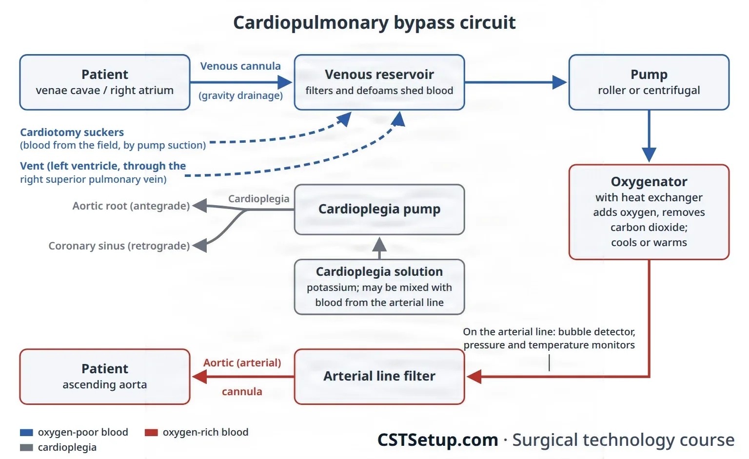
The perfusionist primes the circuit with fluid, clears it of air and runs its safety devices, such as a bubble detector and an arterial linear-TEER-ee-ul lineA thin catheter placed in an artery, often in the wrist, to measure blood pressure all the time and draw blood.Full entry filter; chapter 23 teaches the machine as equipment. At the field, the scrub helps place and remove the cannulas.
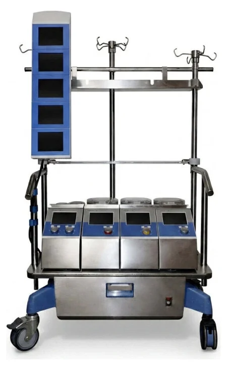
watch the bypass lines, and the heart, for air. Tell the surgeon at once if you see it, because air pumped into the arteries becomes an air embolismEM-buh-liz-umThe blocking of a blood vessel by a mass carried there in the blood (an embolus), such as a clot, fat or an air bubble.embol/o plug-ism conditionFull entry .
The heater-cooler that warms and cools the patient has spread Mycobacterium chimaera infection through its tank water carried in the air, so it is cleaned by the maker's instructions, filled only with sterile or filtered water, and its exhaust is pointed away from the sterile fieldSTAIR-ul feeldAn area set up with sterile drapes and supplies, kept free of all germs, for a sterile procedure.Full entry .
Heparin and the ACT
The patient is given heparinHEP-uh-rinAn anticoagulant drug, given by injection or IV, that keeps blood clots from forming or growing.Full entry before bypass, and the clottingKLOT-ingThe process by which blood thickens into a gel to plug a damaged vessel and stop bleeding; also called coagulation.Full entry time is checked with the activated clotting time (ACT) before bypass starts and at intervalIN-ter-vulThe time or space between two events, such as between two contractions in labor or between two waves on an ECG.Full entry during it. It is kept above about 480 seconds, the target varying with the test device; one reference gives above 500 and above 600 seconds. If heparin must be given again after protaminePROH-tuh-meenAn IV drug that binds heparin and reverses its blood-thinning effect.Full entry , as when bypass has to be restarted, it is confirmed before the pump runs. For a patient with HITaych-eye-tee (said as letters), or HITHeparin-induced thrombocytopenia: an immune reaction to heparin that lowers the platelet count.Full entry (HIT), a planned operation waits until the antibodyAN-tih-bod-eeA Y-shaped protein made by the immune system that sticks to one specific germ or foreign substance and marks it for destruction.anti- againstbody Full entry tests are negative, and an urgent one may use bivalirudin in place of heparin.
Median sternotomy
Most bypass cases open the chest through the sternum.
- The incision runs from the sternal notch to below the xiphoid. The fat and the linea alba are divided with the knife or electrosurgeryee-LEK-troh-SUR-jer-eeUsing high-frequency electric current through a handheld tool to cut tissue or seal bleeding vessels during surgery.Full entry , the surgeon frees the tissue behind the sternal notch and the xiphoid with a finger, and the xiphoid is split in the midlineMID-lineAn imaginary line down the center of the body; also, a type of IV catheter placed in a vein of the upper arm.Full entry with curved Mayo scissors.
- The sternal saw divides the sternum. Army-Navy retractorrih-TRAK-terA surgical instrument that holds back tissue or organs so the surgeon can see and reach the operative site.Full entry lift the edges while bleeding from the periosteum is coagulationkoh-ag-yuh-LAY-shunThe process by which blood changes from a liquid into a gel, forming a clot.coagul clot-ation processFull entry , and bone wax may be pressed on the marrow.
- The sternal retractor is opened, sometimes over moist laps, to show the pericardiumpair-ih-KAR-dee-umThe thin, two-layered sac that surrounds the heart.peri- aroundcardi/o heart-um a structureFull entry .
- The pericardium is lifted with forcepsFOR-sepsA hand instrument with two blades or arms used to grasp, hold or pull tissue or objects.Full entry so the heart is not cut, opened with the ESU or scissors, and its edges sewn up to the chest wall with tractionTRAK-shunA steady pulling force applied to a body part with weights, ropes and pulleys.Full entry sutureSOO-cherA stitch, or the thread used to stitch, that holds the edges of a wound or incision together.Full entry ; this lifts the heart and makes a well that holds irrigationeer-ih-GAY-shunWashing out a wound, body cavity, tube or eye with a steady flow of liquid.Full entry .
For a repeat sternotomy, scar may hold the heart against the back of the sternum. The surgeon uses the oscillating saw, may expose the groin or axillary vessels first so bypass can start quickly, and the perfusionper-FYOO-zhunThe flow of blood through the body's tissues, bringing them oxygen and nutrients.per- throughfus pour-ion processFull entry team is in the room for the opening.
Cannulation
Cannulas go in a set order unless the surgeon says otherwise: the arterial cannula first, so blood can be given at once if needed, then the venous, the retrograde cardioplegia catheter, the vent and the antegrade cardioplegia cannula. TEE may guide and confirm placement. The technique is much the same at every site: a purse-string stitch, a Rummel tourniquet over its ends held with a hemostatHEE-moe-statA clamp with finger rings and a locking ratchet used in surgery to grasp and close off bleeding vessels.hem/o blood-stat stopping, standing stillFull entry , an opening inside the purse-string, the cannula in, and the tourniquet snugged and tied to the cannula with heavy silk.
- The aortic cannula: two purse-strings, with or without pledgets, go in the front of the ascending aorta. The surgeon cuts a small opening between them with a #11 blade (an aortotomy, an incision into the aorta) and covers it with a finger, then inserts the cannula, whose end is clamped so the aorta cannot bleed back through it. It is filled with blood to push out the air before it is joined to the arterial line, under a saline dripDRIPA continuous IV infusion of fluid or a drug, given slowly over time by gravity or a pump.Full entry or while the perfusionist runs a little prime out of the line.
- The venous cannula: through a purse-string in the right atrialAY-tree-ulRelating to an atrium, one of the two upper chambers of the heart.atri/o atrium-al relating toFull entry appendage, held with a clamp (such as a Cooley spoon clamp) while the tip of the appendage is cut off. A two-stage cannula drains both the atrium and, from its tip in the inferior vena cavain-FEER-ee-er VEE-nuh KAY-vuhThe large vein that carries blood from the lower body back to the right atrium of the heart.inferior lowervena veincava hollowFull entry , the lower body; it is enough when the atrium need not be emptied completely, as in many aortic valveay-OR-tik valvThe heart valve between the left ventricle and the aorta, which opens to let blood out to the body.Full entry replacements. Bicaval cannulation places one cannula in each vena cava; with tapes and tourniquets snugged around both cavae, all the venous blood goes to the pump (total bypass), which is used for mitral valveMY-trul valvThe heart valve between the left atrium and the left ventricle.Full entry replacement, septumSEP-tumA wall of tissue that divides two spaces or cavities, such as the two sides of the heart or nose.sept/o wall, partitionFull entry defects, ventricleVEN-trih-kulOne of the two lower chambers of the heart, or one of the fluid-filled spaces inside the brain.Full entry aneurysm repair and transplant, and whenever the right side of the heart is opened. In partial bypass some blood still passes through the heart.
- Retrograde cardioplegia catheter: through a purse-string in the right atrium into the coronary sinusSY-nusA hollow space in the body, most often one of the air-filled spaces in the skull bones around the nose.Full entry , its place confirmed by TEE or by the surgeon's touch; its pressure is monitored.
- The vent: through the right superior pulmonary vein, across the mitral valve into the left ventricle, connected to the pump suction.
- Antegrade cardioplegia cannula: a small needle-tipped cannula through a pledgeted purse-string in the ascending aorta, between the cross-clamp and the heart.
- femoralFEM-er-ulRelating to the thigh or the femur, the thigh bone.Full entry cannulation: through the groin, femoral cannulas are placed in the femoral arteryFEM-er-ul AR-ter-eeThe large artery that carries blood to the leg, running through the groin and down the thigh.Full entry and vein over a guidewire with dilatorDY-lay-terAn instrument used to stretch or widen an opening, canal or passage in the body.Full entry (the Seldinger technique), as in minimally invasive surgery, a dissectiondy-SEK-shunA tear in the inner layer of an artery wall that lets blood split the layers apart; also, the cutting apart of tissue in surgery.Full entry or a repeat sternotomy, or when the aorta cannot be used. The axillary artery is another arterial site, often through a short graftGRAFTTissue or a man-made material moved into place in surgery to replace or repair a damaged part.Full entry sewn onto it, when the aorta is diseased or the brain will be perfused during circulatory arrest.
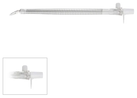
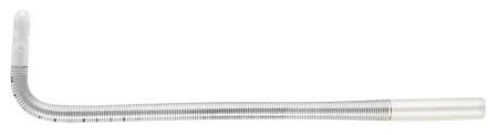
Keep a tubing clamp on each cannula until it is connected, and know which lines bring blood to the patient and which take it away.
Stopping and protecting the heart
Once bypass is running, the surgeon places the aortic cross-clamp across the ascending aorta, between the aortic cannula and the heart. This cuts the heart off from the circulation so cardioplegia can fill the coronary arteryKOR-uh-nair-ee AR-ter-eeOne of the blood vessels on the surface of the heart that bring oxygen-rich blood to the heart muscle.Full entry without being washed out.
Cardioplegia is the planned stopping of all the heart's activity. An arrested heart uses far less energy than a beating or fibrillating one, and cardioplegia protects the muscle while its blood supply is cut off. The solution contains potassium (2 to 50 mEq/L), its most important ingredient, which stops the heart in diastoledy-AS-tuh-leeThe part of each heartbeat when the heart muscle relaxes and the chambers fill with blood.Full entry , with buffers against the acid that builds up; it is usually given cold, at 4 to 10°C, and may be given warm. It is given two ways:
- Antegrade, into the aortic root below the cross-clamp. The pressure closes the aortic valve, so the solution has nowhere to go but the coronary arteries. If the aortic valve leaks, the solution fills and stretches the left ventricle instead, so it is given retrograde. It can also go into finished vein grafts; handheld cannulas can put it straight into the coronary openings, but because they can injure the openings they are kept for when retrograde delivery cannot be used.
- Retrograde, through the coronary sinus, backward through the heart's veins; it helps when the coronary arteries are blocked.
When the electrocardiogramih-lek-troh-KAR-dee-oh-gramA test that records the heart's electrical activity as waves on paper or a screen.electr/o electricitycardi/o heart-gram recordFull entry goes flat, the heart is arrested; the dose is repeated about every 15 to 20 minutes, or when fine fibrillation appears. A last warm dose before the clamp comes off helps the heart recover.
Cooling: the heat exchanger cools the whole body, and cold saline or slush is applied around the heart. Moderate cooling to 28°C halves the body's oxygen use. Slush is kept free of large ice chips, which can injure the phrenic nerves in the pericardium. ventricular fibrillationven-TRIK-yuh-ler fib-ruh-LAY-shunA heart rhythm in which the lower chambers quiver in a fast, chaotic way instead of pumping.ventricul/o ventricle-ar relating tofibrill small fiber, twitching-ation processFull entry is less likely above about 32°C, and cooling harms clotting and wound healingwoond HEE-lingThe body's process of repairing damaged skin and tissue.Full entry , which may slow the end of bleeding after heparin is reversed. Chapter 13 teaches deliberate hypothermiahy-poh-THER-mee-uhA body temperature that has fallen below the normal range.hypo- belowtherm/o heat-ia conditionFull entry .
Circulatory arrest: for complex aortic arch surgery, where no clamp can be placed, the patient is cooled to about 18°C and the pump is stopped, so the field is dry, while the brain is protected as the arch section below describes.
Coming off bypass
- Near the end of the repair the perfusionist rewarms the patient, and the surgeon removes the air from the left ventricle and the aorta, using the vent, a needle in the aorta, the table tilted head down, the lungs inflated and TEE to find any air left.
- The cross-clamp comes off. The heart often starts beating on its own; if it fibrillates, it is defibrillationdee-fib-ruh-LAY-shunAn electric shock given to the heart to stop a chaotic, life-threatening rhythm so a normal rhythm can return.Full entry .
- Temporary pacing wires are sewn to the atrium and ventricle.
- With the heart beating and the lungs mechanical ventilationmeh-KAN-ih-kul ven-tuh-LAY-shunBreathing support from a machine, a ventilator, that pushes air into the lungs through a tube or a mask.Full entry , the perfusionist slowly reduces the venous drainage and the arterial flow until the heart carries the circulation, and bypass ends.
- The vent is usually removed first, before bypass ends, then the venous and aortic cannulas, each purse-string tied as its cannula comes out. Femoral cannulas come out the same way, the vein first, and each vessel is closed with fine polypropylene.
- Protamine reverses the heparin; the pump suckers go off as it starts.
- Tubes drain the pericardium, and the pleuralPLOOR-ulHaving to do with the pleura, the thin two-layer membrane that covers the lungs and lines the chest wall.pleur/o pleura-al relating toFull entry if opened. The pericardium is usually left open, so collecting blood cannot cause tamponade.
Problems after bypass: bleeding from cannulation sites, from heparin not fully reversed or from damaged tissue; atelectasisat-uh-LEK-tuh-sisThe collapse of part or all of a lung, in which the tiny air sacs close and hold no air.atel- incomplete-ectasis expansionFull entry ; brief changes in thinking and perceptionper-SEP-shunThe brain's way of taking in and making sense of what the senses detect; also a person's own view or understanding of something.Full entry . Blood's contact with the circuit can also set off a body-wide inflammatory responsein-FLAM-uh-tor-ee rih-SPONSThe body's protective reaction to injury or infection, which brings blood, fluid and white cells to the area.Full entry , fluid shifts and showers of tiny emboli.
12Coronary artery surgery
Coronary angioplasty
What and why: percutaneous coronary intervention (PCI) is done in the cardiac catheterizationKAR-dee-ak kath-uh-ter-ih-ZAY-shunA procedure in which a thin tube is threaded through a blood vessel into the heart to take pictures, measure pressures or open blocked arteries.Full entry lab. A balloon catheter is passed into the narrowed coronary arteryKOR-uh-nair-ee AR-ter-eeOne of the blood vessels on the surface of the heart that bring oxygen-rich blood to the heart muscle.Full entry and inflated to open it (percutaneous transluminal coronary angioplasty, PTCA), and a bare-metal or drug-eluting stentstentA small tube, often of metal mesh, placed inside a vessel or duct to hold it open.Full entry is usually placed to hold it open. Clot-dissolving drugs can reopen a freshly blocked artery, and antiplateletan-tee-PLAYT-litA drug that keeps platelets from sticking together and forming clots.anti- againstplatelet a blood cell that helps clottingFull entry drugs, such as aspirin and clopidogrelkloh-PID-oh-grelAn antiplatelet drug that keeps platelets from clumping together to form clots.Full entry , help keep it from narrowing again. PCI is often used during a myocardial infarctionmy-oh-KAR-dee-ul in-FARK-shunDeath of part of the heart muscle because its blood supply is blocked; a heart attack.my/o musclecardi/o heart-al having to do withinfarct area of dead tissueFull entry and can spare a patient bypass surgery, though coronary disease keeps progressing and some patients need a bypass later. For disease of the left main coronary artery or of all three vessels, bypass surgery is still the standard of careSTAN-derd uv kairThe level of care that a reasonably careful nurse with the same training would give in the same situation.Full entry . In a hybrid room, a surgeon may sew a bypass graftGRAFTTissue or a man-made material moved into place in surgery to replace or repair a damaged part.Full entry and a cardiologist place stents in the same session. Chapter 22 teaches balloons, stents and catheter work. A catheter case through the skin enters no tract, so it fits Class I.
Coronary artery bypass grafting (CABG)
What and why: CABG routes blood around a narrowed coronary artery with a graft, a conduit (a channel for blood) taken from the patient's own body, to relieve anginaan-JY-nuhChest pain or pressure that happens when the heart muscle does not get enough blood and oxygen.Full entry and lengthen life in patients with disease of several vessels. The internal mammary artery stays attached at its upper end to the subclavian artery, so it brings its own blood; a vein or a free artery graft is sewn from the aorta to the coronary artery beyond the blockage. Chapter 5 teaches the grafts' anatomy.
The conduits:
- The left internal mammary artery (LIMA) to the left anterior descending (LAD) artery is the first choice: it stays open longest. The STS guideline recommends an internal mammary artery whenever the LAD needs a graft.
- A second arterial graft, the right internal mammary or a radial artery, should be considered in suitable patients, though taking both mammary arteries may add to the risk of a sternal infection.
- The radial artery is taken from the arm the patient uses less, after a test such as the Allen testAL-en testA bedside check of blood flow to the hand through the ulnar artery, done by pressing and releasing the wrist arteries.Full entry or a Doppler ultrasoundDOP-ler UL-truh-soundAn ultrasound that uses sound waves to hear or show blood flowing through vessels or the fetal heartbeat.Full entry shows the ulnar artery alone can feed the hand; a drug is used to prevent spasmSPAZ-umA sudden, involuntary tightening of a muscle or of the muscle in a hollow organ or tube.Full entry . More and more often it, too, is taken with an endoscopeEN-duh-skohpA thin tube with a light and a camera that is passed into the body to look inside an organ or cavity.endo- within-scope instrument for viewingFull entry .
- The great saphenous vein remains the workhorse when several grafts are needed, usually taken through small incisions with an endoscope. It is reversed, or its valves are cut with a valvulotome, so they cannot block the flow, and it is flushed with heparinized blood or saline and kept moist until it is used. Chapter 40 teaches the vein itself and the open harvest for a leg bypass.
- The right gastroepiploic artery, from the stomach, is rarely used, when other conduits are poor.
endoscopicen-doh-SKOP-ikDone with an endoscope, a thin tube with a light and camera passed into the body.endo- within-scopic viewingFull entry vein harvesting (EVH) has become the standard way to take the saphenous vein for a bypass in the United States: compared with one long leg incision, it means less pain and far fewer wound infections, collections of blood or fluid, and wound breakdowns. An emergency may still call for the faster open harvest. Many centers map the vein with Doppler ultrasound before surgery, to find it and check its size.
- With the knee bent and supported, a cut of about 2 cm is made over the vein on the inner leg near the knee, and the vein is found under direct vision.
- A sealing port goes into the incision, and the endoscope is passed along the vein inside its cannulaKAN-yoo-luhA thin tube placed into the body or a body opening to deliver or remove fluid or air.Full entry while carbon dioxideKAR-bun dy-OK-sideA gas made by the body's cells as waste, carried in the blood to the lungs and breathed out.Full entry holds the tunnel open.
- The side branches are sealed and cut with the kit's cautery device, or with clips and bipolarby-POH-lerHaving two poles or two opposite ends; most often used as a short form of bipolar disorder.bi- twopolar having polesFull entry energy, and small cuts at the groin and ankle let the vein's ends be divided; the vein is drawn out through the knee incision.
- The tunnel is checked for bleeding, a drain is placed if needed, the incisions are closed and the leg is wrapped firmly.
- On the back table the vein is checked for tears and loose branches, which are clipped or repaired, and it is kept in heparinized saline until it is sewn in.
The harvester can injure the saphenous nerve beside the vein, leaving pain or numbness on the inner leg, and the carbon dioxide can spread under the skin or into the abdomen, or cause an air embolismEM-buh-liz-umThe blocking of a blood vessel by a mass carried there in the blood (an embolus), such as a clot, fat or an air bubble.embol/o plug-ism conditionFull entry .
Position, prep and drape: supine; a prep from the jaw to the toes, the legs all the way around, with a urinary catheter.
Setup: a major cardiac set, a coronary artery set, a vascular set, vessel loops, clips and Rummel tourniquets; an IMA retractorrih-TRAK-terA surgical instrument that holds back tissue or organs so the surgeon can see and reach the operative site.Full entry , coronary dilatorDY-lay-terAn instrument used to stretch or widen an opening, canal or passage in the body.Full entry , an aortic punch and fine polypropylene sutureSOO-cherA stitch, or the thread used to stitch, that holds the edges of a wound or incision together.Full entry .
Key steps (on bypass):
- The surgeon opens the sternum while the assistant takes the vein or radial artery.
- The IMA takedown: a retractor lifts one side of the sternum, and the artery is freed from the chest wall with fine forcepsFOR-sepsA hand instrument with two blades or arms used to grasp, hold or pull tissue or objects.Full entry and the electrosurgeryee-LEK-troh-SUR-jer-eeUsing high-frequency electric current through a handheld tool to cut tissue or seal bleeding vessels during surgery.Full entry or ultrasonic scalpel, its small branches clipped. heparinHEP-uh-rinAn anticoagulant drug, given by injection or IV, that keeps blood clots from forming or growing.Full entry is given before it is cut at its lower end, the artery is checked for good flow, papaverine is put on or into it, and a small bulldog closes its tip; it is wrapped in a papaverine-soaked sponge and tucked into the pleural spacePLOOR-ul SPAYSThe thin space between the two layers of the pleura, the membrane that covers the lungs and lines the chest wall.Full entry until needed.
- Bypass starts, usually with light cooling; the aorta is cross-clamped and cardioplegia is given.
- Distal anastomosisuh-nas-tuh-MOH-sisA connection between two tube-shaped structures, such as two ends of bowel or two blood vessels, made by surgery or found naturally.Full entry : the surgeon clears fat from the coronary artery with a #64 Beaver or #15 blade, opens it with a #11 or Beaver blade and extends the opening with Potts scissors. A dilator may size it; the graft end is cut on a slant and sewn on with 7-0 polypropylene, then filled with cardioplegia to check for leaks and its length.
- proximalPROK-sih-mulNearer the center of the body, or nearer the place where a part is attached.Full entry anastomoses: a #11 blade and the aortic punch make small holes in the aorta, and the vein grafts are sewn to them, either during the cross-clamp or with a partial occlusionuh-KLOO-zhunA blockage or closing off of a passage, such as a blood vessel, tube or IV line.Full entry clamp on the aorta after the cross-clamp comes off. Epiaortic ultrasound may find a spot free of plaquePLAKA deposit that builds up on a surface in the body, such as a sticky film of bacteria on teeth or a fatty buildup in an artery wall.Full entry first.
- A fine needle, 25 or 27 gauge, lets trapped air out of each vein graft, and every join is checked for leaks.
- The aortic ends of the vein grafts may be marked with radiopaqueray-dee-oh-PAYKBlocking x-rays, so the material shows up white on an x-ray image.radi/o radiation, x-rayopaque not letting throughFull entry rings or clips, so they can be found at a later catheterization.
- Bypass ends, pacing wires and chest tubechest toobA tube placed through the chest wall into the space around the lung to drain air, blood or fluid.Full entry are placed and the chest is closed.
After surgery: bleeding, arrhythmiauh-RITH-mee-uhA heart rhythm that is not normal: too fast, too slow or irregular.a- withoutrhythm regular beat-ia conditionFull entry (atrial fibrillationAY-tree-ul fib-rih-LAY-shunAn irregular heart rhythm in which the upper chambers of the heart quiver instead of beating in an organized way.atri/o atrium (upper heart chamber)fibrill/o small fibers, quivering-ation processFull entry is common after CABG), stroke, infection and ischemiais-KEE-mee-uhToo little blood flow to a part of the body, so the tissue does not get enough oxygen.isch- hold back-emia blood conditionFull entry are the main complications, and a graft that clots can cause an MI.
Wound class: I; no tract is entered in the chest or the leg.
Off-pump and minimally invasive bypass
Off-pump coronary artery bypass (OPCAB), beating-heart surgery, avoids bypass, cardiac arrestKAR-dee-ak uh-RESTThe sudden stop of the heart's pumping, so blood no longer reaches the brain and body.Full entry and cooling. A suction stabilizer, attached to the sternal retractor, holds still the small patch of heart around the artery being grafted, and a suction cup on the apex lifts the heart to reach the arteries on its side and back. Blood from the opened artery is cleared with a mist of humidified carbon dioxide from a blower, elastic coronary tourniquets or a small Silastic shuntSHUNTA passage that moves blood or other fluid from one place to another, either made by a surgeon or present in the body.Full entry placed inside the artery. Bypass stands by, with the perfusionist assigned and the circuit ready, in case the heart fibrillates or the patient becomes unstableun-STAY-bulDescribes a client whose condition is changing quickly or is hard to predict.Full entry . Off-pump techniques are not used when a chamber of the heart must be opened, because air in the open chamber would travel to the brain. Long-term studies have found some off-pump disadvantages, such as less complete grafting and grafts that stay open less well.
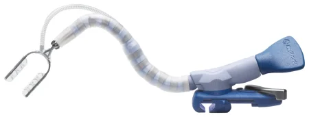
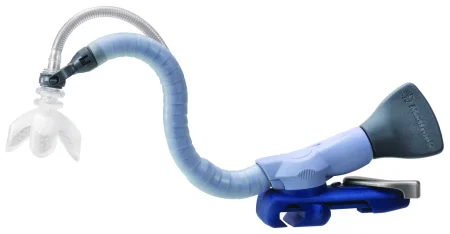
Minimally invasive direct coronary artery bypass (MIDCAB) joins the LIMA to the LAD on the beating heart through a short left anterior thoracotomythor-uh-KOT-uh-meeA surgical cut through the chest wall to reach the lungs, heart or other organs in the chest.thorac/o chest-tomy cutting intoFull entry , about 10 cm, in the fourth rib space, for disease of one vessel. Some surgeons have the legs prepped too, in case the LIMA cannot be used.
Wound class (OPCAB and MIDCAB): I, as for CABG; no tract is entered.
13Valve surgery
Valve prostheses
- Mechanical valves, most with a tilting disk design, last a long time but need lifelong anticoagulationan-tee-koh-ag-yoo-LAY-shunThe use of drugs that slow the blood's clotting, to prevent or treat blood clots.anti- againstcoagul clotting-ation processFull entry , and a clot can suddenly block one. Protect them from scratches.
- Biological valves (bioprostheses) are made from pig valves or from cow or horse pericardiumpair-ih-KAR-dee-umThe thin, two-layered sac that surrounds the heart.peri- aroundcardi/o heart-um a structureFull entry . Most patients need no long-term anticoagulation, but a tissue valve can wear out and need replacing.
- Allografts (homografts), human donor valves, resist infection and need no anticoagulation; they arrive frozen and are thawed by the supplier's protocol.
- Valved conduits join a valve to a tube graftGRAFTTissue or a man-made material moved into place in surgery to replace or repair a damaged part.Full entry , to replace the aortic valveay-OR-tik valvThe heart valve between the left ventricle and the aorta, which opens to let blood out to the body.Full entry and the ascending aorta together.
- Annuloplasty rings and bands are sewn around a valve to repair it rather than replace it.
Valves are very expensive. Handle them as little as possible, and verify the type, size and serial number with the circulator, who records them. Each prosthesispros-THEE-sisAn artificial body part that replaces one that is missing or does not work, such as an artificial leg, joint, eye or heart valve.Full entry has its own sizers, holders and handles. Biological valves stored in glutaraldehydegloo-tuh-RAL-duh-hideA liquid chemical used for high-level disinfection, or sterilization with longer soaking, of heat-sensitive medical devices.Full entry are rinsed in saline baths before they are sewn in; the number of baths, the volume and the time differ between makers, so follow the instructions for use every time. Keep a biological valve moist with saline from then on, because dry leaflets are damaged, and never soak it in antibiotic solution. Check that a valve's temperature indicator shows it was stored correctly.
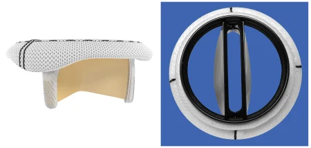
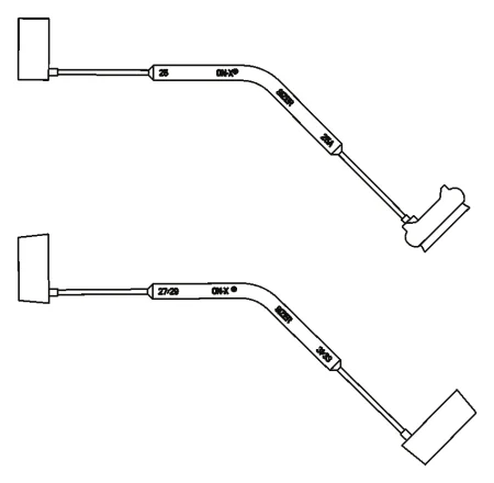
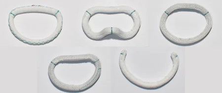
The patient's choice matters, including any social, cultural or religious objection to animal tissue. After any valve operation the risks include clots and stroke, bleeding from the anticoagulantan-tee-koh-AG-yoo-luntA drug that slows the blood's ability to form clots.anti- againstcoagul/o clotting-ant an agent that doesFull entry , infection of the prosthesis (prostheticpros-THET-ikDescribes an artificial part made to replace a missing or damaged body part, such as a limb, joint, eye or heart valve.prosthe an addition (from Greek)-tic relating toFull entry valve endocarditisen-doh-kar-DY-tisInflammation of the inner lining of the heart, most often on the heart valves.endo- insidecardi/o heart-itis inflammationFull entry ), a leak around the valve, a valve too small for the patient's needs and failure of the prosthesis.
Aortic valve replacement (AVR)
What and why: the aortic valve is replaced for stenosisstuh-NOH-sisNarrowing of a passage or opening in the body, such as a blood vessel, heart valve or the spinal canal.sten/o narrow-osis conditionFull entry , when calcified, stiff leaflets narrow the opening to a slit, or for insufficiencyin-suh-FISH-un-seeA state in which an organ or part does not work well enough to meet the body's needs.in- notsufficient enoughFull entry , when a leaking valve lets blood fall back into the left ventricleLEFT VEN-trih-kulThe heart's lower left chamber, which pumps oxygen-rich blood into the aorta and out to the body.Full entry ; either way the ventricleVEN-trih-kulOne of the two lower chambers of the heart, or one of the fluid-filled spaces inside the brain.Full entry overworks and can fail. A partial sternotomy may be used for an aortic valve alone.
Setup: an open-heart set, aortic valve instruments, the valves, sizers and holders the surgeon prefers, a retrograde cardioplegia catheter and vents, and basins of saline to rinse a tissue valve.
Key steps:
- Bypass with a two-stage venous cannulaKAN-yoo-luhA thin tube placed into the body or a body opening to deliver or remove fluid or air.Full entry ; a vent through the right superior pulmonary veinPUL-muh-nair-ee VAYNOne of the four veins that carry oxygen-rich blood from the lungs to the left atrium of the heart.Full entry and a retrograde catheter into the coronary sinusSY-nusA hollow space in the body, most often one of the air-filled spaces in the skull bones around the nose.Full entry .
- The aorta is cross-clamped. With stenosis, the first dose of cardioplegia can go into the aortic root; with a leaking valve it goes retrograde, because antegrade solution would fill the ventricle.
- The aorta is opened and the diseased cusps are cut out; calcium is removed with rongeurron-ZHURA strong surgical instrument with sharp, cup-shaped jaws used to bite away small pieces of bone.Full entry . Narrow packing in the ventricle catches loose bits, and you wipe each instrument with a moist sponge, so no fragment is left to embolize.
- The annulus is measured with the valve's own sizers, and the circulator opens the chosen valve.
- Interrupted, often pledgeted, sutureSOO-cherA stitch, or the thread used to stitch, that holds the edges of a wound or incision together.Full entry are placed through the annulus and then through the valve's sewing ring, in three groups, one for each cusp, and held in order.
- The valve is lowered into place and the sutures tied; the aorta is closed.
- Air is cleared from the left heart through the vent, by tilting the table head down and inflating the lungs, and the heart is not allowed to eject until the surgeon, helped by TEE, is satisfied that no air is left. The cross-clamp comes off, and the heart is defibrillationdee-fib-ruh-LAY-shunAn electric shock given to the heart to stop a chaotic, life-threatening rhythm so a normal rhythm can return.Full entry if needed.
- Pacing wires and chest tubechest toobA tube placed through the chest wall into the space around the lung to drain air, blood or fluid.Full entry are placed and the chest is closed.
After surgery: bleeding, arrhythmiauh-RITH-mee-uhA heart rhythm that is not normal: too fast, too slow or irregular.a- withoutrhythm regular beat-ia conditionFull entry , stroke, infection, ischemiais-KEE-mee-uhToo little blood flow to a part of the body, so the tissue does not get enough oxygen.isch- hold back-emia blood conditionFull entry and failure of the valve.
Mitral valve repair and replacement
What and why: a stenotic mitral valveMY-trul valvThe heart valve between the left atrium and the left ventricle.Full entry is opened, or a leaking one made to close. Repair is preferred whenever possible, because it avoids a prosthesis and lifelong anticoagulation. Instruments and valves for replacement are always ready, since the surgeon decides only after looking at the valve. Bicaval cannulation decompresses the heart and improves the view, and TEE checks the repair once the heart beats again.
Repairs:
- Commissurotomy: fused leaflets are cut or spread apart, sometimes with a dilatorDY-lay-terAn instrument used to stretch or widen an opening, canal or passage in the body.Full entry , for stenosis.
- Annuloplasty: a complete or partial (C-shaped) ring, sized with its obturatorOB-too-ray-terA smooth-tipped rod that fits inside a tube, such as a tracheostomy tube or cannula, to guide it in and then is taken out.Full entry , is sewn into a stretched annulus to make it smaller.
- Leaflet and chord repair: a floppy section of the posterior leaflet is cut out and the edges sewn, and torn or stretched chordae are replaced with PTFE suture.
- Edge-to-edge (Alfieri) repair: a stitch, or a clip placed through a catheter, joins the middle of the two leaflets.
The repair is tested by filling the ventricle with saline from a bulb syringe and watching the leaflets close.
Replacement: the left atriumAY-tree-umOne of the two upper chambers of the heart, which receive blood coming back to the heart.atri/o atrium-um noun endingFull entry is opened along the groove between the atria and held open with an atrialAY-tree-ulRelating to an atrium, one of the two upper chambers of the heart.atri/o atrium-al relating toFull entry retractorrih-TRAK-terA surgical instrument that holds back tissue or organs so the surgeon can see and reach the operative site.Full entry . The anterior leaflet is cut out, but the posterior leaflet and its chordae are kept when possible to preserve the shape and pumping of the ventricle. Fifteen to 20 or more pledgeted sutures go through the annulus and the valve's sewing ring, kept moist and in order; the valve is seated and tied, the atrium closed, and air removed with the table head down and the lungs inflated.
Mitral surgery can also be done through a right mini-thoracotomythor-uh-KOT-uh-meeA surgical cut through the chest wall to reach the lungs, heart or other organs in the chest.thorac/o chest-tomy cutting intoFull entry or with the robot, usually with femoralFEM-er-ulRelating to the thigh or the femur, the thigh bone.Full entry cannulation, and the aorta is closed off with a cross-clamp or a balloon inside it.
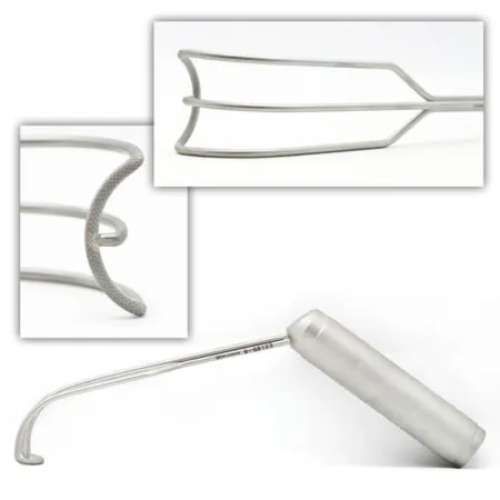
The tricuspid valvetry-KUS-pid VALVThe heart valve between the right atrium and the right ventricle.tri- threecusp point, flapFull entry is most often operated on at the same time as a mitral valve and is repaired, for example with a ring, or replaced with a mitral prosthesis, its stitches kept clear of the conductionkun-DUK-shunThe passing of something, such as an electrical signal or heat, from one place to another through a material.Full entry tissue near the AV nodeay-VEE nohdAtrioventricular node: a small group of cells between the upper and lower chambers of the heart that passes on the heartbeat signal.atri/o atriumventricul/o ventricle-ar relating toFull entry .
Two valves: when the aortic and mitral valves are both replaced, the mitral goes in first, because the firm ring of an aortic prosthesis already in place could injure the heart and make the mitral harder to insert; when a CABG is added to an aortic valve, the distal grafts are sewn before the new valve goes in and the proximalPROK-sih-mulNearer the center of the body, or nearer the place where a part is attached.Full entry grafts last.
Transcatheter aortic valve replacement (TAVR)
What and why: a tissue valve compressed onto a catheter is placed inside the diseased aortic valve without opening the chest, for severe aortic stenosis with symptoms in patients at high risk for open surgery, and more and more in lower-risk patients. It is done in a hybrid room or the catheterization lab, usually through the femoral arteryFEM-er-ul AR-ter-eeThe large artery that carries blood to the leg, running through the groin and down the thigh.Full entry ; the axillary artery or the apex of the heart is used when the leg arteries are too narrow or diseased.
Setup: endovascular valve and femoral access instruments, the valve and its deployment system, guidewires and catheters with the catheter lubricants their makers name, contrastKON-trastA dye or other substance given before an imaging test so body parts show up more clearly.Full entry , lead aprons and fluoroscopyfloo-ROS-kuh-peeAn x-ray method that shows moving pictures of the inside of the body in real time.fluor/o glowing (from the fluorescent screen)-scopy looking, viewingFull entry . The valve is rinsed or kept moist by its maker's instructions.
Key steps:
- A sheathSHEETHA covering that wraps around a structure; in procedures, a short hollow tube left in a blood vessel to pass catheters through.Full entry goes into the femoral artery over a guidewire, and the catheter is threaded up the aorta to the valve.
- Landmarks are confirmed on fluoroscopy, and the valve is brought into the aortic annulus.
- A self-expanding valve is released; a balloon-expanded one is opened by inflating its balloon.
- The surgeon looks for leaks around the valve. A self-expanding valve can be adjusted; with a balloon-expanded valve, a second one is placed inside the first.
- The access is closed.
Be ready to convert from the catheter method to open surgery in every case; injury to the heart or to the access vessel is one of the risks of a valve placed through a catheter.
Wound class (valve surgery, TAVR included): I, no tract is entered.
14Aortic aneurysm and dissection
The aortic arch
What and why: an aneurysmAN-yuh-riz-umA weak spot in the wall of an artery that bulges outward like a balloon.aneurysm a widening (Greek)Full entry or a dissectiondy-SEK-shunA tear in the inner layer of an artery wall that lets blood split the layers apart; also, the cutting apart of tissue in surgery.Full entry of the arch can cut off blood to the brain and arms, because the arch gives off the brachiocephalic, left carotidkuh-ROT-idHaving to do with the carotid arteries, the two large arteries on each side of the neck that carry blood to the head and brain.Full entry and left subclavian arteries; in the ascending aorta, one may burst or keep the aortic valveay-OR-tik valvThe heart valve between the left ventricle and the aorta, which opens to let blood out to the body.Full entry from closing.
Position and setup: supine for a median sternotomy, or a modified thoracotomythor-uh-KOT-uh-meeA surgical cut through the chest wall to reach the lungs, heart or other organs in the chest.thorac/o chest-tomy cutting intoFull entry position for the far end of the arch. Aneurysm instruments and woven or collagenKOL-uh-jenThe main structural protein of the body, which gives strength to skin, bones, tendons, ligaments and scar tissue.coll/a glue-gen producingFull entry -sealed graftGRAFTTissue or a man-made material moved into place in surgery to replace or repair a damaged part.Full entry in a range of sizes, with valve and coronary instruments, valves and valved conduits ready. Have a basin for clot and debris. Since the weakened aorta may not take a cannulaKAN-yoo-luhA thin tube placed into the body or a body opening to deliver or remove fluid or air.Full entry , the femoralFEM-er-ulRelating to the thigh or the femur, the thigh bone.Full entry or axillary artery is often used for arterial inflow, and the femoral arteryFEM-er-ul AR-ter-eeThe large artery that carries blood to the leg, running through the groin and down the thigh.Full entry and vein may be isolated before the chest is opened in case the aneurysm ruptures.
Key steps:
- The right atriumAY-tree-umOne of the two upper chambers of the heart, which receive blood coming back to the heart.atri/o atrium-um noun endingFull entry and the right axillary or a femoral artery are cannulated, and the patient is cooled for circulatory arrest; the head may be packed in ice bags, padded against frostbite.
- At the planned temperature the surgeon clamps the arch vessels one by one or stops the circulation, and the brain is perfusionper-FYOO-zhunThe flow of blood through the body's tissues, bringing them oxygen and nutrients.per- throughfus pour-ion processFull entry through an arch branch or backward through the superior vena cavasuh-PEER-ee-er VEE-nuh KAY-vuhThe large vein that carries blood from the head, neck, arms and upper chest back to the right atrium of the heart.superior uppervena veincava hollowFull entry .
- The tissue holding all three arch vessels is cut out as an "island," and a graft, straight or with side branches, is sewn first to the descending aorta.
- An opening cut in the graft is sewn to the island of arch vessels.
- The graft is de-aired and bypass restarts, perfusing the head and the lower body again; the graft is then joined to the ascending aorta.
- Glue may reinforceree-in-FORSTo repeat and support teaching or care that has already been started, so the client remembers and follows it.Full entry the sutureSOO-cherA stitch, or the thread used to stitch, that holds the edges of a wound or incision together.Full entry lines, the patient is rewarmed and weaned from bypass, and the chest is closed.
After surgery: bleeding, stroke, infection and death are the risks of aortic surgery, and a join that keeps bleeding may need repair.
Dissection, the aortic root and stent grafts
A Type A dissection, one that involves the ascending aorta, is an emergency because the false channel can rupture, cut off the aorta's branches or keep the aortic valve from closing. It is repaired at once on bypass, with the arterial cannula often in the femoral or axillary artery, by replacing the torn aorta with a woven graft, or with a valved conduit when the aortic root is involved.
When the root is involved, the Bentall procedure replaces the aortic root, the valve and the ascending aorta with a valved conduit, and the coronary arteryKOR-uh-nair-ee AR-ter-eeOne of the blood vessels on the surface of the heart that bring oxygen-rich blood to the heart muscle.Full entry are sewn into openings that the electrocautery makes in the graft, sealing its cut edges at the same time.
Most aneurysms and dissections of the descending aorta are now treated by endovascular repair (TEVAR): a stentstentA small tube, often of metal mesh, placed inside a vessel or duct to hold it open.Full entry graft goes in through the femoral artery under fluoroscopyfloo-ROS-kuh-peeAn x-ray method that shows moving pictures of the inside of the body in real time.fluor/o glowing (from the fluorescent screen)-scopy looking, viewingFull entry , its guidewires, which can be more than 6 feet long, are anchored on the field with wet sterile towels, and the open set stays ready in case of bleeding; chapter 40 teaches stent grafts.
Left ventricular aneurysm
What and why: the thin scar of an old MI is removed to reduce the risk of rupture and of clots breaking off.
Setup: the major cardiac set, with a Dacron patch and Teflon felt.
Key steps: through a median sternotomy, on bypass with the aorta cross-clamped, the surgeon opens the aneurysm and gently removes any clot; keep the instruments clean so no clot gets into the circulation. Most surgeons now sew a patch of Dacron, Teflon or pericardiumpair-ih-KAR-dee-umThe thin, two-layered sac that surrounds the heart.peri- aroundcardi/o heart-um a structureFull entry inside, on the border of healthy muscle (endoaneurysmorrhaphy), because it keeps the ventricleVEN-trih-kulOne of the two lower chambers of the heart, or one of the fluid-filled spaces inside the brain.Full entry 's shape better. The ventricle is vented and de-aired before the last stitch is tied.
cutting the scar out and closing the ventricle by sewing its edges together between strips of Teflon felt, which one reference still describes.
After surgery: the patient is watched closely in the intensive care unit for a low cardiac outputKAR-dee-ak OWT-putThe amount of blood the heart pumps out in one minute.Full entry and ventricular arrhythmiauh-RITH-mee-uhA heart rhythm that is not normal: too fast, too slow or irregular.a- withoutrhythm regular beat-ia conditionFull entry ; failure of the left ventricleLEFT VEN-trih-kulThe heart's lower left chamber, which pumps oxygen-rich blood into the aorta and out to the body.Full entry is the most common cause of death in the hospital.
Wound class (aorta and ventricle): I.
15Surgery for rhythm problems
Many, but not all, pacemakerPAYS-may-kerA small device that sends electrical pulses to the heart to keep it beating at a set rate; also the heart's own natural rhythm center.Full entry and defibrillators now go in in the electrophysiology (EP) lab or the catheterization lab, and ablationab-LAY-shunA procedure that destroys a small area of tissue, often with heat or cold, to stop it from causing a problem.Full entry for atrial fibrillationAY-tree-ul fib-rih-LAY-shunAn irregular heart rhythm in which the upper chambers of the heart quiver instead of beating in an organized way.atri/o atrium (upper heart chamber)fibrill/o small fibers, quivering-ation processFull entry is normally done in the EP lab. Chapter 20 teaches energy safety around implanted devices.
Temporary pacing wires
At the end of heart surgery, temporary wires are sewn to the surface of the right atriumAY-tree-umOne of the two upper chambers of the heart, which receive blood coming back to the heart.atri/o atrium-um noun endingFull entry , the right ventriclerite VEN-trih-kulThe lower right chamber of the heart, which pumps blood to the lungs.Full entry or both, before bypass ends, while the heart is easy to reach and the pump still protects the circulation if handling the heart causes an arrhythmiauh-RITH-mee-uhA heart rhythm that is not normal: too fast, too slow or irregular.a- withoutrhythm regular beat-ia conditionFull entry . The needle at the heart end is cut off after it passes through the muscle, the wire is sewn in place, and the needle at the other end brings it out through the skin, where it is stitched down; an alligator cable connects it to the external pacemaker, so the anesthesia provideran-us-THEE-zhuh pruh-VY-derThe licensed professional who gives anesthesia and watches over the client's breathing and vital functions during a procedure.Full entry can pace the heart. The wires stay in after surgery for a slow or fast rhythm.
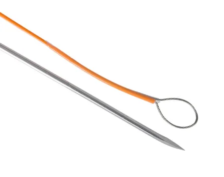
Permanent pacemaker
What and why: a pulse generator and its leads start the contraction of the atrium, the ventricleVEN-trih-kulOne of the two lower chambers of the heart, or one of the fluid-filled spaces inside the brain.Full entry or both, most often for complete heart block and slow rhythms; a pacemaker is a device that helps control abnormal heart rhythms.
Before surgery: local anesthesiaLOH-kul an-es-THEE-zhuhNumbing medicine applied to or injected into one small area, so a procedure there is not felt while the person stays awake.Full entry with monitored anesthesiaan-us-THEE-zhuhLoss of feeling or sensation, often brought on by drugs so that surgery or a procedure can be done without pain.an- withoutesthesi/o feeling, sensation-ia conditionFull entry care; fluoroscopyfloo-ROS-kuh-peeAn x-ray method that shows moving pictures of the inside of the body in real time.fluor/o glowing (from the fluorescent screen)-scopy looking, viewingFull entry , a defibrillator in the room and a pacing system analyzer, often brought by the maker's representative. The table is tilted head down to fill the vein and to prevent an air embolismEM-buh-liz-umThe blocking of a blood vessel by a mass carried there in the blood (an embolus), such as a clot, fat or an air bubble.embol/o plug-ism conditionFull entry .
Key steps (transvenous):
- A needle enters the subclavian, axillary or internal jugular veinin-TUR-nul JUG-yuh-ler vaynA large vein deep on each side of the neck that carries blood from the brain, face and neck back toward the heart.Full entry , and a guidewire is passed into the right atrium under fluoroscopy.
- A pocket for the generator is made under the skin below the collarbone.
- A sheathSHEETHA covering that wraps around a structure; in procedures, a short hollow tube left in a blood vessel to pass catheters through.Full entry and dilatorDY-lay-terAn instrument used to stretch or widen an opening, canal or passage in the body.Full entry go over the wire, and the lead, stiffened with a stylet, is advanced into the right ventricle and fixed at its apex, by a small screw or by lodging in the muscle ridges.
- The lead is tested with the analyzer through alligator cables, then sewn to the muscle; a second lead goes into the right atrium for a dual-chamber pacemaker.
- The pocket is irrigationeer-ih-GAY-shunWashing out a wound, body cavity, tube or eye with a steady flow of liquid.Full entry with an antimicrobialan-tee-my-KROH-bee-ulA drug or substance that kills or slows the growth of microorganisms such as bacteria, viruses, fungi or parasites.anti- againstmicro- smallbi/o life-al relating toFull entry solution, the generator is connected and sewn into the pocket, and the incision is closed in layers.
After surgery: the complications include bleeding into the pocket (a hematomahee-muh-TOH-muhA collection of blood, often clotted, that has leaked out of the blood vessels into the tissue.hem/o blood (here as hemat-)-oma swelling or massFull entry ), which raises the risk of infection, infection of the pocket or the leads, a lead that moves or breaks, and, rarely, a lead that punctures the heart.
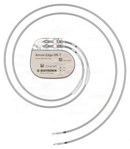

Other options: a leadless pacemaker, placed through a catheter into the right ventricle, needs no leads or pocket. Epicardial leads are fixed onto the heart through a subxiphoid incision, a small thoracotomythor-uh-KOT-uh-meeA surgical cut through the chest wall to reach the lungs, heart or other organs in the chest.thorac/o chest-tomy cutting intoFull entry or a sternotomy. Cardiac resynchronization therapy adds a lead through the coronary sinusSY-nusA hollow space in the body, most often one of the air-filled spaces in the skull bones around the nose.Full entry to pace the left ventricleLEFT VEN-trih-kulThe heart's lower left chamber, which pumps oxygen-rich blood into the aorta and out to the body.Full entry too, for heart failurehart FAYL-yerA long-term condition in which the heart cannot pump enough blood to meet the body's needs.Full entry .
Wound class: I.
Implantable cardioverter defibrillator (ICD)
An ICD watches the heart's rhythm and shockshokA life-threatening state in which blood flow is too low to bring enough oxygen to the body's tissues.Full entry it when it senses ventricular tachycardiaven-TRIK-yuh-ler tak-ih-KAR-dee-uhA fast heart rhythm that starts in the lower chambers of the heart (the ventricles), usually over 100 beats per minute.ventricul/o ventricletachy- fastcardi/o heart-ia conditionFull entry or fibrillationfib-rih-LAY-shunFast, uncoordinated twitching of muscle fibers, most often in the heart, so the muscle quivers instead of contracting together.fibr/o fiber-ation processFull entry , while a pacemaker responds when the heart fails to beat; ICDs can pace as well. The operation is much like a transvenous pacemaker: the leads go in through a vein under fluoroscopy, the generator goes in a pocket in the chest wall, and the device is tested; EP studies before and after show the rhythm and check the device.
Ablation and the Maze procedure
Catheter ablation: in the EP lab, the electrophysiologist threads a catheter from the femoralFEM-er-ulRelating to the thigh or the femur, the thigh bone.Full entry vein or artery into the heart, provokes the arrhythmia to find where it starts, and destroys that spot with radiofrequency energy or freezing (cryoablation). Radiofrequency ablation destroys tissue with the heat of high-frequency energy.
The Maze procedure treats atrial fibrillation in the OR. Lines of scar are made in the atria; electrical signals cannot cross scar, so they are forced along one path, the "maze," from the SA nodeES-AY NOHDSinoatrial node: a small cluster of cells in the right atrium that starts each heartbeat.sin/o sinusatri/o atriumFull entry to the AV nodeay-VEE nohdAtrioventricular node: a small group of cells between the upper and lower chambers of the heart that passes on the heartbeat signal.atri/o atriumventricul/o ventricle-ar relating toFull entry . Today the lines are made with cryothermal, radiofrequency or ultrasonic energy rather than cut and sewn, especially around the pulmonary veinPUL-muh-nair-ee VAYNOne of the four veins that carry oxygen-rich blood from the lungs to the left atrium of the heart.Full entry .
- Through a sternotomy: on total bypass with the heart arrested, the lesionLEE-zhunAn area of tissue that has been damaged or has changed from normal, such as a sore, wound, lump or spot.Full entry are made through an opening in the right atrium and then one in the left.
- Through a right mini-thoracotomy: the patient tilted slightly left with a roll under the right chest, femoral cannulation, and the right atrialAY-tree-ulRelating to an atrium, one of the two upper chambers of the heart.atri/o atrium-al relating toFull entry lesions made before bypass starts, the left ones after the heart is arrested.
After surgery the rhythm is watched, and it can take up to 3 months for the heart to beat normally.
Wound class (ICD, ablation and Maze): I.
16Congenital heart surgery
Children's heart surgery uses the open-heart setup scaled to the child's size, with patch material ready. Chapter 13 teaches the child under anesthesiaan-us-THEE-zhuhLoss of feeling or sensation, often brought on by drugs so that surgery or a procedure can be done without pain.an- withoutesthesi/o feeling, sensation-ia conditionFull entry , and chapter 18 the child and family as people.
Patent ductus arteriosus (PDA)
What and why: when the ductus stays open after birth, blood shuntSHUNTA passage that moves blood or other fluid from one place to another, either made by a surgeon or present in the body.Full entry from the aorta into the pulmonary arteryPUL-muh-nair-ee AR-ter-eeThe large blood vessel that carries blood low in oxygen from the right side of the heart to the lungs.Full entry , overloading the lungs and the heart, which enlarges and can fail. It is common in premature babies. A ductus is not closed when it is keeping the child alive, as with some other defects such as severe pulmonary stenosisstuh-NOH-sisNarrowing of a passage or opening in the body, such as a blood vessel, heart valve or the spinal canal.sten/o narrow-osis conditionFull entry .
Before surgery: for a newborn, the operation may be done at the bedside in the intensive care nursery, since the operation is short and the baby need not be moved; older children come to the OR. No bypass is needed. A thoracoscopic closure with clips is an option, with the open instruments ready.
Key steps (open):
- Right side down, a left posterolateral thoracotomythor-uh-KOT-uh-meeA surgical cut through the chest wall to reach the lungs, heart or other organs in the chest.thorac/o chest-tomy cutting intoFull entry through the third or fourth rib space, with a Finochietto spreader; the lung is held back with a moist pack.
- The pleuralPLOOR-ulHaving to do with the pleura, the thin two-layer membrane that covers the lungs and lines the chest wall.pleur/o pleura-al relating toFull entry over the ductus is opened between the phrenic and vagus nerveVAY-gus nervThe 10th cranial nerve, which runs from the brain stem to the chest and belly and slows the heart and helps digestion.Full entry , and the recurrent larynxLAIR-inksThe voice box: the part of the airway in the neck, above the windpipe, that holds the vocal cords.laryng/o larynxFull entry nerve is found and protected.
- The ductus is tied twice, close to the aorta and close to the pulmonary artery, or clamped with ductus clamps, divided and both ends sewn closed; in a tiny baby it may simply be tied or clipped.
- A small chest tubechest toobA tube placed through the chest wall into the space around the lung to drain air, blood or fluid.Full entry is placed and the chest is closed.
After surgery: the complications include infection, bleeding, injury to the recurrent laryngeal nerve with a paralysispuh-RAL-uh-sisLoss of the ability to move a part of the body, and sometimes loss of feeling in it.para- beside, abnormal-lysis looseningFull entry vocal cord, injury to the vagus nerve or the thoracicthuh-RAS-ikRelating to the chest, or to the middle part of the spine.thorac/o chest-ic relating toFull entry duct, and a ductus that is not fully closed.
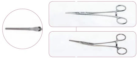
Coarctation of the aorta
What and why: a narrowing of the aorta, usually near where the ductus joined it, blocks flow to the lower body; the heart enlarges from pumping against it, and the lower body may be underdeveloped.
Setup: a cardiac set, with coarctation clamps, bulldogs and moist umbilical tape.
Key steps:
- Lateral position and a posterolateral thoracotomy; a moist lap protects the retracted lung.
- The mediastinal pleura is opened, a moist umbilical tape is passed around the aorta, and the ductus or its remnant (the ligamentum arteriosum) is tied and divided.
- The aorta is clamped above and below the narrowing, with bulldog clamps on vessels between the clamps.
- The narrowed segment is cut out and the ends are joined; interrupted sutureSOO-cherA stitch, or the thread used to stitch, that holds the edges of a wound or incision together.Full entry in a child allow growth. A tube graftGRAFTTissue or a man-made material moved into place in surgery to replace or repair a damaged part.Full entry , or a flap of the left subclavian artery, is used when the ends will not meet.
- The clamps come off, restoring flow to the lower body, and the chest is closed over a chest tube.
After surgery, the cross-clamp can starve the spinal cord of blood and cause paraplegiapair-uh-PLEE-juhParalysis of both legs and the lower part of the body.para- beside, abnormal-plegia paralysisFull entry , the most devastating complication, and adults often have high blood pressure for the first few days.
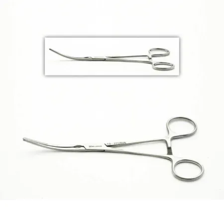
Atrial and ventricular septal defects
What and why: an atrial septal defectAY-tree-ul SEP-tul DEE-fektA hole in the wall between the two upper chambers of the heart.atri/o atrium (upper heart chamber)sept/o wall, partition-al relating toFull entry (ASDay-es-DEE (said as letters)An abbreviation with several meanings, most often autism spectrum disorder or atrial septal defect.Full entry ) is a hole between the atriumAY-tree-umOne of the two upper chambers of the heart, which receive blood coming back to the heart.atri/o atrium-um noun endingFull entry , so blood shunts from the left atrium to the right. Children often have no symptoms, but over years the right heart and the lung vessels are overloaded, with pulmonary hypertensionPUL-muh-nair-ee hy-per-TEN-shunHigh blood pressure in the arteries that carry blood from the heart to the lungs.pulmon/o lung-ary relating tohyper- abovetension pressureFull entry , heart failurehart FAYL-yerA long-term condition in which the heart cannot pump enough blood to meet the body's needs.Full entry and arrhythmiauh-RITH-mee-uhA heart rhythm that is not normal: too fast, too slow or irregular.a- withoutrhythm regular beat-ia conditionFull entry , and the shunt can reverse. Some septumSEP-tumA wall of tissue that divides two spaces or cavities, such as the two sides of the heart or nose.sept/o wall, partitionFull entry defects can be closed with a device in the catheterization lab.
Key steps (ASD):
- A median sternotomy (or a right anterolateral thoracotomy) and bypass, usually with bicaval cannulation.
- The right atrium is opened and the defect examined.
- It is closed with a running stitch or a patch of pericardiumpair-ih-KAR-dee-umThe thin, two-layered sac that surrounds the heart.peri- aroundcardi/o heart-um a structureFull entry or synthetic material; the left atrium is filled with blood to push out air before the last stitches are tied.
- Bypass ends and the chest is closed.
After surgery: the child is watched for arrhythmias; the complications include infection, bleeding, failure of a synthetic patch, a leaking mitral valveMY-trul valvThe heart valve between the left atrium and the left ventricle.Full entry and, rarely, heart block.
A ventricular septal defectven-TRIK-yuh-ler SEP-tul DEE-fektA hole in the wall between the heart's two lower chambers, present from birth.Full entry (VSD), a hole between the ventricleVEN-trih-kulOne of the two lower chambers of the heart, or one of the fluid-filled spaces inside the brain.Full entry , is one of the most common heart defects, and a large one floods the lungs at high pressure, so it is closed to prevent pulmonary hypertension. It is often reached through the right atrium and the tricuspid valvetry-KUS-pid VALVThe heart valve between the right atrium and the right ventricle.tri- threecusp point, flapFull entry and closed with a patch, the stitches placed with great care near the conductionkun-DUK-shunThe passing of something, such as an electrical signal or heat, from one place to another through a material.Full entry system, because damage causes heart block.
Tetralogy of Fallot
What and why: four defects together: a VSD, narrowing below or at the pulmonary valve, an aorta that sits over the septum and a thickened right ventriclerite VEN-trih-kulThe lower right chamber of the heart, which pumps blood to the lungs.Full entry . Oxygen-poor blood shunts from right to left, so the child is cyanosissy-uh-NOH-sisA blue or gray color of the skin, lips or nail beds.cyan/o blue-osis abnormal conditionFull entry , with spells of severe cyanosis ("tet spells"), clubbingKLUB-ingA widening and rounding of the fingertips, with the angle where the nail meets the skin flattened to 180 degrees or more.Full entry and too many red cells. A complete repair is preferred; when it must wait, a shunt from a systemicsis-TEM-ikAffecting the whole body rather than one part.Full entry artery to the pulmonary artery improves the oxygen until the child is bigger.
Key steps:
- A median sternotomy, bypass with cooling and cardioplegia.
- The right ventricle is opened over its outflowOWT-flohThe flow of a fluid out of an organ, space or vessel.Full entry tract, and the thick muscle blocking it is cut away; a fused pulmonary valve is opened.
- The VSD is closed with a patch, the stitches kept clear of the conduction system.
- If the outflow is still too narrow, a patch widens it, sometimes across the valve ring onto the pulmonary artery.
- The pressures in the pulmonary artery and right ventricle are measured to judge the repair, and bypass ends.
After surgery: for the first 24 hours, bleeding in the chest is watched closely, because the long-standing extra red cells raise its risk, along with blood collecting around the heart (tamponade) and the cardiac outputKAR-dee-ak OWT-putThe amount of blood the heart pumps out in one minute.Full entry ; the complications include infection, bleeding, a low cardiac output and, rarely, heart block.
Other repairs include a valvulotomy, which opens a narrowed pulmonary valve on bypass, and, for babies with only one working ventricle, as in hypoplastic left heart syndrome, a series of three operations ending with the Fontan procedure, which sends the blood from the vena cavaVEE-nuh KAY-vuhOne of the two large veins that return oxygen-poor blood from the body to the right atrium of the heart.Full entry straight to the lungs.
Wound class (congenitalkun-JEN-ih-tulPresent at birth.con- withgen birth, origin-al relating toFull entry heart repairs): I.
17Heart failure: support devices and transplant
When the heart is failing, devices can support it for a short time, a long time or for good, and a transplant replaces it when they cannot reverse the decline.
Intra-aortic balloon pump (IABP)
What and why: a balloon on a catheter sits in the descending thoracicthuh-RAS-ikRelating to the chest, or to the middle part of the spine.thorac/o chest-ic relating toFull entry aorta and works by counterpulsation. A console fills it with helium as the ventricleVEN-trih-kulOne of the two lower chambers of the heart, or one of the fluid-filled spaces inside the brain.Full entry relaxes, pushing blood into the coronary arteryKOR-uh-nair-ee AR-ter-eeOne of the blood vessels on the surface of the heart that bring oxygen-rich blood to the heart muscle.Full entry and the organs, and empties it just as the ventricle contracts, lowering the pressure the heart must pump against. It reduces the heart's work after an MI and helps patients who cannot come off bypass.
Key steps:
- The balloon goes in through a femoral arteryFEM-er-ul AR-ter-eeThe large artery that carries blood to the leg, running through the groin and down the thigh.Full entry , under local anesthesiaLOH-kul an-es-THEE-zhuhNumbing medicine applied to or injected into one small area, so a procedure there is not felt while the person stays awake.Full entry or at the end of heart surgery.
- A needle punctures the artery, a guidewire is passed and the needle removed; dilatorDY-lay-terAn instrument used to stretch or widen an opening, canal or passage in the body.Full entry widen the track and a sheathSHEETHA covering that wraps around a structure; in procedures, a short hollow tube left in a blood vessel to pass catheters through.Full entry goes in.
- The air is removed from the balloon with its one-way valve and a syringe.
- The balloon catheter is advanced over the wire to just below the left subclavian artery; the surgeon marks the length with a silk tie.
- Its connections are passed off the field and joined to the console.
After: complications include ischemiais-KEE-mee-uhToo little blood flow to a part of the body, so the tissue does not get enough oxygen.isch- hold back-emia blood conditionFull entry of the leg and compartment syndromekum-PART-ment SIN-drohmA buildup of pressure inside a closed group of muscles that cuts off their blood supply.Full entry , embolismEM-buh-liz-umThe blocking of a blood vessel by a mass carried there in the blood (an embolus), such as a clot, fat or an air bubble.embol/o plug-ism conditionFull entry , dissectiondy-SEK-shunA tear in the inner layer of an artery wall that lets blood split the layers apart; also, the cutting apart of tissue in surgery.Full entry of the aorta or iliacIL-ee-akHaving to do with the ilium, the large, wing-shaped upper part of the hip bone.Full entry artery, bleeding and failure of the balloon.
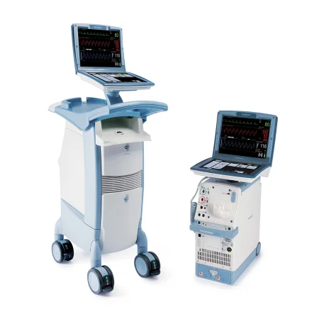
Ventricular assist device (VAD)
What and why: a pump that takes blood from a failing ventricle and sends it on, as a bridge to a transplant, as permanent destination therapy for a patient who cannot have one, or to help a patient off bypass. Devices support the left ventricleLEFT VEN-trih-kulThe heart's lower left chamber, which pumps oxygen-rich blood into the aorta and out to the body.Full entry (LVAD), the right (RVAD) or both (BiVAD). An LVAD has the pump, an inflow cannulaKAN-yoo-luhA thin tube placed into the body or a body opening to deliver or remove fluid or air.Full entry in the left ventricle's apex, an outflowOWT-flohThe flow of a fluid out of an organ, space or vessel.Full entry graftGRAFTTissue or a man-made material moved into place in surgery to replace or repair a damaged part.Full entry to the aorta and a driveline that leaves the body through the skin to its power source.
Key steps (LVAD):
- A median sternotomy, and bypass with the heart still beating.
- The atrialAY-tree-ulRelating to an atrium, one of the two upper chambers of the heart.atri/o atrium-al relating toFull entry septumSEP-tumA wall of tissue that divides two spaces or cavities, such as the two sides of the heart or nose.sept/o wall, partitionFull entry is checked for a hole, which is closed if found, so blood cannot shuntSHUNTA passage that moves blood or other fluid from one place to another, either made by a surgeon or present in the body.Full entry once the pump runs.
- The apex of the left ventricle is cored out with the device's coring knife, and pledgeted sutureSOO-cherA stitch, or the thread used to stitch, that holds the edges of a wound or incision together.Full entry , sometimes with a felt strip, are placed around the hole.
- The inflow cuff is sewn into the apex and the pump attached.
- The outflow graft is measured, a side-biting clamp is placed on the ascending aorta and the graft is sewn end to side onto it.
- The driveline is tunneled under the skin and out through the abdominal wall.
- The pump is started as bypass is withdrawn.
Bleeding, clots, infection and device failure are the main complications, and a total artificial heart is used for some patients whose two sides both fail.
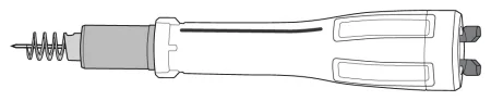
Extracorporeal membrane oxygenation (ECMO)
What and why: a pump and an oxygenator outside the body that take over the lungs' work, or the heart's and lungs', for a time, while the organs recover. Venovenous (VV) ECMO drains blood from a vein and returns it to a vein, supporting the lungs only; venoarterial (VA) ECMO returns it to an artery, supporting the heart and the lungs, for cardiogenic shockkar-dee-oh-JEN-ik SHOKA type of shock in which the heart cannot pump enough blood to supply the body's organs.cardi/o heart-genic produced byFull entry or cardiac arrestKAR-dee-ak uh-RESTThe sudden stop of the heart's pumping, so blood no longer reaches the brain and body.Full entry .
Cannulation:
- Peripheral VA, in an emergency or for cardiogenic shock: a drainage cannula in a femoralFEM-er-ulRelating to the thigh or the femur, the thigh bone.Full entry vein and a return cannula in a femoral artery. A small extra cannula is often placed to carry blood down the leg beyond the arterial cannula, because without it the leg can lose its blood supply.
- Central VA, after heart surgery: the bypass cannulas in the right atriumAY-tree-umOne of the two upper chambers of the heart, which receive blood coming back to the heart.atri/o atrium-um noun endingFull entry and aorta are moved over to the ECMO circuit; a patient who cannot be weaned from bypass can be switched to ECMO in the OR.
- In children, VA ECMO usually uses the right common carotidkuh-ROT-idHaving to do with the carotid arteries, the two large arteries on each side of the neck that carry blood to the head and brain.Full entry artery and the internal jugular veinin-TUR-nul JUG-yuh-ler vaynA large vein deep on each side of the neck that carries blood from the brain, face and neck back toward the heart.Full entry in the neck.
Setup: ECMO often starts in the ICU or the emergency department. Plan room at the bedside for the ECMO pump, a sterile table, the electrosurgeryee-LEK-troh-SUR-jer-eeUsing high-frequency electric current through a handheld tool to cut tissue or seal bleeding vessels during surgery.Full entry , a headlight and a defibrillator with external and internal paddles; everyone wears surgical attire and traffic is limited; keep the instruments sterile until an X-ray confirms the cannulas are in the right place. A continuous heparinHEP-uh-rinAn anticoagulant drug, given by injection or IV, that keeps blood clots from forming or growing.Full entry infusion prevents clots, and bleeding and clots are the most common complications.
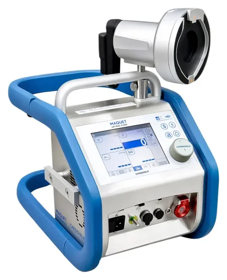
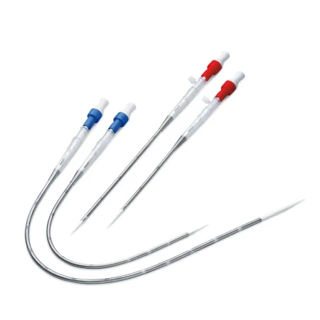
Heart transplant
What and why: a donor heart replaces a heart with end-stageEND-stayjDescribes the last stage of a long-term disease, when an organ has almost stopped working.Full entry disease, such as coronary artery diseaseKOR-uh-nair-ee AR-tuh-ree dih-ZEEZNarrowing of the arteries that supply blood to the heart muscle, most often from fatty plaque buildup.Full entry , a congenitalkun-JEN-ih-tulPresent at birth.con- withgen birth, origin-al relating toFull entry defect, valve disease or rejection of an earlier transplant. Orthotopic transplant, replacing the heart in its own place, is the usual operation. The donor and the recipient each need their own instrument setup, and the donor heart arrives cooled in saline in a transport container. Chapter 18 teaches organ donation.
Key steps (recipient):
- Supine, prepped from the chin to the knees; a median sternotomy and heparin.
- Bicaval cannulation with tapes around each vena cavaVEE-nuh KAY-vuhOne of the two large veins that return oxygen-poor blood from the body to the right atrium of the heart.Full entry ; the patient is cooled to about 32 to 34°C, then the cross-clamp goes on the aorta and the tapes are snugged.
- The aorta and pulmonary arteryPUL-muh-nair-ee AR-ter-eeThe large blood vessel that carries blood low in oxygen from the right side of the heart to the lungs.Full entry are divided above their valves, and the atria are cut so the back walls of the recipient's atria remain; the heart is removed.
- On the back table, in a basin, the surgeon trims the donor heart.
- The donor heart is placed in the pericardiumpair-ih-KAR-dee-umThe thin, two-layered sac that surrounds the heart.peri- aroundcardi/o heart-um a structureFull entry , and the left atria are joined first; then the venae cavae (the bicaval technique, which causes fewer rhythm and tricuspid problems than joining the right atria), the pulmonary artery and the aorta.
- Air is removed, the cross-clamp comes off, and a single shockshokA life-threatening state in which blood flow is too low to bring enough oxygen to the body's tissues.Full entry usually starts the heart; pacing wires and chest tubechest toobA tube placed through the chest wall into the space around the lung to drain air, blood or fluid.Full entry are placed and the chest is closed.
A combined heart-lung transplant also spares the donor's SA nodeES-AY NOHDSinoatrial node: a small cluster of cells in the right atrium that starts each heartbeat.sin/o sinusatri/o atriumFull entry and the recipient's phrenic, vagus and recurrent larynxLAIR-inksThe voice box: the part of the airway in the neck, above the windpipe, that holds the vocal cords.laryng/o larynxFull entry nerves. Afterward the patient takes immunosuppressantim-yoo-noh-suh-PRES-untA drug that lowers the activity of the immune system.immun/o immunesuppress hold down-ant agentFull entry for life, with regular biopsies of the myocardiummy-oh-KAR-dee-umThe thick middle layer of the heart wall, made of heart muscle that contracts to pump blood.my/o musclecardi/o heart-um structureFull entry to look for rejection; rejection and infection are the main complications.
Wound class (support devices and transplant): I.
18Closing the chest and after surgery
Closing a sternotomy
- Before the chest is closed, the pacing wires and usually two chest tubechest toobA tube placed through the chest wall into the space around the lung to drain air, blood or fluid.Full entry are in place.
- The surgeon places six to eight stainless steel wires through the sternal edges with a heavy sternal needle holder. As each wire is passed, hold its free ends so they stay under control and off anything unsterile.
- The holes are checked for bleeding, and the wires are tightened and twisted to bring the halves of the sternum together, cut with wire cutters, and their twisted ends buried in the periosteum. Some surgeons use small metal crimpers instead of twisting; a frail sternum may get extra wires running along each edge, bands or plates.
- The fasciaFASH-ee-uhA sheet or band of tough connective tissue that covers and separates muscles and organs.fasci/o bandFull entry over the sternum, the linea alba, the fat and the skin are closed. If the skin is stapled, a staple remover goes with the patient.
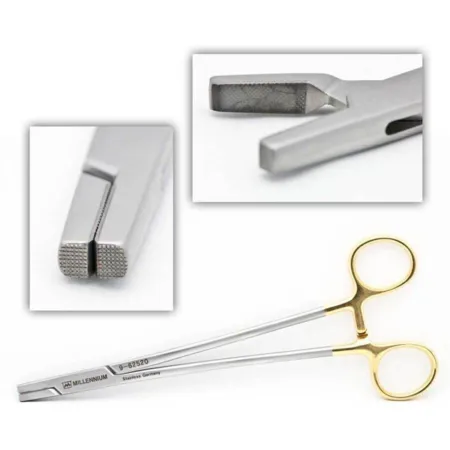
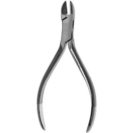
Before the patient leaves, the circulator phones a handoff reportHAND-off ree-PORTThe passing of information and responsibility for a client's care from one nurse or team to another.Full entry to the intensive care unit, covering, among other things, the graftGRAFTTissue or a man-made material moved into place in surgery to replace or repair a damaged part.Full entry used, the cross-clamp and pump times, the blood loss, the blood and drugs given and whether the heart is being paced; chapter 16 teaches handoffs.
After heart surgery
After heart surgery, patients go to a cardiovascular intensive care unit, where the blood pressure, the heart rhythm, the chest tube drainage and the temperature are watched closely. The breathing tube may come out within a few hours or stay until the patient can breathe alone. Many patients go home after about 3 to 7 days, and a sternotomy takes about 4 to 12 weeks to recover from, compared with 1 to 2 weeks after a minimally invasive operation.
- Bleeding and tamponade: chest tube drainage of more than 100 mL an hour is investigated, and the surgeon is told before the patient leaves the room; the chest may have to be reopened to stop bleeding. Sudden bleeding that causes tamponade needs a fast, organized response, through the chest tubes or by reopening the chest. This is one reason the sterile setup stays intact until the patient has left the room.
- arrhythmiauh-RITH-mee-uhA heart rhythm that is not normal: too fast, too slow or irregular.a- withoutrhythm regular beat-ia conditionFull entry : atrial fibrillationAY-tree-ul fib-rih-LAY-shunAn irregular heart rhythm in which the upper chambers of the heart quiver instead of beating in an organized way.atri/o atrium (upper heart chamber)fibrill/o small fibers, quivering-ation processFull entry is common after bypass surgery, and beta blockerBAY-tuh BLOK-erA drug that blocks the effects of adrenaline on beta receptors, slowing the heart rate and lowering blood pressure.Full entry or amiodaroneam-ee-OH-duh-roneA drug used to treat and prevent serious abnormal heart rhythms.Full entry help prevent it; the pacing wires treat slow rhythms.
- Sternal wound infection (mediastinitis, an organ or space SSIes-es-EYE (said as letters)An abbreviation that can mean surgical site infection or sliding scale insulin.Full entry from chapter 11) can mean a return to the OR to clean the wound and cover it with a muscle flap, an operation that is Class IV because the infection is already present. Its risks include diabetes, obesity, COPDsee-oh-pee-dee (said as letters)Chronic obstructive pulmonary disease: a long-term lung disease that makes it hard to move air out of the lungs.chronic long-lastingobstructive blockingpulmon/o lung-ary relating toFull entry , smoking, use of both mammary arteries, a balloon pump, a long operation and a return for bleeding; blood glucose is kept below 200 mg/dL.
- Other complications: stroke, renal failureREE-nul FAYL-yerLoss of the kidneys' ability to filter waste and extra fluid from the blood.ren kidney-al relating toFull entry , a long time on the mechanical ventilationmeh-KAN-ih-kul ven-tuh-LAY-shunBreathing support from a machine, a ventilator, that pushes air into the lungs through a tube or a mask.Full entry , pneumonianoo-MOHN-yuhAn infection of one or both lungs in which the air sacs fill with fluid or pus.pneum/o lung, air-ia conditionFull entry , clots in the legs and the lungs, infection and death.
- Teaching: a patient with a mechanical valve needs to understand lifelong anticoagulationan-tee-koh-ag-yoo-LAY-shunThe use of drugs that slow the blood's clotting, to prevent or treat blood clots.anti- againstcoagul clotting-ation processFull entry and the signs of bleeding to report.
After lung surgery
The priorities are breathing and pain control. The patient breathes deeply, coughs, uses the incentive spirometer every 1 to 2 hours while awake, and gets up on the day of surgery; some go home on oxygen. An enhanced recovery (ERAS) pathway moves the chest tube to water seal after a short time on suction. An air leak or a lung infection can delay going home.
What to take from this chapter
- Chest surgery is planned around negative pressure: an opened chest collapses the lung, so it is closed over chest tubes and a drainage unit kept upright and below the chest. If a tube comes out, petroleum gauze covers the hole at once.
- Most lung operations are now VATS, with the patient lateral on a double-lumen tube and the open thoracotomy set ready. The bronchus is opened under suction, lung staple lines are tested for air leaks under warm saline, and every lymph node is labeled by its station.
- A heart field runs on two back tables and three Mayos, a separate setup and light for the leg, clean instruments free of calcium and clot, cold and warm solutions, and every drug labeled, heparin and papaverine above all.
- Bypass drains venous blood to the heart-lung machine, which pumps it through the oxygenator and back into the aorta. Heparin and an adequate ACT come first; the cannulas go in arterial first; the cross-clamp isolates the heart, and potassium cardioplegia, antegrade or retrograde, stops it in diastole. Watch the lines for air.
- Coming off bypass: de-air, remove the clamp, defibrillate if needed, place pacing wires, wean, decannulate, then protamine, with the pump suckers turned off as it starts.
- CABG joins the aorta to a coronary artery beyond the blockage; the LIMA to the LAD is the first choice, and OPCAB works on the beating heart with stabilizers, bypass on standby. Valves are handled little, verified, and, if biological, rinsed by the maker's instructions and kept moist.
- Arch surgery cools the patient to about 18°C, stops the circulation and perfuses the brain; rhythm devices go in through a vein under fluoroscopy and are tested with an analyzer; congenital repairs close shunts and open narrowings, guarding the conduction system and the nerves.
- The IABP inflates in diastole and deflates before systole; a VAD bridges to transplant or replaces it; VA ECMO supports heart and lungs, VV ECMO the lungs. After heart surgery, watch for bleeding over 100 mL an hour and tamponade, and keep the setup sterile until the patient leaves.
Check yourself6 quick questions
Free for everyone. Pick an answer for each, then check them all at once.
In the ORWhere you'll use this
The chapter's whole question pool in sets of 10, with the answer explained after each one.
Part of the educators programThe chapter's words on spaced cards: the ones you miss come back sooner.
Part of the educators programGiven by your class's teacher. Each class draws its own 25 from the chapter's pool.
Part of the educators program
Suggest a fix
Something wrong, unclear or out of date in this chapter? Tell us and we will fix it for everyone. Your email is only used to reply.