Sutures, needles and supplies
The sutures, needles, sponges, drains and other supplies you open, prepare and pass on every case, and the reason behind the way each one is handled.
1Why it matters
Every operation ends the same way: something has to hold the tissue together while it heals, and most of the time that something is suture. Suture is any strand used to sew tissue together; bringing two cut edges back together this way is called approximation. A strand tied around a vessel or duct to close it is a ligature, or tie, and tying it off is called ligating.
The surgeon chooses the suture. Your job is to know what comes next, read a packet in a second, open only what the case needs, load each needle ready to use, and get every needle back.
Suture needles are also the sharps that hurt surgical teams most. In one large study of sharps injuries in surgery, suture needles caused more than four in ten, and surgical technologists were most often hurt while passing sharps, taking them apart or throwing them away.
Sponges, drains, drapes, blades, staplers, syringes and suction tips arrive on your back table too, and each has a right way to be handled. Counting them is in chapters 28 and 29, dressings in chapter 30, electrosurgery safety in chapter 20, and drugs on the field in chapter 12.
2The words of suture
Suture has its own vocabulary, and AST's Core Curriculum expects you to know it.
How a strand is built
- Monofilament: one solid strand. It glides through tissue and gives germs nowhere to hide, but it is stiffer and its knots slip more easily.
- Multifilament: many fine fibers twisted or braided into one strand. It is soft and ties firm knots, but the spaces between its fibers can hold fluid and bacteria.
- Coated: a covering, usually on a braided strand, so it slides more easily and frays less. Some coatings carry an antibacterial agent. An uncoated strand has none.
- Dyed or undyed: a strand may be colored with an approved dye, as violet polyglactin and blue polypropylene are, or left undyed, clear or in its natural color.
What makes a good suture
- Tensile strength: how much pull a strand takes before it breaks. A suture should be about as strong as the tissue it holds.
- Uniform size: a good strand has the same diameter all along its length, because a thin spot is a weak spot.
- Knot security: how well a knot holds without slipping.
- Memory: a strand's habit of springing back into its packet coils. High memory means tangles and knots that loosen. Monofilaments have the most; silk and braided polyester the least.
- Pliability: how easily a strand bends and handles. Silk is the classic soft suture.
- Capillarity, or wicking: soaking up fluid and carrying it along the strand. Braided strands wick most, and wicking can carry bacteria along the suture line.
- Tissue drag: friction as the strand is pulled through tissue, measured as its coefficient of friction. Coating lowers it, though a very slippery strand lets knots loosen.
- Elasticity: stretching and coming back, so a suture line can give a little as tissue swells.
- Plasticity: stretching and keeping the new shape. It helps knots stay secure; polypropylene has high plasticity.
- Tissue reaction (bioactivity): the body treats almost every suture as a foreign body and reacts with inflammation. A suture that causes little reaction is inert. Steel, polypropylene and nylon are among the most inert. Gut causes a strong reaction, and silk the most of the nonabsorbable sutures.
Absorbable and nonabsorbable
- Absorbable suture is broken down by the body. It loses its strength first, usually within weeks, then slowly disappears. Ideally it lasts as long as the tissue needs support.
- Natural absorbable suture (surgical gut) is digested by enzymes from the body's cells. Synthetic absorbable suture is broken down by water in the tissues, called hydrolysis, which is more predictable and causes less inflammation.
- Nonabsorbable suture stays. The body walls it off in fibrous tissue, called encapsulation, or it is taken out later, as skin sutures often are.
- Infection speeds up the breakdown. Most sutures, gut among them, break down very quickly in infected tissue, one reason an actively infected wound is not sutured until the infection is treated.
3Suture size
Size is a strand's diameter. In the United States, every suture needs FDA approval and has to meet the standards of the United States Pharmacopeia (USP), which cover size, tensile strength and sterility. Because of the USP standard, a 3-0 strand is the same thickness in silk, nylon or polyglactin. The numbers run two ways from 0:
- Below 0, more zeros mean a finer strand. 2-0 is finer than 0, and 4-0 is finer than 3-0. The finest go down to 11-0 and even 12-0. Size 11-0, used in microsurgery, is light enough to float in the air.
- Above 0, bigger numbers mean a thicker strand. Size 2 is heavier than size 1. The heaviest, size 5, is about as thick as ordinary string.
Finer strands are weaker, so the surgeon uses the finest one that will hold: it does less damage, makes smaller knots and leaves less foreign material. Sizes 0 to 4-0 cover most general surgery. As a rough guide, though each surgeon's preference card has the final word:
- 1 and 0: abdominal fascia and orthopedic wounds, where the pull is greatest.
- 2-0 and 3-0: most stick ties. Ties from a reel are usually 2-0, 3-0 or 4-0.
- 3-0 and 4-0: most subcuticular skin closures.
- 4-0 and 5-0: joining the aorta; 4-0 also closes the dura.
- 6-0 and 7-0: small arteries, such as the coronary and carotid arteries.
- 8-0 to 11-0: microvascular and eye surgery.
Stainless steel wire was long sized by the Brown and Sharpe (B&S) gauge, where a bigger number means thinner wire. Steel now has USP sizes too, but some surgeons still ask for wire by gauge, and the package prints it.
More zeros, finer strand. 5-0 is far finer than 0, and size 2 is thicker than 0.
4Suture materials
Sutures fall into four groups: natural or synthetic, and absorbable or nonabsorbable. Learn the groups first. Trade names differ by company, and most facilities buy most of their suture from one company, so learn its products.
Natural absorbable: surgical gut
- Surgical gut is collagen from the intestines of sheep or cattle, the only natural absorbable suture. Synthetic sutures have largely replaced it, but it still suits fast-healing tissue such as the lining of the mouth.
- Plain gut is untreated. It loses its strength within about 7 to 10 days, is gone in about 70 days, and causes a strong inflammatory reaction.
- Chromic gut is treated with chromium salts, so it reacts less and lasts longer: more than half its strength at 7 to 10 days, some up to 3 weeks, fully absorbed in about 90 days.
- Where gut is not used: plain gut is not used in heart, vessel or brain and nerve surgery, chromic gut is kept out of the skin, where its reaction can scar and invite infection, and any gut breaks down fast in an infected wound.
- Gut is packed in an alcohol solution that can catch fire on the field, so open it away from the incision and use it soon. If it has stiffened, dip it in room-temperature saline for a few seconds just before use. Never soak or heat it, and handle it as little as you can.
Synthetic absorbable
These polymers break down by hydrolysis at a steady rate with little reaction. Keep them dry: moisture weakens them, so they are never dipped.
- Polyglactin 910 (Vicryl) and polyglycolic acid (Dexon): braided and easy to handle. They hold for about 2 to 3 weeks, for soft tissue, ties and many layers in general and gynecologic surgery. An antibacterial version that carries triclosan (Vicryl Plus) is also made.
- Poliglecaprone 25 (Monocryl): a smooth monofilament that loses about half its strength in a week and nearly all by 3 weeks. It is used for buried stitches, such as a subcuticular skin closure, and is not recommended for fascia or other tissue under strain.
- Polydioxanone (PDS) and polyglyconate (Maxon): monofilaments that hold much longer, about half their strength at 4 weeks, a common choice for fascia.
- Fast-absorbing versions, such as polyglactin 910 Rapide, lose most of their strength in a week and suit skin.
Natural nonabsorbable: silk
Silk is spun by silkworms, braided or twisted, and treated to wick less. It handles beautifully and its knots hold, so it is a favorite for ties and for bowel and vessels. It loses strength when wet, and buried silk slowly loses its strength over about a year. As a braided strand, it is a poor choice in a contaminated wound. Cotton is another natural nonabsorbable suture, rarely used now.
Synthetic nonabsorbable
- Nylon (Ethilon): strong and nearly inert. Monofilament nylon is a common skin suture, and its finest sizes, down to 11-0, serve eye surgery and microsurgery. Its knots slip, so surgeons tie extra throws.
- Polypropylene (Prolene): a smooth, very inert monofilament that can be used in the presence of infection, a mainstay of vascular and plastic surgery. High memory makes it springy in larger sizes.
- Polyester (Ethibond, Mersilene): braided and very strong, with little reaction, for heart valves, grafts and fascia.
- Stainless steel: very strong and among the most inert, for bone, tendon and jaw surgery. It kinks, can tear out of tissue, and its barbed ends can damage tissue and gloves. It is never bent before use.
- ePTFE (expanded polytetrafluoroethylene): a nonabsorbable monofilament used in heart surgery, for example to replace the cords that hold the mitral valve.
Barbed and antibacterial sutures
Barbed suture has tiny barbs that grip tissue in one direction, so the surgeon can close without knots and the strand does not slide back. It comes in absorbable and nonabsorbable materials. Antibacterial sutures carry an agent such as triclosan in their coating to help prevent infection.
5Choosing a suture
The surgeon chooses, and the choice is on the preference card. It rests on the type of procedure, the tissue, the state of the wound and of the patient, and the surgeon's own experience. Knowing why lets you predict the next request and catch a mistake.
- Healing speed. Mucosa, the subcutaneous layer and other quick-healing tissue can take absorbable suture. Fascia, tendon and ligament carry heavy loads, and fascia heals slowly, so they need nonabsorbable suture, or a slowly absorbed one, in a larger size.
- Critical tissue. The heart and blood vessels get nonabsorbable suture.
- Wound status. An actively infected wound is not sutured. Where the risk of infection is high, because of the wound's class or the patient's condition, the surgeon wants a strong suture line that resists absorption: nonabsorbable suture, or an absorbable one that lasts a long time. Monofilaments are preferred, because braided strands wick germs. A grossly contaminated wound, such as one from a traumatic injury, may be left open to heal by second intention (see First, second and third intention, below).
- The urinary tract. Suture in contact with urine can seed stones, so absorbable suture is used.
- Scarring. For a cosmetic closure of the face and other exposed skin, the surgeon picks an inert suture, usually a monofilament, which does the least injury as it passes through.
- The patient. Age and illness such as diabetes, lung disease or long-term steroid use slow healing and change the choice.
The surgeon picks the suture. You read the card, ask when the plan changes, and have the next one ready before anyone asks.
6The suture packet and its label
Suture comes in dispenser boxes of one to three dozen packets. Each packet is two layers: a sterile primary packet of foil or paper, which may hold fluid, sealed inside a peel-open overwrap whose outside is not sterile. A packet stays sterile while it is whole, and some carry an expiration date because the suture itself can break down. Good packaging does three jobs: it keeps the suture sterile, it protects the strand and needle from damage, and it lets the suture reach the field and come out of the packet quickly without tangling.
Boxes and packets are color coded by material, but the colors are not the same at every company, so always read the label.
Suture boxes are kept on carts or racks in a supply area, or in closed cabinets in the OR, and a large facility may have a room just for suture. Suture carts are never brought into the operating room, where blood and body fluids could soil them.
Reading the label
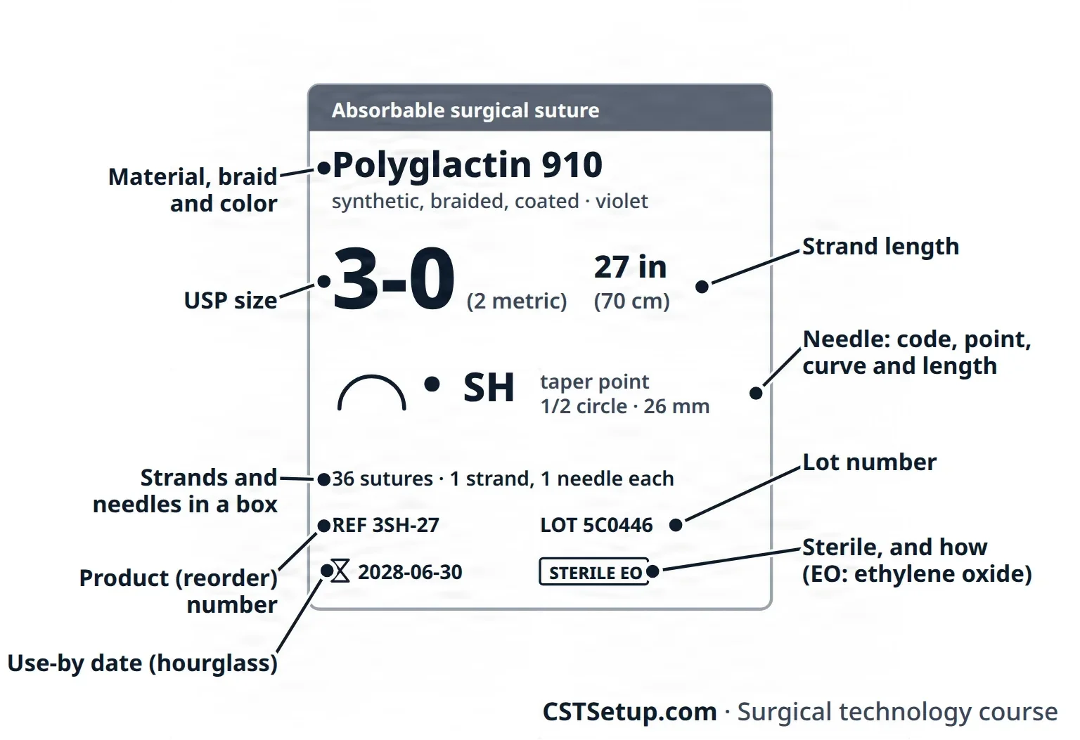
Find the size, the material, and the type and size of the needle first.
- Material, by name, with the strand's color.
- Size: the USP size, often with its metric diameter.
- Needle: a silhouette of its shape, the number of needles, and the maker's needle code.
- Strand length and number of strands.
- Product (reorder) number, lot number and expiration date.
Suture styles
- Pre-cut strands: several needleless lengths, used as free ties, from about 12 to 60 inches.
- Reels: one long strand, often 54 inches, on a spool for many ties in a row. Reels show on x-ray, and many facilities count them because they are easily lost in a wound. The holes in the side of the reel show its size.
- Single-armed: one needle swaged to one end.
- Double-armed: a needle at each end, for joining vessels and other round structures, sewing from the middle out both ways.
- Multi-strand packs: several needle-and-strand sutures in one packet, for a row of interrupted stitches. They replaced the old way of threading eyed needles one after another (rapid-sequence suturing), but they put more needles and more paper on the field, which raises the risk of a lost needle and the cost.
- Control release (detachable, pop-off): the needle comes off with a straight tug of the needle holder, so many interrupted stitches can be placed quickly, as in an anastomosis. Other multi-strand packs have needles that are cut off with suture scissors.
- Looped: the strand comes as a loop, for a running stitch that is tied to itself. The label says when a suture is looped.
- Barbed and knotless: barbed suture holds without knots (see Barbed and antibacterial sutures above), and its label says so.
7Opening and preparing suture
Open only the suture the case needs. Leftover packets cannot be sterilized again, and every extra needle is one more to count.
- Check each box against the preference card: material, size, needle and length.
- The circulator peels the overwrap and flips the primary packet onto the field, or holds it for you to lift out. The sealed edge is the line between sterile and non-sterile.
- Tear the primary packet at the notch near its sealed edge, or unfold it if it comes ready to load.
- Count multi-needle packets with the circulator as you open them. Your facility's policy says whether they are opened and counted before the incision.
- Arrange packets in the order the surgeon will use them, with the sharps grouped near the sharps container.
To take out a tie, hold the packet and draw the strand out by its loose end. Take out packet kinks by holding both ends and gently pulling your hands apart. Never strip a braided strand through your gloved fingers, which frays it, and never test it with a sharp jerk.
To make ties from a full-length strand, rest the coil across one hand and draw the loose end out gently until the strand is uncoiled. Then take an end in each hand and pull the middle out into thirds before cutting.
For a suture with a needle, grip the needle with the needle holder first, then draw the strand out without tugging on the swage. To straighten it, hold the strand an inch or two from the needle and pull gently on the far end. Nylon can be drawn gently between gloved fingers to take out its memory. Straighten a control-release suture with care, or the needle pops off and the suture is wasted.
Too long a strand drags below the field; too short is hard to tie. As a guide, a running suture is about 24 inches, an interrupted stitch 12 to 14 inches, and a tie for a deep vessel 24 to 30 inches.
Rules for handling suture
- Never wet a synthetic absorbable, and never soak or heat gut.
- Never crush a strand with an instrument. A damaged monofilament can look fine and still break after surgery.
- Never bend steel wire.
- Empty packets and used ties go in the suture bag on the side of the Mayo stand away from the wound, never in the kick bucket, where a needle could hide among the sponges.
8Needles
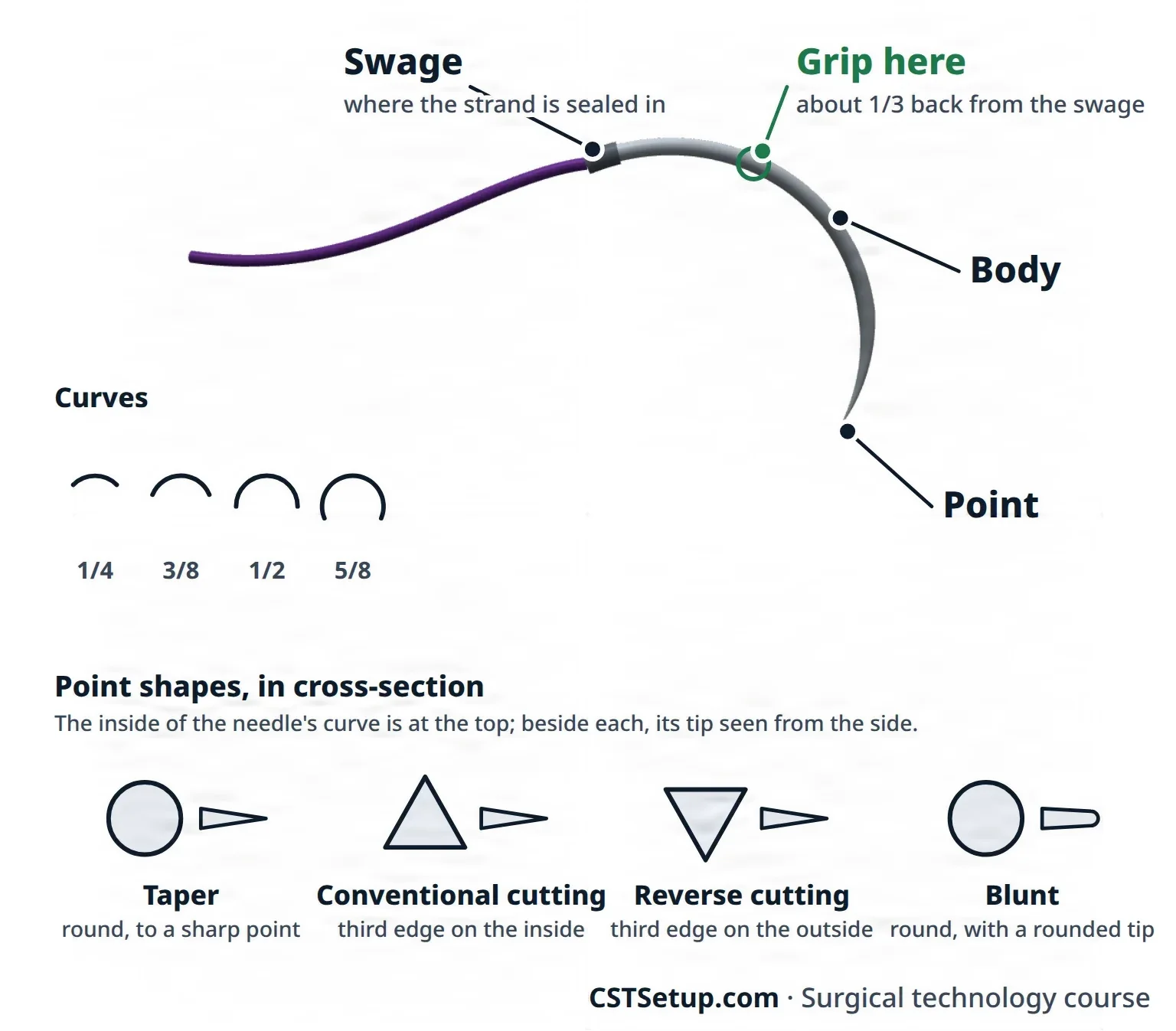
Surgical needles are made of high-grade steel alloy. Every suture needle has three parts: the eye or swage, where the strand attaches; the body, which the needle holder grips; and the point.
Swaged and eyed needles
Nearly all suture today is swaged: the strand is sealed into the hollow end of the needle at the factory. A single strand follows the needle and fills the hole, so the tissue is barely torn. That is why a swaged needle is called atraumatic. Eyed needles are rare today, but you still need to know them:
- A closed-eye needle is threaded by hand like a sewing needle, so a double strand passes through the tissue and tears it more.
- A French-eye (spring-eye) needle has a slit above the eye. The strand is pulled down over the slit until it snaps in. Never pull the strand through the eye once it is set: that strips the strand and can break it.
Most scrubs never thread a needle, but learn how in case you must. Pass the strand through the eye from the inside of the needle's curve to the outside, which keeps the short end from slipping back out. Leave a short end of about 3 to 4 inches on a 24-inch running suture, or 2 to 3 inches on a 12- to 14-inch interrupted stitch; one reference leaves about a third of the strand. Before passing it, lay both ends between the tips of the needle holder's jaws so the strand cannot back out of the eye. In heart valve surgery, keep French-eye needles ready in case the surgeon must place a suture again after its needles were cut off.
Shape and size
A straight needle, such as a Keith needle, is used by hand, with no needle holder. Curved needles are named for the part of a circle they make: 1/4, 3/8, 1/2 or 5/8 circle. A compound curve changes its curve along its length, for delicate work such as the eye. The deeper and tighter the space, the more curve the surgeon needs. A needle's size is its length and the diameter of its body: fine needles for microsurgery, very heavy ones for closing the sternum. Each maker codes its needles by size and shape, and you learn your surgeons' codes by working with them.
Point
- Taper: a round body narrowing to a sharp point that pierces and spreads tissue without cutting. The most used point, for soft tissue such as bowel, vessels, peritoneum, muscle, fat and dura.
- Conventional cutting: a triangular point with its third cutting edge on the inside of the curve, facing the wound edge, for tough tissue such as skin, tendon and joint capsule.
- Reverse cutting: the third edge is on the outside of the curve, away from the wound edge, so the stitch is less likely to tear out. Stronger than conventional cutting, and a common skin needle.
- Taper-cut: a cutting tip on a round body, for dense fascia, tendon and periosteum.
- Side-cutting (spatula): flat on top and bottom, cutting at the sides, to split thin layers of the eye without diving deep.
- Blunt: a rounded tip that pushes between tissue fibers. Used for friable organs such as the liver and kidney, and now recommended for fascia and muscle because it greatly cuts needlestick injuries.
Taper points for soft tissue, cutting points for tough tissue, blunt points for friable organs and fascia.
9Loading and passing the needle
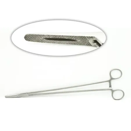
Choose a needle holder whose length suits the depth of the wound and whose jaws suit the needle: fine for fine, heavy for heavy. Chapter 24 covers the needle holders themselves.
Loading
- Load the needle while it is still in the packet, using the packet to steady it. Never set a needle in place with your fingers.
- Grip the needle with the tips of the jaws, set square (at a right angle) to them, about one-third of the way back from the swage; anywhere from one-third to one-half is acceptable. Tough tissue may call for a grip nearer the middle, and the surgeon asks when another angle is wanted.
- Never clamp the swage. It weakens the join and puts the needle holder too far back for good balance.
- Draw the strand out gently, without pulling on the needle, and straighten it.
On a needle holder with curved jaws, such as a Heaney, load the needle so the angled tip of the jaws points toward the swage, not toward the needle point. A locking micro needle holder, such as a Castroviejo, opens with a gentle squeeze on its bowed handles, takes the needle, and locks again with another squeeze. Some have no lock; keep light pressure on their handles as you pass them so the needle cannot drop.
The neutral zone
A neutral zone is a set spot on the field, such as a magnetic pad, an instrument mat or a basin, where sharps are put down and picked up, so two people never hold the same sharp at once. AST recommends it for every operation, and AORN and the American College of Surgeons recommend it too. Agree on its place with the surgeon before the incision, say "sharp" each time you place one, and take your hand completely away. Lay the needle holder so it can be picked up by the handle without turning it, and keep only one sharp in the zone at a time.
The surgeon puts the used needle holder back in the zone; never pull a loaded one out of the surgeon's hand. When the surgeon cannot use the zone, for example under a microscope, say each pass out loud and say when your hand is out of the way. A surgeon who is not using the zone turns the needle with forceps toward the box lock of the needle holder before setting it down.
Some references still teach handing every loaded needle holder straight into the surgeon's palm as the usual pass, and some teams still work that way.
Passing
- Load and present the needle holder so the surgeon can use it without shifting it or looking up: needle point up, on the surgeon's thumb side, and aimed back toward the surgeon. When it is handed directly, as when the neutral zone cannot be used, place it firmly in the surgeon's palm.
- Hold the free end of the strand, or lay it over the back of your hand, so it does not tangle in the surgeon's hand or slide off the field.
- A right-handed surgeon drives the needle clockwise and a left-handed surgeon counterclockwise, so load it as a mirror image for the left hand. A backhand stitch reverses the direction. Where the surgeon stands, across from you or beside you, also changes how you hold it out, so practice both, with either hand.
- Hand tissue forceps to the surgeon's other hand with the needle holder. The surgeon uses them to steady the tissue and to pull the needle through and reposition it, so the needle is moved with instruments, never fingers. Choose them for the tissue: forceps with teeth for skin, tendon and fascia; smooth forceps for mucosa, organs and any tissue that bleeds easily; and vascular forceps, whose scored tips grip without puncturing, for vessels.
AST's 2017 sharps guideline has the scrub pass forceps with teeth with every needle holder.
Every needle comes back
- Pass on an exchange basis: a loaded needle holder goes out as the used one comes back, so you always know where every needle is.
- Look at each returned needle: it must be whole. A broken or missing needle is reported to the surgeon and the circulator at once. Every piece is found and accounted for, then kept in the sharps container or passed off to the circulator.
- Used needles go straight onto the magnetic pad or into the needle counter. A needle is never loose: it is on a needle holder, in its packet or in the counter.
- Load needles just before they are needed, so no bare needle waits on the Mayo stand or back table. In a trauma case, holders may be loaded early with the needle tips still tucked in the packet.
Some references still teach keeping a loaded needle holder ready at all times, or loading ahead of time: stick ties when heavy bleeding is expected, and in a routine case the sutures, lined up in order of use.
- Before the surgeon ties, cut the needle off or use a control-release suture, so no needle swings free during the knot.
- Keep the second needle of a double-armed suture in sight, for example on a rubber-shod hemostat, so it does not snag the drapes.
- Pick up a needle dropped on the field with a needle holder, never fingers. One on the floor is the circulator's to pick up the same way.
- After the case: once the skin is closed and the dressing is on, look over the field for any loose sharp before the drapes come off. Needles and blades go into the small sharps container you set up on the back table, never overfilled so it closes tight, and that whole container goes into the room's large puncture-resistant sharps container.
Never touch a suture needle with your fingers, and never pass a sharp hand to hand when the neutral zone can be used. Suture needles cause more sharps injuries in surgery than any other device.
10How suture is used
Halsted's principles
More than 100 years ago, the Johns Hopkins surgeon William Halsted set out rules for treating tissue that still shape good surgery:
- Handle tissue gently.
- Control bleeding (hemostasis).
- Keep the blood supply.
- Keep tissue moist.
- Keep strict sterile technique.
- Keep tension off the tissue.
- Close dead space, the gaps between layers where blood and serum can pool.
Halsted also favored fine sutures placed close together, which is why surgeons still use the finest strand that will hold.
How the team exposes and holds tissue during the case, with retraction, traction and countertraction, is taught in chapter 29.
Stitches
- Continuous (running): one strand, tied only at the start and end. Quick, with tension spread evenly, but a break anywhere can loosen the whole line. It suits layers with little tension that need a tight seal, such as the peritoneum or a vessel.
- Locking running: each pass goes under the loop before it, for extra strength and better control of bleeding. The person following the suture keeps light tension on the strand.
- Interrupted: each stitch placed, tied and cut on its own. Slower but strongest: if one knot fails, the rest hold, and an infection is less able to travel along the line. Used for bowel and vessel joins. Interrupted stitches may be simple, figure-of-eight, vertical or horizontal mattress, or buried; buried and mattress stitches add security to the line.
- Subcuticular: a running stitch inside the dermis, just under the epidermis, for a neat closure with few marks, usually absorbable so nothing needs taking out.
- Purse-string: a running stitch in a circle around an opening, drawn tight like a drawstring, as around the base of the appendix or around a tube.
Ties and ligatures
- Free tie: a pre-cut strand placed in the surgeon's open hand to tie around a clamped vessel.
- Reel: the whole reel goes into the palm with 2 or 3 inches of strand pulled free, then back to its place; a loose reel can fall into the wound.
- Tie on a passer: for a deep vessel, the end of a tie is clamped in the tip of a curved or right-angle clamp so the surgeon can pass it around the vessel. Lay the strand over the back of your hand as you pass it.
- Suture ligature (stick tie): a needle and strand on a needle holder. The needle passes through the vessel before the tie, so the tie cannot slip off a large vessel.
- Tag: a hemostat on the ends of a suture, to hold it for later or pull on it. Traction (stay) sutures hold tissue out of the way.
- Drain stitch: a nonabsorbable stitch, such as nylon, that holds a drain to the skin so it stays in place.
Suturing through a scope
In laparoscopic and robotic surgery, the surgeon sutures through ports. In extracorporeal suturing the knot is tied outside the body and pushed down the port to the tissue; in intracorporeal suturing it is tied inside with instruments. Endoscopic suture devices help: a pre-tied loop on a long plastic pusher (such as the Endoloop) slips over a pedicle and is cinched tight, and a stitching device (such as the Endo Stitch) passes its needle between two jaws and doubles as a knot pusher. A port closure device (such as the Carter-Thomason) can close the fascia at a port site. Chapter 21 covers the scope and ports themselves.
Retention sutures and other helpers
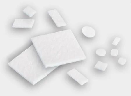
Retention sutures are heavy, nonabsorbable, interrupted sutures through all layers of the abdominal wall, a few centimeters out from the incision. They take strain off a wound at risk of splitting open (dehiscence) or of the bowel pushing through (evisceration). Where they cross the skin, they run through bolsters of rubber or plastic tubing, or tie over an adjustable bridge, so they do not cut in. A pledget, a small square of felt on a stitch, keeps it from cutting through a fragile vessel wall. A tendon suture may be tied over a button for the same reason, and a suture anchor, a small device of metal or absorbable material (such as a screw) set into bone, holds a suture that fixes tendon or ligament to the bone.
Cutting suture
Use suture scissors, never tissue scissors. Steady the blades with your index finger, open them slightly, slide the tips down to the knot and turn them to about a 45-degree angle, which leaves a short tail. When you can, keep the strand and knot in view so you cut nothing else. Leave the tail the surgeon wants: long enough that the knot holds, short enough to leave little foreign material. Monofilament knots slip more easily, so their tails are usually left a little longer. Cut too short, and you may cut the knot itself; then the stitch has to come out and be placed again.
When you must cut while doing other tasks, you can palm the scissors. Pick cut ends out of the wound area so they cannot fall in. To take a stitch out, lift it with a straight clamp, cut it below the knot on one side only, and pull it through; cut both sides, and the strand can be drawn into the wound, where it may not be found without opening the wound. A straight hemostat, such as a Crile or Kelly, is the best tool for pulling out the remains of a cut suture. When the surgeon removes old sutures from an earlier operation, often buried in scar, a fine straight hemostat works best: lay a folded towel on the field to hold the pieces so none drop back into the wound, then take the towel off the field.
First, second and third intention
Surgical wounds are also named by how they are closed, which decides how they heal. How the body heals a wound is taught in chapter 11.
- First intention (primary): a clean wound sutured closed, so its cut edges touch and heal together.
- Second intention (granulation): a wound left open, such as an infected or badly contaminated one. It fills in slowly from its base with granulation tissue and leaves a larger scar, and a large one may need a skin graft.
- Third intention (delayed primary closure): an infected or contaminated wound is treated and packed open first, then sutured once enough granulation tissue has grown.
11Blades
Scalpel blades fit handles by number: #10, #11, #12 and #15 fit the #3 and #7 handles, and #20 and up fit the #4 handle.
- #10: a curved edge, for skin incisions.
- #11: for small puncture (stab) incisions, such as port sites.
- #12: curved, cutting on the inside of its curve, for tonsil and palate surgery.
- #15: for short, delicate cuts: small vessels, plastic surgery skin incisions and the hand.
- #20 and #22: larger versions of the #10; the #20 for orthopedic surgery, the #22 for chest and heart incisions.
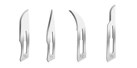
Never load or remove a blade with your fingers. To load one, grip the blade's dull side with a hemostat or a needle holder and fit it into the handle's slots. Skip any needle holder whose jaws are lined with diamond dust or special metal: loading blades can knock its jaws out of line. To take a blade off, use a single-handed blade remover, or use a safety scalpel instead, such as one with a retractable blade or a single-use handle that is never taken apart.
Some references still teach taking the blade off with a needle holder: lift the lower corner of the blade slightly, then pull it out of the slot.
With a safety scalpel, AST suggests the scrub uncovers the blade, places the scalpel in the neutral zone, and covers it again when it returns. Pass every knife through the neutral zone, drop used blades straight into the sharps container, and pick up a blade dropped on the field with forceps.
12Sponges
Sponges soak up blood, cushion tissue under retractors, hold bowel aside and help separate tissue. Every sponge used in a wound has a radiopaque marker that shows on x-ray, and gauze without one never goes in. AST and AORN guidance say sponges are never cut or altered.
- Raytec (4 by 4): woven gauze folded into a 4-inch square, for shallow wounds. In a deep wound or body cavity, it is always mounted on sponge forceps as a sponge stick: fold it in thirds one way and in half the other, with the folded edge at the tip. Any loose Raytec comes off the field before the peritoneum is opened.
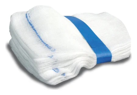
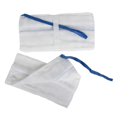
- Laparotomy sponge (lap): a large pad with a loop of tape and a radiopaque marker at one corner, for the abdomen and chest. Wet it in warm saline and wring it out before passing, unless the surgeon wants it soaking wet or dry.
- Dissectors (peanuts, Kittners, cherries, pushers): small, tightly rolled sponges for blunt dissection, always passed on a clamp. Spares stay in their holder, never loose on the field.
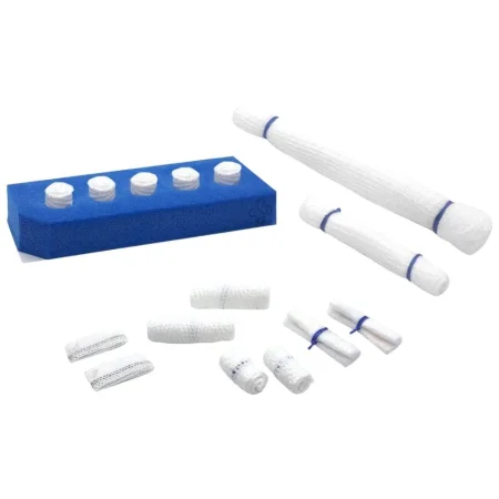
- Tonsil sponges: gauze-covered balls on a string, for the throat. The string stays outside the mouth so a sponge cannot fall into the airway; check it is still attached when the sponge comes back.
- Cottonoids (patties): flat squares of pressed cotton or rayon with a string, for brain, spine, ear and vascular surgery. They are usually wet with saline or a drug such as thrombin, kept moist the whole case, and offered from a basin or on a flat ribbon retractor, and they can sit between a suction tip and delicate tissue. Every one comes back with its string; never cut the strings.
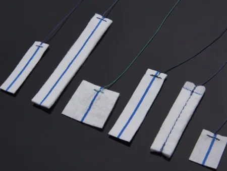
- Cellulose spears (such as Weck-Cel): small cellulose sponges used in eye and eyelid surgery to dab tiny bleeders, so the surgeon has a dry view and can find each bleeder to cauterize.
- Dental sponges: small 2 by 2 gauze sponges, with cotton rolls or dental pellets, for mouth and tooth surgery. A gauze throat pack with a radiopaque thread is moistened and placed in the throat; it is counted, and it must come out before the breathing tube does.
Keep one basin of saline just for wetting sponges, because lint comes off them. Drop used sponges, never throw them, into the kick bucket for the circulator to count.
13Drains
Blood and serum that collect in a wound give germs a place to grow, and a drain gives that fluid, or air, a way out. Most drains leave through a small stab wound beside the incision and are stitched to the skin with nonabsorbable suture. They are made of PVC, silicone (Silastic is a silicone rubber), rubber or latex, so check for a latex allergy. Before a drain goes in, look it over and make sure it is open all the way through, because a faulty drain can leave a piece behind when it is pulled.
All but the simplest drains empty into a reservoir, which keeps the fluid contained so infection does not spread and lets the team measure it. Keep every drain's collection unit below the level where its tube leaves the body, so fluid cannot run back in.
Drains are flat or tube-shaped, with or without a reservoir, and they are open or closed. An open drain, such as a Penrose, lets outside air reach the wound. A closed drain is sealed from the air and works by suction or a pressure difference.
Passive drains
Passive drains work by gravity and capillary action.
- Penrose: a soft, flat, thin-walled tube that lets fluid run out into the dressing, with no reservoir. A Penrose is also looped around a structure, such as the esophagus, to lift and hold it.
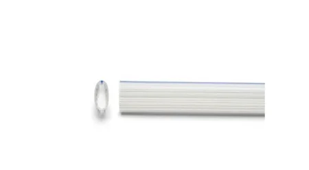
- T-tube: drains bile from the bile duct into a bag.
- Catheters such as the Foley, Malecot and Pezzer also drain passively, as after genitourinary surgery (chapter 34). Drains such as the Pezzer, Malecot and Penrose are usually set in place in the wound with a blunt clamp, such as a Mayo or Pean.
- Sump drain: a tube inside a tube. Fluid seeps into the outer tube, which has many holes, and is drawn off through the inner one, a design that keeps the holes from clogging. It handles large amounts of fluid deep in the abdomen. AST's Core Curriculum groups it with the passive drains, though it works best on low suction.
Active drains
- Closed suction drains, such as the Jackson-Pratt (a soft bulb) and the Hemovac (a canister that is pressed flat): perforated tubing in the wound connects to a reservoir. Once the wound is closed, squeezing the reservoir flat and closing its plug creates suction that draws fluid out as it slowly opens up. As the dressing is taped, the circulator connects the drains' nonsterile ends (dressings are in chapter 30).
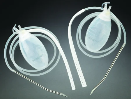
- The tubing leaves the skin on a sharp trocar. Take off its guard with an instrument, pass it through the neutral zone, and put the cut-off trocar straight into the sharps container.
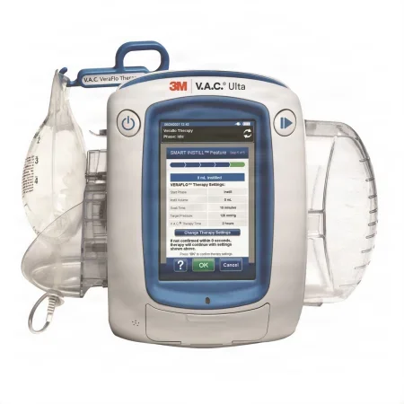
- Negative pressure wound therapy (a wound VAC): foam in the wound, sealed under film and connected to a pump that keeps up steady suction. It removes fluid, keeps the wound moist and reduces swelling.
- A stoma pouch, which collects drainage from an opening made onto the skin in surgery, goes on with the dressings (chapter 30).
Chest drainage
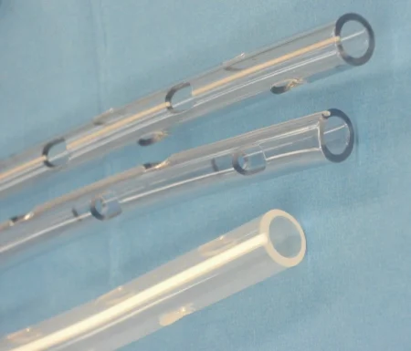
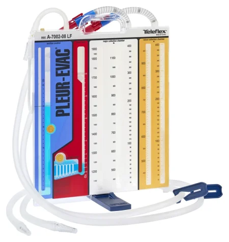
The space around the lungs normally holds a negative pressure that keeps them open. After chest surgery or injury, a chest tube connects to a chest drainage unit (the Pleur-evac is one brand) with three chambers: one collects drainage, one holds a water seal that keeps air from being drawn back into the chest, and one controls suction. Keep the unit upright and below the chest at all times, because fluid running back can collapse the lung. Chest tubes are stiff Silastic or PVC tubes with many holes near the tip, sewn to the skin with heavy nonabsorbable suture and dressed with petroleum gauze, fluffs and flat gauze; two tubes may be joined with a Y-connector to one unit. Connections are secured with bands or tape, and each tube is labeled, such as anterior and posterior. If a tube comes out, cover the hole at once with sterile petroleum gauze, so air cannot rush in. Tubes come out when the air leak has stopped and the drainage has stayed low for 24 hours.
14Drapes
Drapes form a barrier between the prepped incision site and everything that is not sterile. Draping itself comes in chapters 26 and 28; these are the materials.
- Nonwoven disposable drapes are impervious or nearly so. Woven cloth drapes are treated to resist moisture but are not waterproof, so they need extra layers where fluid will be. AST calls for impervious, fluid-resistant drapes, especially around the opening.
- Towels square off the incision site as the first layer.
- Adhesive barrier drapes: plastic towel drapes (such as the Steri-Drape) with a strip of adhesive along one edge, used to wall off an area, such as the eyes, nose and mouth in ear surgery.
- Fenestrated drapes have a window (fenestration) over the incision, made for each area: laparotomy, thyroid, eye, extremity and more.
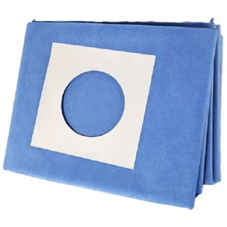
- Plain sheets (half, three-quarter or full), also called utility drapes, cover large areas, and split or U-drapes wrap around a limb or the hip. Universal drapes are used with towels in place of a fenestrated sheet on some cases.
- Fluid-collection pouch: a pouch, built into a drape or added to it, that catches and channels large amounts of irrigation and blood, with ports to drain it during the case, as in orthopedic, brain, gynecologic and urologic surgery.
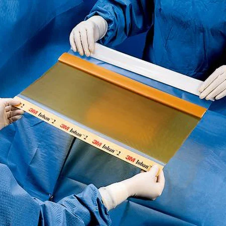
- Incise drapes: clear adhesive plastic pressed onto the prepped skin over the incision, plain or impregnated with an iodine antiseptic. The CDC finds them not necessary to prevent infection, so whether one is used follows the surgeon and your facility's policy. When one is used, some guidelines call for the iodine type, except for a patient allergic to iodine.
- Stockinette: a fabric tube, impervious or not, rolled up over an arm or leg. Coban, a stretchy wrap that sticks to itself, may be wound over it.
- Equipment covers: sterile plastic sleeves shaped for the microscope, C-arm, cameras, probes and robot.
15Electrosurgical supplies
Chapter 20 explains electrosurgery and its safety. These are the supplies you set up.
- Active electrode: the handheld pencil, often called the Bovie, with changeable tips: blade, needle, ball, loop and extended, some insulated or coated. Check the cord and insulation, and seat the tip firmly. The tip counts as a sharp: it is part of the sharps count and goes into the sharps container at the end.
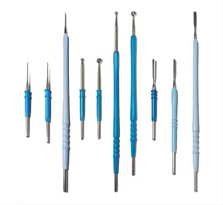
- Safety holster: a nonconductive holder on the drapes, in plain sight and within the surgeon's reach, preferably fastened with a clamp that is not metal. The pencil goes back in it after every use, never onto the patient or drapes.
- Scratch pad: an abrasive pad for cleaning char (eschar) off the tip, kept handy for the surgeon but away from the wound. A coated, nonstick tip is wiped with a wet sponge instead, because the pad ruins the coating. Never scrape a tip with a blade; it pits the tip and risks a cut.
- Patient return electrode: the adhesive, gel-coated pad that carries current back to the generator in monopolar electrosurgery. Check its expiration date, pick the right size, never cut it, and place it on clean, dry skin over a large muscle near the surgical site. Bipolar instruments need no pad.
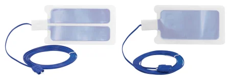
- Other energy handpieces you will set up: bipolar forceps, some with an irrigation channel at the tips; vessel-sealing devices (such as LigaSure); the harmonic (ultrasonic) scalpel, which coagulates and divides tissue between its jaws; the argon beam coagulator; radiofrequency ablation probes; and suction coagulators. Chapter 20 explains how each kind of energy works.
16Staplers and clips
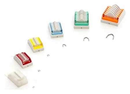
Staplers close or join tissue quickly with staples of stainless steel, titanium or absorbable material. Each staple closes into a B shape that does not crush, so blood still reaches the tissue edges. Staplers come disposable or reusable, for open or minimally invasive surgery, and most take color-coded reloads. A reusable stapler is put together by the scrub during setup; a disposable one comes assembled and sterile, and new cartridges load into it so one stapler can fire again. Each company's stapler works differently, so learn each one through the maker's training. Chapter 32 shows them at work.
Stapling is faster than suturing, with less blood loss and less time under anesthesia, but it costs more, and a misplaced staple line is much harder to fix than a stitch. Besides the open and endoscopic kinds, staplers come powered (battery driven), robotic (fired from the robot's arm) and for skin and subcuticular closure.
- Linear stapler: straight, staggered rows that close tissue without cutting it.
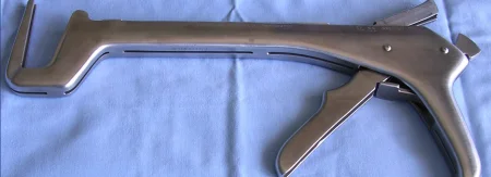
- Linear cutter: two double rows with a knife between them, sealing and dividing in one firing, as in bowel surgery.
- Circular (intraluminal) stapler: a double ring of staples with a circle trimmed inside it, to join two ends of bowel, often low in the colon.
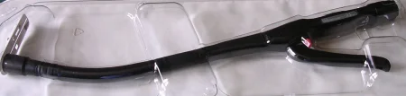
- Staple line reinforcement: strips of absorbable synthetic or biologic material (a buttress) fitted to the stapler's jaws, a glue, or stitches sewn over the line, used to cut bleeding and leaks along a staple line, as in a sleeve gastrectomy.
- Skin stapler: one staple per squeeze, the arrow on the stapler head lined up on the skin edges while the surgeon turns them outward with toothed forceps. Quick, low in tissue reaction and even in tension, though it leaves more marks than a subcuticular closure and the staples must be taken out later. AST's position is that a CST has the training to run the skin stapler while the surgeon or assistant everts the edges.
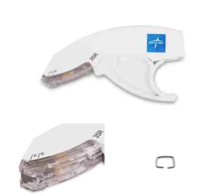
Staplers can misfire, fail to fire or form staples badly. When you reload a linear cutter, wipe off the other side of its jaws so no staple from the last firing is left behind, because a leftover staple can make it misfire or not fire at all. The FDA asks teams to read the maker's instructions, keep a range of staple sizes on hand, use the cartridge that matches the tissue's type and thickness, and, if a stapler fails on tissue, clamp or tie the vessels before it is released.
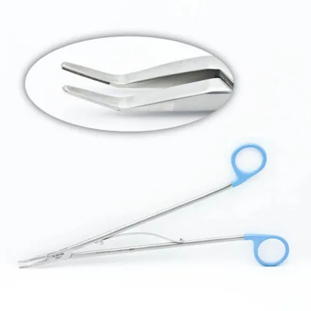
Ligating clips close a vessel or duct in place of a tie. They are stainless steel, titanium or absorbable, come in sizes from micro to large, each matched to its own applier, and are usually placed on both sides before the structure is cut between them. Appliers come disposable and preloaded, or reusable and loaded one clip at a time, with long versions for laparoscopy.
17Syringes and injection needles
- Fittings: a Luer-lock tip twists on and locks, so a needle or tubing cannot pop off. A slip tip pushes on and pulls off, for low-pressure uses. Both ends of a connection must match.
- Sizes: 1 mL to 60 mL, each marked along the barrel in milliliters. The 1 mL tuberculin syringe measures tiny doses; never confuse it with an insulin syringe, which is marked in units. A 60 mL syringe is for flushing and irrigation, not measuring. Most syringes are plastic; glass ones are used when a very smooth injection matters, such as injecting local anesthetic into tissue.
- Control syringe: rings for the thumb and fingers, for better control while injecting, such as local anesthetic.
- Prefilled cartridges: some drugs come in a ready-filled cartridge that fits a holder with a plunger. The older Tubex system was replaced by the Carpuject.
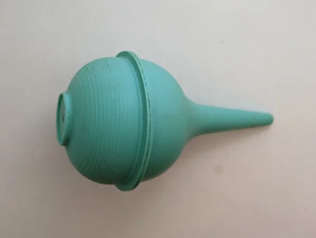
- Irrigation: the Asepto, a bulb on a barrel holding about 120 mL, is the usual irrigator in open cases. The bulb syringe has no barrel, for small areas and an infant's mouth and nose. The Toomey is a large syringe, often used through a catheter to irrigate the bladder. Keep track of the irrigation you use, because it mixes with blood in the suction canister and the team needs it to estimate blood loss.
- Needles: hypodermic needles are sized by gauge and length. The higher the gauge, the thinner the needle: an 18-gauge is wider than a 22-gauge. A blunt needle draws a drug from the circulator's vial without risk of a stick, and a filter needle keeps glass out when drawing from an ampule. Spinal needles are 3 to 4 inches long with a stylet. An angiocatheter (such as the Angio-Cath) uses a needle to place a plastic tube that stays in a vein or artery. Chapter 12 covers drawing up and labeling drugs on the field.
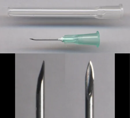
- Recapping: never with two hands. If the same needle will be used again on the same patient, scoop the cap up from the Mayo stand one-handed and leave it loose.
18Containers and field items
- Basins: one for irrigation and a second only for wetting sponges, placed so the circulator can pour without reaching over the field.
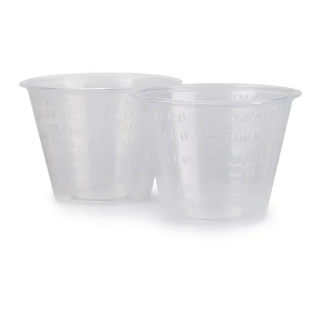
- Medicine cups: set at the edge of the back table to be filled, then moved in. Label every cup, basin and syringe that holds a solution or drug.
- Specimen cups: hold tissue in a safe spot on the back table until it goes off the field.
- Pitchers, trays and petri dishes: sterile containers come in many shapes, and many carry graduated marks for measuring. A graduated pitcher sits at the edge of the back table to be filled, like a medicine cup, and a petri (culture) dish can hold a small piece of tissue, such as a donor cornea.
- Light handle covers: sterile covers that let the sterile team move and focus the overhead lights.
- Magnetic mat or needle counter: holds used needles and small sharps where you can see and count them.
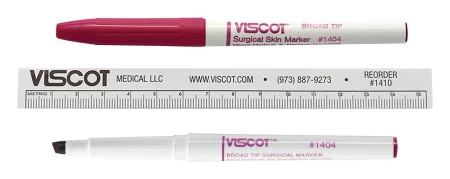
- Sterile marking pen, ruler and labels: for marking the skin and labeling drugs and solutions.
- Suture bag: a paper bag from the back table pack for empty packets and used ties.
19Suction
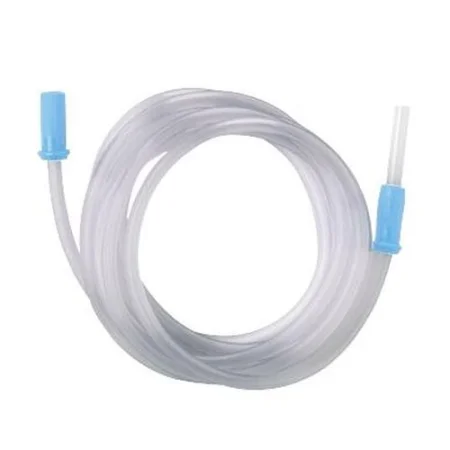
Tubing joins the tip on the field to a canister. Set it up with the tip attached, and draw sterile water through it now and then so blood does not clot inside. Firm, large-bore tubing is used with high-power suction, as in liposuction.
- Yankauer (tonsil suction): a rigid, angled tube with a rounded tip, the most used tip, which suctions without grabbing tissue.
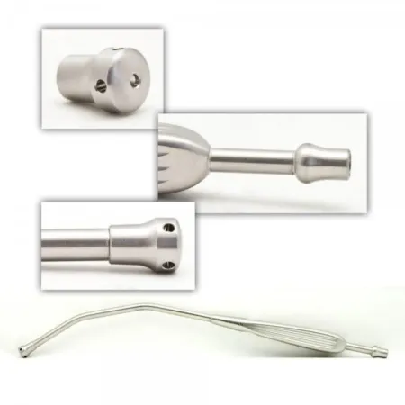
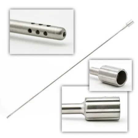
- Frazier: a thin, angled tip with a thumb hole; covering the hole increases the pull. For small spaces such as the nose, spine and skull, with a wire stylet to clear it.
- Poole: an outer sheath with many holes over an inner tube, for large amounts of fluid in the abdomen. The holes spread the suction so bowel is not drawn against the tip.
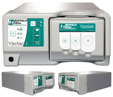
- Smoke evacuation: electrosurgery and lasers make a smoke plume that carries chemicals, cells and germs. A smoke evacuator with a filter removes it. Hold the wand as close as you can to where the tip meets the tissue, always within 2 inches (5 cm), without blocking the surgeon's view.
AST's 2012 electrosurgery standard says to hold the wand less than 1 centimeter from where the electrode touches the tissue.
- Cell salvage: a cell saver suctions the patient's own blood, washes and filters it, and returns it, reducing the need for donor blood. Topical hemostatic agents and some antibiotics should not be suctioned into it, and amniotic fluid needs its own separate suction.
20Tissue repair and replacement
Closing skin without suture
- Skin closure tapes (such as Steri-Strips): adhesive strips that hold small wounds closed or support a subcuticular closure. They need dry skin, and a liquid adhesive such as benzoin helps them stick.
- Skin adhesive (a cyanoacrylate glue, such as Dermabond; the Core Curriculum also names Cavilon): painted in layers over skin edges held together. It seals out germs, needs no removal and wears off on its own in about 5 to 10 days. Only for wounds not under tension, and never on an infected wound or for a patient allergic to cyanoacrylate or formaldehyde.
- Wound zipper: two adhesive strips joined by a zipper. The strips go on clean, dry skin on each side of the incision, and closing the zipper pulls the edges together, with no needle holes and no needles to stick anyone.
Mesh
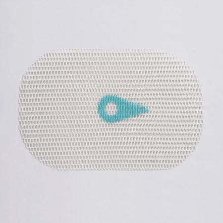
Mesh bridges or strengthens tissue that cannot be closed without tension, most often in hernia repair. Synthetic meshes include polypropylene, which tolerates infection; polyester, which does not; PTFE; and absorbable polyglactin 910 for temporary support. Biologic meshes, made from animal tissue, let the patient's own tissue grow in.
Grafts
- Autograft: tissue moved within the same patient, such as skin, bone, cartilage or fat.
- Allograft (homograft): tissue from another person, kept by a tissue bank, such as skin, bone or cartilage.
- Xenograft: tissue from another species, such as pig or cow skin.
Protect an autograft: keep it moist in saline unless the surgeon says otherwise, never on a dry, absorbent surface, in a safe spot on the back table, and point it out when you hand off to another scrub. Implants such as mesh are opened only once the surgeon confirms the exact type and size, and, as with any sterile package, the wrapper is checked for damage and the expiration date confirmed first. Each one goes in the facility's implant log with its lot number and expiration date.
21Other supplies
- Vessel loops: colored silicone loops that lift and hold vessels, nerves and ducts; red usually marks arteries, blue veins, and white or yellow nerves and ducts. Umbilical tape, a cotton tape used wet on a hemostat, does the same job.
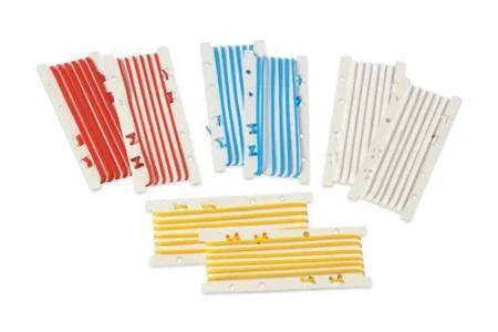
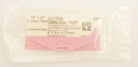
- Pain pumps: a small catheter left in the wound or joint and a disposable pump that delivers local anesthetic for 2 or 4 days, depending on the pump.
- Latex-free supplies: for a patient with a latex allergy, every item must be latex-free, ideally from a dedicated latex allergy cart, and the patient goes first in the day.
- Emergency supplies: know where the malignant hyperthermia cart and the other emergency carts are and what they hold, so you can fetch them without stopping to think.
Specialty supplies
Each specialty has supplies of its own, and you meet them with the procedures that use them: general surgery (chapter 32), laparoscopic and robotic surgery (chapter 21), obstetrics and gynecology (33), urology, with its many catheters and stents (34), eye (35), ear, nose and throat (36), oral and maxillofacial (37), plastic and reconstructive (38), orthopedic (39), heart, chest and vascular surgery (40 and 41) and neurosurgery (42). Anesthesia supplies are in chapter 13.
22Handling any supply
The same habits apply to every item on this page. Know whether it is disposable or reusable and what it does well or poorly. Inspect it before it goes on the field, because a single-use item can be damaged too: check the package is whole and the date is good. For a device such as a stapler, learn from the maker's training how to prepare, load, reload and unload it. Keep track of what is used, in the count, the implant log and the record.
Know how each device can fail and what to do about it, as with a stapler that misfires (see Staplers and clips). At the end, dispose of each item by its type and your facility's policy: sharps go into the sharps container, and soiled drapes, gowns and gloves are placed, never thrown, into the impervious biohazard bag. Breaking down the field is in chapter 30.
Lo que te llevas de este capítulo
- Sutures are absorbable or nonabsorbable, natural or synthetic, and monofilament or multifilament. Braided strands handle well but wick; monofilaments glide and resist infection but hold knots less well.
- More zeros mean a finer strand: 4-0 is finer than 2-0, and 2 is thicker than 0. The surgeon uses the finest strand that will hold.
- Read the label for size, material, needle and length, and open only what the case needs.
- Never wet synthetic absorbables, never soak gut, never crush a strand, and never bend steel wire.
- Taper points for soft tissue, cutting points for tough tissue, blunt points for friable organs and fascia.
- Load the needle in its packet, one-third back from the swage, never with fingers. Pass it ready to use, through the neutral zone, one for one, and check that every needle comes back whole.
- Every sponge in a wound is radiopaque and never cut. Dissecting sponges always go up on a clamp.
- Passive drains work by gravity; active drains use suction. A chest drainage unit stays upright and below the chest.
Ponte a prueba5 preguntas rápidas
Gratis para todos. Elige una respuesta para cada una y luego revísalas todas juntas.
En el quirófanoDónde vas a usar esto
Todo el banco de preguntas del capítulo en series de 10, con la respuesta explicada después de cada una.
Parte del programa para educadoresLas palabras del capítulo en tarjetas de estudio: las que fallas vuelven antes.
Parte del programa para educadoresLo asigna tu docente. Cada grupo saca sus propias 25 preguntas del banco del capítulo.
Parte del programa para educadores
Sugerir una corrección
¿Algo está mal, confuso o desactualizado en este capítulo? Avísanos y lo corregimos para todos. Tu correo solo se usa para responderte.