Orthotopic Liver Transplantation
This complex surgery involves the total hepatectomy of the diseased native liver and the subsequent implantation of a donor liver into the same anatomical position. The procedure requires multiple vascular anastomoses, including the vena cava, portal vein, and hepatic artery, along with biliary reconstruction.
In 30 seconds
Scan this outside the room.
- anesthesiaan-us-THEE-zhuhLoss of feeling or sensation, often brought on by drugs so that surgery or a procedure can be done without pain.an- withoutesthesi/o feeling, sensation-ia conditionFull entry and Positioning: Patient is put under general anesthesiaJEN-er-ul an-es-THEE-zhuhDrug-induced loss of consciousness and feeling over the whole body, so a person feels nothing and is not aware during surgery.an- withoutesthesi/o feeling, sensation-ia conditionFull entry and positioned on their back with arms extended.
- Incision and Exposure: A large incision is made in the upper abdomen to expose the liver.
- Liver Mobilization: The surgeon divides ligaments and blood vessels to mobilize the liver.
- dissectiondy-SEK-shunA tear in the inner layer of an artery wall that lets blood split the layers apart; also, the cutting apart of tissue in surgery.Full entry : The hepatichih-PAT-ikRelating to the liver.hepat/o liver-ic relating toFull entry artery, portal vein, and bile ductbyle duktOne of the tubes that carry bile from the liver and gallbladder to the small intestine.Full entry are dissectdy-SEKT, or dih-SEKTTo cut apart or separate tissues carefully during surgery.Full entry .
- Hepatectomy: The diseased liver is removed by dividing the blood vessels and bile ductbyle duktOne of the tubes that carry bile from the liver and gallbladder to the small intestine.Full entry that connect it to the body.
- graftGRAFTTissue or a man-made material moved into place in surgery to replace or repair a damaged part.Full entry Preparation: The healthy liver is trimmed and prepared for implantationim-plan-TAY-shunThe attachment of a fertilized egg to the lining of the uterus, the start of a pregnancy.im- inplant to set-ation the process ofFull entry .
- implantationim-plan-TAY-shunThe attachment of a fertilized egg to the lining of the uterus, the start of a pregnancy.im- inplant to set-ation the process ofFull entry : The graftGRAFTTissue or a man-made material moved into place in surgery to replace or repair a damaged part.Full entry is implanted by connecting the blood vessels and bile ductbyle duktOne of the tubes that carry bile from the liver and gallbladder to the small intestine.Full entry to those of the recipient.
- hemostasishee-moh-STAY-sisThe stopping of bleeding, either by the body's own clotting process or by medical or surgical methods.hem/o blood-stasis stopping, standing stillFull entry : The surgeon ensures there is no bleeding from the surgical site and that blood flow is adequate.
- Closure: The incision is closed with sutureSOO-cherA stitch, or the thread used to stitch, that holds the edges of a wound or incision together.Full entry or staples, and sterile dressings are applied to the wound.
The tray
11 instruments, by what they do. Tap a name for its card.
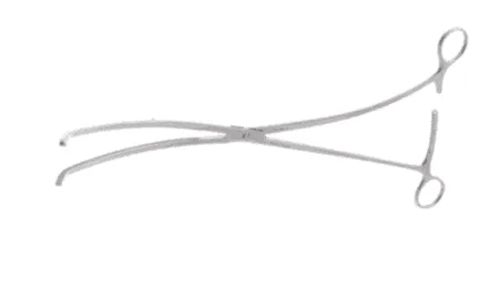
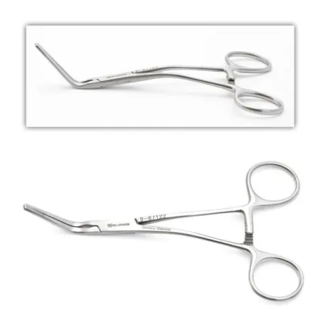
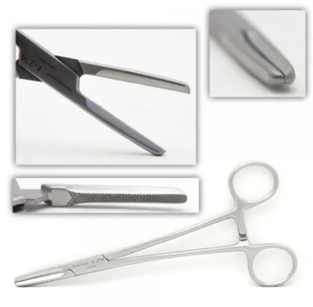
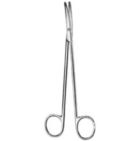
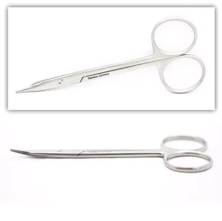
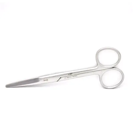
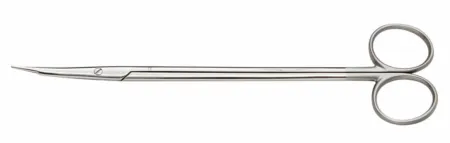
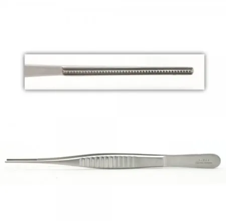
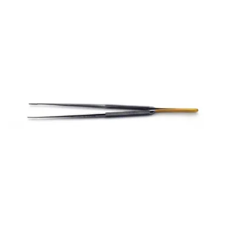
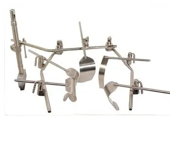
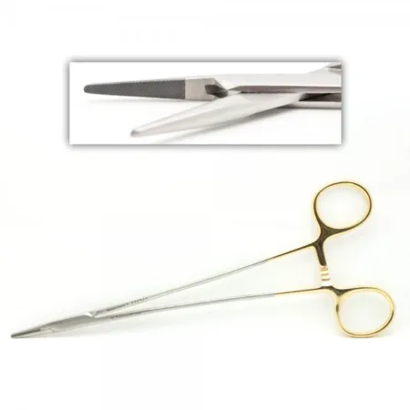
Clamping & Occluding3
Cutting & Dissecting4
Grasping & Holding2
Retracting & Exposing1
Suturing & Stapling1
Know this tray before you scrub in.
With Pro, for every setup in every specialty. Cancel any time.
Or scrub in free: every General setup and 5 questions a day.
Step by step
9 steps, one line each.
- 1
Anesthesia and Positioning
The first step in an Orthotopic Liver Transplantation is to put the patient under general anesthesiaJEN-er-ul an-es-THEE-zhuhDrug-induced loss of consciousness and feeling over the whole body, so a person feels nothing and is not aware during surgery.an- withoutesthesi/o feeling, sensation-ia conditionFull entry and position them on their back with their arms extended (supine). This position allows the surgical team to have access to the abdomen.
- 2
Incision and Exposure
A large incision is made with a #10 knife blade on a #3 knife handle in the upper abdomen to expose the liver. This incision allows the surgical team to have a clear view of the liver and access to the blood vessels and ducts that need to be dissectdy-SEKT, or dih-SEKTTo cut apart or separate tissues carefully during surgery.Full entry and divided during the surgery. Common instruments to use during dissectiondy-SEK-shunA tear in the inner layer of an artery wall that lets blood split the layers apart; also, the cutting apart of tissue in surgery.Full entry are hemostatHEE-moe-statA clamp with finger rings and a locking ratchet used in surgery to grasp and close off bleeding vessels.hem/o blood-stat stopping, standing stillFull entry , right angles, and a variety of scissors such as Tenotomys or Metz.
- 3
Liver Mobilization
The surgeon carefully mobilizes the liver by dividing the ligaments and blood vessels that connect it to the surrounding structures. This step allows the liver to be lifted and moved to give the surgical team better access to the blood vessels and ducts that need to be dissectdy-SEKT, or dih-SEKTTo cut apart or separate tissues carefully during surgery.Full entry and divided.
- 4
Dissection of the Hepatic Artery, Portal Vein, and Bile Duct
The surgeon dissectdy-SEKT, or dih-SEKTTo cut apart or separate tissues carefully during surgery.Full entry the hepatichih-PAT-ikRelating to the liver.hepat/o liver-ic relating toFull entry artery, portal vein, and bile ductbyle duktOne of the tubes that carry bile from the liver and gallbladder to the small intestine.Full entry , which are the blood vessels and ducts that supply the liver with blood and bilebileA yellow-green fluid made by the liver and stored in the gallbladder that helps digest fats.chol/e bileFull entry . This step is critical to the success of the transplant as it ensures that the graftGRAFTTissue or a man-made material moved into place in surgery to replace or repair a damaged part.Full entry has proper blood flow and biliary drainage. Crawford clamps and pediatric vascular clamps are commonly used during this portion, along with a variety of silk tie sizes, vessel loops, and sometimes a 45 or 60 Ethicon stapler for veins. 6-0 BVI is commonly used on veins, and 7-0 Prolene on arteries.
- 5
Hepatectomy
The diseased liver is removed by dividing the blood vessels and bile ductbyle duktOne of the tubes that carry bile from the liver and gallbladder to the small intestine.Full entry that connect it to the body. This step requires precision and care to avoid damage to surrounding organs and structures.
- 6
Graft Preparation
The healthy liver (graftGRAFTTissue or a man-made material moved into place in surgery to replace or repair a damaged part.Full entry ) is carefully prepared for implantationim-plan-TAY-shunThe attachment of a fertilized egg to the lining of the uterus, the start of a pregnancy.im- inplant to set-ation the process ofFull entry , which includes trimming it to the appropriate size and reconnecting the hepatichih-PAT-ikRelating to the liver.hepat/o liver-ic relating toFull entry artery, portal vein, and bile ductbyle duktOne of the tubes that carry bile from the liver and gallbladder to the small intestine.Full entry . This step ensures that the graft is the correct size for the recipient and that it has the proper blood flow and bilebileA yellow-green fluid made by the liver and stored in the gallbladder that helps digest fats.chol/e bileFull entry drainage.
- 7
Implantation
The graftGRAFTTissue or a man-made material moved into place in surgery to replace or repair a damaged part.Full entry is implanted by connecting the blood vessels and bile ductbyle duktOne of the tubes that carry bile from the liver and gallbladder to the small intestine.Full entry to those of the recipient, using surgical sutureSOO-cherA stitch, or the thread used to stitch, that holds the edges of a wound or incision together.Full entry to create a secure connection. This step requires precision and care to ensure that the graft has proper blood flow and bilebileA yellow-green fluid made by the liver and stored in the gallbladder that helps digest fats.chol/e bileFull entry drainage.
- 8
Hemostasis
The surgeon ensures that there is no bleeding from the surgical site and that blood flow to the liver is adequate. This step is critical to the success of the transplant and requires careful monitoring and management.
- 9
Closure
The incision is closed with sutureSOO-cherA stitch, or the thread used to stitch, that holds the edges of a wound or incision together.Full entry or staples, and sterile dressings are applied to the wound. This step is important to prevent infection and promote healing. Common sutures used are 3-0 and 4-0 SHes-aych (said as letters)Social history: the part of a health history about a person's living situation, work, habits and supports.Full entry Prolene, 5-0 C1Prolene, and 6-0 PDS.
The case
An Orthotopic Liver Transplantation is a surgical procedure where a diseased liver is removed and replaced with a healthy liver from a donor. The procedure is typically performed in patients with end-stage liver disease or liver failure, and aims to restore liver function and prolong the patient's life.
Built by the techs who scrub it.
Every surgeon runs it a little differently. Send your table, or a fix for this page.
 Tayelor LittlefieldReviewer
The rest of this setup is Pro.
Everything unlocked above stays free. Pro opens the rest, on every setup: where new techs slip, what to load next at every step, the circulator’s side, the medicines, the specimen, wound class and trays on standby, what to grab if it goes wrong, and practice for every tray.
Free, for good. No card.
- All 2,882 instrument cards
- Every General setup in full
- 5 exam questions a day
- Your own preference cards
Pass your exam. Know every case.
Everything in Free, plus:
- All 260 setups in every specialty: back table, Mayo and every step
- Practice that never runs out and full timed exams for the CST, TS-C, CRCST, CSPDT and CSFA, with the why behind every answer
- Flashcards on any tray, timed to come back before you forget
- Every instrument card in full: the 3D model, cautions, inspection, how to pass it
Cancel any time from your account. Not worth it? Email us within 30 days of a charge for a refund, one per customer. Refund policy
Pro is also in the CSTSetup app, on the same account. Already have Pro? Log in
Send your table
Your Mayo, your back table, your surgeon’s way, or anything wrong on this page. Your email is only used to credit you. Submissions stay private and are reviewed by our team; accepted ones earn a Contributor credit here.