Jejunostomy Tube (J-Tube) Insertion
A feeding tube is surgically or laparoscopically placed directly into the lumen of the proximal jejunum through the abdominal wall. This creates a permanent or semi-permanent tract for enteral nutrition, bypassing the upper gastrointestinal tract.
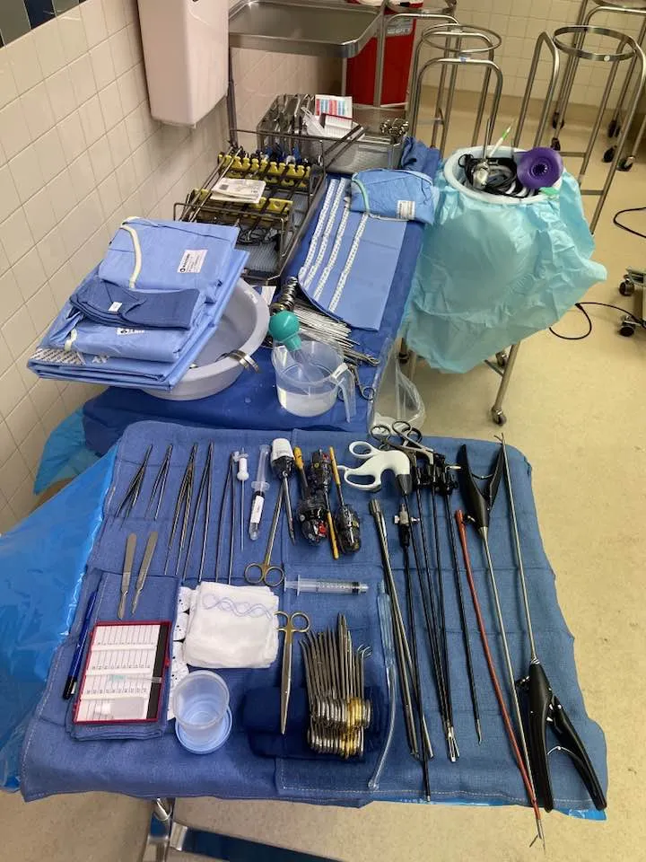
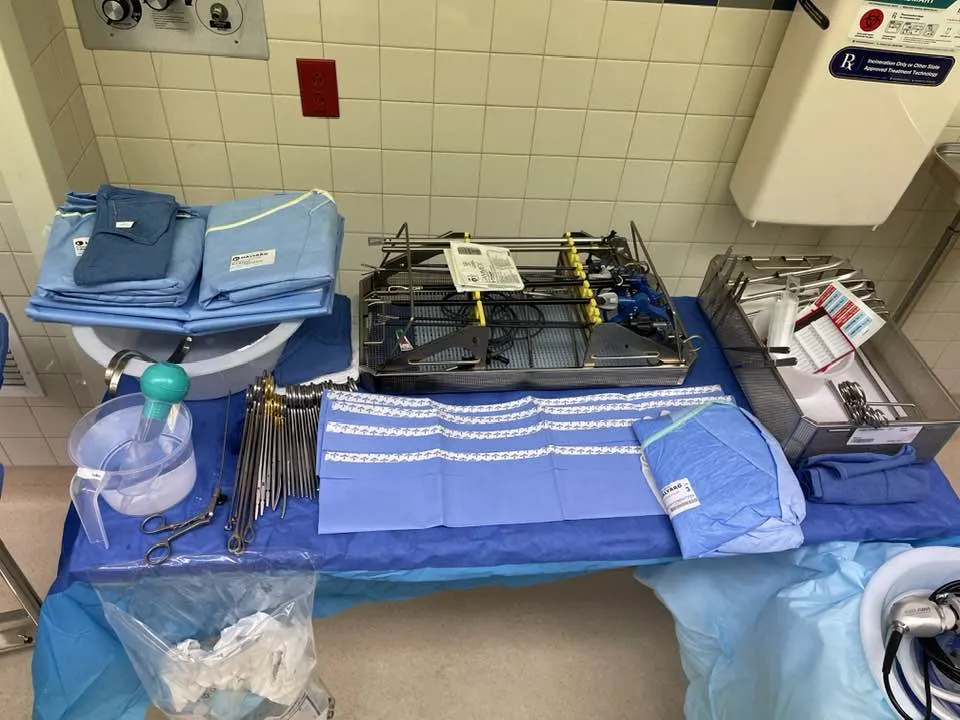
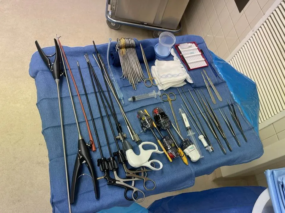
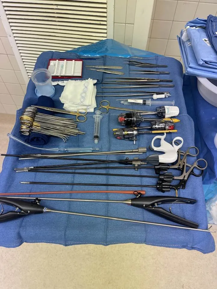
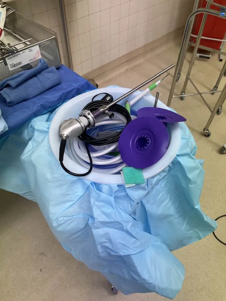
- Position
- Reverse Trendelenburg
In 30 seconds
Scan this outside the room.
- Mark the ideal placement position on the abdomen.
- Create space by making a small incision and filling the abdomen with carbon dioxideKAR-bun dy-OK-sideA gas made by the body's cells as waste, carried in the blood to the lungs and breathed out.Full entry .
- Insert trocarTROH-karA sharp or bladed instrument inside a tube (cannula), used to pierce a body wall and leave the tube in place as a port.Full entry and ports for laparoscopiclap-uh-roh-SKOP-ikDone through a few small cuts in the abdomen with a thin camera and long instruments, instead of one large opening.lapar/o abdomen-scopic viewingFull entry instrument access.
- Manipulate the colon to locate the Ligament of Treitz.
- Use sutureSOO-cherA stitch, or the thread used to stitch, that holds the edges of a wound or incision together.Full entry to bring the colon up to the abdominal wall.
- Place the J-tube using a percutaneous needle and guide wire.
- Tie sutureSOO-cherA stitch, or the thread used to stitch, that holds the edges of a wound or incision together.Full entry to secure the J-tube and inflate the balloon.
- Close incisions with sutureSOO-cherA stitch, or the thread used to stitch, that holds the edges of a wound or incision together.Full entry or skin glue.
The tray
13 instruments, by what they do. Tap a name for its card.
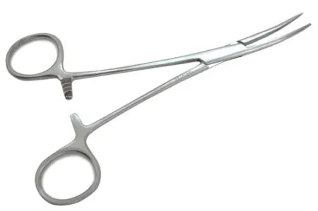
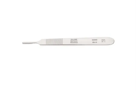
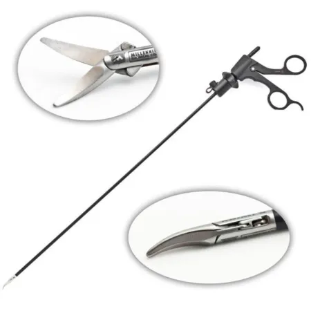
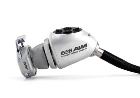
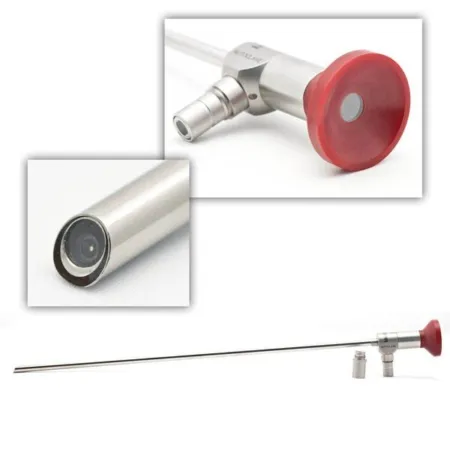
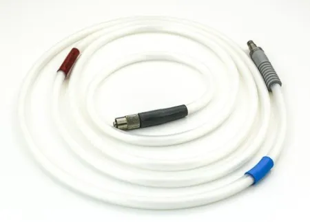
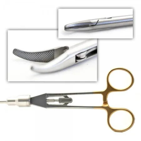
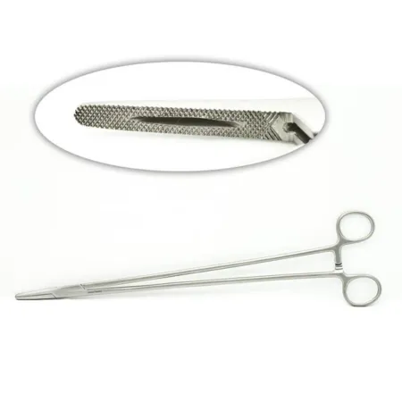
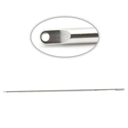
Clamping & Occluding1
Cutting & Dissecting4
Grasping & Holding2
Specialty3
Suturing & Stapling3
Know this tray before you scrub in.
With Pro, for every setup in every specialty. Cancel any time.
Or scrub in free: every General setup and 5 questions a day.
The room
The equipment, the prep and drape, the suture and the medicines.
Folded blue towels or utility drapes and a laparotomy drape
Step by step
7 steps, one line each.
- 1
Marking the Ideal Placement Position
Before beginning the Jejunostomy Tube insertion surgery, the surgical technologistSUR-jih-kul tek-NOL-uh-jistA member of the operating room team who prepares the sterile field and instruments and hands them to the surgeon.Full entry will assist in finding the ideal placement position for the J-tube. Typically, this position is in the upper left quadrant of the abdomen. Using a skin markerMAR-kerSomething that can be measured, such as a substance in the blood, that shows a disease or body process is present.Full entry , the technologist will mark the site for incision.
- 2
Preparing the Abdominal Wall
To create space for the procedure, an 11 blade loaded on a #3 knife handle will be used to make a small incision in the marked area. Once the incision is made, a Verres needle will be inserted to fill the abdomen with carbon dioxideKAR-bun dy-OK-sideA gas made by the body's cells as waste, carried in the blood to the lungs and breathed out.Full entry . This process helps to lift the abdominal wall away from the organs, providing better visibility for the surgery.
- 3
Inserting Trocars and Ports
Using the same 11 blade, the surgical technologistSUR-jih-kul tek-NOL-uh-jistA member of the operating room team who prepares the sterile field and instruments and hands them to the surgeon.Full entry will make small incisions to insert trocarTROH-karA sharp or bladed instrument inside a tube (cannula), used to pierce a body wall and leave the tube in place as a port.Full entry and their ports. Typically, 2 or 3 5mm ports and a 10mm port are used. These ports serve as entry points for the laparoscopiclap-uh-roh-SKOP-ikDone through a few small cuts in the abdomen with a thin camera and long instruments, instead of one large opening.lapar/o abdomen-scopic viewingFull entry instruments that will be used during the surgery. The trocars and ports facilitate access to the abdomen, enabling the surgical team to work effectively.
- 4
Manipulating the Colon and Locating the Ligament of Treitz
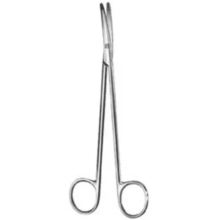
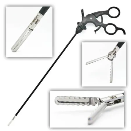
With the laparoscopiclap-uh-roh-SKOP-ikDone through a few small cuts in the abdomen with a thin camera and long instruments, instead of one large opening.lapar/o abdomen-scopic viewingFull entry instruments, such as bowel graspers, wave graspers, and lap Metz scissors, the surgeon will manipulate the colon to locate the Ligament of Treitz. This ligament serves as a crucial landmark for determining the placement of the J-tube. The technologist must have hemostatHEE-moe-statA clamp with finger rings and a locking ratchet used in surgery to grasp and close off bleeding vessels.hem/o blood-stat stopping, standing stillFull entry readily available to tag the sutureSOO-cherA stitch, or the thread used to stitch, that holds the edges of a wound or incision together.Full entry , ensuring they do not slip back into the abdomen.
- 5
Creating Suture Configuration and Bringing the Colon to the Abdominal Wall
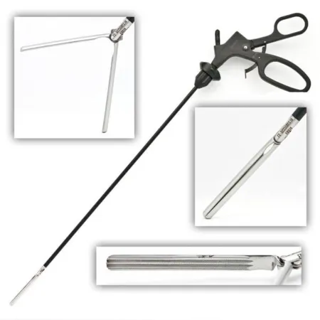
Once the Ligament of Treitz is found, the surgeon will move approximately 30-40 cm away from it to place the J-tube. An EndoStitch and EndoClose may be used to create a diamond configuration of sutureSOO-cherA stitch, or the thread used to stitch, that holds the edges of a wound or incision together.Full entry that will ultimately bring the colon up to the abdominal wall. Alternatively, the surgeon may choose to use laparoscopiclap-uh-roh-SKOP-ikDone through a few small cuts in the abdomen with a thin camera and long instruments, instead of one large opening.lapar/o abdomen-scopic viewingFull entry needle holders and a knot pusher in place of the EndoStitch and EndoClose. Laparoscopic scissors or EndoShears will be used to cut the sutures as needed.
- 6
Placing the J-tube
A percutaneous needle will be inserted through the bowel, and the J-tube guide wire will be threaded through the needle. The introducer and tear away sheathSHEETHA covering that wraps around a structure; in procedures, a short hollow tube left in a blood vessel to pass catheters through.Full entry will be advanced into the abdomen and then into the bowel, following the guide wire. Afterward, the tear away sheath will be removed, and the J-tube will be placed over the introducer. The introducer will be gently removed, and the sutureSOO-cherA stitch, or the thread used to stitch, that holds the edges of a wound or incision together.Full entry will be tied to secure the J-tube in place. A syringe, provided in the J-tube pack, will be used to inflate the balloon on the J-tube, ensuring it remains securely positioned.
- 7
Closing the Incisions
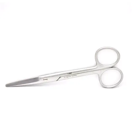
To complete the surgery, the port incisions will need to be closed. The surgical technologistSUR-jih-kul tek-NOL-uh-jistA member of the operating room team who prepares the sterile field and instruments and hands them to the surgeon.Full entry may use a 4-0 Monocryl or Nylon sutureSOO-cherA stitch, or the thread used to stitch, that holds the edges of a wound or incision together.Full entry with a Mayo needle driver to close the incisions. Alternatively, skin glue may be used as an alternative closure method. Straight Mayo scissors will be utilized to cut the sutures as necessary.
The case
Jejunostomy Tube (J-Tube) Insertion is a surgical procedure in which a flexible feeding tube is placed directly into the jejunum, a part of the small intestine. This tube allows for nutrition, fluids, and medications to be delivered directly into the digestive system when a person cannot eat or drink normally due to medical conditions like severe swallowing difficulties, gastrointestinal issues, or malnutrition. The procedure is typically done under anesthesia, using minimally invasive techniques or open surgery, and is often recommended for long-term feeding support.
Built by the techs who scrub it.
Every surgeon runs it a little differently. Send your table, or a fix for this page.
 Diana Lei-alohaReviewer
The rest of this setup is Pro.
Everything unlocked above stays free. Pro opens the rest, on every setup: where new techs slip, what to load next at every step, the circulator’s side, the medicines, the specimen, wound class and trays on standby, what to grab if it goes wrong, and practice for every tray.
Free, for good. No card.
- All 2,882 instrument cards
- Every General setup in full
- 5 exam questions a day
- Your own preference cards
Pass your exam. Know every case.
Everything in Free, plus:
- All 260 setups in every specialty: back table, Mayo and every step
- Practice that never runs out and full timed exams for the CST, TS-C, CRCST, CSPDT and CSFA, with the why behind every answer
- Flashcards on any tray, timed to come back before you forget
- Every instrument card in full: the 3D model, cautions, inspection, how to pass it
Cancel any time from your account. Not worth it? Email us within 30 days of a charge for a refund, one per customer. Refund policy
Pro is also in the CSTSetup app, on the same account. Already have Pro? Log in
Send your table
Your Mayo, your back table, your surgeon’s way, or anything wrong on this page. Your email is only used to credit you. Submissions stay private and are reviewed by our team; accepted ones earn a Contributor credit here.