Laparoscopic Hiatal Hernia Repair
Through small abdominal incisions, the surgeon returns the herniated stomach from the thoracic cavity back into the abdomen and narrows the esophageal hiatus in the diaphragm. A fundoplication is often performed simultaneously to strengthen the lower esophageal sphincter.
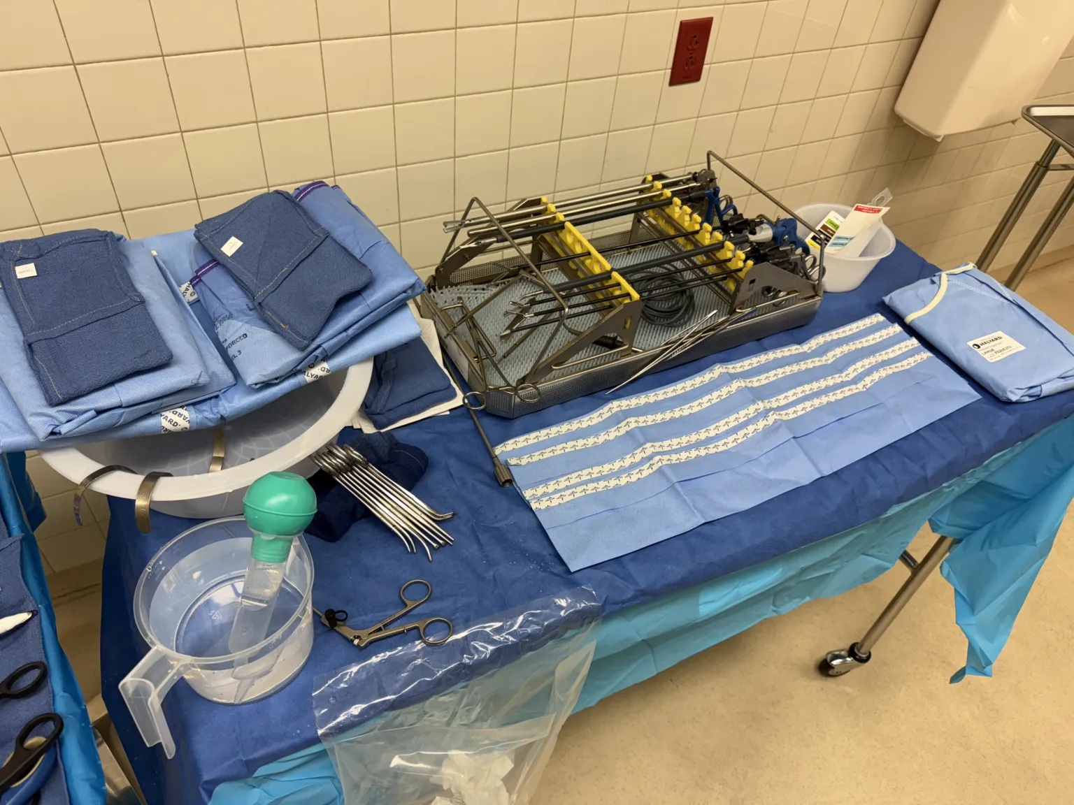
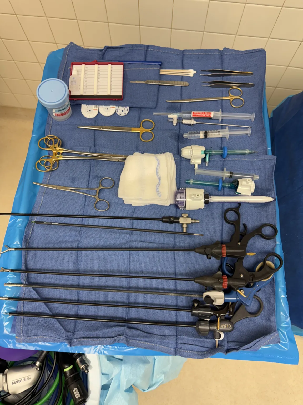
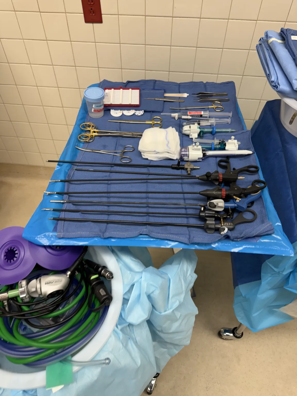
- Position
- Supine
- Anesthesia
- General Anesthesia
In 30 seconds
Scan this outside the room.
- Prep patient, make incision for 10mm port, insert insufflationin-suh-FLAY-shunBlowing a gas into a body cavity, such as carbon dioxide into the abdomen during laparoscopic surgery.Full entry tubing, insert laparoscope.
- Place 5mm ports, insert liver retractorrih-TRAK-terA surgical instrument that holds back tissue or organs so the surgeon can see and reach the operative site.Full entry .
- Find hiatal herniaHER-nee-uhA bulge of an organ or tissue through a weak spot in the muscle or tissue wall that normally holds it in.Full entry , use laparoscopiclap-uh-roh-SKOP-ikDone through a few small cuts in the abdomen with a thin camera and long instruments, instead of one large opening.lapar/o abdomen-scopic viewingFull entry instruments to take down adhesionad-HEE-zhunA band of scar tissue that joins two surfaces inside the body that are normally separate.ad- towardhes stick-ion conditionFull entry .
- Mobilize esophagusih-SOF-uh-gusThe muscular tube that carries food and liquid from the throat to the stomach.esophag/o esophagusFull entry , divide short gastric vessels, perform gastroplasty.
- Use EndoStitch to partially close window, use mesh to close defect, trim excess with Metz.
- Perform fundoplication with EndoStitch.
- Check for leaks with endoscopeEN-duh-skohpA thin tube with a light and a camera that is passed into the body to look inside an organ or cavity.endo- within-scope instrument for viewingFull entry , irrigationeer-ih-GAY-shunWashing out a wound, body cavity, tube or eye with a steady flow of liquid.Full entry , remove ports and retractorrih-TRAK-terA surgical instrument that holds back tissue or organs so the surgeon can see and reach the operative site.Full entry , close incisions.
The tray
19 instruments, by what they do. Tap a name for its card.
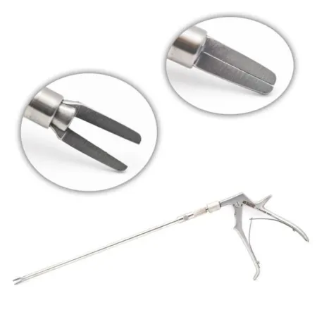 Endo clip applierClamping
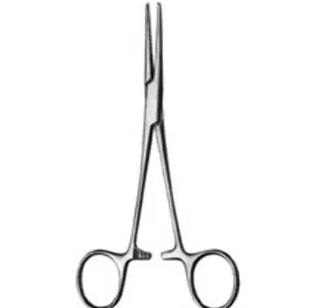 Kelly ForcepsClamping
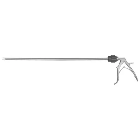 Laparoscopic Clip AppliersClamping
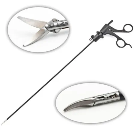 Laparoscopic Metz ScissorCutting
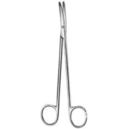 Metzenbaum ScissorCutting
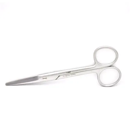 Straight Mayo ScissorCutting
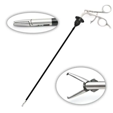 Cobra Laparoscopic GrasperGrasping
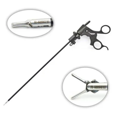 Dissecting Laparoscopic Grasper ForcepsGrasping
All 19 instruments
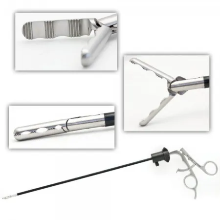 Fundus Laparoscopic Grasping ForcepsGrasping
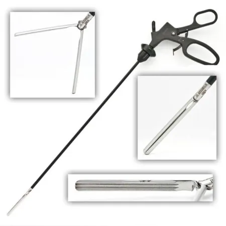 Laparoscopic Bowel GrasperGrasping
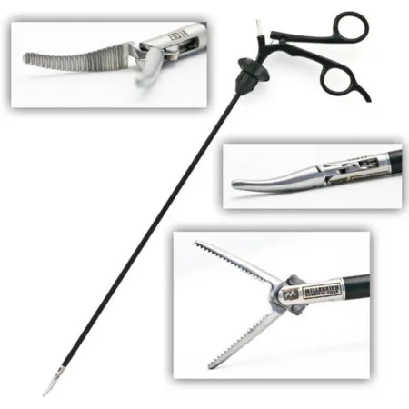 Laparoscopic Maryland Dissector GrasperGrasping
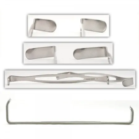 Army-Navy RetractorRetracting
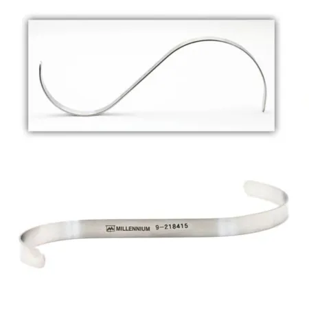 Hasson S RetractorRetracting
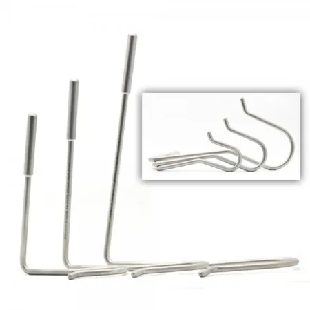 Nathanson Hook Liver RetractorRetracting
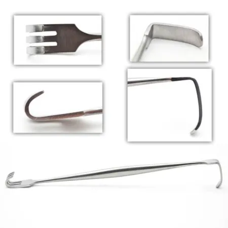 Senn RetractorRetracting
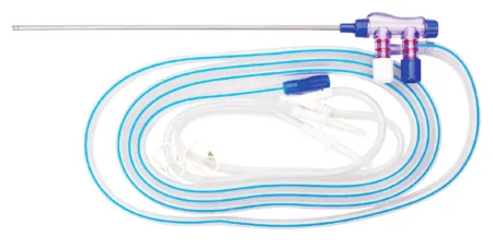 Suction IrrigatorSuctioning
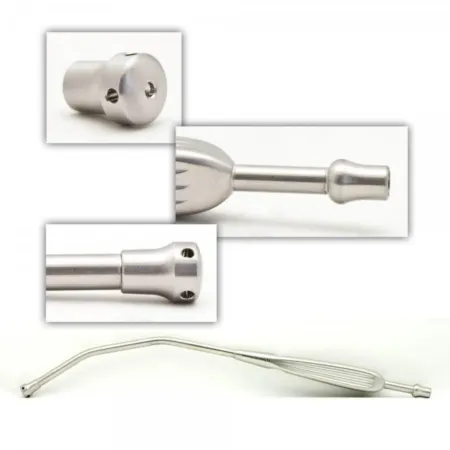 Yankauer SuctionSuctioning
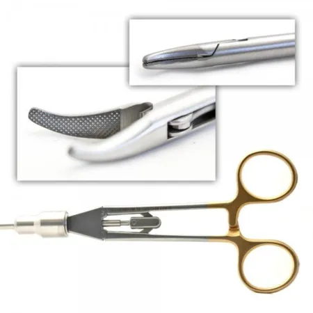 Laparoscopic Needle HolderSuturing
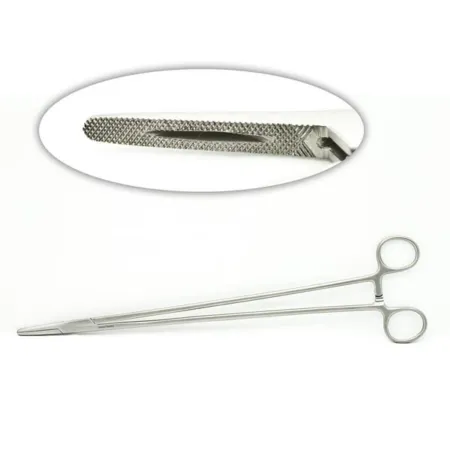 Mayo-Hegar Needle HolderSuturing
Clamping & Occluding3
Cutting & Dissecting3
Grasping & Holding5
Retracting & Exposing4
Suctioning2
Suturing & Stapling2
Know this tray before you scrub in.
With Pro, for every setup in every specialty. Cancel any time.
Or scrub in free: every General setup and 5 questions a day.
The room
The equipment, the prep and drape, the suture and the medicines.
Folded towels or utility drapes and laparoscopy drape
Step by step
7 steps, one line each.
- 1
Preparing for Surgery
After the patient has been put under general anesthesiaJEN-er-ul an-es-THEE-zhuhDrug-induced loss of consciousness and feeling over the whole body, so a person feels nothing and is not aware during surgery.an- withoutesthesi/o feeling, sensation-ia conditionFull entry , a indwelling urinary catheterin-DWEL-ing YUR-ih-nair-ee KATH-uh-terA tube passed through the urethra into the bladder and left in place to drain urine into a bag.Full entry will be inserted, and they will be prepped and draped for the procedure. The patient's bed should be placed in reverse Trendelenburg positionree-VERS tren-DEL-un-burg puh-ZISH-unA position lying on the back on a bed or table that is tilted so the head is higher than the feet, with the body kept straight.Full entry , and the footboard should be attached to the bed.
- 2
Creating Ports and Insufflation
An 11 or 15 blade (surgeon's preference) is loaded onto a #3 knife handle and used to make the first incision for a 10mm port. insufflationin-suh-FLAY-shunBlowing a gas into a body cavity, such as carbon dioxide into the abdomen during laparoscopic surgery.Full entry tubing will be connected to the port, and the carbon dioxideKAR-bun dy-OK-sideA gas made by the body's cells as waste, carried in the blood to the lungs and breathed out.Full entry will be turned on by the circulator. A laparoscope will be connected to the camera and light cord and inserted through the port to look around. A couple of 5mm ports will be placed for the instruments to be placed through.
- 3
Finding the Hiatal Hernia
The hiatal herniaHER-nee-uhA bulge of an organ or tissue through a weak spot in the muscle or tissue wall that normally holds it in.Full entry will be located after placing the bed in reverse Trendelenburg positionree-VERS tren-DEL-un-burg puh-ZISH-unA position lying on the back on a bed or table that is tilted so the head is higher than the feet, with the body kept straight.Full entry . A liver retractorrih-TRAK-terA surgical instrument that holds back tissue or organs so the surgeon can see and reach the operative site.Full entry will be inserted through one of the ports to hold it out of the way for the entire case.
- 4
Dissecting Adhesions and Mobilizing the Esophagus
A variety of laparoscopiclap-uh-roh-SKOP-ikDone through a few small cuts in the abdomen with a thin camera and long instruments, instead of one large opening.lapar/o abdomen-scopic viewingFull entry instruments such as wave graspers, Debakey graspers, Maryland dissectors, and a Harmonic scalpel will be used to take down adhesionad-HEE-zhunA band of scar tissue that joins two surfaces inside the body that are normally separate.ad- towardhes stick-ion conditionFull entry and get to the herniaHER-nee-uhA bulge of an organ or tissue through a weak spot in the muscle or tissue wall that normally holds it in.Full entry . The surgeon will dissectdy-SEKT, or dih-SEKTTo cut apart or separate tissues carefully during surgery.Full entry until the hernia sac is reduced into the abdomen. Once that is done, the esophagusih-SOF-uh-gusThe muscular tube that carries food and liquid from the throat to the stomach.esophag/o esophagusFull entry will be mobilized to get the length that is needed. The surgeon will divide the short gastric vessels and move the fat pad off of the stomach to see the gastroesophageal junction (GEJ).
- 5
Gastroplasty and Mesh Insertion
A bougie will be placed to prevent GEJ constriction. An endo stapler (commonly several 16mm loads) will be used to perform a gastroplasty to lengthen the esophagusih-SOF-uh-gusThe muscular tube that carries food and liquid from the throat to the stomach.esophag/o esophagusFull entry even more. Once the stapler has been fired, a wedge will be made. An EndoStitch will be used to partially close the window that was made. A piece of mesh will then be used to close the rest of the defect. The mesh is cut into a U shape and put around the esophagus. A tacking instrument will be used to attach the mesh to the diaphragmDY-uh-framThe dome-shaped sheet of muscle under the lungs that is the main muscle of breathing; also the flat side of a stethoscope.Full entry , holding it in place. Excess mesh can be trimmed away with the laparoscopiclap-uh-roh-SKOP-ikDone through a few small cuts in the abdomen with a thin camera and long instruments, instead of one large opening.lapar/o abdomen-scopic viewingFull entry Metz.
- 6
Fundoplication and Inspection
Fundoplication will be done using the EndoStitch again. An endoscopeEN-duh-skohpA thin tube with a light and a camera that is passed into the body to look inside an organ or cavity.endo- within-scope instrument for viewingFull entry will be used to check for any leaks in the staple line. Once there are no leaks found, a suction irrigator can be used to irrigationeer-ih-GAY-shunWashing out a wound, body cavity, tube or eye with a steady flow of liquid.Full entry , and the liver retractorrih-TRAK-terA surgical instrument that holds back tissue or organs so the surgeon can see and reach the operative site.Full entry and ports can be removed.
- 7
Closing Incisions
0 or 2-0 Vicryl is commonly used to close the deep layer of the larger incision, and 3-0 or 4-0 Nylon or Monocryl can be used to close the skin layers.
The case
Laparoscopic hiatal hernia repair is a surgical procedure used to fix a type of hernia that occurs when a portion of the stomach protrudes through the diaphragm into the chest cavity. The procedure involves using specialized instruments and a laparoscope to repair the hernia, reduce the protrusion of the stomach, and prevent acid reflux. It is a minimally invasive surgery that is usually performed under general anesthesia.
Built by the techs who scrub it.
Every surgeon runs it a little differently. Send your table, or a fix for this page.
 Diana Lei-alohaReviewer
 Reviewer
 Jessica CarrPhotos
The rest of this setup is Pro.
Everything unlocked above stays free. Pro opens the rest, on every setup: where new techs slip, what to load next at every step, the circulator’s side, the medicines, the specimen, wound class and trays on standby, what to grab if it goes wrong, and practice for every tray.
Free, for good. No card.
- All 2,882 instrument cards
- Every General setup in full
- 5 exam questions a day
- Your own preference cards
Pass your exam. Know every case.
Everything in Free, plus:
- All 260 setups in every specialty: back table, Mayo and every step
- Practice that never runs out and full timed exams for the CST, TS-C, CRCST, CSPDT and CSFA, with the why behind every answer
- Flashcards on any tray, timed to come back before you forget
- Every instrument card in full: the 3D model, cautions, inspection, how to pass it
Cancel any time from your account. Not worth it? Email us within 30 days of a charge for a refund, one per customer. Refund policy
Pro is also in the CSTSetup app, on the same account. Already have Pro? Log in
Send your table
Your Mayo, your back table, your surgeon’s way, or anything wrong on this page. Your email is only used to credit you. Submissions stay private and are reviewed by our team; accepted ones earn a Contributor credit here.