Rectal Exam Under Anesthesia (EUA)
While the patient is under general or regional anesthesia to ensure sphincter relaxation, the surgeon performs an intensive manual and visual inspection of the anal canal and rectum. This allows for the assessment of pathologies such as fistulae, fissures, or neoplasms that cannot be fully evaluated in an awake patient.
9 instruments to master.
Master this setup: test yourself on every instrument, earn the tier, hold the record.
No site record yet · target 3:57
Study flashcards
Card by card, grade yourself · 9 cards
Practice test
Answer, see why · all 11
Take the exam
11 questions · target 3:57
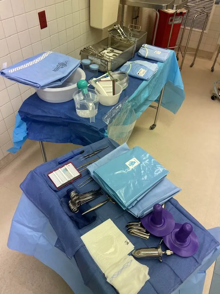
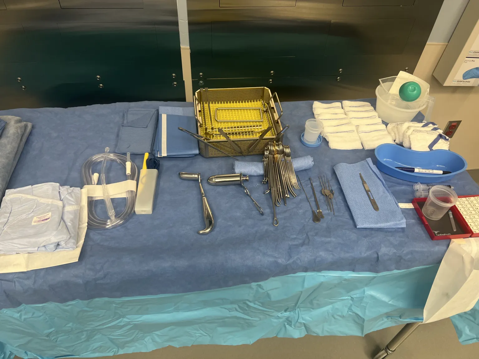
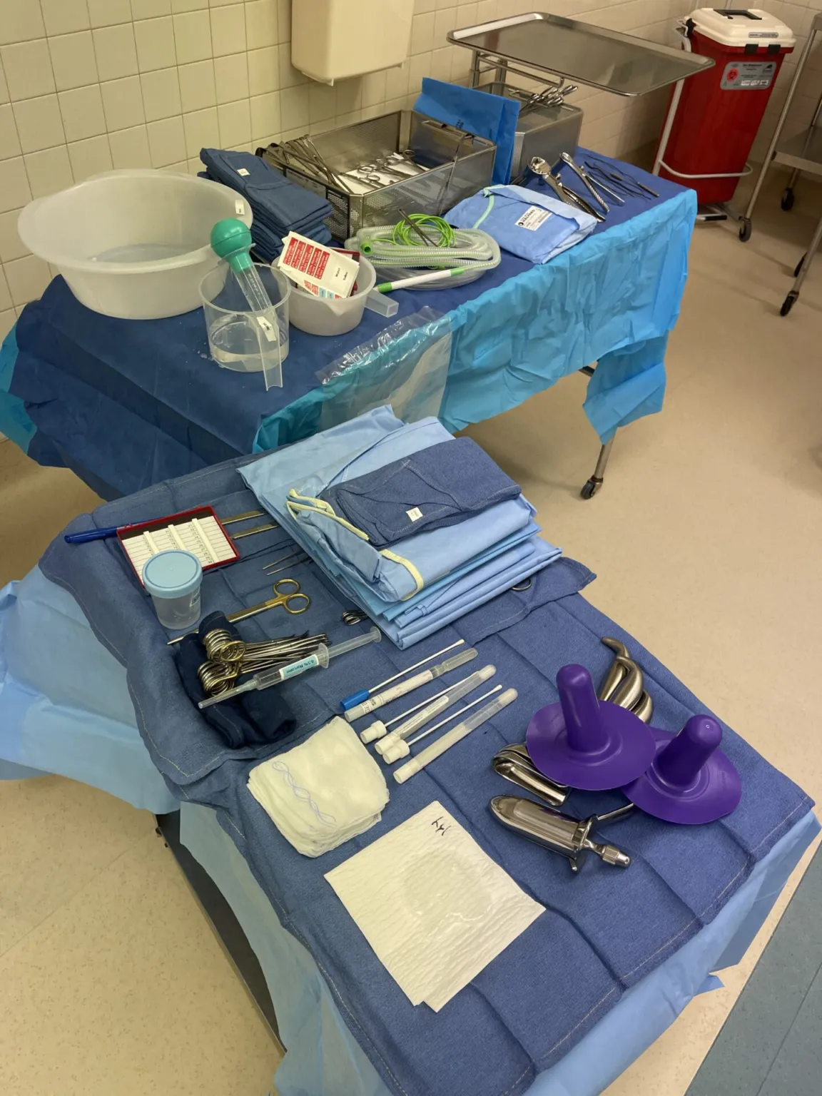
Surgical instruments
9 on this table. Tap a photo to zoom, the name for its card.
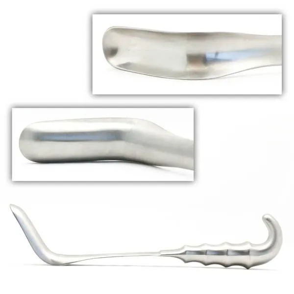
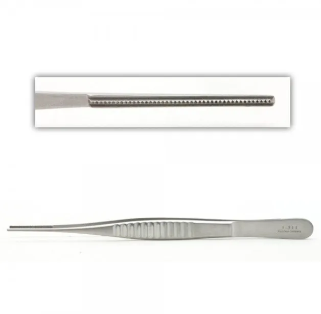
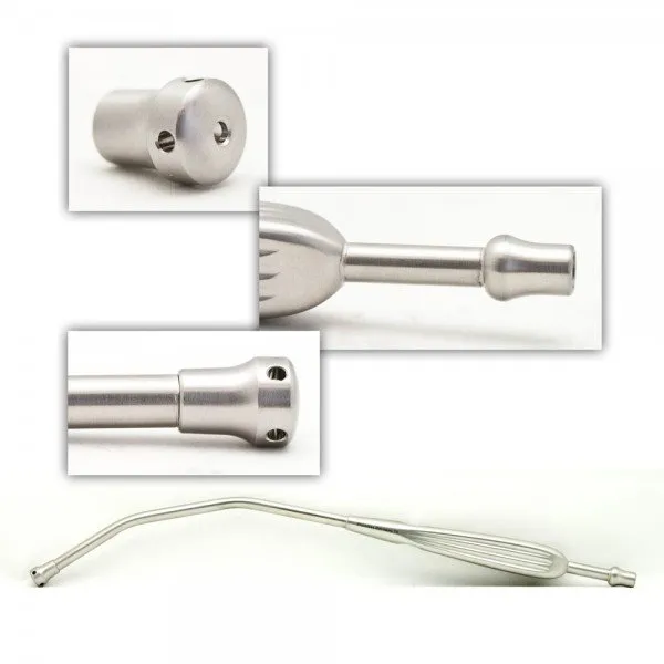
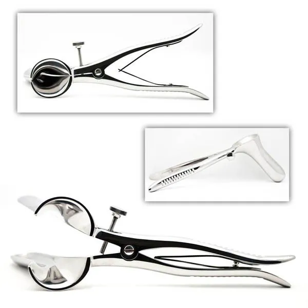
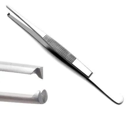
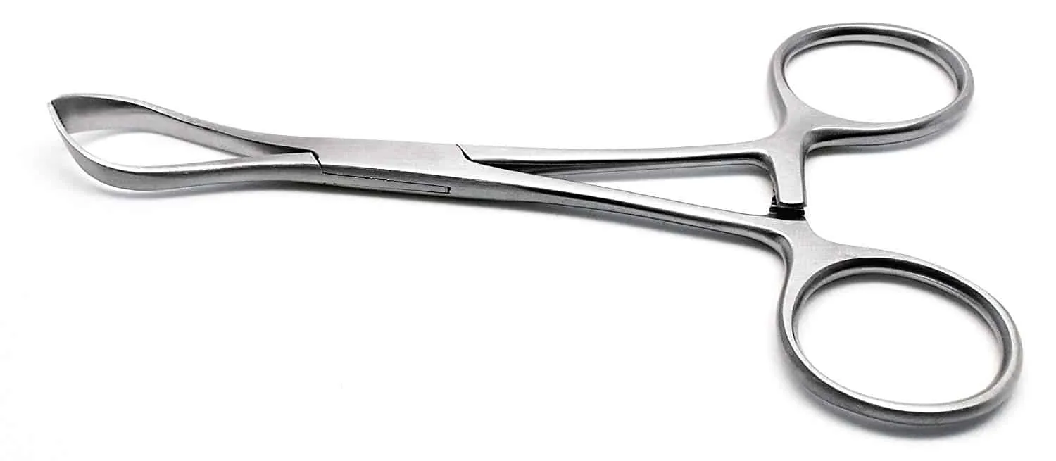

The case
Spark notes
The 30-second rundown. Scan this outside the room.
Walkthrough
What the surgeon does, and what to have in your hand before they ask.
Preoperative Preparation
Ensure the patient is either given a regional anesthetic or general anesthesia, placing them in the lithotomy position using stirrups or Yellofin leg holders. Utilize Betadine as a standard prep solution for colorectal surgery. Drape the patient with lithotomy drapes and, if applicable, pass off the ends of the suction and cautery to the circulator. Note that these items may not be needed for a digital rectal exam (DRE), typically performed in an office setting but occasionally required in the operating room.
Patient Draping
Proceed with draping the patient appropriately for the procedure, keeping in mind the lithotomy position.
Initial Examination
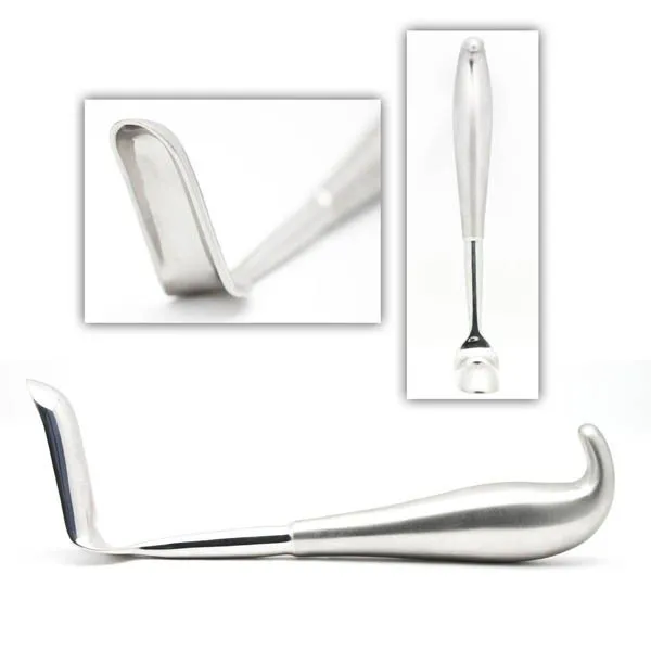
The surgeon begins the examination by inserting a lubricated rectal speculum, such as a Hill Ferguson or Pratt, into the rectum. This speculum facilitates continuous visualization throughout the case. If a self-retaining retractor is not used, the surgical technologist or another scrubbed-in individual may need to assist in holding the retractor. The surgeon visually inspects and palpates for any abnormalities, potentially making a diagnosis. In cases where consent is obtained, the surgeon may address and correct fissures or hemorrhoids during the same anesthesia period.
Instrumentation for Examination
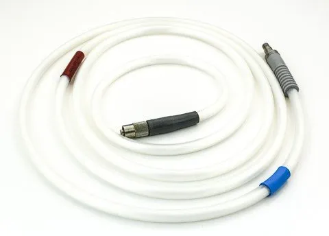
Ensure the availability of essential instruments for a straightforward rectal exam. These include the specified rectal speculums, sponge sticks with raytecs for cleaning and visualization, Debakeys pickups, toothed forceps, and a yankauer for suction. Confirm the surgeon's preference regarding the use of a sigmoidoscope for visualization and secure its availability before the patient enters the operating room. If a sigmoidoscope is used, be prepared with a light cord and a light source to illuminate the rectum.
Sigmoidoscope Considerations
In cases where the surgeon opts for a sigmoidoscope, inquire about the use of gas during the procedure. Ensure the availability of necessary equipment, such as a light cord with a light source, to adequately illuminate the rectum during the examination.
Postoperative Cleanup
Upon completion of the rectal exam, remove the drapes and wash off the Betadine or other prep solution from the patient. Use a wet towel or lap for this purpose, ensuring a thorough and efficient cleanup.
Add your table
Every surgeon runs it a little differently. Submissions stay private, are reviewed by our team, and accepted contributions earn a credit in the contributor list.