Bowel Resection (Colectomy/Hemicolectomy)
This procedure involves the surgical excision of a diseased segment of the large intestine, followed by the re-anastomosis of the remaining healthy bowel ends. The anatomical goal is the removal of compromised colonic tissue while restoring intestinal continuity.
29 instruments to master.
Master this setup: test yourself on every instrument, earn the tier, hold the record.
No site record yet · target 10:42
Study flashcards
Card by card, grade yourself · 29 cards
Practice test
Answer, see why · 25 at a time
Take the exam
30 questions · target 10:42
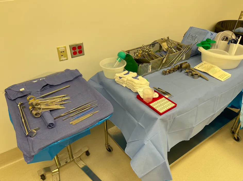
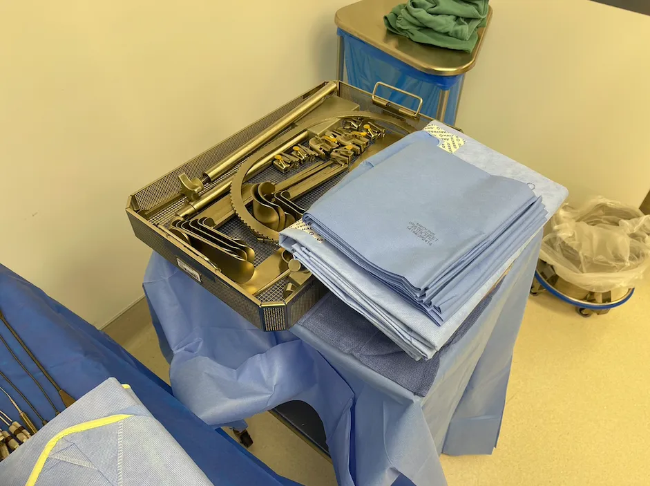
Surgical instruments
29 on this table. Tap a photo to zoom, the name for its card.
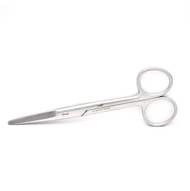
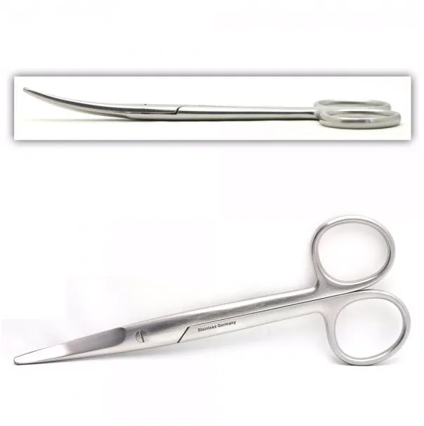
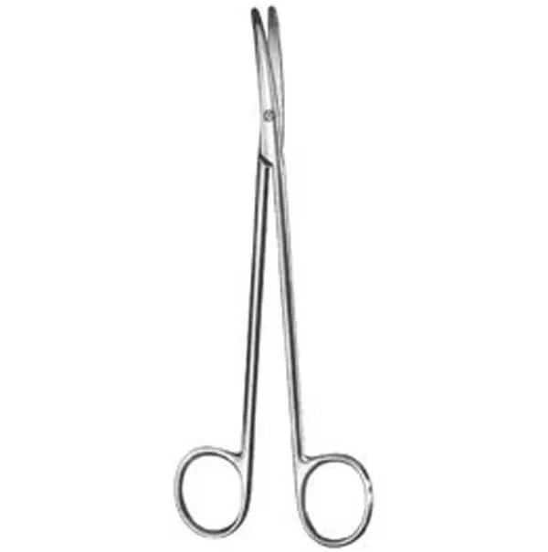
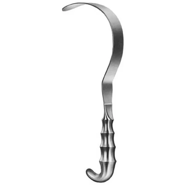
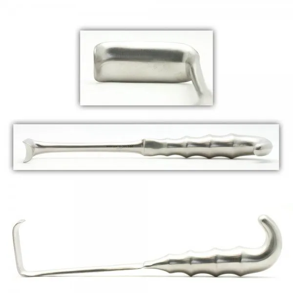
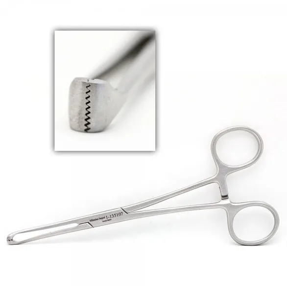
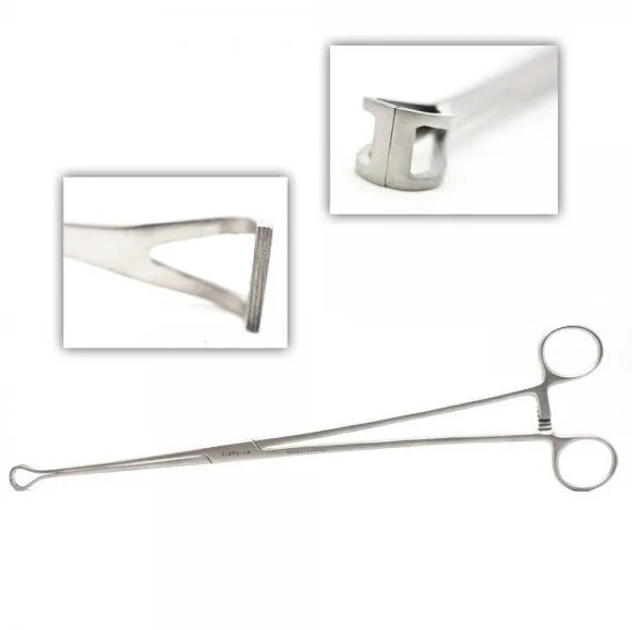
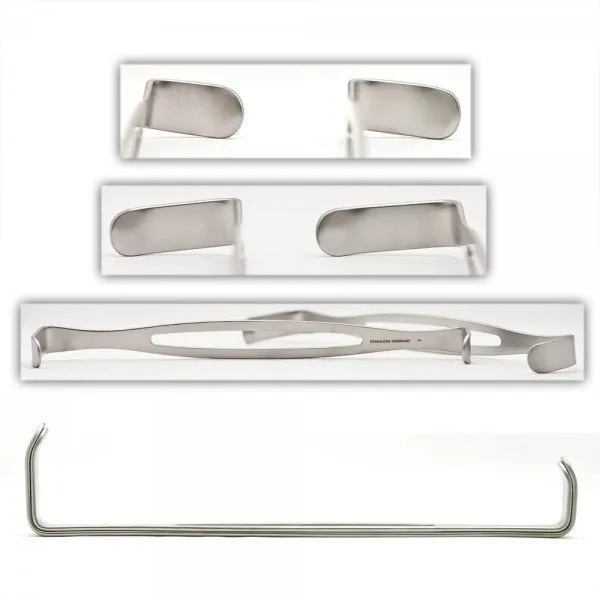
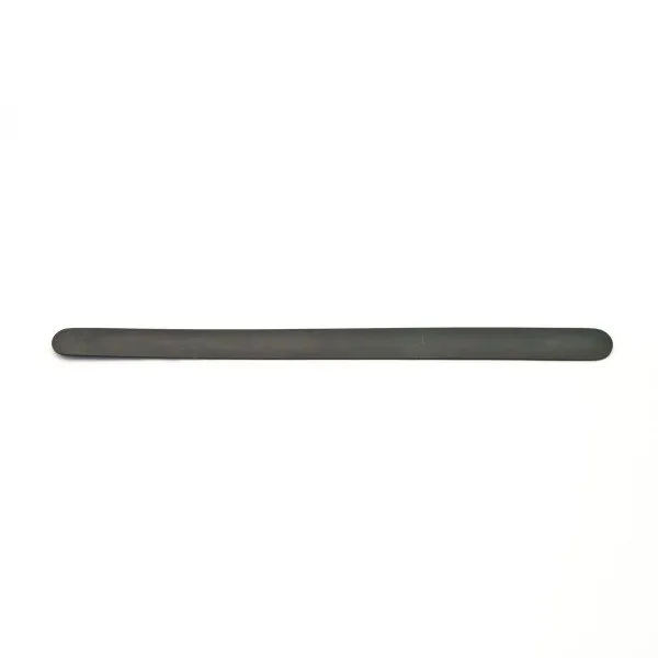
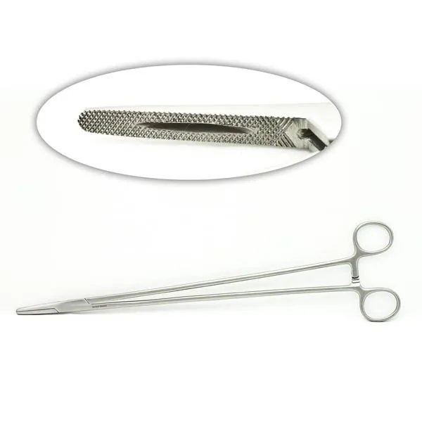
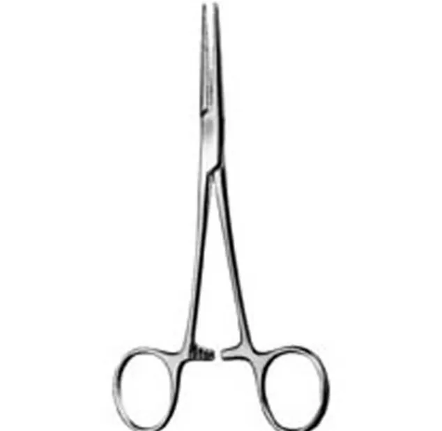
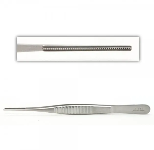
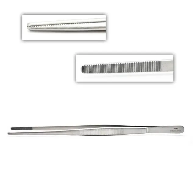
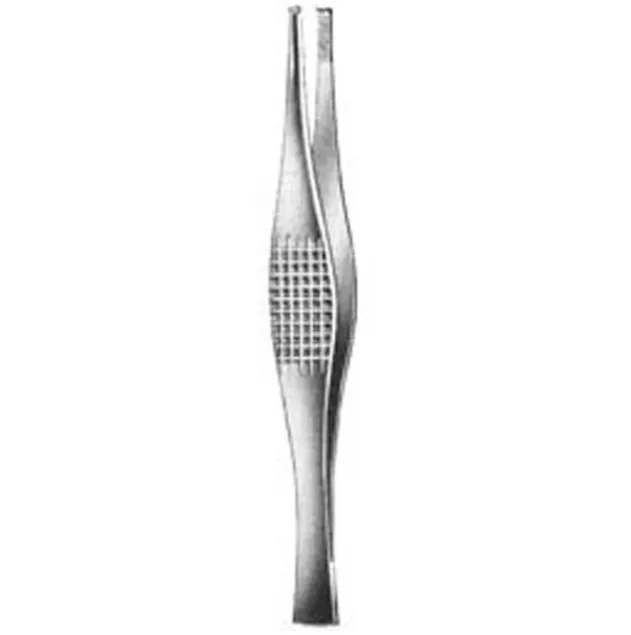
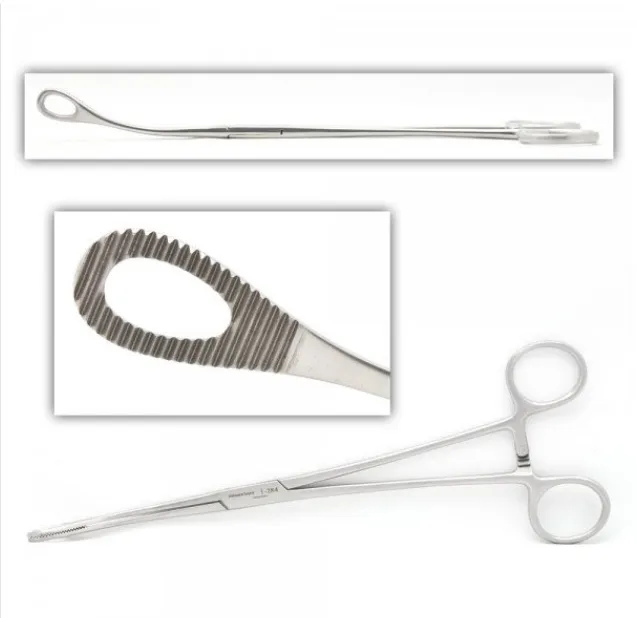
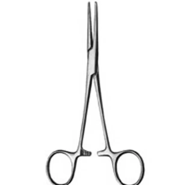
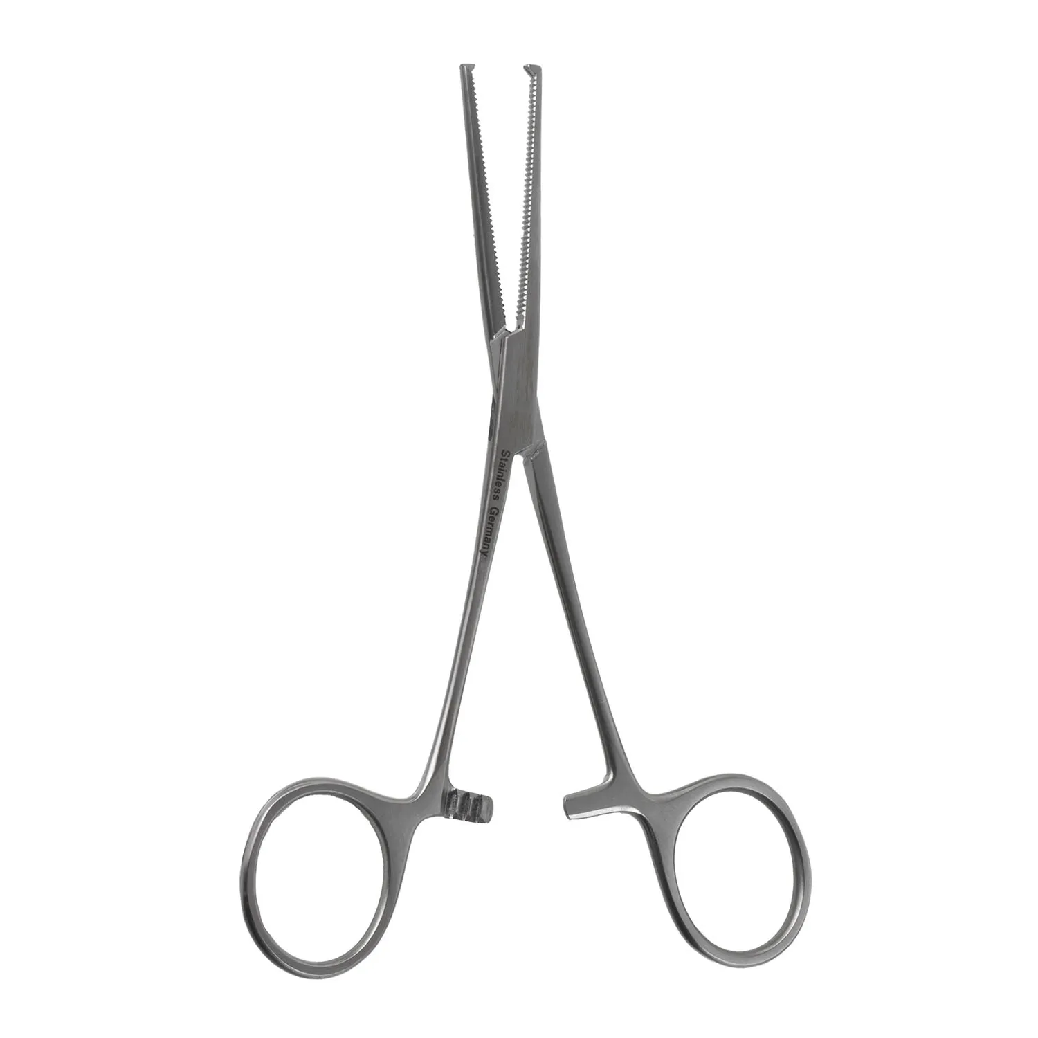
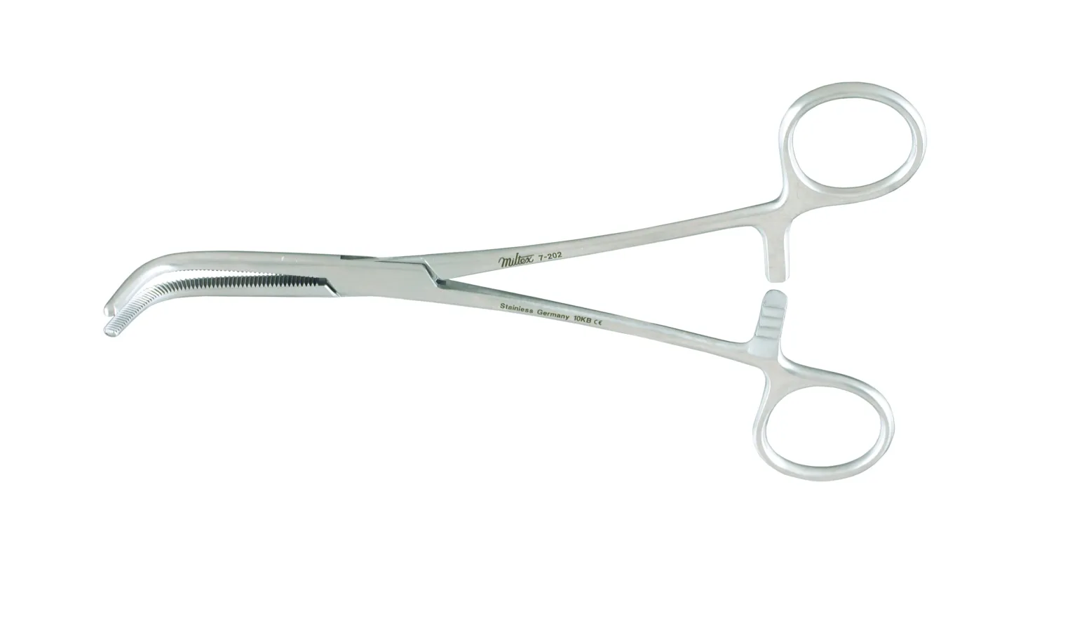
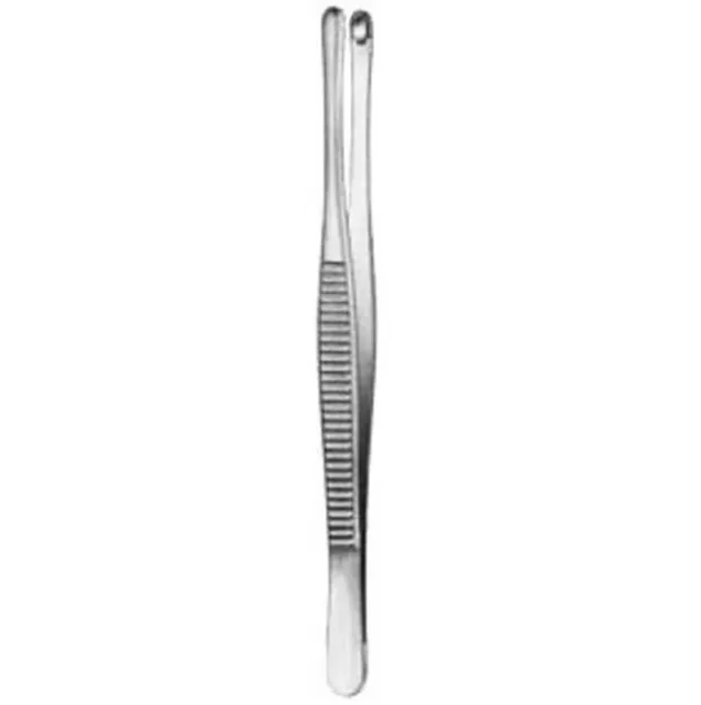
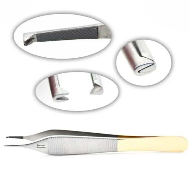
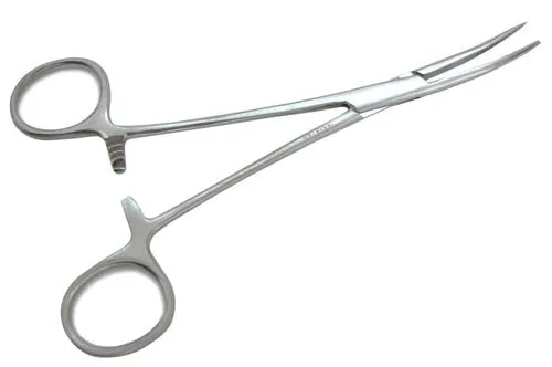
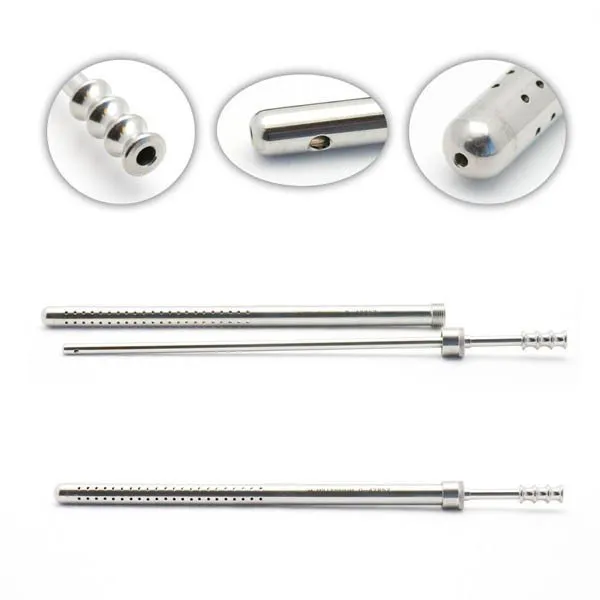
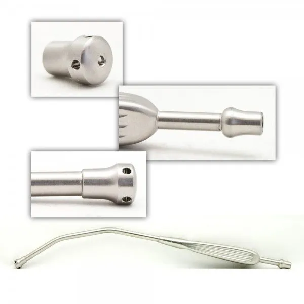
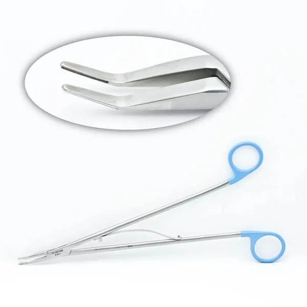
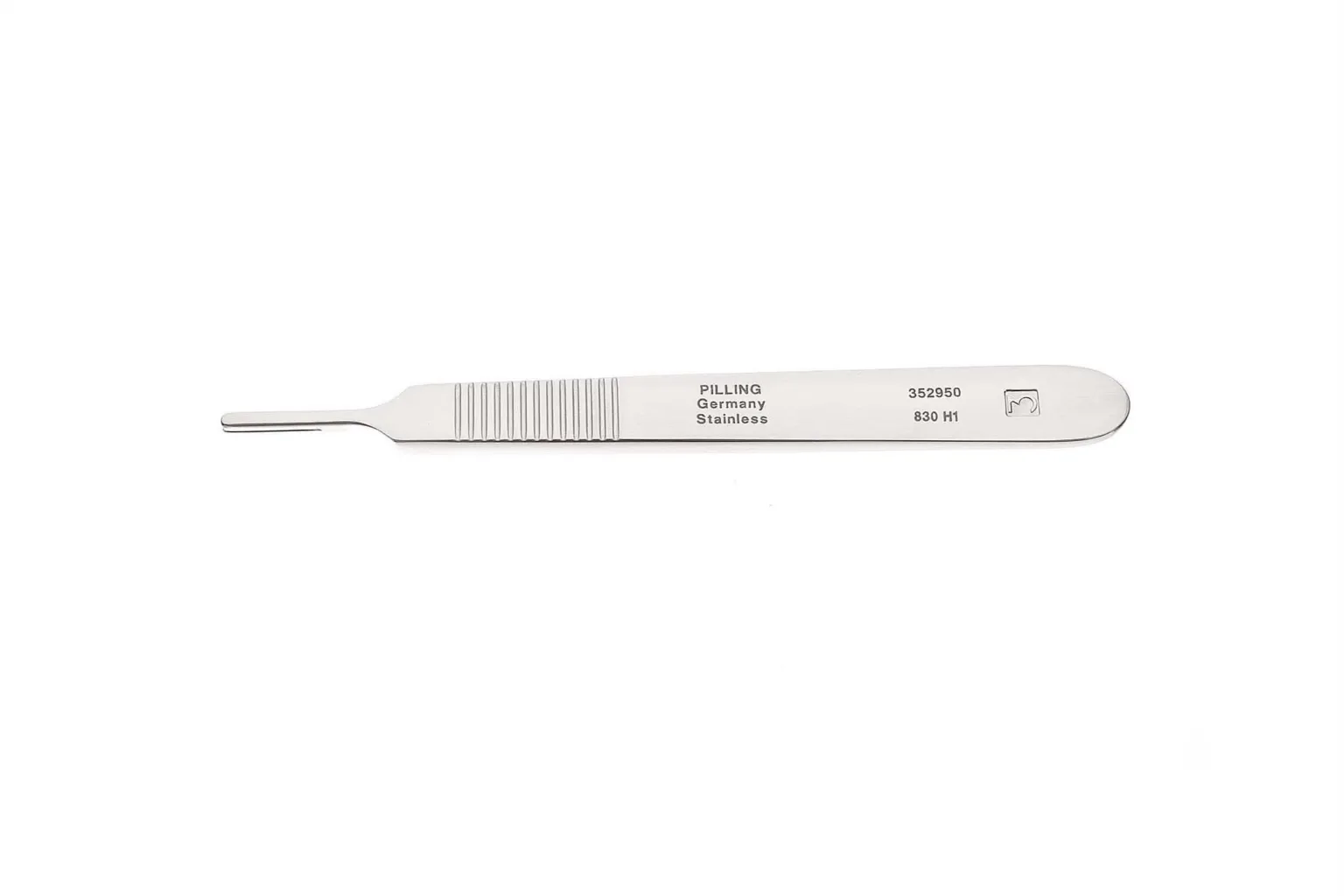
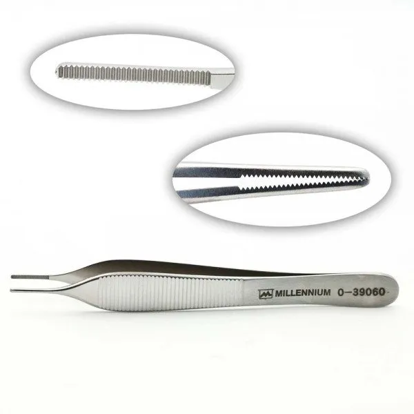
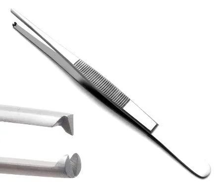
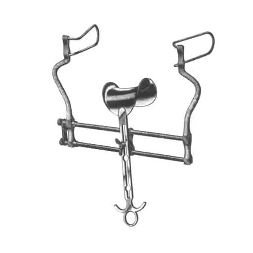
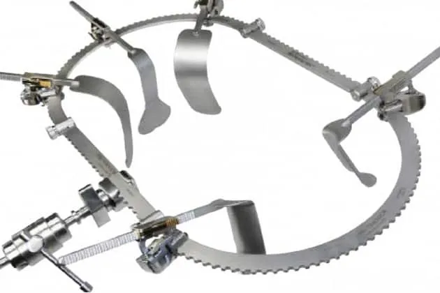
The case
Spark notes
The 30-second rundown. Scan this outside the room.
- Make incision with 10 blade to access small bowel.
- Cut and dissect mesentery while maintaining hemostasis.
- Identify and remove affected small bowel using TA or GIA stapler.
- Hand off resected piece as specimen.
- Closure begins in layers: peritoneum, fascia, skin.
- Scar after Hemicolectomy surgery
Walkthrough
What the surgeon does, and what to have in your hand before they ask.
Incision and Mesentery Dissection
The first step in a bowel resection surgery is making an incision to gain access to the small bowel. A 10 blade is typically used for this purpose. Once the incision is made, the mesentery is cut into and dissected. It is important to maintain hemostasis throughout this process to prevent excessive bleeding.
Identification and Removal of Affected Small Bowel
After the mesentery is dissected, the affected part of the small bowel is identified and removed. This is typically done using either a TA or GIA stapler to connect the two ends of small bowel back together. The resected piece of small bowel should be handed off as a specimen for further analysis.
Closure
Once the affected section of small bowel has been removed, closure begins in layers. The first layer of closure is typically the peritoneum, which is closed using a running absorbable suture. Next, the fascia is closed with interrupted non-absorbable sutures. Finally, the skin is closed using either a running subcuticular suture or staples.
Tips and tricks
Have long instrument trays available, as well as a Balfour and Bookwalter retractor. Always have different size clips easily accessible. The resected part of the bowel being removed should be kept separate in order to avoid contaminating the rest of the sterile setup. Any instruments that become contaminated with bowel contents should also be taken off of the sterile field. A separate sterile field may be requested for the end of the case for closing, containing extra gowns, gloves, and a minor procedure tray.
Add your table
Every surgeon runs it a little differently. Submissions stay private, are reviewed by our team, and accepted contributions earn a credit in the contributor list.