Kidney Transplant
The surgical implantation of a healthy donor kidney into the iliac fossa of a recipient with end-stage renal disease. The procedure involves establishing vascular anastomoses between the donor vessels and the recipient's iliac artery and vein, followed by a ureteroneocystostomy to connect the donor ureter to the bladder.
26 instruments to master.
Master this setup: test yourself on every instrument, earn the tier, hold the record.
No site record yet · target 9:38
Study flashcards
Card by card, grade yourself · 26 cards
Practice test
Answer, see why · 25 at a time
Take the exam
27 questions · target 9:38
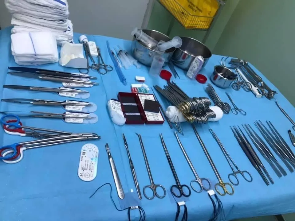
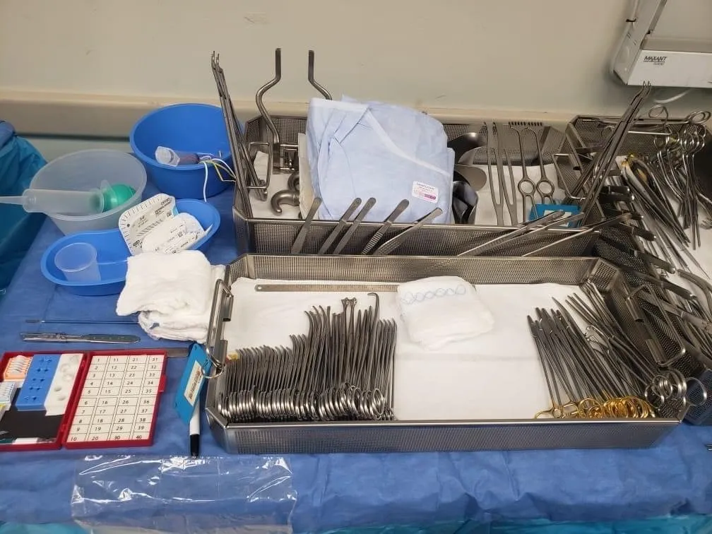
Surgical instruments
26 on this table. Tap a photo to zoom, the name for its card.

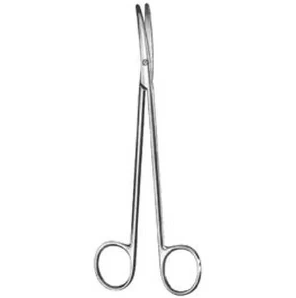
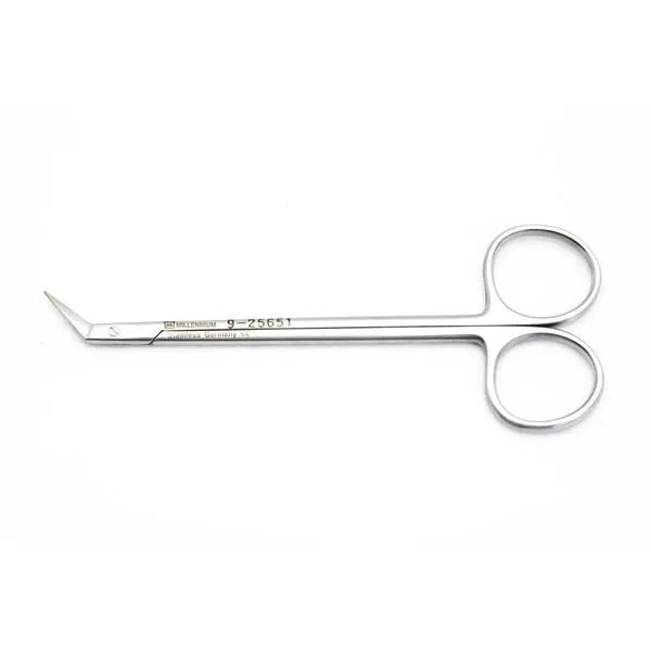
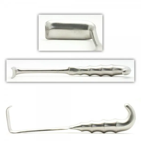
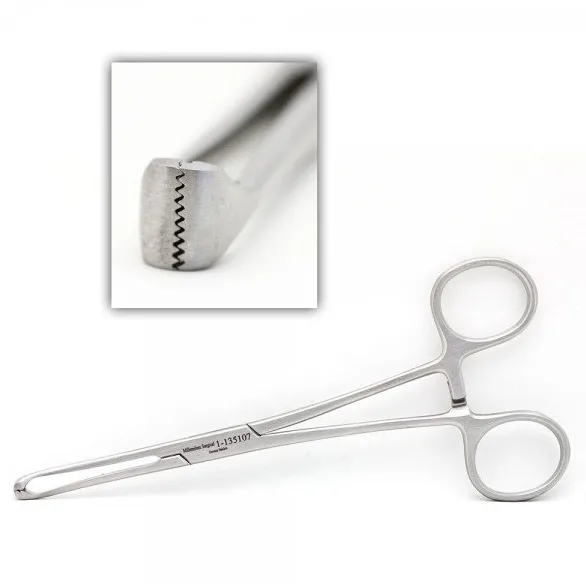
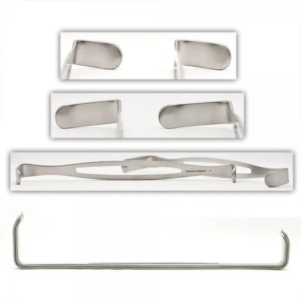
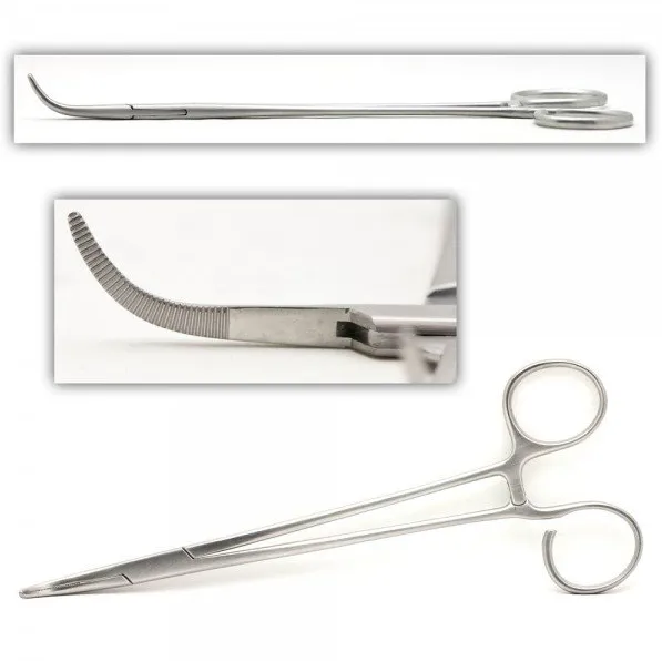
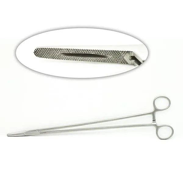
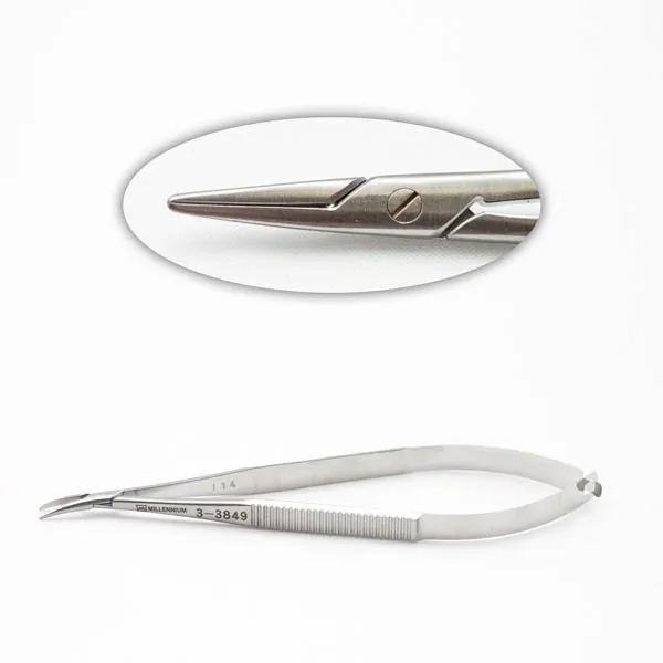
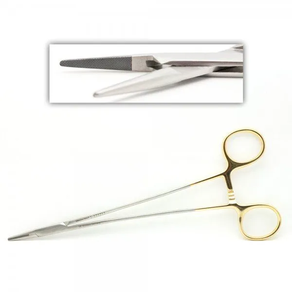
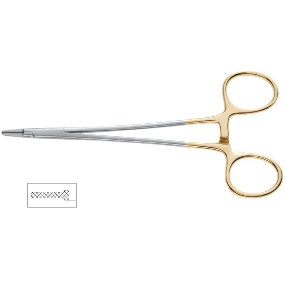
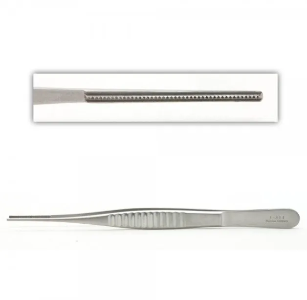
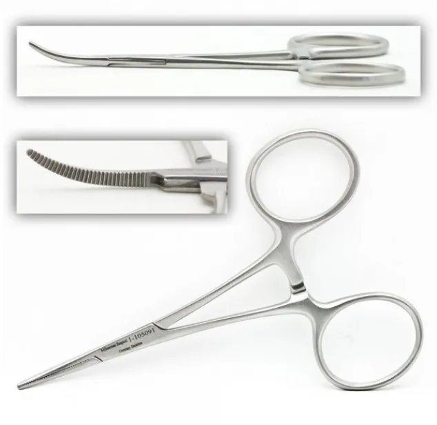
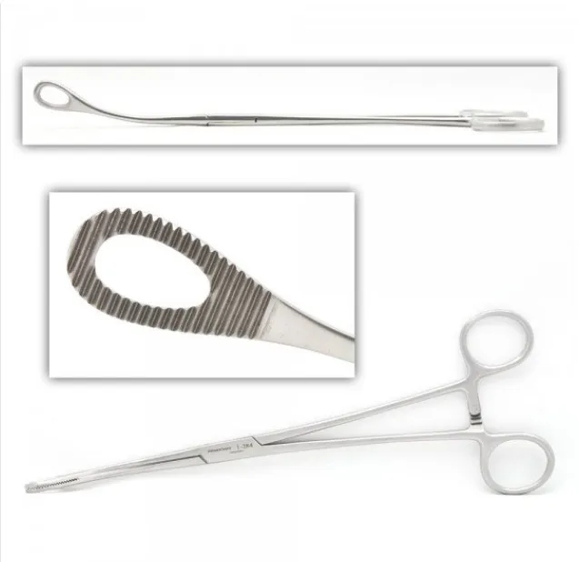
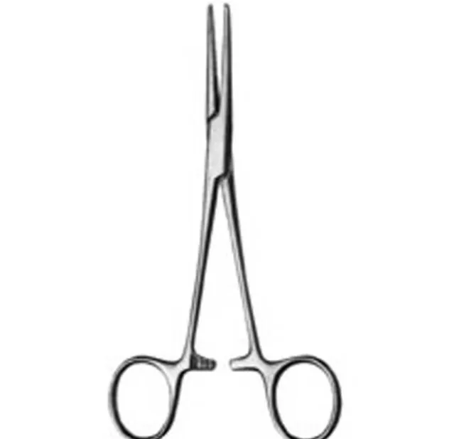
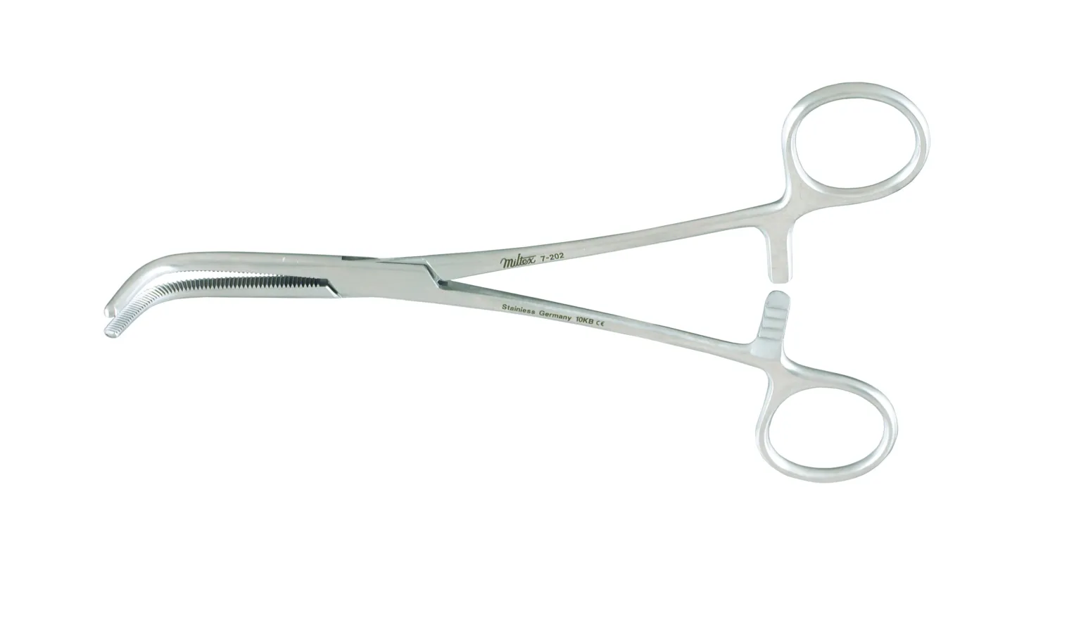
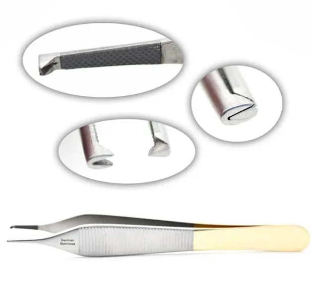
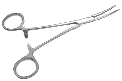
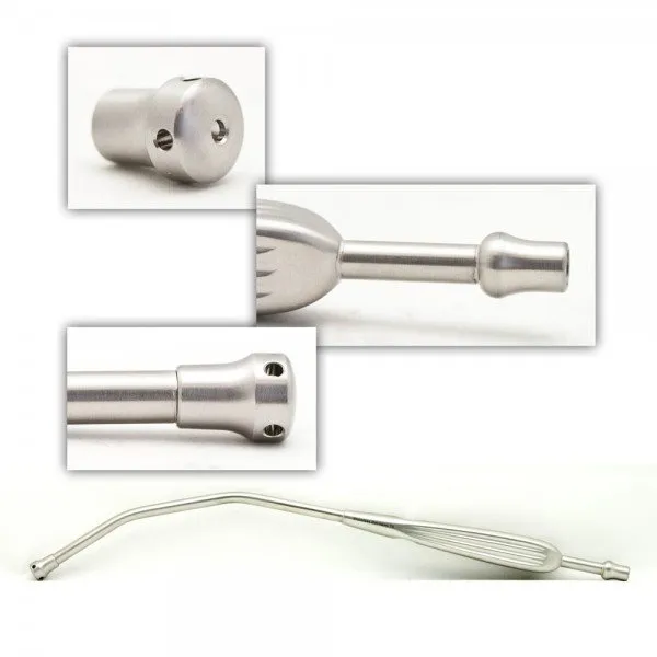
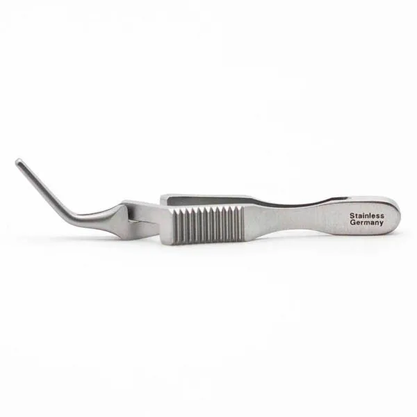
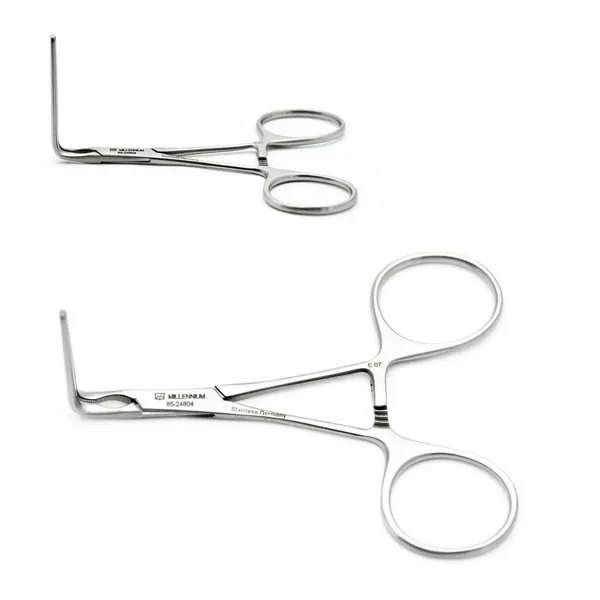
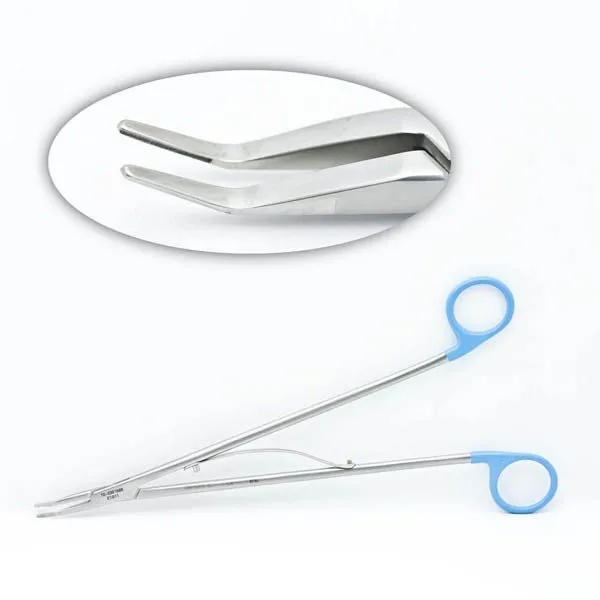
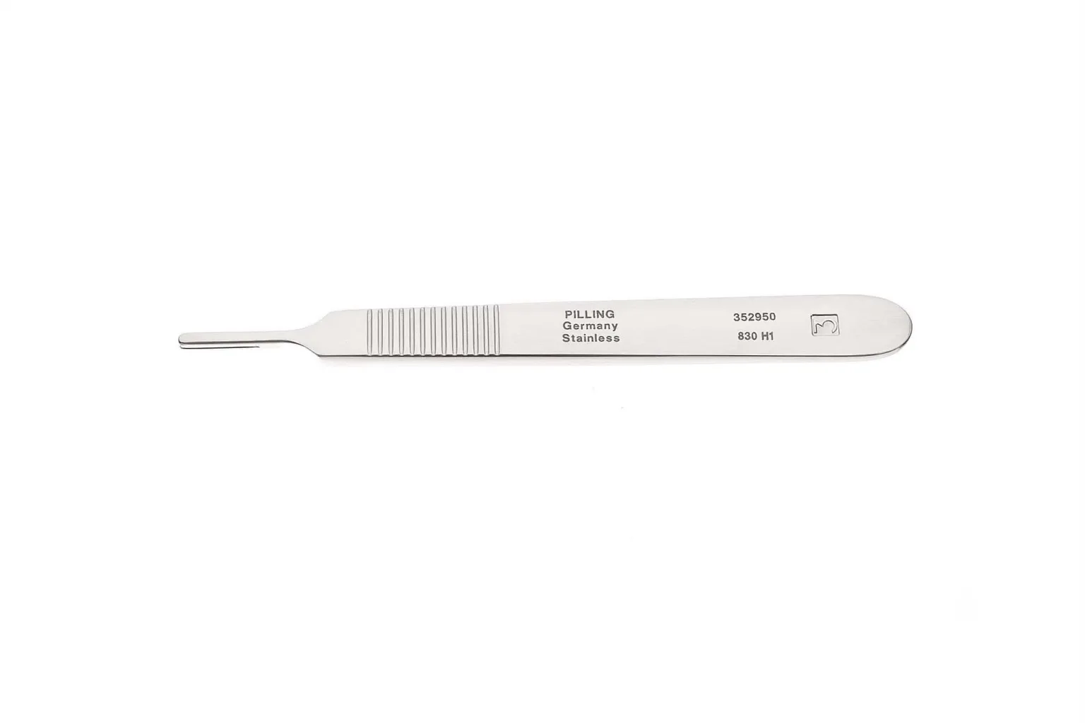
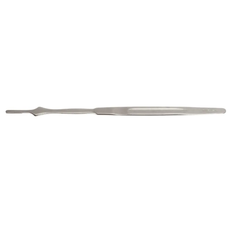
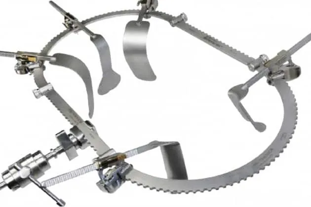
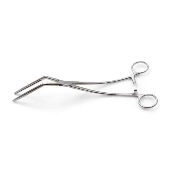
The case
Spark notes
The 30-second rundown. Scan this outside the room.
- Prepare donor kidney by cleaning off fat and shortening vessels.
- Place kidney in iliac fossa or retroperitoneum through large incision.
- Dissect external oblique muscle and fascia, divide internal oblique and transverse muscles to expose peritoneum, ligate inferior epigastric and preserve spermatic cord.
- Anastomose renal artery and vein to external iliac artery and vein using small Prolene suture.
- Trim and fit aortic cuff to renal artery and attach it to arteriotomy in external iliac artery.
- Reconstruct urinary tract by incising bladder, finding patient's ureter, making an incision in it, feeding donor ureter through it, and anastomosing it with absorbable suture.
- Close bladder incision and check for leaks.
- Reapproximate tissue and close with suture.
- Use booties for clamping suture and ensure ties are available on the sterile field.
Walkthrough
What the surgeon does, and what to have in your hand before they ask.
Preparing the Donor Kidney
The donor kidney will be brought to the operating room and will be prepared for implantation into the recipient. This involves cleaning off any excess fat and dissecting the vessels clean. The vessels may be shortened to make the anastomosis easier. If the kidney came from a deceased patient, an aortic cuff may be used.
Reconstructing the Veins
The veins need to be reconstructed, and there will be branches that will need to be ligated, but a large renal vein will be kept.
Placing the Kidney
The kidney will either be placed in the iliac fossa or in the retroperitoneum.
Making the Incision
A large incision with a 10 blade will be made, and the external oblique muscle and fascia are dissected. The internal oblique and transverse muscles will be divided to expose the peritoneum. The inferior epigastric will be ligated, and the spermatic cord will be preserved.
Anastomosing the Renal Artery and Vein
The renal artery and vein will be anastomosed. The renal vein will be attached to the external iliac vein, usually with a small 5-0 Prolene suture. The artery will be attached to the external iliac artery. The aortic cuff will be trimmed and fitted to the renal artery and then attached to the arteriotomy in the external iliac artery.
Reconstructing the Urinary Tract
Now, the urinary tract will be reconstructed. The bladder will be incised, and the patients' ureter is found. An incision is made in it. The donor ureter will be fed through the incision into the bladder and anastomosed with absorbable suture. The bladder incision is then closed, and the bladder is filled to check for leaks.
Closing the Incision
The tissue will be reapproximated and closed with suture.Common sutures used for 5-0 and 6-0 Prolene, 1 and 4-0 PDS, 2-0 and 3-0 silk ties, and #5 Ethibond or Nylon.
Tips and tricks
It's important to place booties on some mosquitos for clamping suture, and ties should always be available on the sterile field.
Add your table
Every surgeon runs it a little differently. Submissions stay private, are reviewed by our team, and accepted contributions earn a credit in the contributor list.