Laparoscopic Cholecystectomy (Lap Chole)
Minimally invasive removal of the gallbladder through small keyhole incisions using a camera and specialized instruments.
18 instruments to master.
Master this setup: test yourself on every instrument, earn the tier, hold the record.
No site record yet · target 7:08
Study flashcards
Card by card, grade yourself · 18 cards
Practice test
Answer, see why · all 20
Take the exam
20 questions · target 7:08
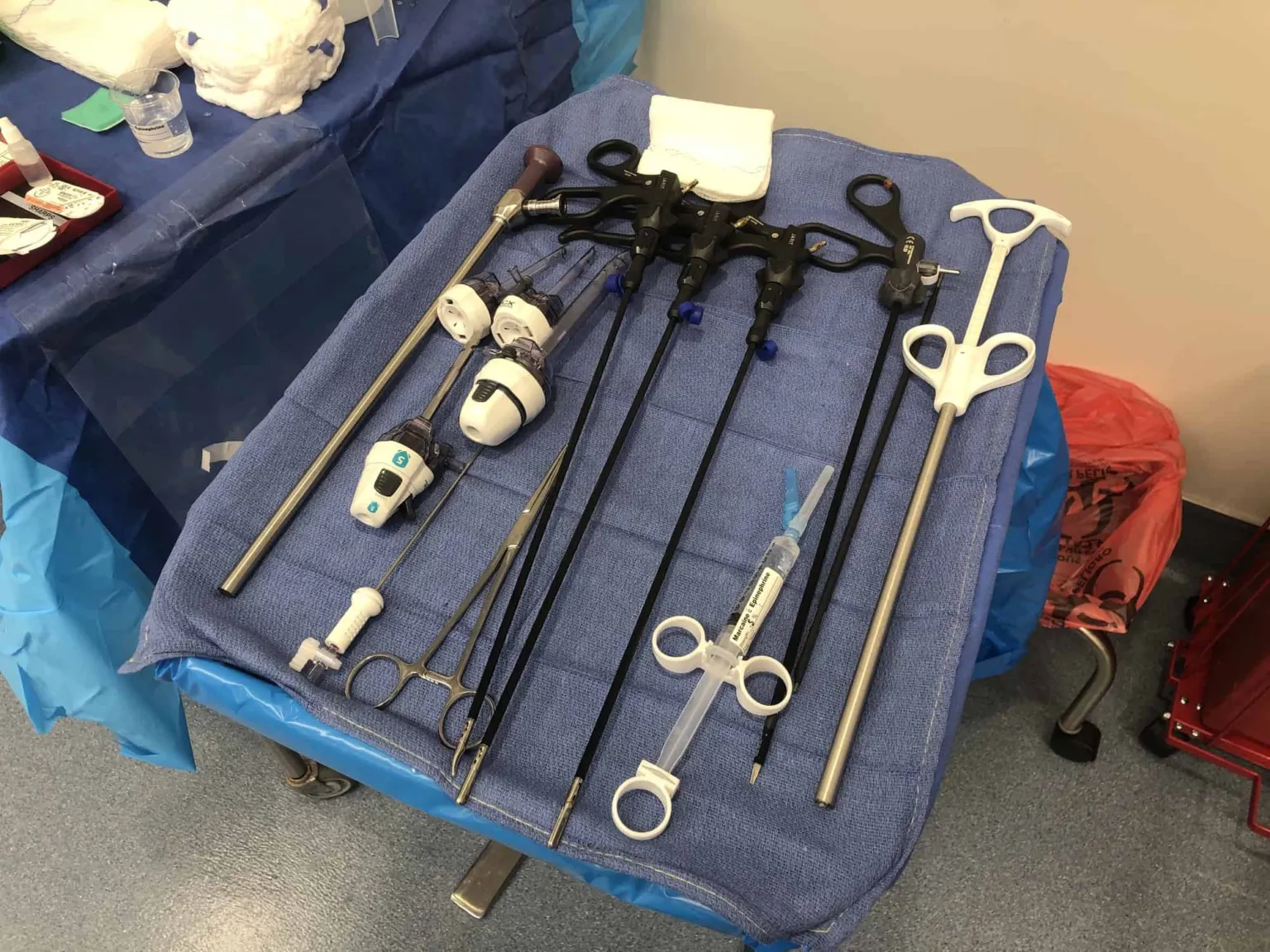
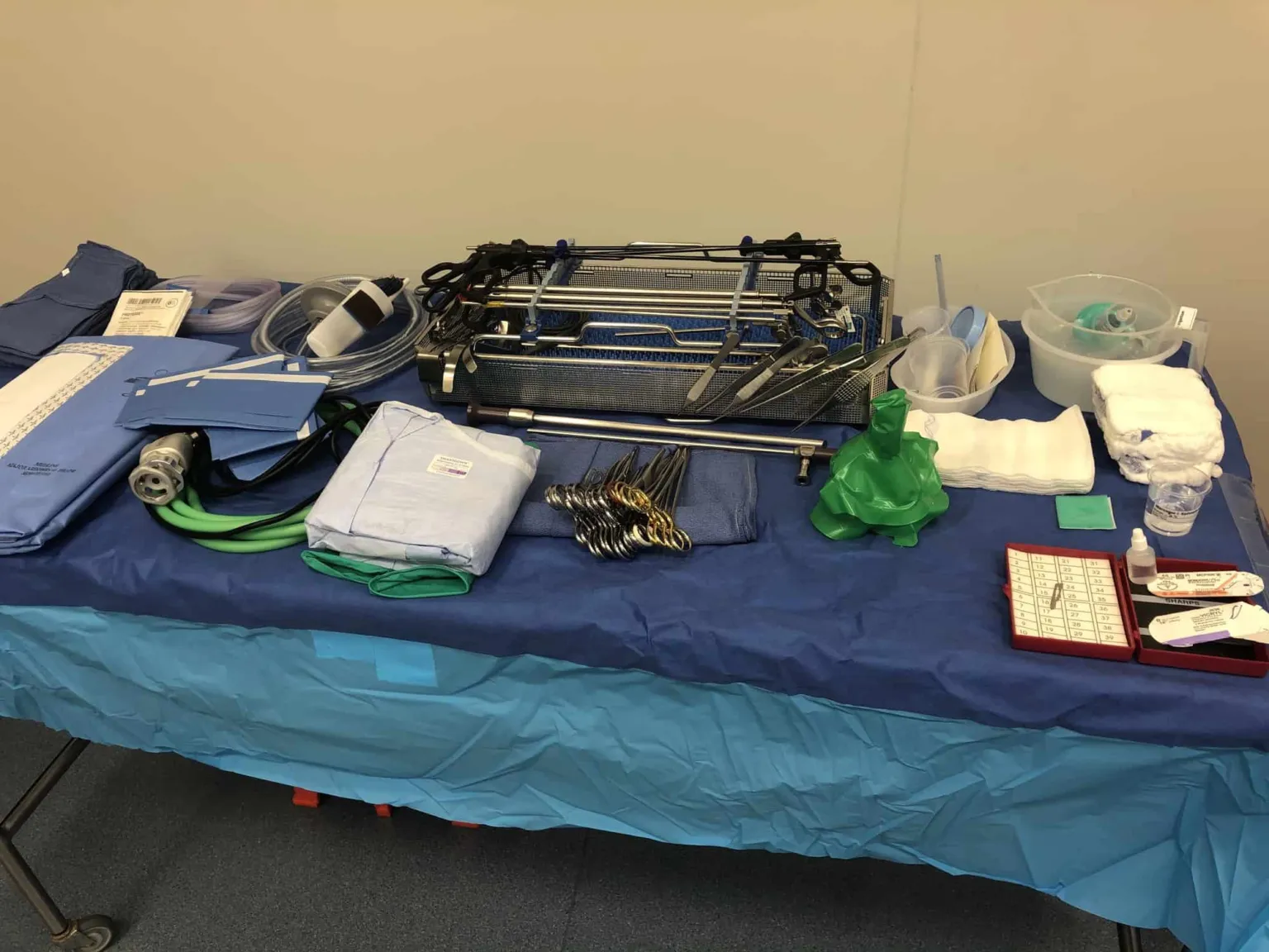
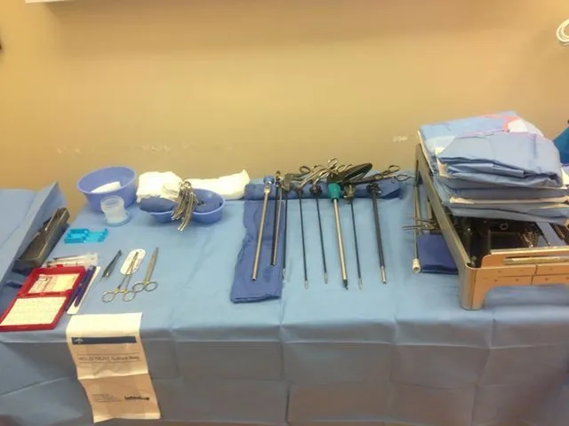
Surgical instruments
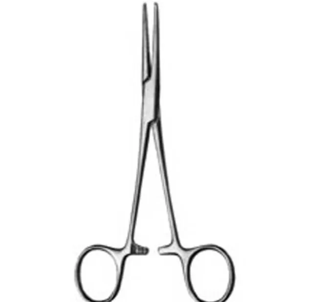
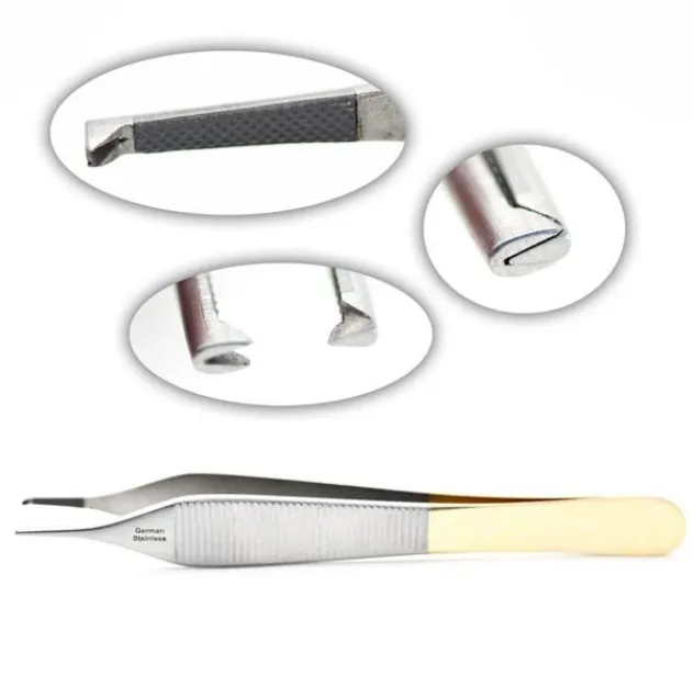
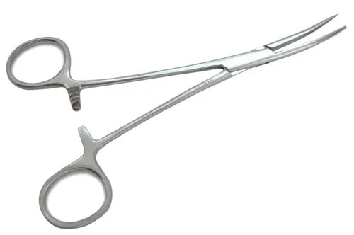
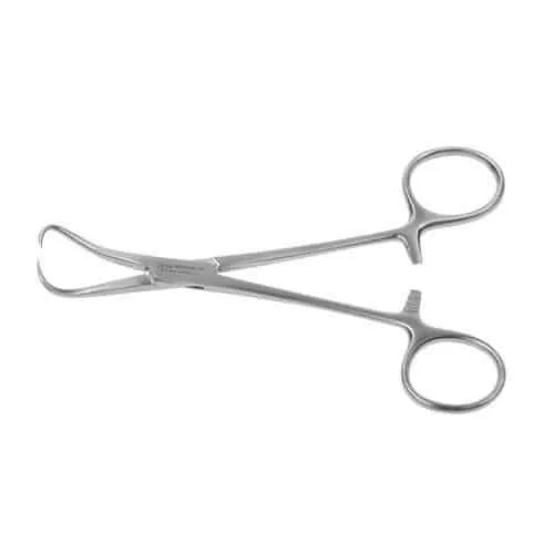
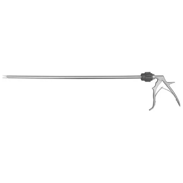
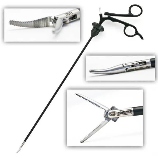
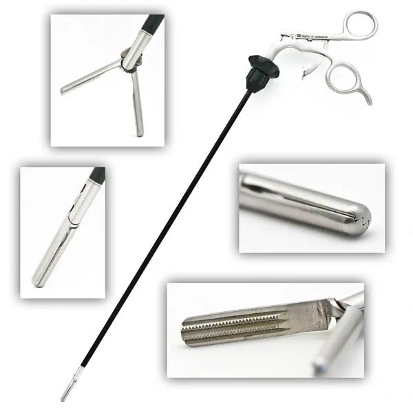
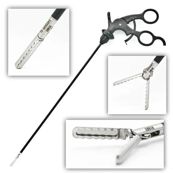
18 on this table. Tap a photo to zoom, the name for its card.
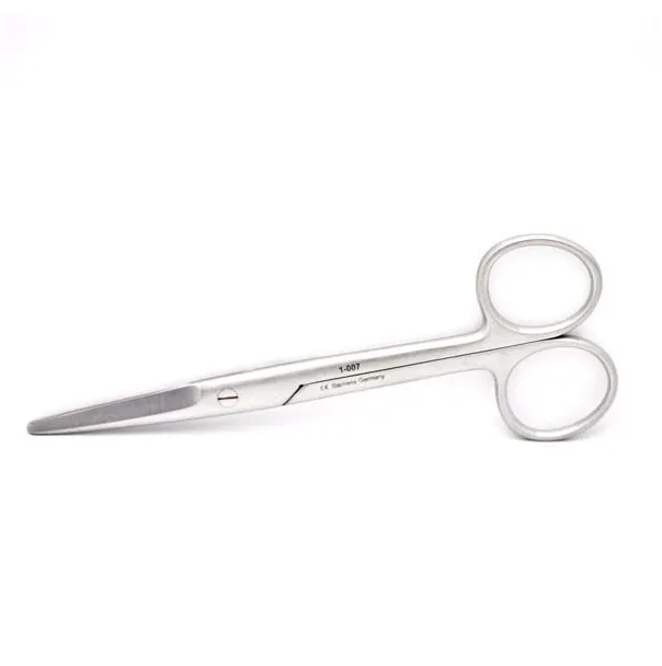
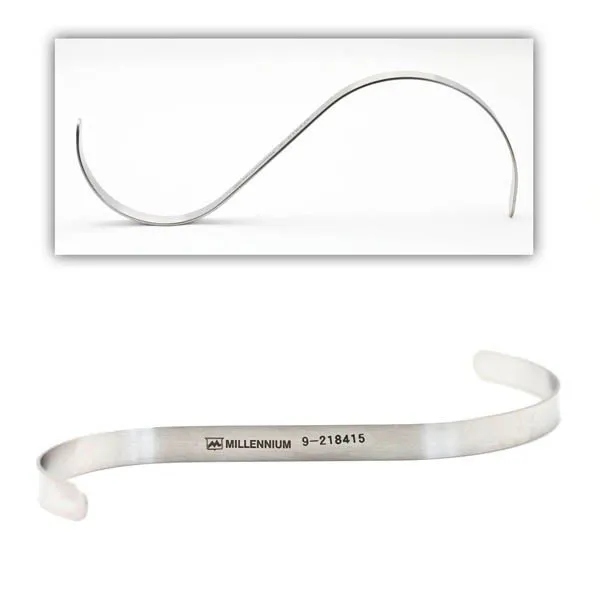
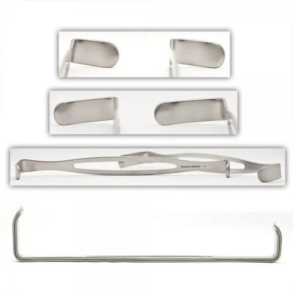
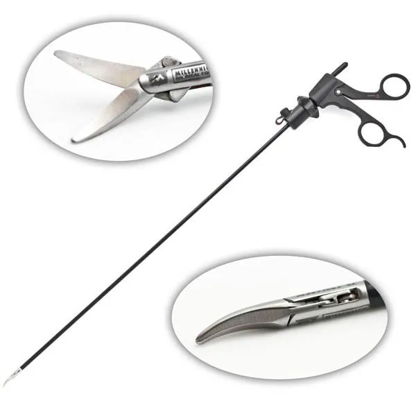
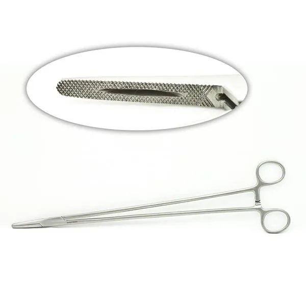
The case
Spark notes
The 30-second rundown. Scan this outside the room.
Walkthrough
What the surgeon does, and what to have in your hand before they ask.
Incision and Trocar Insertion
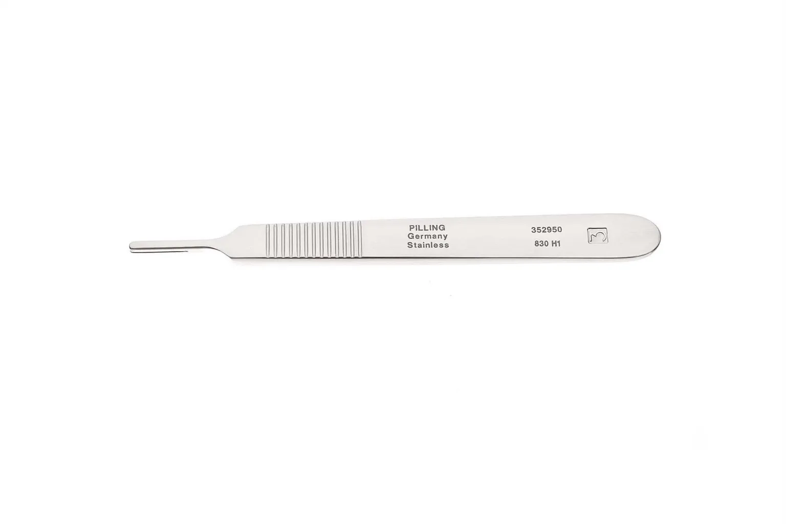
The first step in a laparoscopic cholecystectomy is to make approximately 3-4 small incisions. The scalpel is used to create these incisions, which are then used to insert trocars. The S retractors or Army-Navys may be used to retract the incision area while inserting the trocars.
Verres Needle Insertion
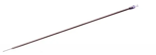
A Verres needle may be introduced into the umbilical incision to introduce CO2 into the abdomen. To insert the Verres needle, a Kelley and two towel clips should be available. Once the needle is inserted, a syringe of saline may be needed to check for proper placement.
Pneumoperitoneum
Another method for creating pneumoperitoneum is to insert a trocar and then attach the CO2 tubing. This method can be used if the Verres needle is not used.
Gallbladder Dissection
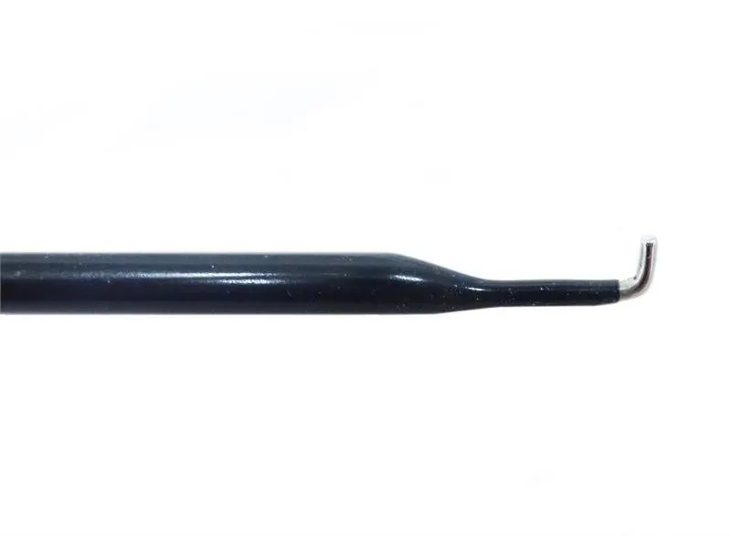
Once the pneumoperitoneum is established, a locking grasper is used to grasp the gallbladder and retract it. Fat or adhesions can be dissected with graspers, hook cautery, and Maryland. When properly dissected, the gallbladder hilum is visualized along with the cystic duct and artery.
Cystic Artery Clip Applier
The clip applier is then used on the cystic artery two times. This is when a cholangiogram may be performed. The laparoscopic scissors will be used to incise the cystic duct. The cholangiogram catheter will be inserted into the duct’s incision, and the clip applier will be used again to hold it in place. Saline and radiopaque dye should be available and drawn up into syringes. X-ray will be used for visualization. The cholangiogram catheter will then be removed, and the clip applier will be used again on the duct.
Gallbladder Removal
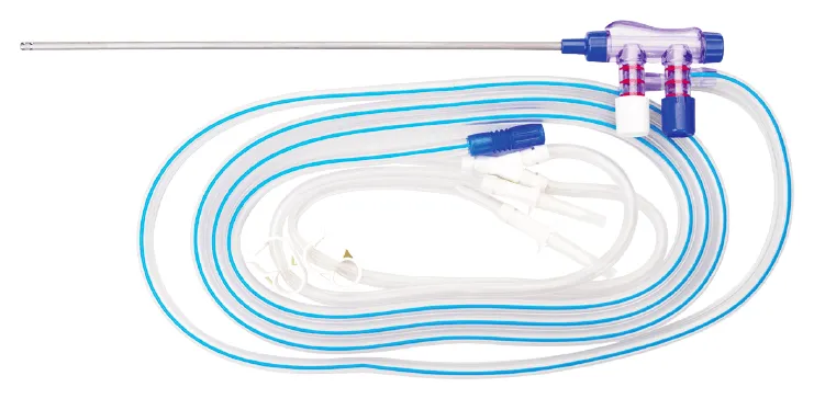
The rest of the gallbladder will then be dissected from the liver, most likely using the hook cautery and wave graspers. The suction/irrigator should then be used to visualize any bleeding on the liver. An endopouch will then be inserted, and the gallbladder will be removed from the port. If there are stones in the gallbladder preventing it from fitting through the port site, the gallbladder may be aspirated, and the stones may need to be removed.
Suturing and Closure
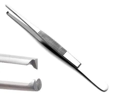
Before skin closure, the doctor may use the suction irrigator one last time. The trocars will then be removed, and the port sites will be closed. A needle driver and an adson pick-up will be needed for suturing, and the straight mayo scissors should be available for cutting sutures.
Add your table
Every surgeon runs it a little differently. Submissions stay private, are reviewed by our team, and accepted contributions earn a credit in the contributor list.