Laparoscopic Transhiatal Esophagectomy
The esophagus is mobilized through the diaphragmatic hiatus and excised, followed by the creation of a gastric pull-up to restore gastrointestinal continuity.
- Position
- Reverse Trendelenburg
In 30 seconds
Scan this outside the room.
- Place five ports: 12mm above bellybutton for laparoscope, two 5mm ports for retractionrih-TRAK-shunA drawing back or pulling in of tissue; in breathing, the skin sinking in between, below or above the ribs with each breath.Full entry , and two 12mm ports for grasping and dissectiondy-SEK-shunA tear in the inner layer of an artery wall that lets blood split the layers apart; also, the cutting apart of tissue in surgery.Full entry .
- Use grasping instruments for dissectiondy-SEK-shunA tear in the inner layer of an artery wall that lets blood split the layers apart; also, the cutting apart of tissue in surgery.Full entry of tissue and nodes.
- Ligating left gastric artery and vein using clips, L hook cautery, and scissors.
- Use GIA stapler for gastric tubulization and mobilize the esophagusih-SOF-uh-gusThe muscular tube that carries food and liquid from the throat to the stomach.esophag/o esophagusFull entry .
- Perform an esophagogastric anastomosisuh-nas-tuh-MOH-sisA connection between two tube-shaped structures, such as two ends of bowel or two blood vessels, made by surgery or found naturally.Full entry after pulling the esophagusih-SOF-uh-gusThe muscular tube that carries food and liquid from the throat to the stomach.esophag/o esophagusFull entry and gastric tubuleTOO-byoolA very small tube in the body, most often one of the tiny tubes in the kidney that turn filtered fluid into urine.Full entry through the cervicotomy.
- Place a nasogastric tubenay-zoh-GAS-trik toobA thin, flexible tube passed through the nose and down the esophagus into the stomach.nas/o nosegastr/o stomach-ic having to do withFull entry and enteral feeding tubeEN-ter-ul FEE-ding toobA tube placed into the stomach or small intestine to give liquid food, water and drugs to a person who cannot eat enough by mouth.Full entry , and close the incisions.
The tray
19 instruments, by what they do. Tap a name for its card.
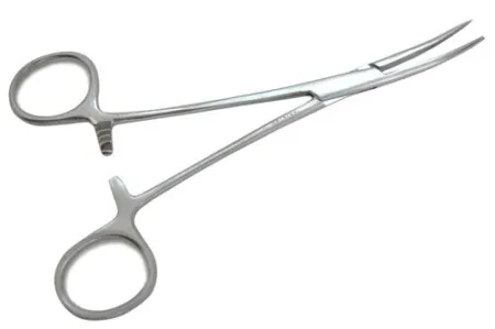 Curved HemostatsClamping
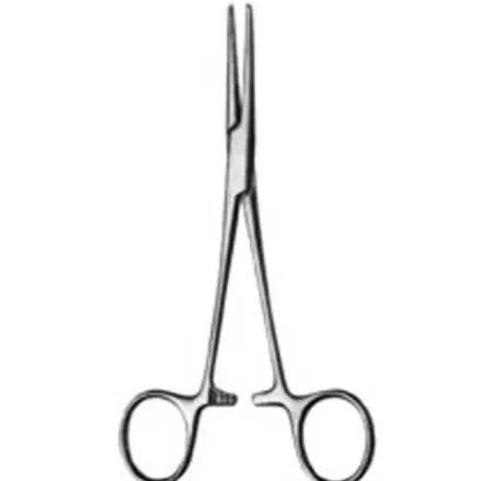 Kelly ForcepsClamping
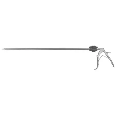 Laparoscopic Clip AppliersClamping
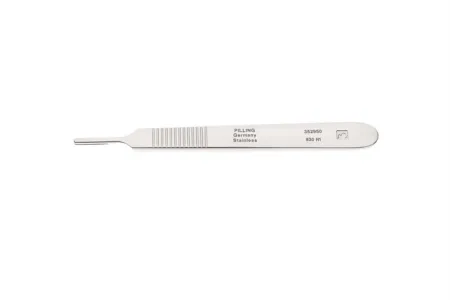 #3 Knife HandleCutting
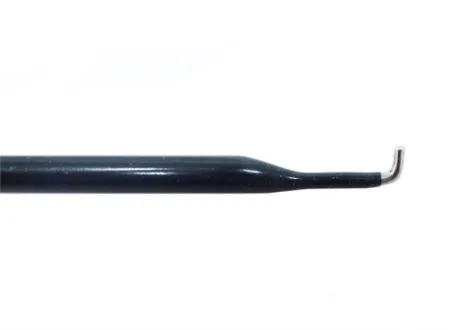 L Hook CauteryCutting
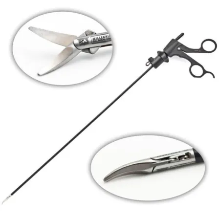 Laparoscopic Metz ScissorCutting
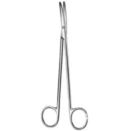 Metzenbaum ScissorCutting
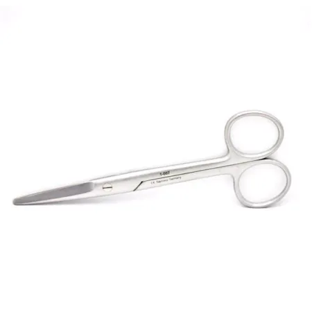 Straight Mayo ScissorCutting
All 19 instruments
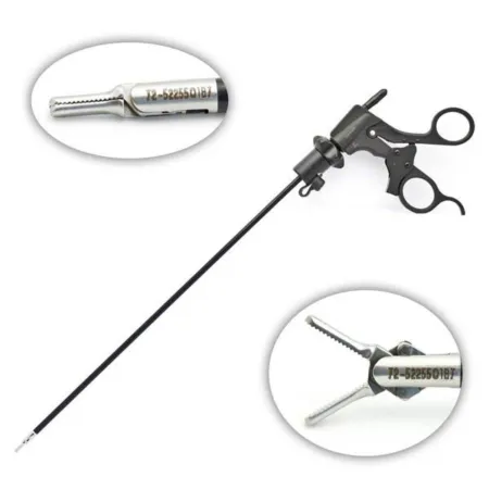 Dissecting Laparoscopic Grasper ForcepsGrasping
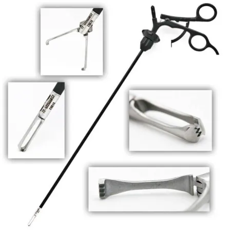 Laparoscopic Allis GrasperGrasping
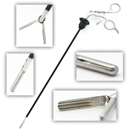 Laparoscopic DeBakey ForcepsGrasping
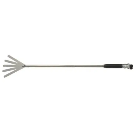 Fan RetractorRetracting
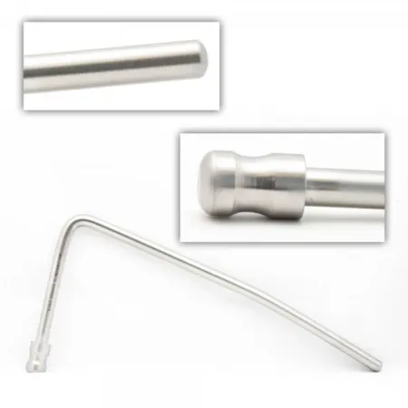 Fisher Style Liver RetractorRetracting
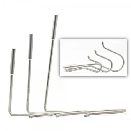 Nathanson Hook Liver RetractorRetracting
 Camera HeadSpecialty
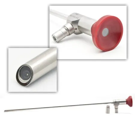 LaparoscopeSpecialty
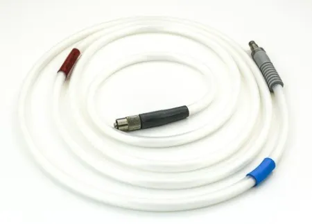 Light CordSpecialty
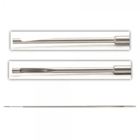 Appel Laparoscopic Knot PusherSuturing
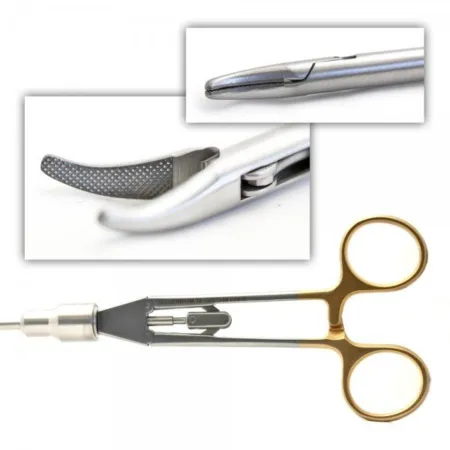 Laparoscopic Needle HolderSuturing
Clamping & Occluding3
Cutting & Dissecting5
Grasping & Holding3
Retracting & Exposing3
Specialty3
Suturing & Stapling2
Know this tray before you scrub in.
With Pro, for every setup in every specialty. Cancel any time.
Or scrub in free: every General setup and 5 questions a day.
The room
The equipment, the prep and drape, the suture and the medicines.
- Camera and light box
- suction
- CO2
- surgeons preference staplers
Step by step
5 steps, one line each.
- 1
Port Placement
The first step of the procedure involves placing five ports. The initial port, a 12mm port, will be placed above the bellybutton for the laparoscope insertion. The insufflationin-suh-FLAY-shunBlowing a gas into a body cavity, such as carbon dioxide into the abdomen during laparoscopic surgery.Full entry tubing will then be attached, and a 30-degree laparoscope will be inserted. Two 5mm ports will be placed; one for liver retractionrih-TRAK-shunA drawing back or pulling in of tissue; in breathing, the skin sinking in between, below or above the ribs with each breath.Full entry with the use of a liver retractorrih-TRAK-terA surgical instrument that holds back tissue or organs so the surgeon can see and reach the operative site.Full entry and the other for additional retraction. The remaining two ports will be 12mm ports for grasping and dissectiondy-SEK-shunA tear in the inner layer of an artery wall that lets blood split the layers apart; also, the cutting apart of tissue in surgery.Full entry .
- 2
Dissection
After port placement, the lesser omentum is entered, and grasping instruments are used for dissecting tissue and nodes. The left gastric artery and vein are then ligated, which may involve using clips, L hook cautery, and laparoscopiclap-uh-roh-SKOP-ikDone through a few small cuts in the abdomen with a thin camera and long instruments, instead of one large opening.lapar/o abdomen-scopic viewingFull entry scissors. More dissectiondy-SEK-shunA tear in the inner layer of an artery wall that lets blood split the layers apart; also, the cutting apart of tissue in surgery.Full entry will be done as required.
- 3
Tubulization
The next step involves the use of a GIA stapler for gastric tubulization. The esophagusih-SOF-uh-gusThe muscular tube that carries food and liquid from the throat to the stomach.esophag/o esophagusFull entry will be mobilized a little bit more, followed by more dissectiondy-SEK-shunA tear in the inner layer of an artery wall that lets blood split the layers apart; also, the cutting apart of tissue in surgery.Full entry , eventually leading to a cervicotomy and the esophagus being isolated.
- 4
Anastomosis
The esophagusih-SOF-uh-gusThe muscular tube that carries food and liquid from the throat to the stomach.esophag/o esophagusFull entry and gastric tubuleTOO-byoolA very small tube in the body, most often one of the tiny tubes in the kidney that turn filtered fluid into urine.Full entry (made with the stapler) will then be pulled through the cervicotomy, and an esophagogastric anastomosisuh-nas-tuh-MOH-sisA connection between two tube-shaped structures, such as two ends of bowel or two blood vessels, made by surgery or found naturally.Full entry will be performed.
- 5
Closure
Finally, a nasogastric tubenay-zoh-GAS-trik toobA thin, flexible tube passed through the nose and down the esophagus into the stomach.nas/o nosegastr/o stomach-ic having to do withFull entry will be placed, followed by a enteral feeding tubeEN-ter-ul FEE-ding toobA tube placed into the stomach or small intestine to give liquid food, water and drugs to a person who cannot eat enough by mouth.Full entry , and the incisions will be closed.
The case
A Laparoscopic Transhiatal Esophagectomy is a minimally invasive surgical procedure used to treat esophageal cancer and strictures. It involves using a laparoscope and a few small incisions to remove part of the esophagus and reconnect it to the stomach. This technique has replaced the older three-incision method, resulting in lower complication rates and morbidity. This is typically done to treat cancer of the esophagus or to address other conditions that may affect the function of the esophagus. The surgery involves the use of a laparoscope and various instruments to access the esophagus and perform the necessary dissection, ligations, and anastomosis.
Built by the techs who scrub it.
Every surgeon runs it a little differently. Send your table, or a fix for this page.
 Diana Lei-alohaReviewer
The rest of this setup is Pro.
Everything unlocked above stays free. Pro opens the rest, on every setup: where new techs slip, what to load next at every step, the circulator’s side, the medicines, the specimen, wound class and trays on standby, what to grab if it goes wrong, and practice for every tray.
Free, for good. No card.
- All 2,882 instrument cards
- Every General setup in full
- 5 exam questions a day
- Your own preference cards
Pass your exam. Know every case.
Everything in Free, plus:
- All 260 setups in every specialty: back table, Mayo and every step
- Practice that never runs out and full timed exams for the CST, TS-C, CRCST, CSPDT and CSFA, with the why behind every answer
- Flashcards on any tray, timed to come back before you forget
- Every instrument card in full: the 3D model, cautions, inspection, how to pass it
Cancel any time from your account. Not worth it? Email us within 30 days of a charge for a refund, one per customer. Refund policy
Pro is also in the CSTSetup app, on the same account. Already have Pro? Log in
Send your table
Your Mayo, your back table, your surgeon’s way, or anything wrong on this page. Your email is only used to credit you. Submissions stay private and are reviewed by our team; accepted ones earn a Contributor credit here.