Understanding power ports
What a power port is, how it differs from a standard port, and what the placement involves. Written for surgical techs, in plain language.

What a power port is.
A power port is an implanted port that can take a high-pressure injection. Like any port it is a small chamber placed under the skin of the chest with a catheter running into a central vein, so treatment goes in without repeatedly sticking a peripheral vein. What makes it a power port is that it is reinforced to withstand a power injector, which means the same device handles chemotherapy and the contrast for a CT scan. A standard port cannot do the second job.
That single difference is why they have become the default in oncology. Someone on chemotherapy is being scanned regularly to see whether the treatment is working, and a standard port means a second needle in a peripheral vein for every one of those scans. A power port removes that entirely.
If you are the patient reading this, the practical version is short: it sits under the skin below your collarbone, nothing shows except a small scar and a slight raised area, and it is accessed with a special needle through a self-sealing membrane that closes behind itself thousands of times.
Power port or standard port.
Built for pressure
Reinforced to take a power injection, so contrast can be pushed through it at the rate a CT scanner needs.
- Takes both treatment and contrast imaging
- Marked so it can be identified on an X-ray
- Usually triangular, often with three palpable bumps
- One access point covers chemo and scans
Built for infusion
Fine for medication and blood draws, but not rated for the pressure a contrast study needs.
- Infusions, medication and blood draws
- No power injection, so imaging needs separate access
- Simpler construction, no reinforcement
- Often carries no identifying marker
How a port is placed.
Placement is short, minimally invasive and usually done under local anaesthetic with sedation, in an operating room or interventional suite with fluoroscopy. From the scrub's point of view it is four movements.
Access the vein
Usually the internal jugular or subclavian, found with ultrasound. Micropuncture needle, then a guidewire.
Make the pocket
A small incision below the clavicle, and blunt dissection to create a pocket in the subcutaneous tissue.
Tunnel the catheter
The catheter is tunnelled from the pocket to the venotomy, trimmed to length, and attached to the port body.
Confirm and close
Fluoroscopy confirms the tip at the cavoatrial junction. The port is secured, flushed with heparin, and the pocket closed.
What is actually on the table.
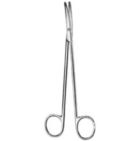
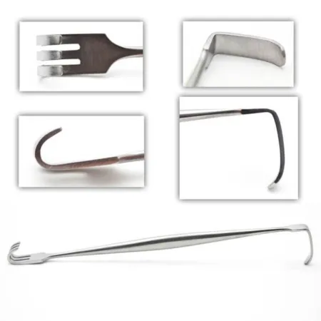
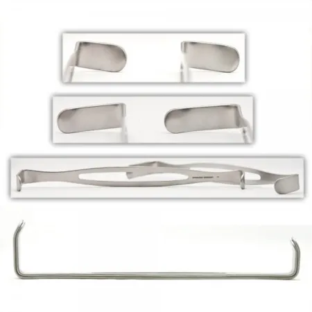
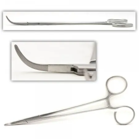
Pulled live from our Port Placement setup, so this is the real tray rather than a list somebody typed out. Tap any instrument for its full card.

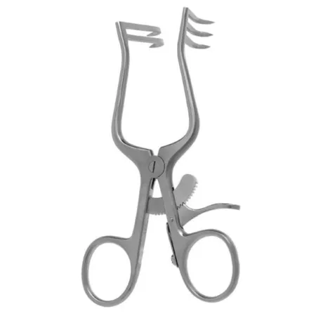
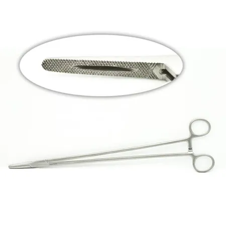
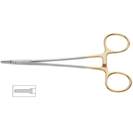
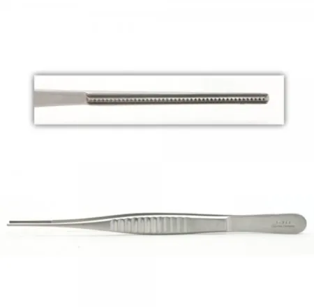
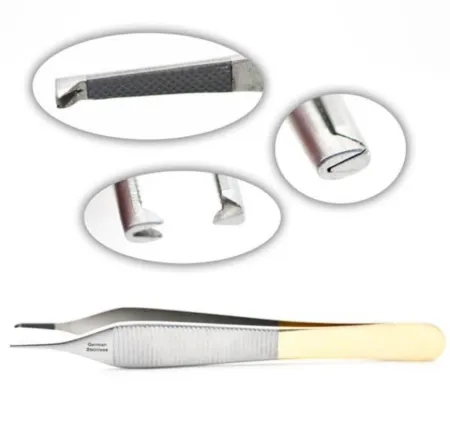
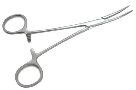
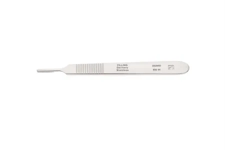
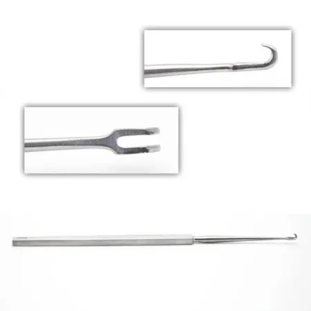
After it is in.
A port is accessed with a non-coring needle, which has an angled tip that parts the septum rather than punching a plug out of it. That is the whole reason the membrane survives a thousand sticks, and it is why a standard hypodermic needle must never go into one.
Flushing
The port is flushed after each use and periodically when it is not in use, to keep the catheter patent.
The site
Kept clean and dry while the incision heals. Redness, swelling, warmth or fever gets reported, because a port infection is a bloodstream infection.
Living with it
Once healed there are almost no restrictions. It stays until the treatment it was placed for is finished, then comes out in a shorter procedure than the one that put it in.
Behind the scenes of the case.
Curious how the room gets ready for a procedure like this one, or studying to work in one? Start here.
The Port Placement setup
Every instrument on the table for this case, with photos.
How the back table is set up
The back table and Mayo stand, step by step.
The instrument library
2,800+ surgical instruments with real photos.
Today's instrument quiz
Five questions with pictures. How many do you know?
Walk in already knowing the table.
Port placements are quick, and quick cases are where being half prepared shows. Every setup here is a real back table and Mayo stand, in the free CSTSetup app too.