Abdominal Laparoscopy
This is a diagnostic or therapeutic procedure where an endoscope is inserted into the peritoneal cavity to visualize the intra-abdominal organs. It allows for the inspection of the viscera and the performance of minor surgical maneuvers without the need for a large laparotomy incision.
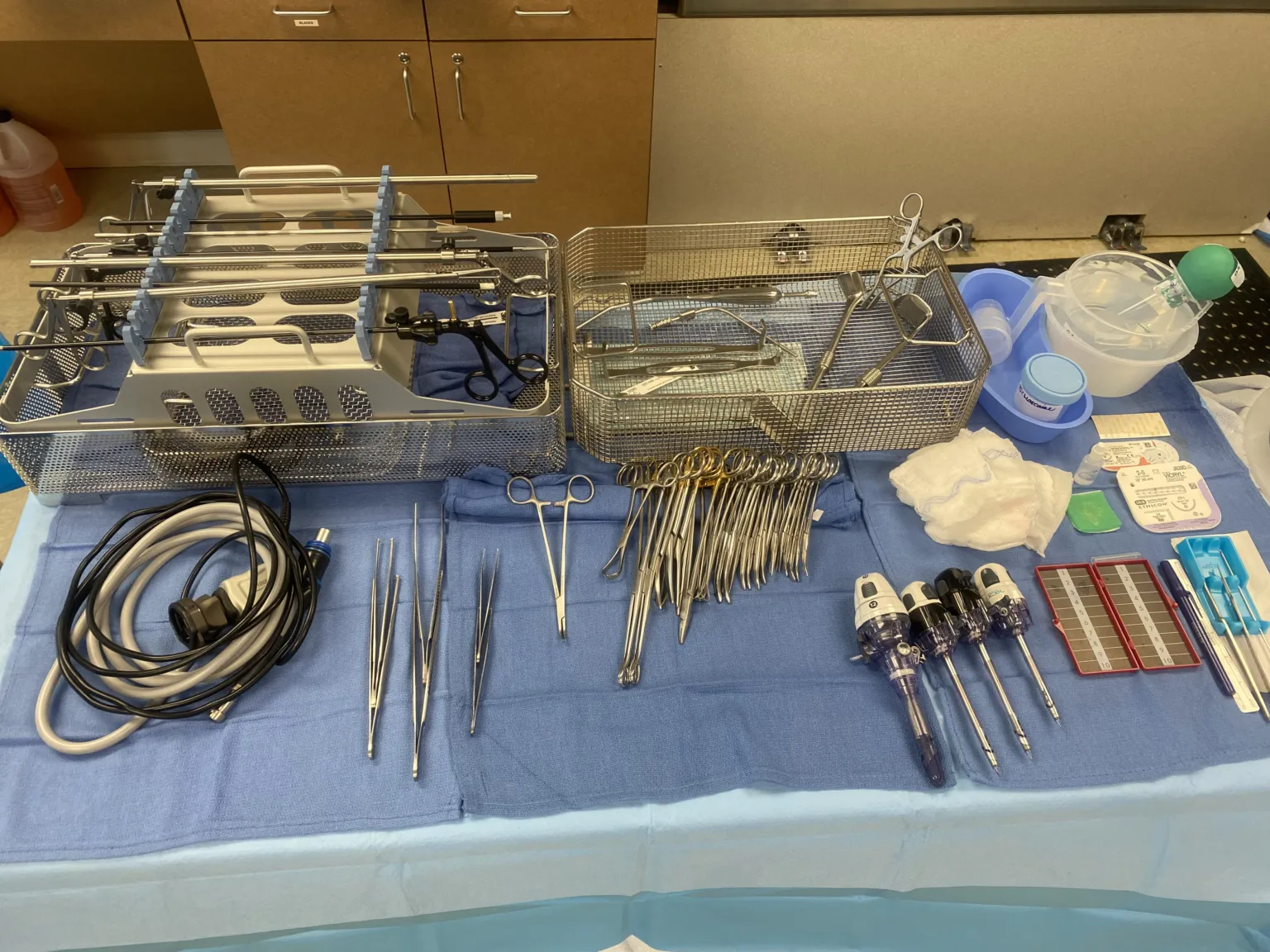
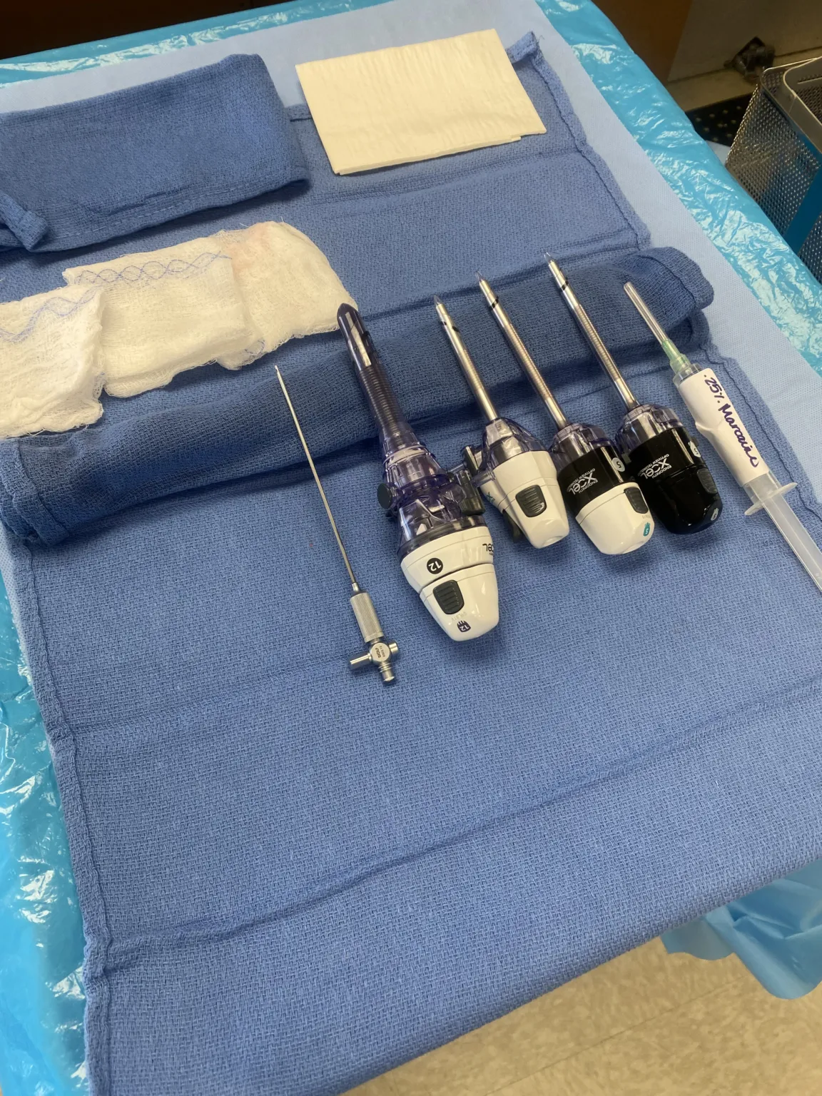
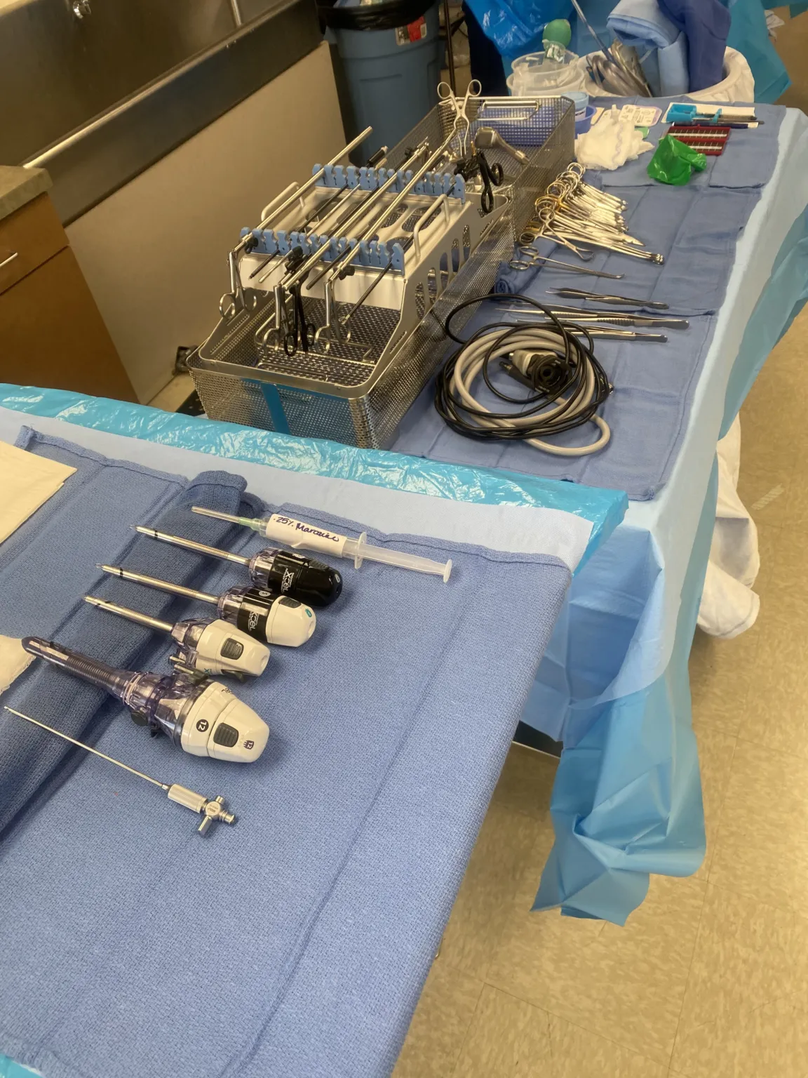
In 30 seconds
Scan this outside the room.
- Insert Veress needle.
- Place trocarTROH-karA sharp or bladed instrument inside a tube (cannula), used to pierce a body wall and leave the tube in place as a port.Full entry with S retractorrih-TRAK-terA surgical instrument that holds back tissue or organs so the surgeon can see and reach the operative site.Full entry or Army-Navys or use pneumoperitoneum method.
- Insert laparoscope for viewing and use graspers, hook cautery, Maryland, or laparoscopiclap-uh-roh-SKOP-ikDone through a few small cuts in the abdomen with a thin camera and long instruments, instead of one large opening.lapar/o abdomen-scopic viewingFull entry scissors to dissectdy-SEKT, or dih-SEKTTo cut apart or separate tissues carefully during surgery.Full entry fat or adhesionad-HEE-zhunA band of scar tissue that joins two surfaces inside the body that are normally separate.ad- towardhes stick-ion conditionFull entry .
- Explore abdomen and surrounding organs for pathologypuh-THOL-uh-jeeThe study of disease and its causes and effects, or the disease changes found in a person or tissue.path/o disease-logy study ofFull entry or abnormalities.
- Close incisions with needle driver and adson pick-up for suturing and straight mayo scissors for cutting sutureSOO-cherA stitch, or the thread used to stitch, that holds the edges of a wound or incision together.Full entry .
The tray
18 instruments, by what they do. Tap a name for its card.
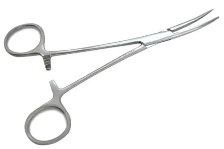 Curved HemostatsClamping
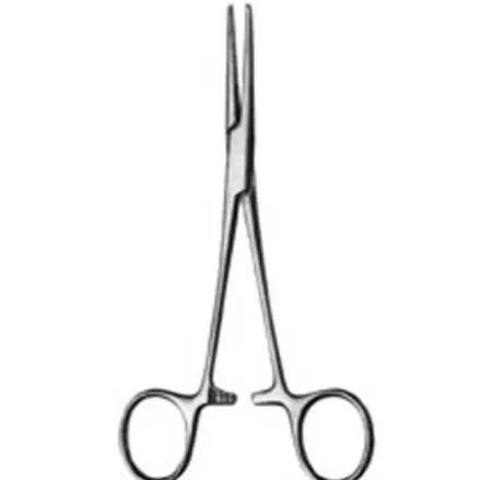 Kelly ForcepsClamping
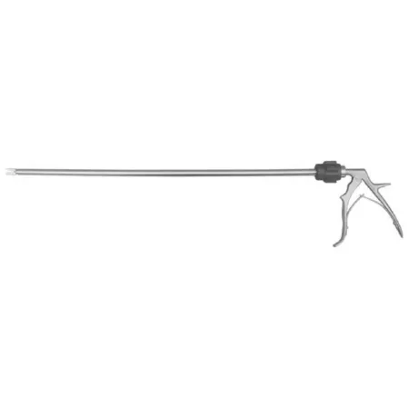 Laparoscopic Clip AppliersClamping
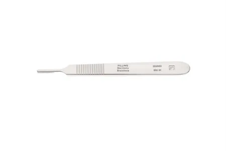 #3 Knife HandleCutting
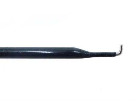 L Hook CauteryCutting
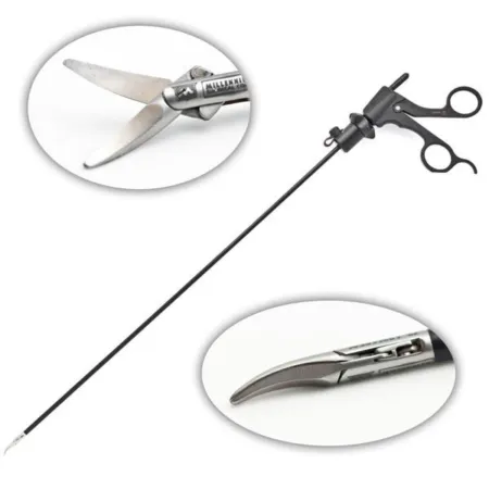 Laparoscopic Metz ScissorCutting
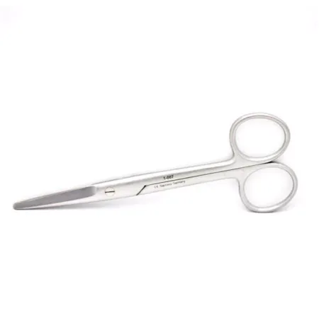 Straight Mayo ScissorCutting
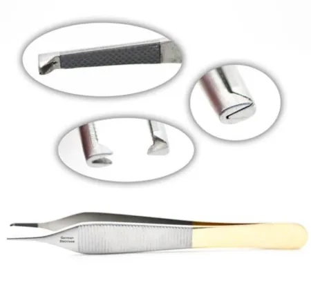 Adson ForcepsGrasping
All 18 instruments
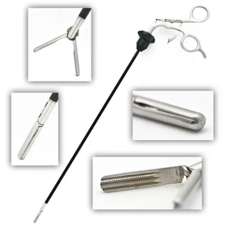 Laparoscopic DeBakey ForcepsGrasping
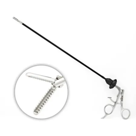 Laparoscopic Fenestrated GrasperGrasping
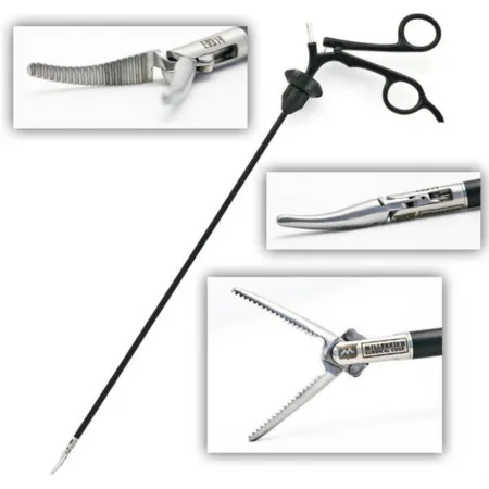 Laparoscopic Maryland Dissector GrasperGrasping
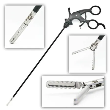 Laparoscopic Wave GrasperGrasping
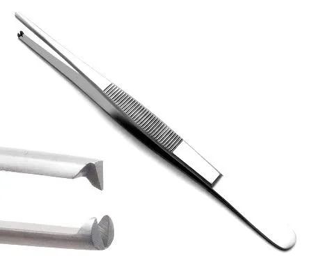 Toothed ForcepsGrasping
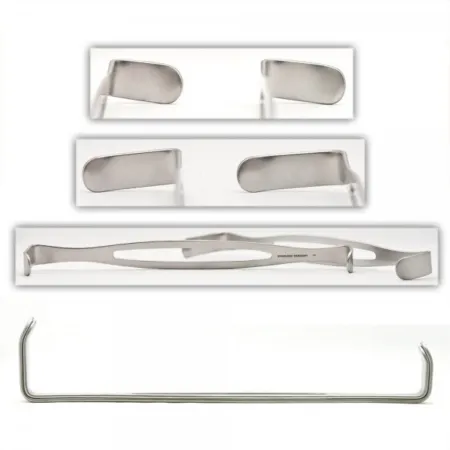 Army-Navy RetractorRetracting
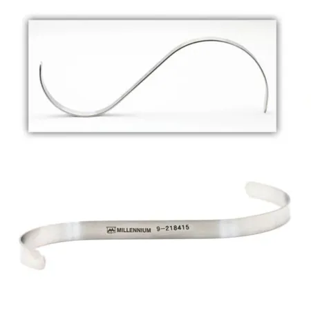 Hasson S RetractorRetracting
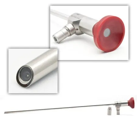 LaparoscopeSpecialty
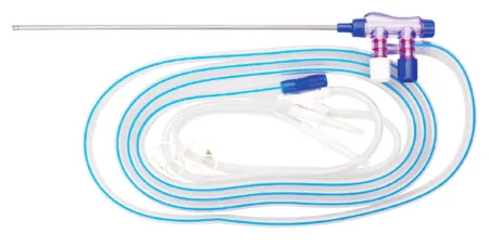 Suction IrrigatorSuctioning
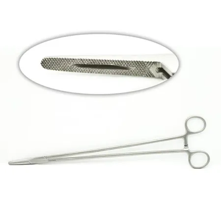 Mayo-Hegar Needle HolderSuturing
Clamping & Occluding3
Cutting & Dissecting4
Grasping & Holding6
Retracting & Exposing2
Specialty1
Suctioning1
Suturing & Stapling1
Know this tray before you scrub in.
With Pro, for every setup in every specialty. Cancel any time.
Or scrub in free: every General setup and 5 questions a day.
The room
The equipment, the prep and drape, the suture and the medicines.
- Camera
- light cord
- CO2 tubing
- suction tubing
- Endopouch
- bovie
- poss. port closure device
- poss. Veress needle
- trocars
- poss. Cholangiogram device
- telescopes
- poss aspirating needle
Four folded towels and laparotomy drape
Step by step
5 steps, one line each.
- 1
Insertion of Veress Needle
To introduce carbon dioxideKAR-bun dy-OK-sideA gas made by the body's cells as waste, carried in the blood to the lungs and breathed out.Full entry into the abdomen, a Veress needle may be introduced into the umbilical incision. A Kelly and two towel clips should be available to insert the Veress needle. After inserting the Veress needle, a syringe of saline may also be needed to confirm correct placement.
- 2
Placement of Trocars
S retractorrih-TRAK-terA surgical instrument that holds back tissue or organs so the surgeon can see and reach the operative site.Full entry or Army-Navys may be used to retract the abdominal wall to insert trocarTROH-karA sharp or bladed instrument inside a tube (cannula), used to pierce a body wall and leave the tube in place as a port.Full entry . Alternatively, the first trocar can be inserted and then attached to the carbon dioxideKAR-bun dy-OK-sideA gas made by the body's cells as waste, carried in the blood to the lungs and breathed out.Full entry tubing, achieving pneumoperitoneum. The rest of the trocars will then be placed.
- 3
Insertion of Laparoscope
The laparoscope will then be inserted into the abdomen for viewing. The surgeon may use a variety of graspers, hook cautery, Maryland, or laparoscopiclap-uh-roh-SKOP-ikDone through a few small cuts in the abdomen with a thin camera and long instruments, instead of one large opening.lapar/o abdomen-scopic viewingFull entry scissors to dissectdy-SEKT, or dih-SEKTTo cut apart or separate tissues carefully during surgery.Full entry any fat or adhesionad-HEE-zhunA band of scar tissue that joins two surfaces inside the body that are normally separate.ad- towardhes stick-ion conditionFull entry that may obstruct the view.
- 4
Surgical Exploration
Once the laparoscope is in place, the doctor will view the contents of the abdomen using their preference of graspers. The surgeon may explore the abdomen and surrounding organs to assess for any pathologypuh-THOL-uh-jeeThe study of disease and its causes and effects, or the disease changes found in a person or tissue.path/o disease-logy study ofFull entry or abnormal findings.
- 5
Closure of Incisions
After the exploration is complete, the ports will be removed, and the incisions will be closed. A needle driver and an adson pick-up will be needed for suturing, and the straight mayo scissors should be available for cutting sutureSOO-cherA stitch, or the thread used to stitch, that holds the edges of a wound or incision together.Full entry . Tips and tricks With a laparoscopylap-uh-ROS-kuh-peeSurgery or an exam done through small cuts in the belly, using a thin, lighted tube with a camera.lapar/o abdomen-scopy looking with an instrumentFull entry , there is a high chance it may turn into a bigger case requiring the surgeon to open the abdomen. This will require more instrument pans. Always try to have a major procedure pan and a bookwalter retractorrih-TRAK-terA surgical instrument that holds back tissue or organs so the surgeon can see and reach the operative site.Full entry pan in your room just in case. Lay the carbon dioxideKAR-bun dy-OK-sideA gas made by the body's cells as waste, carried in the blood to the lungs and breathed out.Full entry insufflationin-suh-FLAY-shunBlowing a gas into a body cavity, such as carbon dioxide into the abdomen during laparoscopic surgery.Full entry tubing on the back table with the camera and light cord so all three pass off together, and coil the suction tubing in a basin so it is ready to hand up.
The case
Abdominal laparoscopy is a minimally invasive surgical procedure that allows a surgeon to examine the organs inside the abdomen with the help of a laparoscope, a thin tube with a camera and light. The procedure is performed through small incisions in the abdomen, which reduces recovery time and scarring compared to traditional open surgery. Abdominal laparoscopy can be used to diagnose and treat a variety of conditions, such as abdominal pain, infertility, and tumors.
Built by the techs who scrub it.
Every surgeon runs it a little differently. Send your table, or a fix for this page.
 Diana Lei-alohaReviewer
 Chantel BrownPhotos
 Alex JayneReviewer
The rest of this setup is Pro.
Everything unlocked above stays free. Pro opens the rest, on every setup: where new techs slip, what to load next at every step, the circulator’s side, the medicines, the specimen, wound class and trays on standby, what to grab if it goes wrong, and practice for every tray.
Free, for good. No card.
- All 2,882 instrument cards
- Every General setup in full
- 5 exam questions a day
- Your own preference cards
Pass your exam. Know every case.
Everything in Free, plus:
- All 260 setups in every specialty: back table, Mayo and every step
- Practice that never runs out and full timed exams for the CST, TS-C, CRCST, CSPDT and CSFA, with the why behind every answer
- Flashcards on any tray, timed to come back before you forget
- Every instrument card in full: the 3D model, cautions, inspection, how to pass it
Cancel any time from your account. Not worth it? Email us within 30 days of a charge for a refund, one per customer. Refund policy
Pro is also in the CSTSetup app, on the same account. Already have Pro? Log in
Send your table
Your Mayo, your back table, your surgeon’s way, or anything wrong on this page. Your email is only used to credit you. Submissions stay private and are reviewed by our team; accepted ones earn a Contributor credit here.