Interventional radiology
How doctors find and treat disease through a needle and a catheter under live images, how each kind of medical image is made, and how you keep the patient and the team safe from radiation and from the MRI magnet.
1Why it matters
Interventional radiology, IR for short, finds and treats disease through a needle or a thin tube called a catheter, steered by medical images, instead of through an open incision. Many conditions that once needed open surgery are now treated this way, with far less trauma to the patient and a faster recovery.
IR procedures need surgical asepsisSUR-jih-kul ay-SEP-sisPractices that keep an area or object completely free of microorganisms; also called sterile technique.a- withoutsepsis decay, infectionFull entry , so surgical technologistSUR-jih-kul tek-NOL-uh-jistA member of the operating room team who prepares the sterile field and instruments and hands them to the surgeon.Full entry may be asked to work in them. In the operating room you help take images too: x-rays taken during surgery are used most often in orthopedic surgeryor-thuh-PEE-dik SUR-juh-reeSurgery on the bones, joints, ligaments, tendons and muscles.ortho- straightped child (the specialty began with straightening children's bones)-ic relating toFull entry , bilebileA yellow-green fluid made by the liver and stored in the gallbladder that helps digest fats.chol/e bileFull entry procedures and vascular surgery, and an x-ray may be taken when a count cannot be resolved.
Every one of those x-ray images gives a dose of ionizing radiationray-dee-AY-shunEnergy that travels as waves or particles, such as X-rays, used to make images or treat cancer; also pain that spreads from its starting point.radi/o rays, radiation-ation processFull entry , which can damage living tissue. Its effects depend on the dose and add up over time: the larger the dose, or the more often you are exposed, the greater the risk. As x-ray imaging has spread through interventional surgery, the radiation that OR staff receive has risen sharply. The magnet of an magnetic resonance imagingmag-NET-ik REZ-uh-nunts IM-uh-jingAn imaging test that uses a strong magnet and radio waves to make detailed pictures of the inside of the body.Full entry scanner brings a different danger: it can pull metal objects, even one as large as a hospital bed, into the scanner. This chapter teaches how the images are made, how IR works, and how you keep the patient, the team and yourself safe.
AST expects every surgical technology student to learn the effects of radiation on the body, radiation safety, shielding devices, dosimeters and what the regulating agencies require, and every working surgical technologist to keep up with yearly continuing education on it. The facility writes its own radiation safety policies, so learn yours.
Other chapters hold the pieces around this one: the vessels that catheters travel in chapter 5; blood tests in chapter 5 and urine tests in chapter 8; how tissue biopsies are taken and handled in chapters 11 and 29; contrastKON-trastA dye or other substance given before an imaging test so body parts show up more clearly.Full entry as drugs (allergy, labeling at the field) in chapter 12; sedationsih-DAY-shunA calm, relaxed or sleepy state brought on by drugs, or the use of drugs to cause it.sed calm, settle-ation processFull entry in chapter 13; the disaster side of a radiological event, and decontaminationdee-kun-tam-ih-NAY-shunRemoving or destroying germs, chemicals or radioactive material from a person, surface or object to make it safe.de- removecontamination being made uncleanFull entry , in chapter 17; navigation systems in chapter 21; the C-arm as room equipment in chapter 23; draping it in chapter 28; and the vascular, heart and other procedures done in a hybrid room in chapters 40 and 41.
2What interventional radiology is
An interventional radiologist is a physician who diagnoses and treats disease with image-guided techniques that need only a puncture or a tiny incision. Those treatments reach heart disease, stroke, cancer and fibroids of the uterus, among others. IR uses x-ray images, magnetic resonance imagingmag-NET-ik REZ-uh-nunts IM-uh-jingAn imaging test that uses a strong magnet and radio waves to make detailed pictures of the inside of the body.Full entry , computed tomographykum-PYOO-tid toh-MOG-ruh-feeAn imaging test that uses x-rays and a computer to make cross-section pictures of the inside of the body.tom/o cut, slice-graphy process of recordingFull entry , ultrasound and other imaging to guide its instruments into vessels and organs.
Two words come up again and again. Percutaneous means "through the skin": no incision is made, and a catheter or other device goes in through a puncture. Endovascular means inside a blood vessel.
To find the problem, to fix it, or both
- Diagnostic: the procedure shows what is wrong. Catheter angiographyan-jee-OG-ruh-feeAn imaging test that shows blood vessels by injecting a contrast dye and taking x-ray, CT or MRI pictures.angi/o vessel-graphy process of recordingFull entry , an injection of contrastKON-trastA dye or other substance given before an imaging test so body parts show up more clearly.Full entry into an artery to make it show on x-ray, is the classic example, and it remains the best test for many problems of the arteries. Angiography to make a diagnosis before surgery is commonly done in the IR department's angiography suite. For many diagnostic studies of the arteries, though, CTAsee-tee-ay (said as letters)Computed tomography angiography: a CT scan with IV contrast dye that shows blood vessels in detail.Full entry has taken its place.
- Therapeutic: the procedure treats the problem, as when a narrowed artery is opened with a balloon or a stentstentA small tube, often of metal mesh, placed inside a vessel or duct to hold it open.Full entry is placed.
- Both: one procedure can do both jobs. When the diagnosis is uncertain, a drain placed through the skin into the gallbladder (percutaneous cholecystostomy) can settle it and treat the problem too; chapter 32 teaches gallbladder surgery.
The Seldinger technique (below), described in 1953, opened the way for catheter angiography and for IR as a specialty.
Where IR is done
The Core Curriculum names two settings, the IR suite and the standard OR, and the hybrid OR joins the two.
The IR suite. The angiography suite is its own department of the hospital. It holds the x-ray imaging equipment, monitors for the patient's oxygen level, blood pressure and heart rate, a place to store medications, a resuscitationrih-sus-ih-TAY-shunEmergency treatment to restore breathing, heartbeat or blood circulation.Full entry cart, a scrub sink, and a surgical light hung from the ceiling. Its staff are radiologists, radiology technologists trained in interventional angiography, nurses and anesthesia provideran-us-THEE-zhuh pruh-VY-derThe licensed professional who gives anesthesia and watches over the client's breathing and vital functions during a procedure.Full entry . You may get the chance to work there, but the angiography technologist is usually the one who assists.
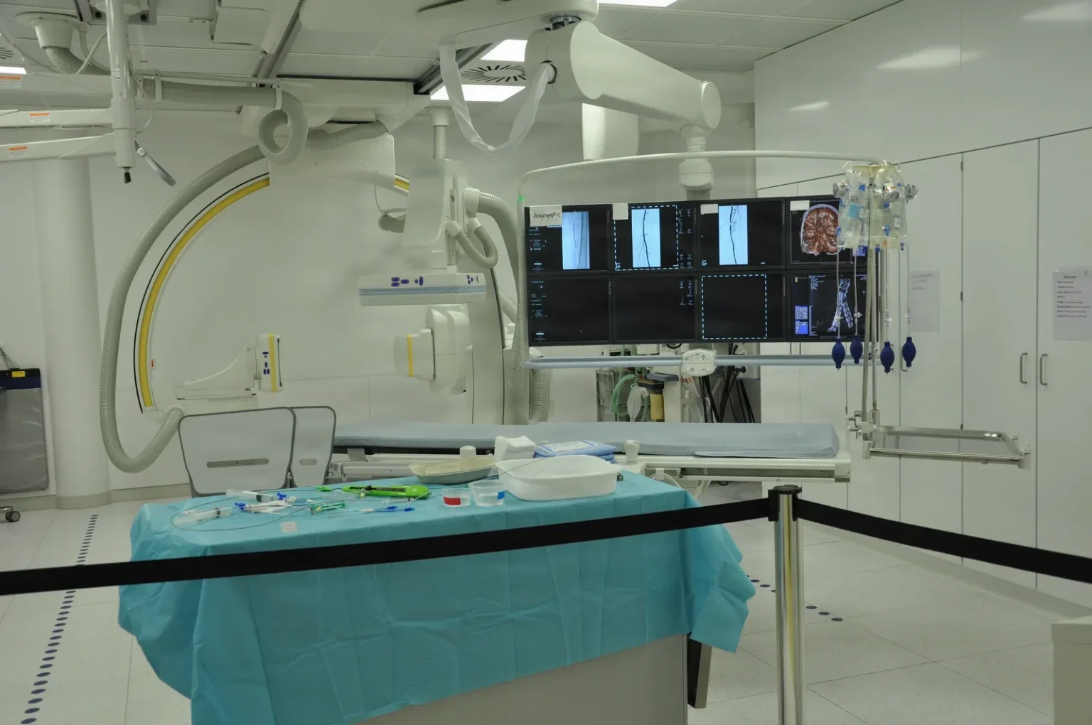
The hybrid OR. A hybrid operating room is specially equipped for both endovascular and open procedures. It combines a full surgical environment with the fluoroscopic imaging of an interventional lab. That gives the team advanced imaging and, in the same room, the instruments and supplies needed if the case turns into an emergency. Stent graftGRAFTTissue or a man-made material moved into place in surgery to replace or repair a damaged part.Full entry for aneurysmAN-yuh-riz-umA weak spot in the wall of an artery that bulges outward like a balloon.aneurysm a widening (Greek)Full entry and dissectiondy-SEK-shunA tear in the inner layer of an artery wall that lets blood split the layers apart; also, the cutting apart of tissue in surgery.Full entry of the aorta, and aortic valveay-OR-tik valvThe heart valve between the left ventricle and the aorta, which opens to let blood out to the body.Full entry placed through a catheter, are among the procedures done there.
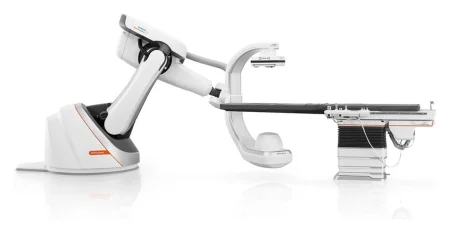
The standard OR. In an ordinary operating room, a mobile C-arm brings live x-ray images to the table. Kyphoplasty, for example, needs a C-arm, a table that x-rays pass through, and x-ray vests for the staff.
3Preparing the patient
Most patients arrive with their diagnostic studies already done, and those results guide the plan. Before an IR procedure, the team also checks these points:
- Allergies. The physician must know the patient's allergies, especially any reaction to iodinated contrastKON-trastA dye or other substance given before an imaging test so body parts show up more clearly.Full entry . A careful history asks about any earlier reaction to contrast. A shellfish allergy is not a reason to expect a reaction to contrast.
- Kidney function. The kidneys clear contrast given into a vessel: when they work normally, up to all of it is gone within 24 hours, and when they do not, clearing it can take several days. So a blood urea nitrogenblud yoo-REE-uh NY-truh-jenA blood test that measures urea nitrogen, a waste product made when the body breaks down protein.Full entry (BUN) and a creatininekree-AT-uh-neenA waste product of muscle that the kidneys remove from the blood; its blood level is used to measure how well the kidneys work.Full entry , the best measures of kidney function, are drawn, and the results must be back before the procedure. Preexisting kidney disease, diabetes, dehydration, older age, heart failurehart FAYL-yerA long-term condition in which the heart cannot pump enough blood to meet the body's needs.Full entry , liver diseaseLIV-er dih-ZEEZAny condition that damages the liver or keeps it from working normally.hepat/o liverFull entry and drugs that harm the kidneys all raise the risk of kidney injury from contrast. metforminmet-FOR-minAn oral drug for type 2 diabetes that lowers blood sugar, mainly by reducing the sugar the liver releases.Full entry , a diabetes drug, is stopped for a time when a patient has a study with iodinated contrast given into a vessel, because the contrast can suddenly change how the kidneys work.
- Pregnancy. Physicians and nurses make sure the patient is not pregnant, so that a fetus is not exposed to radiationray-dee-AY-shunEnergy that travels as waves or particles, such as X-rays, used to make images or treat cancer; also pain that spreads from its starting point.radi/o rays, radiation-ation processFull entry . When a pregnancy is possible or known, the surgeon is told and decides whether the procedure should be rescheduled.
- Earlier radiation. AST asks the assistant circulator to review the chart for past radiation exposure, report problems such as an earlier radiation skin injury to the surgeon, and confirm whether the patient has had a previous radionuclide (radioactive tracer or treatment) procedure. The radiation safety officer is consulted when that could expose the team.
- Access and sedationsih-DAY-shunA calm, relaxed or sleepy state brought on by drugs, or the use of drugs to cause it.sed calm, settle-ation processFull entry . A nurse or technologist makes sure the patient has a working IV line, and the access site is cleaned and numbed. Most patients need only short-acting IV sedative-hypnoticSED-uh-tiv hip-NOT-ikA drug that calms a person (sedative) or helps them fall asleep (hypnotic); the same drug can do either, depending on the dose.Full entry plus local anestheticLOH-kul an-es-THET-ikA drug that blocks feeling in one part of the body without making the person unconscious.Full entry at the puncture site; a very anxious patient may have general anesthesiaJEN-er-ul an-es-THEE-zhuhDrug-induced loss of consciousness and feeling over the whole body, so a person feels nothing and is not aware during surgery.an- withoutesthesi/o feeling, sensation-ia conditionFull entry . Many vascular patients also have heart, vessel or kidney disease, so close monitoring matters whatever the anesthetican-us-THET-ikA drug that causes loss of feeling, with or without loss of consciousness.an- withoutesthesi/o feeling, sensation-ic relating toFull entry . Chapter 13 teaches the levels of sedation.
As for any invasive procedure, informed consentin-FORMD kun-SENTA person's agreement to a treatment or procedure, given freely after the risks, benefits and other choices have been explained to them.Full entry , the time-outTYM-owtA pause just before a procedure when the whole team confirms the right client, procedure and site; also, a short break to calm a child.Full entry and the rest of the safety checks apply in the IR suite as they do in the OR; chapters 15 and 28 teach them. The procedure is sterile, so the team wears caps, masks, sterile gowns and gloves.
4Getting into a vessel
Choosing the way in
A catheter can travel against the flow of blood (retrograde) or with it (antegrade). The vessel used for access may not be the one that is diseased: it is chosen for its size and condition, avoiding scarring or hardened plaquePLAKA deposit that builds up on a surface in the body, such as a sticky film of bacteria on teeth or a fatty buildup in an artery wall.Full entry at the puncture, and the patient is positioned for that site.
- Arteries: the common femoral arteryFEM-er-ul AR-ter-eeThe large artery that carries blood to the leg, running through the groin and down the thigh.Full entry in the groin is used most often; the brachial and axillary arteries in the arm are other choices.
- Veins: the internal jugularJUG-yuh-lerRelating to the neck, especially the large veins in the neck.Full entry , common femoralFEM-er-ulRelating to the thigh or the femur, the thigh bone.Full entry , brachial and popliteal veins.
Ultrasound shows the tissue around the needle as it goes in, and once access is secure, fluoroscopyfloo-ROS-kuh-peeAn x-ray method that shows moving pictures of the inside of the body in real time.fluor/o glowing (from the fluorescent screen)-scopy looking, viewingFull entry guides the devices inside the vessel. Using ultrasound to guide arterial access has cut down on complications at the puncture site.
The Seldinger technique
This is how a catheter or sheathSHEETHA covering that wraps around a structure; in procedures, a short hollow tube left in a blood vessel to pass catheters through.Full entry goes into a vessel through the skin:
- A needle punctures the vessel, often an 18-gauge needle guided by ultrasound.
- A guidewire is threaded through the needle.
- The needle is withdrawn, leaving the wire in the vessel.
- The catheter or sheath is slid over the wire into place.
- For a central venous catheterSEN-trul VEE-nus KATH-uh-terAn IV catheter whose tip sits in a large vein near the heart, used for long-term drugs, fluids, nutrition or blood draws.Full entry , the wire is then taken out and the catheter flushed; in an endovascular case, a wire stays in to guide the devices that follow, each passed in a set order.
The common femoral artery can be reached this way, with a needle and wire through the skin, or by a small surgical cutdown, or by exchanging the wire through an arterial linear-TEER-ee-ul lineA thin catheter placed in an artery, often in the wrist, to measure blood pressure all the time and draw blood.Full entry already in place.
The supplies
The Core Curriculum's endovascular case lists the supply families you will see: arteriogram supplies (an angiocatheter, an arterial needle, IV extension tubing, syringes and three-way stopcocks), balloons, catheters, guidewires, needles, pressure bags with high-pressure tubing, sheaths, snares, stentstentA small tube, often of metal mesh, placed inside a vessel or duct to hold it open.Full entry graftGRAFTTissue or a man-made material moved into place in surgery to replace or repair a damaged part.Full entry , and vascular closure devices. Its outline for IR singles out three: catheters, contrastKON-trastA dye or other substance given before an imaging test so body parts show up more clearly.Full entry and guidewires.
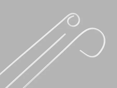
Guidewires. Once the needle is in the vessel, the guidewire goes in first and acts as the pilot for every device that follows. It also holds the place in the vessel while catheters and other devices are exchanged over it. Wires come in lengths from 50 to 260 cm. A catheter's channel is sized to match its wire. The Core Curriculum sorts wires as exchange, introducing, steerable and non-steerable, with flexible, J-curved, rigid or stiff tips.
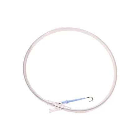
Sheaths. An access sheath is a tube slid into the vessel over the wire. Catheters, stents and other devices pass through it one after another while it protects the vessel wall. It has an introducer (dilatorDY-lay-terAn instrument used to stretch or widen an opening, canal or passage in the body.Full entry ), a side port for flushing with heparinized saline, and a stopcock that stops bleeding.
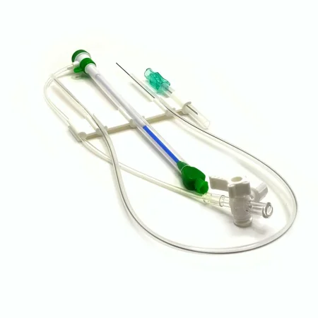
Catheters. Every endovascular catheter has a channel (lumenLOO-menThe open space inside a tube, such as a blood vessel, the intestine or a catheter.Full entry ) for a guidewire or for injecting contrast. The Core Curriculum lists five kinds:
- Sizing: radiopaqueray-dee-oh-PAYKBlocking x-rays, so the material shows up white on an x-ray image.radi/o radiation, x-rayopaque not letting throughFull entry marks every centimeter measure a vessel on the screen.
- Directional: a shaped tip steers the wire into the right branch where a vessel divides.
- Exchange: holds the place in the vessel while the surgeon swaps one wire for another.
- Balloon: carries a balloon filled with contrast, or contrast mixed with heparinized saline, so it shows under fluoroscopy.
- Pigtail: its tip curls, as the name says.
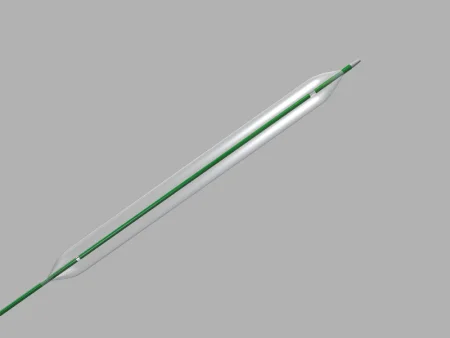
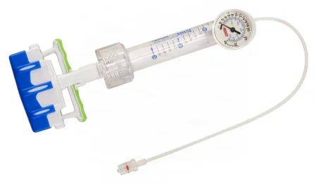
Balloons. An angioplasty balloon is inflated to an exact pressure with an inflation device loaded with contrast solution (the Core Curriculum calls it an insufflationin-suh-FLAY-shunBlowing a gas into a body cavity, such as carbon dioxide into the abdomen during laparoscopic surgery.Full entry device). Every bit of air comes out of the device, its tubing and the balloon first, because trapped air blurs the image and could become an air embolismEM-buh-liz-umThe blocking of a blood vessel by a mass carried there in the blood (an embolus), such as a clot, fat or an air bubble.embol/o plug-ism conditionFull entry if the balloon burst.
The rest of the list, from snares and stent grafts to closure devices, belongs to vascular surgery: chapter 40 teaches how each is chosen and used, stents included.
Contrast in IR
Iodinated contrast is a clear liquid that shows white on x-ray, used for x-ray images, computed tomographykum-PYOO-tid toh-MOG-ruh-feeAn imaging test that uses x-rays and a computer to make cross-section pictures of the inside of the body.tom/o cut, slice-graphy process of recordingFull entry and fluoroscopy. Lower-osmolality agents cause fewer reactions and are better tolerated in vessels, so they are replacing the high-osmolality agents. A power injector delivers contrast at a set rate and volume, which is often hard to do by hand.
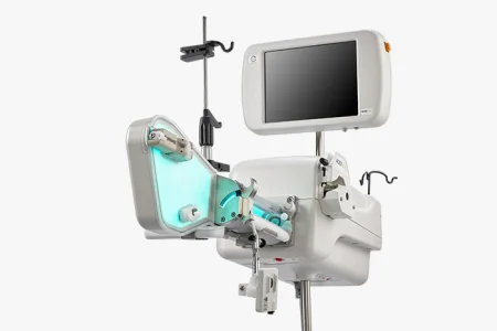
As with every drug used in surgery and IR, keep track of the total amount and strength of contrast used, so it can be reported at the end. Contrast is clear, and so is the heparinized saline beside it, so every syringe and container must be labeled, contrast included. Chapter 12 teaches contrast as a drug and the labeling steps at the field.
5Common IR procedures
You will meet these in the IR suite, the hybrid OR and the standard OR. Each specialty chapter teaches its own procedures step by step; here is what each one is.
- angiographyan-jee-OG-ruh-feeAn imaging test that shows blood vessels by injecting a contrast dye and taking x-ray, CT or MRI pictures.angi/o vessel-graphy process of recordingFull entry : imaging of arteries or veins, done to diagnose, during surgery, or as part of a treatment. contrastKON-trastA dye or other substance given before an imaging test so body parts show up more clearly.Full entry is injected into a chosen artery and its branches to find exactly where a vessel is narrowed, blocked or malformed. It is done under fluoroscopyfloo-ROS-kuh-peeAn x-ray method that shows moving pictures of the inside of the body in real time.fluor/o glowing (from the fluorescent screen)-scopy looking, viewingFull entry or computed tomographykum-PYOO-tid toh-MOG-ruh-feeAn imaging test that uses x-rays and a computer to make cross-section pictures of the inside of the body.tom/o cut, slice-graphy process of recordingFull entry . Digital subtraction angiography (DSA) takes away the bone and soft tissue so only the vessels show, and road mapping lays a saved picture of the vessels over the live image to help steer through them; both raise the radiationray-dee-AY-shunEnergy that travels as waves or particles, such as X-rays, used to make images or treat cancer; also pain that spreads from its starting point.radi/o rays, radiation-ation processFull entry dose, so they are kept to what is needed.
- Angioplasty: a balloon catheter is passed into an artery, to the narrowed part, and inflated to open it and restore blood flow.
- stentstentA small tube, often of metal mesh, placed inside a vessel or duct to hold it open.Full entry placement: a stent, a tube of mesh that fits against the artery wall, may be placed after the balloon has opened the artery, to hold it open.
- Stent graftGRAFTTissue or a man-made material moved into place in surgery to replace or repair a damaged part.Full entry for aneurysmAN-yuh-riz-umA weak spot in the wall of an artery that bulges outward like a balloon.aneurysm a widening (Greek)Full entry : a covered stent lines an aneurysm from the inside, as in endovascular repair of an abdominal aortic aneurysmab-DOM-ih-nul ay-OR-tik AN-yuh-riz-umA weak, bulging section of the aorta where it passes through the belly.Full entry (EVAR), which chapter 40 teaches.
- Embolization: a vessel is blocked from the inside on purpose, for example to shrink a tangle of abnormal brain vessels (an arteriovenousar-teer-ee-oh-VEE-nusRelating to both arteries and veins, or joining an artery to a vein.arteri/o arteryven/o vein-ous relating toFull entry malformationmal-for-MAY-shunA body part or organ that formed in an abnormal way before birth.mal- badformation shapingFull entry ) before surgery; chapter 42 teaches brain surgery.
- vena cavaVEE-nuh KAY-vuhOne of the two large veins that return oxygen-poor blood from the body to the right atrium of the heart.Full entry filter: a small metal filter placed in the inferior vena cavain-FEER-ee-er VEE-nuh KAY-vuhThe large vein that carries blood from the lower body back to the right atrium of the heart.inferior lowervena veincava hollowFull entry to stop clots from reaching the lungs, which chapter 40 teaches.
- Central venous lines: a catheter placed through the skin into a large vein, its tip in the vena cava, guided by fluoroscopy or ultrasound; chapter 40 teaches venous access.
- Drains, enteral feeding tubeEN-ter-ul FEE-ding toobA tube placed into the stomach or small intestine to give liquid food, water and drugs to a person who cannot eat enough by mouth.Full entry and biopsies: a gastrostomy tubegas-TROS-tuh-mee toobA feeding tube placed through the belly wall directly into the stomach.gastr/o stomach-stomy surgical openingFull entry , a needle biopsy, a stereotactic procedure, or a drain such as a percutaneous cholecystostomy.
- Kyphoplasty and vertebroplasty: bone cement is injected through a needle into a vertebra broken by compression, at the level confirmed on fluoroscopy; chapter 42 teaches spine surgery.
6When things go wrong
The complications of catheter angiographyan-jee-OG-ruh-feeAn imaging test that shows blood vessels by injecting a contrast dye and taking x-ray, CT or MRI pictures.angi/o vessel-graphy process of recordingFull entry are uncommon. They fall into four groups: at the puncture site, from the catheter, in the whole body, and from radiationray-dee-AY-shunEnergy that travels as waves or particles, such as X-rays, used to make images or treat cancer; also pain that spreads from its starting point.radi/o rays, radiation-ation processFull entry .
At the puncture site
- A hematomahee-muh-TOH-muhA collection of blood, often clotted, that has leaked out of the blood vessels into the tissue.hem/o blood (here as hemat-)-oma swelling or massFull entry : a swelling of blood, usually clotted, that has leaked from the vessel into the tissue at the access site.
- A pseudoaneurysm (a bulge at the vessel that looks like an aneurysmAN-yuh-riz-umA weak spot in the wall of an artery that bulges outward like a balloon.aneurysm a widening (Greek)Full entry ), an abnormal channel between the artery and a vein (an arteriovenous fistulaar-teer-ee-oh-VEE-nus FIS-chuh-luhA connection made by surgery between an artery and a vein, usually in the arm, to give access for hemodialysis.arteri/o arteryven/o vein-ous relating tofistula an abnormal passage between two body partsFull entry ), a split in the vessel wall (dissectiondy-SEK-shunA tear in the inner layer of an artery wall that lets blood split the layers apart; also, the cutting apart of tissue in surgery.Full entry ) or a clot can also form there.
- When a venous sheathSHEETHA covering that wraps around a structure; in procedures, a short hollow tube left in a blood vessel to pass catheters through.Full entry comes out, pressure is held on the site for at least 10 minutes. An arterial site is closed with fine vascular sutureSOO-cherA stitch, or the thread used to stitch, that holds the edges of a wound or incision together.Full entry or with a closure device that pulls a purse-string suture tight around the hole.
Bleeding and a hematoma at the site are what you watch for when the sheath comes out at the end of the case.
From the catheter or the devices
- A wire or catheter can split the vessel wall (dissection) or knock loose material that travels on (embolization).
- A clot can form on a wire or catheter and be pushed into the bloodstream, which is why every device is flushed and wiped as it goes in and out.
- Air left in a balloon system can blur the image and become an air embolismEM-buh-liz-umThe blocking of a blood vessel by a mass carried there in the blood (an embolus), such as a clot, fat or an air bubble.embol/o plug-ism conditionFull entry if the balloon bursts.
Reactions to contrast
- Nausea, vomiting and vasovagal reactions, which can make the patient faint, are the most frequent whole-body problems. A vasovagal reaction is not an allergy and must not be mistaken for one.
- Allergic reactions, such as urticariaur-tih-KAIR-ee-uhHives: raised, red, itchy welts on the skin.urtic nettle, the stinging plant-aria conditionFull entry , swelling and wheezingWEEZ-ingA high-pitched, whistling sound made as air moves through narrowed airways.Full entry , are more common with high-osmolality contrastKON-trastA dye or other substance given before an imaging test so body parts show up more clearly.Full entry . Severe reactions include low blood pressure, an irregular heartbeat, severe bronchospasmBRONG-koh-spaz-umA sudden tightening of the muscles around the airways that narrows them.bronch/o bronchus-spasm sudden involuntary contractionFull entry , swelling of the larynxLAIR-inksThe voice box: the part of the airway in the neck, above the windpipe, that holds the vocal cords.laryng/o larynxFull entry , seizureSEE-zherA sudden burst of abnormal electrical activity in the brain that changes movement, feeling, behavior or awareness.Full entry and fluid in the lungs.
- A reaction can start 20 to 30 minutes after the injection, or up to 7 days after the procedure.
- A crash cart should be ready for every radiographic procedure. Chapter 13 teaches anaphylaxisan-uh-fuh-LAK-sisA sudden, severe allergic reaction that affects the whole body.ana- away, against-phylaxis protectionFull entry and its treatment.
Kidney injury from contrast
Contrast can injure the kidneys suddenly: a form of acute kidney injuryuh-KYOOT KID-nee IN-juh-reeA sudden loss of kidney function over hours to days.Full entry , also called acute renal failure. Fluids to keep the kidneys flushing, and as little contrast as will do the job, are the main protection.
Radiation injury to the skin
How badly the skin is hurt depends on the dose, the size of the area and the time between exposures. A sudden, large exposure can inflame and redden the skin at the point of exposure (dermatitisder-muh-TY-tisInflammation of the skin.dermat/o skin-itis inflammationFull entry and erythemaair-ih-THEE-muhRedness of the skin caused by more blood flowing in the small vessels near the surface.erythr/o red-ema conditionFull entry ). AST also asks for the patient's skin to be checked, and recorded, before and after a radiation procedure. The Joint Commissionthuh JOYNT kuh-MISH-unAn independent US organization that inspects and accredits hospitals and other health care organizations against safety and quality standards.Full entry counts fluoroscopyfloo-ROS-kuh-peeAn x-ray method that shows moving pictures of the inside of the body in real time.fluor/o glowing (from the fluorescent screen)-scopy looking, viewingFull entry that leaves a permanent tissue injury as a sentinel eventSEN-tih-nul ee-VENTAn unexpected event in health care that causes a client's death, permanent harm or severe temporary harm.Full entry when the steps to keep the dose down were not taken or accepted practice was not followed; chapter 15 teaches sentinel events.
Every catheter, wire and sheath is flushed and wiped as it goes in and as it comes out, and every bit of air leaves the balloon system. A clot or a bubble pushed in can travel on through the bloodstream.
7How the images are made
Imaging studies are tests that make a picture of the inside of the body, to show the shape and work of its parts. The Core Curriculum names five that IR uses: computed tomographykum-PYOO-tid toh-MOG-ruh-feeAn imaging test that uses x-rays and a computer to make cross-section pictures of the inside of the body.tom/o cut, slice-graphy process of recordingFull entry , digital fluoroscopyfloo-ROS-kuh-peeAn x-ray method that shows moving pictures of the inside of the body in real time.fluor/o glowing (from the fluorescent screen)-scopy looking, viewingFull entry , magnetic resonance imagingmag-NET-ik REZ-uh-nunts IM-uh-jingAn imaging test that uses a strong magnet and radio waves to make detailed pictures of the inside of the body.Full entry , nuclear medicine and sonography. They work in very different ways, and only some of them use ionizing radiationray-dee-AY-shunEnergy that travels as waves or particles, such as X-rays, used to make images or treat cancer; also pain that spreads from its starting point.radi/o rays, radiation-ation processFull entry .
| Study | What makes the image | Ionizing radiation? | Often used for |
|---|---|---|---|
| X-ray (radiography) | x-rays passing through the body onto a detector | Yes | bones and fractures, a count that will not resolve |
| Fluoroscopy | a continuous x-ray beam shown as a live picture | Yes | guiding catheters, wires and implants in real time |
| Computed tomography (CT) | x-ray images from all around the body, put together by a computer | Yes, many times more than a plain x-ray | cross sections of soft tissue such as the brain, chest and abdomen |
| Magnetic resonance imaging (MRI) | a strong magnet and radio waves | No | detailed pictures that find structural problems, such as tumors |
| Nuclear medicine and positron emission tomographyPOZ-ih-tron ih-MISH-un toh-MOG-ruh-feeAn imaging test that uses a small amount of a radioactive tracer to show how active the body's tissues are.tom/o cut, slice-graphy recordingFull entry | a radioactive tracer given to the patient | Yes, from the tracer | how an organ is working, such as heart disease or the spread of cancer |
| Ultrasound (sonography) | high-frequency sound waves bouncing back | No | the abdomen, pregnancy, blood flow in vessels |
X-rays and radiography
X-rays are high-energy electromagnetic radiation with a short wavelength that can pass through solid matter. An x-ray image forms when the rays pass through the body and strike a detector lined up behind it. Tissues let x-rays through at different rates according to how dense they are, so a very dense substance shows white and air shows black, with organs and tissues in shades of gray between.
- radiopaqueray-dee-oh-PAYKBlocking x-rays, so the material shows up white on an x-ray image.radi/o radiation, x-rayopaque not letting throughFull entry describes a substance that x-rays cannot pass through, so it shows white.
- Radiolucent describes a substance that x-rays pass through partly or wholly. A radiolucent procedure table lets images be taken through it.
Digital radiography (DR) uses flat panel detectors that turn the x-rays straight into a digital image, ready almost at once. It has replaced film in most facilities, gives far better images, and needs a much lower dose than film did, but radiation precautionprih-KAW-shunA step taken ahead of time to prevent harm, such as the infection-control practices used with every client.Full entry still apply.
x-ray images were once all recorded on film, and digital radiography has replaced film in most facilities, not all. A reference still teaches computed radiography, where the image is caught on a screen inside a cassette that must go to a separate reader, an extra step, before it can be seen.
X-rays in the OR
A standard x-ray in surgery is taken with a fixed or portable x-ray machine; the portable machine is brought to the room on call.
- For a front-to-back view, the technologist slides the cassette or detector into a platform under the patient (a Bucky platform) from the head or foot of the table; done properly, this does not risk contaminating the sterile fieldSTAIR-ul feeldAn area set up with sterile drapes and supplies, kept free of all germs, for a sterile procedure.Full entry .
- The overhead tube is brought to the field only after the area under it is covered with sterile drapes, and the scrub protects the field, including the instrument tables, from the tube as it moves in.
- When an image must be taken on the sterile field, the scrub and the circulator place the cassette in a sterile cover, and a holder, never a hand, keeps it in place.

Contrast studies
A contrastKON-trastA dye or other substance given before an imaging test so body parts show up more clearly.Full entry is a substance given to outline a hollow organ or vessel on an image. It is injected, instilled or swallowed before the images are taken, and it shows as solid white, so the shape and any narrowing of the structure stand out on an x-ray or under fluoroscopy. A contrast medium is either positive, radiopaque like iodinated agents and barium, or negative, radiolucent like air. Common contrast studies include:
- Cholangiography: contrast in the bile ductbyle duktOne of the tubes that carry bile from the liver and gallbladder to the small intestine.Full entry , looking for stones, a tumor or widening.
- angiographyan-jee-OG-ruh-feeAn imaging test that shows blood vessels by injecting a contrast dye and taking x-ray, CT or MRI pictures.angi/o vessel-graphy process of recordingFull entry : contrast in the heart or vessels, looking for narrowing or other problems with blood flow.
- Myelography: contrast in the space around the spinal cord, to show the cord and nerve roots.
- Retrograde pyelography: contrast run up into the bladder, ureterYOOR-uh-terOne of the two tubes that carry urine from the kidneys down to the bladder.Full entry and kidney, looking for stones, narrowing or tumors.
- Gastrointestinal studies: barium, swallowed for the upper gut or given as an enemaEN-uh-muhLiquid put into the rectum and lower bowel through the anus.Full entry for the colon and rectum.
Fluoroscopy and the C-arm
Fluoroscopy uses x-rays and digital technology to give live, moving images on a screen that can be seen in normal room light. It is used to diagnose and, during surgery, to guide work with contrast or the placing of an implant.
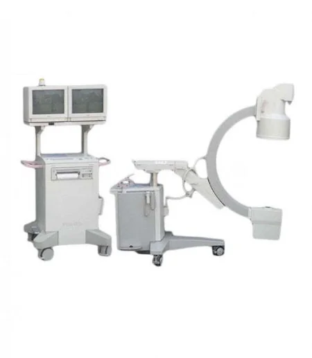
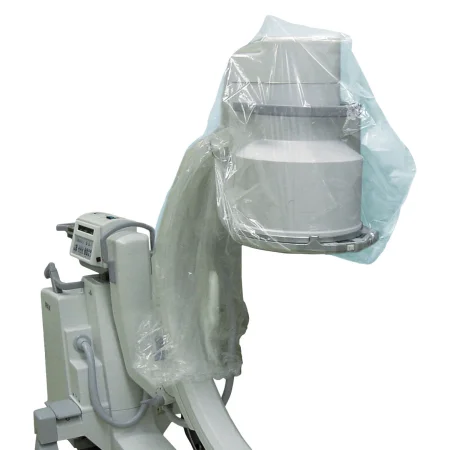
The mobile C-arm brings fluoroscopy to the OR table. The x-ray tube sits at one end of the C and the image intensifier at the other, and the C is moved so that the patient on the table lies centered between them. It can slide along the table to image one level after another. It is draped before it comes to the field, so it can move freely beside it, and in many hospitals it has replaced the portable x-ray machine. Fluoroscopy gives off a lot of radiation, so it calls for extra care; using the C-arm safely is taught below, the C-arm as room equipment in chapter 23, and its drape in chapter 28.
The O-arm
The O-arm is an imaging system shaped like a ring around the patient. Its open tube, called the gantry, houses the x-ray tube and moves along the length of the table, taking up to 391 images around a full circle and building 3-D images in real time. It has become the standard imaging system for spine surgery, and its images are better than a C-arm's, which helps place screws accurately. It also lowers the patient's dose, an important point in surgery on children. Chapter 21 teaches how navigation systems use these images.
Computed tomography (CT)
CT combines x-rays with a computer to make detailed cross sections ("slices") of the body. The patient lies on a moving platform while the scanner turns all the way around them taking x-ray images, and the computer puts them together. CT tells tissues apart precisely, measures structures down to a millimeter, and is especially good for soft tissue such as the brain and the organs of the chest and abdomen. Contrast makes ducts, vessels and the gut stand out. Its main drawback is a radiation dose many times higher than a plain x-ray: children who have CT scans, and adults who have many, have a higher risk of cancer.
Magnetic resonance imaging (MRI)
MRI uses a very strong magnet and radio-frequency signals to make detailed 2-D or 3-D images in cross section, mainly to find structural problems such as tumors. The magnets line up the protons of the water in the body, then flip them, and the scanner captures the tissues as they line up again. MRI uses no ionizing radiation. Its drawbacks are a high cost, a long, noisy scan inside a narrow tube, and a scan room that must be shielded; "open" scanners help patients who cannot bear the tube. MRI is done in the radiology department, and occasionally during surgery in facilities with a dedicated MRI surgical suite. Its gadolinium contrast is taught in chapter 12, and its safety rules near the end of this chapter.
Nuclear medicine and PET
Nuclear medicine uses radioactive materials, or radiopharmaceuticals, to diagnose and treat disease, in a department built and run under strict rules to protect patients and staff. The radioactive elements it uses are called radionuclides or isotopes. Given by vein, by mouth or placed directly in tissue, they give off gamma radiation that can be traced to show the structure and work of an organ or tissue.
Positron emission tomography (PET) pictures a process instead of a structure. A substance the body uses is labeled with radioactive atoms and injected, and the scanner traces the radiation to build a 3-D color image of how a tissue such as the brain or heart is working. PET combines CT with this radioactive scanning. Its tracers give off radiation that is short-lived, and so relatively safe, and PET helps diagnose heart disease, the spread of cancer, infections and brain, bone and thyroid disease.
In the OR you meet it in a sentinel lymph nodelimf nohdA small, bean-shaped organ of the immune system that filters lymph fluid and holds white blood cells.Full entry biopsy, where a gamma probe in a sterile sleeve tracks a radioactive tracer (technetium). Radioactive seeds are implanted directly into a tumor, a kind of brachytherapybrak-ee-THAIR-uh-peeRadiation treatment in which a radioactive source is placed inside the body, in or right next to a tumor.brachy- short (a short distance)-therapy treatmentFull entry , and they may stay in the patient for good. They are used mostly for tumors that cannot be removed because of their size or place, during surgery or in the IR department. Chapter 11 teaches radiation therapyray-dee-AY-shun THAIR-uh-peeTreatment that uses high-energy rays or radioactive material to kill cancer cells or shrink tumors.Full entry , and patients who carry radioactive material are covered below.
Ultrasound (sonography)
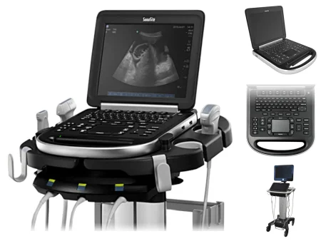
Ultrasound, also called sonography, aims high-frequency sound waves at tissue and turns the echoes into a live image. The waves travel well through liquid but not through air, so a gel is spread between the probe and the skin. Ultrasound is used for the abdominal organs and in pregnancy, and an intraoperativein-truh-OP-er-uh-tivHappening during surgery, from when the client is brought into the operating room until they are moved to recovery.intra- within, duringoperative of surgeryFull entry probe checks vessels and tissue during surgery. It is the least invasive imaging and uses no ionizing radiation, which is why it is the first choice in pregnancy. In IR it guides the needle into a vessel.
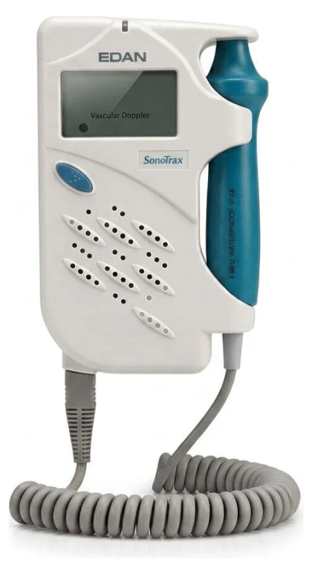
Doppler ultrasoundDOP-ler UL-truh-soundAn ultrasound that uses sound waves to hear or show blood flowing through vessels or the fetal heartbeat.Full entry studies (Doppler ultrasound) use ultrasound to measure blood flow through a vessel. The Doppler probe, held on the skin, turns moving blood into sounds that change in pitch, or into wave signals on a screen.
8Ionizing radiation
radiationray-dee-AY-shunEnergy that travels as waves or particles, such as X-rays, used to make images or treat cancer; also pain that spreads from its starting point.radi/o rays, radiation-ation processFull entry is energy traveling through space or matter. Ionizing radiation carries enough energy to make ions in whatever it strikes; x-rays, gamma rays and beta particles are examples. Visible and ultraviolet light, infrared, microwaves, ultrasound and radio waves are nonionizing. So the x-ray machine and the C-arm give off ionizing radiation, while a laser does not. In surgery, ionizing radiation comes from x-ray machines, fluoroscopes and unshielded radioactive implants, in amounts high enough to damage tissue.
What it does to the body
- Ionizing radiation changes the cell's membrane, its enzymeEN-zimeA protein that speeds up a chemical reaction in the body without being used up.Full entry and proteins, and its genetic material. At the high doses used to treat disease, those changes can lead to cancer of the bone, the thyroid and the reproductive organs, to cataractKAT-uh-raktClouding of the lens of the eye.Full entry and to spontaneous abortionspon-TAY-nee-us uh-BOR-shunThe loss of a pregnancy on its own, before the fetus can live outside the uterus, usually before 20 weeks; a miscarriage.Full entry .
- A cancer can appear years after the exposure, and the chance of one rises with the dose. A US National Research Council committee concluded in 2006 that there is likely no safe level: even a low dose may add a small cancer risk. Radiation can also cloud the lens of the eye (a cataract).
- A sudden, large exposure inflames and reddens the skin where it struck, and a large dose to the whole body causes radiation sickness: nausea, vomiting, diarrhea, weakness and even death. Radiation injury also badly weakens the blood-forming (hematopoietic) and immune systems, so the person cannot fight off infections.
- The thyroid, the lymph nodelimf nohdA small, bean-shaped organ of the immune system that filters lymph fluid and holds white blood cells.Full entry , bone and bone marrowbone MAIR-ohThe soft, spongy tissue inside bones that makes blood cells.Full entry are easily harmed. So are the eyes, where radiation can cause cataracts, ulcerUL-serAn open sore where the skin or the lining of an organ has broken down.Full entry of the corneaKOR-nee-uhThe clear, dome-shaped layer at the front of the eye, over the iris and pupil.Full entry and clouding.
- Children are more sensitive to radiation and have a long life ahead, so the machine's settings can be adjusted for the patient's age. Even low levels can harm a fetus.
How much damage is done depends on how long the exposure lasts, how far you are from the source, and which tissue is exposed.
Why it adds up
The effects of radiation depend on the dose and are cumulative: every exposure adds to the ones before. The risk grows with the number of x-ray exposures over a lifetime, although the cancer risk from one exposure is generally small and an x-ray that is needed is worth far more than its risk. Surgical staff may take many small doses over a whole career.
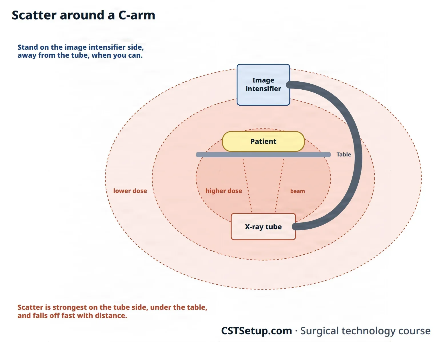
The danger to you is mostly scatter: radiation that bounces off the patient as the beam passes through, especially during fluoroscopyfloo-ROS-kuh-peeAn x-ray method that shows moving pictures of the inside of the body in real time.fluor/o glowing (from the fluorescent screen)-scopy looking, viewingFull entry . In interventional work the team stands close to the patient for long periods, and an angled C-arm can send a high dose of scatter back toward the staff.
How dose is measured
Dose to people is measured in rem or in sieverts (Sv); 5 rem equals 50 millisieverts (mSv). For a sense of scale, natural background radiation gives a person in the continental United States about 3 mSv a year.
- Workers: each US state sets its limits, typically 5 rem (50 mSv) a year for an adult worker. AST's standard uses the same yearly limit. Occupational Safety and Health AdministrationOH-shuh (said as a word)The US federal agency, part of the Department of Labor, that sets and enforces rules to keep workplaces safe.Full entry sets mandatory limits too, requires that staff who work with radiation be monitored, and requires that they be told their dose at least once a year.
- Pregnancy: 0.5 rem (5 mSv) for the whole pregnancy.
One reference gives a lower figure, from the International Commission on Radiological Protection: 20 mSv a year for a radiation worker, averaged over 5 years, and never more than 50 mSv in any one year. Your facility's radiation safety officer checks that the team follows the federal and state rules.
ALARA
Every exposure must be justified, and kept as low as reasonably achievable, or ALARA. It is the guiding idea of radiation safety, and it applies to every healthcare worker who may be exposed. Lowering the patient's dose usually lowers the staff's dose too. Doing only the images that are needed, and doing them well enough that none has to be repeated, is part of ALARA.
The Joint Commissionthuh JOYNT kuh-MISH-unAn independent US organization that inspects and accredits hospitals and other health care organizations against safety and quality standards.Full entry requires each hospital to name a radiation safety officer (RSO), who checks that radiation safety practices are followed, oversees the dosimeter program, and has the authority to stop unsafe practice. The RSO or a physicist reviews the dosimeter results at least every 3 months to see that staff doses stay ALARA and below the limits.
Radiation damage adds up over a lifetime, and the main danger to you is scatter from the patient. Keep every dose as low as reasonably achievable.
9Time, distance and shielding
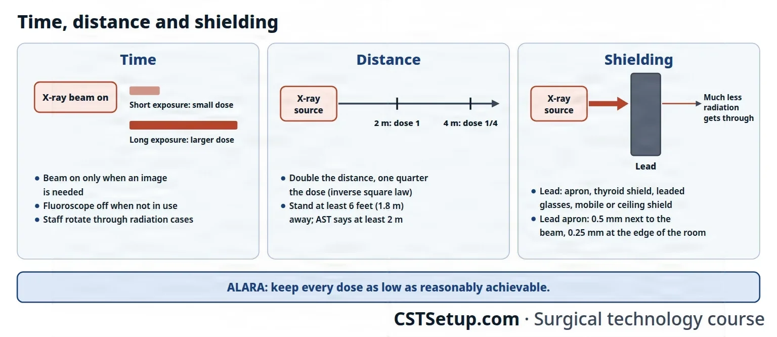
AST calls time, distance and shielding the three fundamental principles of radiationray-dee-AY-shunEnergy that travels as waves or particles, such as X-rays, used to make images or treat cancer; also pain that spreads from its starting point.radi/o rays, radiation-ation processFull entry protection, and every other radiation safety rule is built on them. Occupational Safety and Health AdministrationOH-shuh (said as a word)The US federal agency, part of the Department of Labor, that sets and enforces rules to keep workplaces safe.Full entry puts it the same way: keep the time short, the distance great, and use shielding.
Time
- The dose you receive depends on how long you are exposed.
- Keep each exposure as short as practical. When the fluoroscope is not being used it is turned off, because the patient is exposed the whole time it is on.
- Avoid the direct beam and limit the time you spend near its source.
- The fluoroscope is run at the lowest setting that still gives the surgeon a good image.
- Staff rotate through the cases that use radiation, so the same people are not overexposed.
Distance
A little more distance makes a big difference. By the inverse square law, when you double your distance from the source, your exposure falls to one quarter. A CSTsee-es-tee (said as letters)Certified Surgical Technologist, a credential for surgical technologists; in maternity care, contraction stress test.Full entry standing 4 meters from the source receives a quarter of the dose of a surgical assistant standing at 2 meters.
- Stand as far from the source as you can: at least 6 feet (about 1.8 m) from the source or the patient, by AORN's 2021 guidance and the textbooks, or else behind leaded shielding. AST's 2010 standard says at least 2 meters, a little over 6 1/2 feet. Treat those numbers as the least, and farther is better.
- People not needed in the room leave it. Nonsterile team members step behind a lead screen or out of the room during the exposure.
- The sterile team wears protective garments and moves as far from the source as it can while keeping surgical asepsisSUR-jih-kul ay-SEP-sisPractices that keep an area or object completely free of microorganisms; also called sterile technique.a- withoutsepsis decay, infectionFull entry .
Shielding
Radiation weakens as it passes through material, and leaded shields block x-rays well; lead is the most effective way to block radiation. During x-ray procedures in the OR, everyone either wears a shielding device, stands behind a barrier, or leaves the room.
- Shields in the room: mobile rigid shields on wheels and clear ceiling-mounted barriers, and lead drapes that hang from the table. Rooms in the radiology department have lead in their walls, so staff can step behind a wall while the equipment runs.
- Shields you wear: aprons, vests, skirts, thyroid shields and gloves made with lead or a lead-equivalent material, and leaded eyeglasses with side shields.
Where to stand at the C-arm
The beam enters the patient on the side of the x-ray tube and leaves toward the image intensifier, and the scatter is strongest on the tube side. AST's standard turns that into these rules:
- The tube goes under the table, angled as little as possible, with the beam as near vertical as it can be; the image intensifier sits above the patient.
- Stand on the image intensifier side, away from the tube.
- The patient lies as close to the image intensifier, and as far from the tube, as possible. Keeping the gap between the patient and the image intensifier small also means less scatter, so the machine does not have to turn up its output.
- A larger patient means more scatter, so the team makes every patient's position as good as it can be for imaging.
- Ceiling-mounted shields cut scatter, as long as they do not get in the way of the C-arm's movement.
- Scatter is highest at the level of the table and falls off fast with distance from it.
Never in the direct beam
- Keep your hands out of the primary beam.
- Never hold the patient, or an arm or a leg, in position during an x-ray. Positioning devices such as sandbags, slings and tractionTRAK-shunA steady pulling force applied to a body part with weights, ropes and pulleys.Full entry hold the patient instead, and a holder keeps a cassette in place.
- If holding cannot be avoided, the person holding wears a 0.5 mm lead apron, a thyroid shield, leaded eyeglasses and radiation-attenuating gloves. A pregnant team member never holds the patient or the cassette.
Never put your hand in the beam to hold a limb or a cassette, and never stay beside the table on the tube side while the beam is on. Use a holder, step back, and stand on the image intensifier side.
Talk before the beam goes on
- Before the x-ray equipment is switched on, the radiology technologist checks that everyone is out of the direct beam and at a safe distance, and then tells the team the beam is going on.
- That warning also lets the anesthesia provideran-us-THEE-zhuh pruh-VY-derThe licensed professional who gives anesthesia and watches over the client's breathing and vital functions during a procedure.Full entry hold the patient's breathing for a moment while images of the chest or abdomen are taken.
- During the time-outTYM-owtA pause just before a procedure when the whole team confirms the right client, procedure and site; also, a short break to calm a child.Full entry before radiation is used, the team checks that everyone is wearing their dosimeter.
- Warning signs are posted in plain view at the entrances to rooms where x-ray equipment will be used. They vary in shape and wording, but the radiation symbol is magenta or black on a yellow background.
10Lead aprons and other protective wear
A lead apron must be worn during fluoroscopyfloo-ROS-kuh-peeAn x-ray method that shows moving pictures of the inside of the body in real time.fluor/o glowing (from the fluorescent screen)-scopy looking, viewingFull entry to protect you from scatter. Every member of the sterile team wears one, especially when fluoroscopy will be used throughout the case. Put it on before the surgical scrub, because the sterile gown goes on over it.
- Thickness. Team members next to the main beam wear a 0.5 mm lead apron; those farther out at the edge of the room may wear 0.25 mm. By one reference's figures, a 0.25 mm apron blocks about 96 percent of the dose and a 0.5 mm apron about 99 percent.
- Front and back. The front of an apron holds more lead than the back, and many aprons shield only the front, so face the beam as much as you can. If you may have to turn your back to the beam, wear a wraparound apron.
- Fit. Aprons come in different lengths and widths. The apron must cover the upper legs and the upper chest, including the breasts, to protect the long bones, the bone marrowbone MAIR-ohThe soft, spongy tissue inside bones that makes blood cells.Full entry and the soft tissues of the chest. The people nearest the beam also shield the neck. A well-fitted apron protects better and strains the body less. Aprons are heavy and uncomfortable, and chapter 17 teaches how to lift heavy loads such as these.
- What it misses. An apron protects only what it covers: it does nothing for the eyes or the hands.

- Thyroid shield. The thyroid is sensitive to radiationray-dee-AY-shunEnergy that travels as waves or particles, such as X-rays, used to make images or treat cancer; also pain that spreads from its starting point.radi/o rays, radiation-ation processFull entry , and a shield wraps the neck to protect it. Wear one during fluoroscopy. AST's standard calls for a thyroid shield and leaded eyeglasses for anyone closer than 70 cm (a little over 2 feet) to the direct beam.
- Leaded eyeglasses. Team members next to the main beam, especially during fluoroscopy, wear leaded glasses with side shields to prevent cataractKAT-uh-raktClouding of the lens of the eye.Full entry and other eye damage. They range from heavy 0.5 mm leaded lenses to light tinted ones, and the RSO advises which to use.
- Radiation-attenuating gloves. When the hands will be in the direct beam or close to the patient, sterile radiation-attenuating gloves are worn. They give only partial protection, cutting the dose by about 16 to 22 percent by the figures AST's standard cites, and they are bulky and expensive. One reference also calls for lead-lined gloves when handling radioactive implants or dyes.
Caring for lead aprons
The lead inside an apron cracks when it is folded, and a crack lets radiation through, so aprons and thyroid shields are handled with care and examined regularly.
- Storage. Lay aprons flat, or better, hang them upright from the shoulders on an apron rack. Separate, labeled racks hold the 0.25 mm, 0.5 mm and maternity aprons. Never fold one.
- Cleaning. Wipe the apron down after every procedure with the solution or wipe recommended for it. AST's standard names cold water with a mild detergent, and never solvents or bleach, which damage the cover's nylon, plastics and rubber.
- Inspection. AST's 2010 standard notes, citing a 2006 The Joint Commissionthuh JOYNT kuh-MISH-unAn independent US organization that inspects and accredits hospitals and other health care organizations against safety and quality standards.Full entry manual, that The Joint Commission requires facilities to inspect lead aprons and leaves the method to them. A new apron is tested before it goes into use, and every apron at least once a year and whenever a user asks. First the apron is looked over and felt for tears, holes and thinning creases; one that fails is taken out of use. One that passes is then checked under fluoroscopy on manual settings: the shielded areas look dark, and any defect, seam or stitching shows light.
- When to reject. Each facility sets its own rules. AST's standard gives a guide: remove an apron with a defect larger than 15 square mm over a vital organ, or 670 square mm along a seam, an overlap or the back, and a thyroid shield with a defect larger than 11 square mm.
- Labels. An apron that passes carries an identification number, the date of its inspection, the date the next one is due, and its lead thickness, 0.25 mm or 0.5 mm.
- Disposal. A damaged apron is taken out of service at once. Lead aprons count as hazardous waste and are disposed of by the regulations for it.
Leaded garments are examined regularly, so look yours over before you put it on: a tear, a hole or a thinning crease fails the check, and a damaged apron is taken out of service at once.
11Dosimeters
A dosimeter is a small device, usually a badge, that measures the ionizing radiationray-dee-AY-shunEnergy that travels as waves or particles, such as X-rays, used to make images or treat cancer; also pain that spreads from its starting point.radi/o rays, radiation-ation processFull entry you receive and adds it up over time. Film badges, thermoluminescent dosimeters (TLDs), optically stimulated luminescence (OSL) badges, pocket ionization chambers and finger rings are all used.
- Who wears one. Team members who are routinely exposed to radiation in the OR. A dosimeter is worn whenever you work with x-ray equipment, radioactive materials or a radioactive patient. The RSO decides who needs one; AST recommends monitoring for anyone likely to receive more than a quarter of the yearly limit.
- Where. Always wear it in the same place, so its readings can be compared. With one badge during fluoroscopyfloo-ROS-kuh-peeAn x-ray method that shows moving pictures of the inside of the body in real time.fluor/o glowing (from the fluorescent screen)-scopy looking, viewingFull entry , wear it at the collar, outside the thyroid shield and above the apron. With two, one goes at the collar outside the apron and the other at the waist under it. Staff in high-dose fluoroscopy work wear two. A ring dosimeter goes on the hand closest to the source.
- Reading. The badges are read, and the results recorded, at least every 3 months, and every month for a pregnant team member.
- Keeping it at work. Never take your badge home or out to lunch. It would pick up radiation from other sources, such as the sun, and give a false reading, so it is stored at the facility.
12When a team member is pregnant
Even low levels of radiationray-dee-AY-shunEnergy that travels as waves or particles, such as X-rays, used to make images or treat cancer; also pain that spreads from its starting point.radi/o rays, radiation-ation processFull entry can harm a fetus, so a team member who is or may be pregnant takes extra care.
- Declaring the pregnancy. Telling the RSO is voluntary, but the declaration must be in writing; without it, the person is held to the same dose limits as everyone else. It includes an estimated date of conception.
- The fetal limit. Once the pregnancy is declared, the facility and the RSO must keep the dose to the fetus under 0.5 rem (5 mSv) for the whole pregnancy, spread evenly month by month so it does not come all at once.
- A second badge. A dosimeter is worn at the waist under the apron and read every month.
- The apron. A maternity apron has a double layer of lead in front, but it does not wrap around, so it does not protect the back, and it is heavier and can strain the neck and shoulders. A double-thickness or wraparound apron that covers the whole abdomen also works.
- Holding. A pregnant team member never holds the patient or a cassette. Assignments can be made so the person is not exposed; if exposure cannot be avoided, the person leaves the room or wears a shield that covers the body and the fetus.
13Protecting the patient
Lowering the patient's dose lowers the team's dose too, so every step here protects you as well.
- Keep the time short. Limit how long the patient is exposed, especially to fluoroscopyfloo-ROS-kuh-peeAn x-ray method that shows moving pictures of the inside of the body in real time.fluor/o glowing (from the fluorescent screen)-scopy looking, viewingFull entry , and turn the fluoroscope off when it is not in use.
- Narrow the beam. The beam is narrowed (collimated) to the area that needs to be seen, which lowers the patient's dose and the scatter.
- Get the equipment ready first. The surgeon, the scrub and the radiology technologist set up the x-ray or fluoroscopy equipment together: the C-arm is draped, or the cassette is in its sterile cover, the patient is positioned, and everyone has stepped back before the beam goes on.
- Position for imaging. A supine patient lies as flat as possible, with the arms out on armboards, and as close to the image intensifier as possible.
- Shield what is not being imaged. Keep the parts of the body that do not need to be imaged out of the beam, and place lead shields on the patient whenever they will not spoil the image. A thyroid shield goes on for x-rays of an arm, and for x-rays of a leg a lead shield covers the reproductive organs.
- Ask about pregnancy. Ask a patient who could be pregnant whether they are or might be, and tell the surgeon, who decides whether to reschedule. When the images cannot be avoided, a lead shield goes over the abdomen to protect the fetus.
- Write it down. The assistant circulator records the type of radiationray-dee-AY-shunEnergy that travels as waves or particles, such as X-rays, used to make images or treat cancer; also pain that spreads from its starting point.radi/o rays, radiation-ation processFull entry procedure, the protection used and the body parts covered, and the patient's skin before and after the procedure.
14Patients who carry radioactive material
Some patients come to surgery with radioactive material inside them: a radioactive treatment, a tracer, or implanted seeds. AST's standard asks the team to work with the RSO on each one.
- Warning. The team is told in advance when a patient has received therapeutic radionuclides. The chart carries a "Caution: Radioactive Material" label naming the radionuclide, its activity, the exposure rate at 1 meter and the date the precautionprih-KAW-shunA step taken ahead of time to prevent harm, such as the infection-control practices used with every client.Full entry end, and a wristband on the wrist away from the operative site gives the warning, the radionuclide and its activity.
- During an implant procedure. The RSO keeps the radioactive material under constant watch, and the team helps. Time, distance and shielding apply as in any other radiationray-dee-AY-shunEnergy that travels as waves or particles, such as X-rays, used to make images or treat cancer; also pain that spreads from its starting point.radi/o rays, radiation-ation processFull entry procedure.
- Tissue and body fluids removed from such a patient are labeled "Caution: Radioactive" and treated as hazardous waste, as the RSO directs. Chapter 29 teaches how radioactive specimens are handled.
- Handing off. The circulator tells the receiving unit the type of radionuclide, where it is in the body and its exposure rate.
When such a patient needs emergency surgery, the RSO is told as soon as possible, whatever the amount of material, and advises the team on the precautions for that material. At 5 millicuries (mCi) or less, the team's exposure is minimal and needs no special precautions.
- Radioactive colloid: once the incision is made, the sterile team's faces, and with a large incision their hands, are exposed to intense beta radiation, so they wear 0.5 mm aprons, 0.5 mm leaded glasses with side shields and two pairs of sterile gloveSTAIR-ul gluvA glove that has been sterilized and packed so it can touch a sterile field or an open body area without bringing in germs.Full entry .
- Metal radioactive implants: against high-energy gamma rays, lead gives no protection, so time and distance are all you have.
The team keeps the material from spreading: shoe covers come off in the room, the circulator handles sponges with forcepsFOR-sepsA hand instrument with two blades or arms used to grasp, hold or pull tissue or objects.Full entry , and the scrub keeps irrigationeer-ih-GAY-shunWashing out a wound, body cavity, tube or eye with a steady flow of liquid.Full entry off the floor. If a sharp that may carry radioactive material cuts you, take off your gloves at once, wash the wound under plenty of running water, and report it to the RSO.
15MRI safety
magnetic resonance imagingmag-NET-ik REZ-uh-nunts IM-uh-jingAn imaging test that uses a strong magnet and radio waves to make detailed pictures of the inside of the body.Full entry uses no ionizing radiationray-dee-AY-shunEnergy that travels as waves or particles, such as X-rays, used to make images or treat cancer; also pain that spreads from its starting point.radi/o rays, radiation-ation processFull entry , so its danger is different: the magnet.
The magnet never sleeps
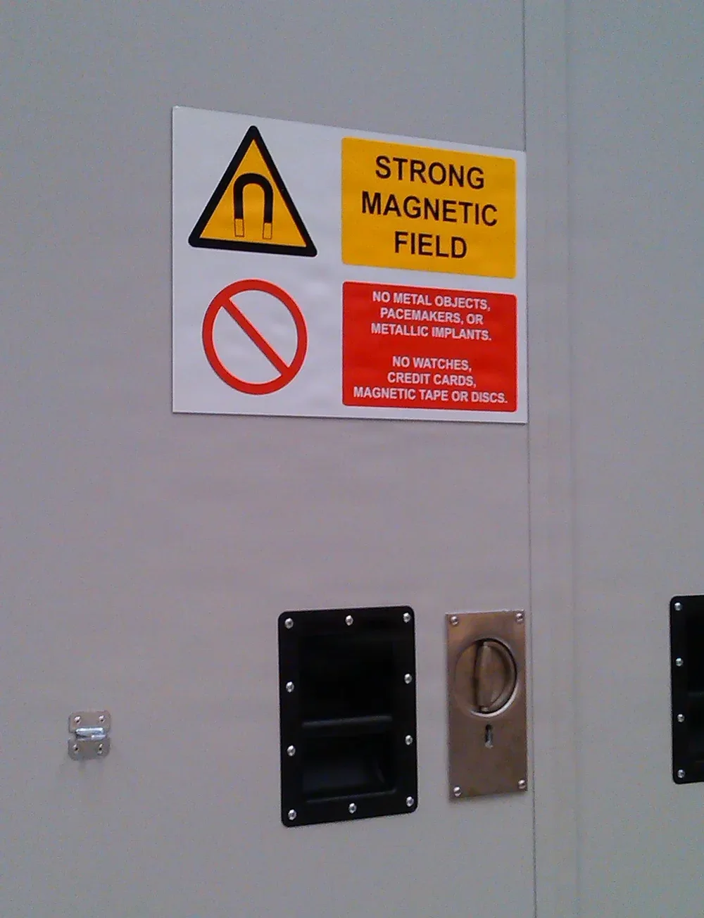
Whenever MRI is used, the main risk is metal, which the magnet can pull out of a hand or a pocket and send flying into its field. The field is strong enough to pull an object as large as a hospital bed into the scanner's tube. Any metal in range can be drawn toward the scanner, a real risk of injury to the patient and to anyone working nearby. On nearly every scanner in use, the field never switches off: it pulls on metal even when no scan is running, and it can reach several meters out from the scanner in every direction.
That is why The Joint Commissionthuh JOYNT kuh-MISH-unAn independent US organization that inspects and accredits hospitals and other health care organizations against safety and quality standards.Full entry requires a sign at the entrance to the scanner room saying that a dangerous magnetic field is present and that the magnet is always on. The only exception is a system built so that the operator can routinely switch its field on and off. The American College of Radiology (ACRay-see-AR (said as letters)An abbreviation for the American College of Radiology or the American College of Rheumatology; in lab reports, albumin-to-creatinine ratio.Full entry ) also asks for a lit red sign at that door reading "The Magnet is Always On," with a batteryBAT-uh-reeIn law, touching a person on purpose without their consent.Full entry backup.
The four MRI zones
The ACR divides an MRI facility into four safety zones, from the public areas of Zone I, where access is not controlled, to the scanner room itself, Zone IV.
- Zone I: everywhere the public may go freely, outside the MRI facility itself. Patients and staff pass through it to reach the MRI area.
- Zone II: the area between the public spaces and the strictly controlled zones. It usually holds the waiting area, patient preparation and locker rooms, and screening is often done here.
- Zone III: the controlled area just before the scanner room, usually where the scanner's operator console sits. Its doors are locked or opened by badge, no unscreened person and no metal object that a magnet attracts (a ferromagnetic object) may enter freely, and staff with the higher level of MRI safety training superviseSOO-per-vyzeTo guide, watch over and check the work of another staff member who is doing a task.Full entry everything in it.
- Zone IV: the scanner room itself, where a ferromagnetic object can become a deadly projectile. It is marked as hazardous, and its door stays closed unless it must be open for patient care or maintenance.
The Joint Commission's 2026 rules restrict the scanner room and the area just before its entrance, which match Zones IV and III. Only people trained in MRI safety, or people screened by staff trained in MRI safety, may enter them, and staff trained in MRI safety control and directly supervise both areas. These MRI zones are separate from the surgical suite's zones in chapter 17. Hospitals also track every time a ferromagnetic object gets into the scanner room by mistake, and every injury such an object causes.
Screening for metal
Everyone who will enter Zone III or IV is screened first. A patient who is awake fills out a written MRI safety screening form before Zone III and is screened at least twice before Zone IV, at least once by trained MRI staff who go over the answers with the patient. Anyone else not trained in MRI safety, an OR team member included, passes a written form and an interview with trained MRI staff before going in. The screening asks about:
- Metal the magnet attracts, on the body or in the pockets, such as jewelry, and items that conduct electricity. Patients take off their own clothes, jewelry and accessories and wear the site's MR Safe gowns, which have no pockets.
- Implants and devices, such as pacemakerPAYS-may-kerA small device that sends electrical pulses to the heart to keep it beating at a set rate; also the heart's own natural rhythm center.Full entry and metal plates, and metal fragments lodged in the body, such as shrapnel.
- Kidney function, because gadolinium contrastKON-trastA dye or other substance given before an imaging test so body parts show up more clearly.Full entry can cause a disease called nephrogenic systemicsis-TEM-ikAffecting the whole body rather than one part.Full entry fibrosisfy-BROH-sisThe thickening and scarring of tissue, which replaces normal tissue with tough fibers.fibr/o fiber-osis abnormal conditionFull entry in patients whose kidneys clear it poorly.
- Fear of closed spaces, because the patient must lie very still in the scanner for up to an hour, and some need sedationsih-DAY-shunA calm, relaxed or sleepy state brought on by drugs, or the use of drugs to cause it.sed calm, settle-ation processFull entry .
Implants matter for two reasons. An implant containing iron can be pulled loose by the magnet, and metal in the body, such as a pacemaker, vascular clips or a tattoo made with metal-based ink, distorts the images. Cardiac pacemakers and ferromagnetic aneurysmAN-yuh-riz-umA weak spot in the wall of an artery that bulges outward like a balloon.aneurysm a widening (Greek)Full entry clips in the brain have long been absolute reasons not to scan. With so many kinds of implants now, including ones with electronics, the instructions of the device's maker and the MRI's maker decide whether and how a patient may be scanned.
MR Safe, MR Conditional, MR Unsafe
Equipment, supplies and implants are labeled for the MRI environment with three terms from an international standard (ASTM F2503) that the FDA has adopted. MRI technologists train every year on which items have been found acceptable there, those labeled MR Safe or MR Conditional.
- MR Safe (a green square label): safe in every MRI environment, with no conditions. Only items that contain no metal, do not conduct electricity and are not magnetic can carry it.
- MR Conditional (a yellow triangle): shown to be safe in the MRI environment only when the conditions its maker lists are met, such as the strength of the magnet. One heart valve, for example, may be scanned only at 1.5 or 3.0 tesla, under the other conditions its maker lists. The conditions hold only for that exact model.
- MR Unsafe (a red circle): the item poses unacceptable risks to the patient, the staff or anyone else in the MRI environment, so it never enters the scanner room; such devices are projectile hazards.
A label decides, never a guess about the metal. Old words such as "nonmagnetic" or "MR compatible" are not one of the three labels, and anything made with metal cannot be MR Safe: it is MR Unsafe or, at best, MR Conditional. Ask the MRI staff before you carry anything past the door.
one reference still teaches that plastic and titanium are the only safe materials for MRI procedures. Today each item is judged by its MR Safe, MR Conditional or MR Unsafe label, and titanium, a metal, cannot be MR Safe.
If you are not sure an item is MR Safe or MR Conditional for that scanner, it stays outside. Scissors, jewelry and anything else metal on you come off before you go in.
The other MRI hazards
The Joint Commission requires MRI technologists to train every year on the hazards beyond flying metal: burns, which careful positioning of the patient and the equipment prevents; the noise, so patients get hearing protection; claustrophobia and anxiety; and emergencies, including the magnet's emergency shutdown (a quench).
MRI has not been shown to harm a fetus or a pregnant patient, so it can be used in pregnancy. Surgical team members may assist with MRI procedures, including during surgery in a dedicated MRI surgical suite, and they are screened like anyone else. In an MRI operating suite, a room can be Zone IV at one time and Zone III at another, as the setup changes.
16Your role in an IR case
IR needs surgical asepsisSUR-jih-kul ay-SEP-sisPractices that keep an area or object completely free of microorganisms; also called sterile technique.a- withoutsepsis decay, infectionFull entry , which is why you may be part of the team. You bring everything from the earlier chapters: the sterile fieldSTAIR-ul feeldAn area set up with sterile drapes and supplies, kept free of all germs, for a sterile procedure.Full entry , the counts, the medications and the specimens. This is what IR adds.
Before the case
- Put on your lead apron, and the thyroid shield and dosimeter the case calls for, before you scrub; the sterile gown goes over them.
- Set up the arteriogram supplies, balloons, catheters, guidewires, needles, sheathSHEETHA covering that wraps around a structure; in procedures, a short hollow tube left in a blood vessel to pass catheters through.Full entry and closure devices on the back table, as the preference card lists them.
- Flush every sheath, catheter and device lumenLOO-menThe open space inside a tube, such as a blood vessel, the intestine or a catheter.Full entry with heparinized saline before passing it, which keeps clot from forming in it and protects the vessel wall.
- Load the balloon inflation device with contrastKON-trastA dye or other substance given before an imaging test so body parts show up more clearly.Full entry solution and get every bubble out of the device, the tubing and the balloon.
- When you accept the contrast, ask the circulator whether the patient has any drug allergies, as chapter 12 teaches.
- Label every syringe and basin: contrast and heparinized saline are both clear.
- The C-arm is draped right before the procedure, then parked a little away from the field until it is needed.
During the case
- Keep guidewires wet and free of clot and debris, and wipe each one with a moist gauze sponge before and after every insertion. Flush the lumen of every wire, catheter and sheath as it goes in and as it comes out.
- Long wires are hard to handle, and devices are made to keep them from falling off the sterile field. Coil each wire quickly and safely after each use.
- Hand the devices up in the order they are used, each over the wire through the sheath.
- Keep a running total of the contrast and other drugs used, and report the totals at the end.
- When the technologist warns that the beam is going on, step back, stand on the image intensifier side, face the beam, and keep your hands out of it.
- Guard the sterile field, the back table included, as the C-arm or an x-ray tube moves over and around it.
At the end
- When a sheath comes out, pressure is held on a venous site for at least 10 minutes, and an arterial site is closed with sutureSOO-cherA stitch, or the thread used to stitch, that holds the edges of a wound or incision together.Full entry or a closure device. Watch the site for bleeding and swelling (a hematomahee-muh-TOH-muhA collection of blood, often clotted, that has leaked out of the blood vessels into the tissue.hem/o blood (here as hemat-)-oma swelling or massFull entry ).
- In an endovascular case, everything stays sterile until the patient leaves the room, and the team is ready to open if the endovascular repair fails.
- As assistant circulator, you record the radiationray-dee-AY-shunEnergy that travels as waves or particles, such as X-rays, used to make images or treat cancer; also pain that spreads from its starting point.radi/o rays, radiation-ation processFull entry procedure, the protection used and the patient's skin before and after, where your facility's policy allows.
In IR you keep the wires wet, wiped and under control, the lumens flushed, the air out, the syringes labeled, and the contrast counted. Then you step back when the beam goes on.
What to take from this chapter
- Interventional radiology finds and treats disease through a needle or a catheter steered by images, in the IR suite, the hybrid OR or the standard OR. It needs sterile technique, so you may be on the team.
- Before IR, the team checks allergies (especially to contrast), kidney function (BUN and creatinine; metformin is stopped for a time), pregnancy, earlier radiation and the plan for sedation.
- In the Seldinger technique a needle enters the vessel, a guidewire goes through it, the needle comes out, and a sheath or catheter slides over the wire. Keep wires wet, wiped and under control, flush every lumen, purge the air from the balloon system, and label and count the contrast.
- Watch for bleeding and a hematoma at the puncture site, contrast reactions (which can come up to 7 days later), kidney injury from contrast and radiation injury to the skin.
- X-rays, fluoroscopy, CT, nuclear medicine and PET use ionizing radiation; MRI and ultrasound do not.
- Radiation damage adds up over a lifetime, and scatter from the patient is the main danger to you. Keep every dose as low as reasonably achievable with time, distance and shielding: doubling your distance cuts your dose to a quarter, and you stand at least 6 feet away (AST says 2 meters), on the image intensifier side, when you can.
- Put the lead apron on before you scrub, face the beam, add a thyroid shield and leaded glasses near the beam, never fold an apron, and wear your dosimeter in the same place every time. A declared pregnancy keeps the fetal dose under 0.5 rem (5 mSv).
- The MRI magnet is always on. Everyone is screened, only trained or screened people enter Zones III and IV (the area before the scanner room and the room itself), and only items labeled MR Safe, or MR Conditional under their conditions, go in.
Check yourself5 quick questions
Free for everyone. Pick an answer for each, then check them all at once.
In the ORWhere you'll use this
The chapter's whole question pool in sets of 10, with the answer explained after each one.
Part of the educators programThe chapter's words on spaced cards: the ones you miss come back sooner.
Part of the educators programGiven by your class's teacher. Each class draws its own 25 from the chapter's pool.
Part of the educators program
Suggest a fix
Something wrong, unclear or out of date in this chapter? Tell us and we will fix it for everyone. Your email is only used to reply.