Minimally invasive and robotic surgery
Scopes, cameras, gas and the robot: how surgery works through small openings, and how you set up, run and protect each part of it.
1Why it matters
In open surgery the surgeon makes an incision big enough to see and touch the organs directly. In minimally invasive surgery (MIS), the surgeon works through a few small incisions instead. A lensed telescope carries the picture to a monitor, and long, thin instruments do the work while the whole team watches the screen.
endoscopyen-DOS-kuh-peeA procedure that uses a thin, flexible tube with a light and camera to look inside the body.endo- within-scopy looking with an instrumentFull entry is the same idea through a natural opening, such as the mouth, the anus or the urethrayoo-REE-thruhThe tube that carries urine from the bladder out of the body.Full entry . A tube that holds the lenses, the light and channels for small instruments goes in without any incision at all.
Facilities and makers use the words endoscopeEN-duh-skohpA thin tube with a light and a camera that is passed into the body to look inside an organ or cavity.endo- within-scope instrument for viewingFull entry and telescope for the same thing, so expect both. In this chapter, a telescope (or scope) is the rigid lensed tube that goes in through an incision, and an endoscope is the tube with built-in channels that goes in through a natural opening.
Why patients and surgeons choose it
Small incisions do less harm to the tissue, and that pays off for the patient.
- Many go home the same day, or within about 23 hours, instead of staying in the hospital.
- Less pain after surgery, and less need for pain medicine.
- Less blood lost during the operation, fewer problems with the incision and a lower rate of surgical site infection.
- Smaller scars and a faster return to normal life. Recovery from an open operation can take 4 to 6 weeks.
Today most operations are done this way. One reference estimates that only about 30 percent of surgical procedures are still done open; the rest use minimally invasive methods, robotic surgery included.
What it costs
MIS brings risks of its own. The gas or fluid that makes room to see can harm the patient, energy devices can burnBURNAn injury to the skin or deeper tissues caused by heat, chemicals, electricity, radiation or friction.Full entry tissue the camera cannot see, and the first needle or trocarTROH-karA sharp or bladed instrument inside a tube (cannula), used to pierce a body wall and leave the tube in place as a port.Full entry goes in partly blind. Not every patient is a good candidate, so patients are screened first: the patient must be able to tolerate the gas and the anesthesiaan-us-THEE-zhuhLoss of feeling or sensation, often brought on by drugs so that surgery or a procedure can be done without pain.an- withoutesthesi/o feeling, sensation-ia conditionFull entry as well as the operation. And every minimally invasive case can turn into an open one, so the team is always ready to convert.
The equipment is complex, delicate and expensive. Everyone on the team has to learn it, keep up with new devices, and handle each piece with care.
Where this chapter fits
This chapter covers the three kinds of minimally invasive system the Core Curriculum names: endoscopy (scopes, the camera chain, gas and fluid), robotics, and navigation, the computer guidance that is also used in open surgery such as joint replacement.
Other chapters hold the pieces around it:
- The video tower as room equipment, and testing it before the case: chapter 23.
- endoscopicen-doh-SKOP-ikDone with an endoscope, a thin tube with a light and camera passed into the body.endo- within-scopic viewingFull entry instruments by name: chapter 24. sutureSOO-cherA stitch, or the thread used to stitch, that holds the edges of a wound or incision together.Full entry through a scope and port closure devices: chapter 25.
- Cleaning and sterilizationstair-uh-luh-ZAY-shunA process that destroys all microorganisms, including spores, on an item; also a permanent birth control procedure.Full entry scopes and robotic instruments: chapter 27.
- electrosurgeryee-LEK-troh-SUR-jer-eeUsing high-frequency electric current through a handheld tool to cut tissue or seal bleeding vessels during surgery.Full entry , stray current and fire: chapter 20. Imaging and radiationray-dee-AY-shunEnergy that travels as waves or particles, such as X-rays, used to make images or treat cancer; also pain that spreads from its starting point.radi/o rays, radiation-ation processFull entry safety: chapter 22.
- Positioning the patient: chapter 31. Anesthesia and its monitors: chapter 13.
- Each operation done laparoscopically or robotically: its procedure chapter, starting with general surgery in chapter 32.
2Kinds of endoscopy
Every endoscopicen-doh-SKOP-ikDone with an endoscope, a thin tube with a light and camera passed into the body.endo- within-scopic viewingFull entry procedure needs the same few things: a way in, a working space, light and a view, instruments to work with, a way to clear fluid and gas, and a way to close.
There are two ways in. The scope either goes through the skin, through a small incision or puncture, or through a natural opening. Either way, the space usually has to be opened up with carbon dioxideKAR-bun dy-OK-sideA gas made by the body's cells as waste, carried in the blood to the lungs and breathed out.Full entry gas or with fluid so the surgeon can see and work.
| Procedure | What it looks into | Way in | What makes the working space |
|---|---|---|---|
| laparoscopylap-uh-ROS-kuh-peeSurgery or an exam done through small cuts in the belly, using a thin, lighted tube with a camera.lapar/o abdomen-scopy looking with an instrumentFull entry | The abdomen and pelvis | Small incisions in the abdominal wall | Carbon dioxide gas |
| Thoracoscopy, or video-assisted thoracoscopic surgery (VATS) | The chest | Small incisions in the chest wall, patient on the side | Gas is needed only when the anesthesia provideran-us-THEE-zhuh pruh-VY-derThe licensed professional who gives anesthesia and watches over the client's breathing and vital functions during a procedure.Full entry wants help collapsing the lung |
| Arthroscopy | A joint, such as the knee or shoulder | Small puncture incisions | Fluid pumped into the joint |
| Cystoscopy | The urethrayoo-REE-thruhThe tube that carries urine from the bladder out of the body.Full entry and bladder | The urethra | Fluid |
| Hysteroscopy | The inside of the uterus | Through the cervix | Fluid |
| bronchoscopybrong-KOS-kuh-peeA procedure in which a thin, lighted tube with a camera is passed through the nose or mouth to look inside the airways.bronch/o airway (bronchus)-scopy looking with an instrumentFull entry | The tracheaTRAY-kee-uhThe windpipe: the tube that carries air from the throat to the lungs.Full entry , bronchusBRONG-kusOne of the large air tubes that branch from the windpipe into each lung.bronch/o airwayFull entry and lungs | The mouth or nose | None: the airway's cartilageKAR-tih-lijFirm, smooth, rubbery tissue that cushions joints and gives shape to parts such as the ear, nose and windpipe.chondr/o cartilageFull entry holds it open |
| colonoscopykoh-luh-NOS-kuh-peeAn exam of the inside of the large intestine with a long, flexible tube that has a camera on its end.col/o colon-scopy looking with an instrumentFull entry and upper GI endoscopyen-DOS-kuh-peeA procedure that uses a thin, flexible tube with a light and camera to look inside the body.endo- within-scopy looking with an instrumentFull entry | The colon, or the esophagusih-SOF-uh-gusThe muscular tube that carries food and liquid from the throat to the stomach.esophag/o esophagusFull entry , stomach and duodenumdoo-uh-DEE-numThe first, short part of the small intestine, just after the stomach.Full entry | The anus, or the mouth | Air blown in through the scope |
| sinusSY-nusA hollow space in the body, most often one of the air-filled spaces in the skull bones around the nose.Full entry endoscopy | The nose and sinuses | The nostril | None: the firm walls of the nose hold the space open |
Scopes come in two main kinds. Flexible scopes include bronchoscopes, colonoscopes, ureteroscopes and some hysteroscopes. Rigid scopes include laparoscopes, arthroscopes, cystoscopes, sinus scopes and some bronchoscopes and hysteroscopes.
A diagnostic scope is for looking only. It has no working channels, and other instruments come in through separate openings if needed. An operative scope has channels for irrigationeer-ih-GAY-shunWashing out a wound, body cavity, tube or eye with a steady flow of liquid.Full entry , suction and instruments, such as a laser fiber.
MIS is used in nearly every specialty, most of all in abdominal, gynecologygy-nuh-KOL-uh-jeeThe branch of medicine that cares for the female reproductive organs and breasts.gynec/o woman-logy study ofFull entry , orthopedicor-thuh-PEE-dikHaving to do with the bones, joints, muscles, ligaments and tendons, and the branch of medicine that treats them.orth/o straightped/o child (the field began with straightening children's bones)-ic relating toFull entry , chest and urologic surgery. A gallbladder, a herniaHER-nee-uhA bulge of an organ or tissue through a weak spot in the muscle or tissue wall that normally holds it in.Full entry , a hysterectomyhis-tuh-REK-tuh-meeSurgery to remove the uterus.hyster/o uterus-ectomy surgical removalFull entry , a knee arthroscopy, a lung resectionrih-SEK-shunSurgery to remove part of an organ or tissue.re- back, againsect to cut-ion processFull entry and a prostatectomy can all be done through a scope.
Other ways in
- Single-incision laparoscopiclap-uh-roh-SKOP-ikDone through a few small cuts in the abdomen with a thin camera and long instruments, instead of one large opening.lapar/o abdomen-scopic viewingFull entry surgery (SILS): one port, usually in the umbilicus, holds several channels for the scope and instruments, leaving one small scar.
- Natural orifice transluminal endoscopic surgery (NOTES): a scope passes through a natural opening such as the mouth, anus or vagina, and the surgeon cuts through the wall of that organ to reach the target, leaving no skin scar.
- Hand-assisted laparoscopy: a sealed port in a small incision lets the surgeon put a hand inside the abdomen to feel and retract while the gas stays in.
A scope that enters sterile tissue, such as a laparoscope, thoracoscope or arthroscope, must be sterile; chapter 27 teaches which scopes may be high-level disinfectiondis-in-FEK-shunCleaning a surface or object with chemicals or heat to kill most germs, though not always all bacterial spores.Full entry instead.
3The scopes
Rigid telescopes
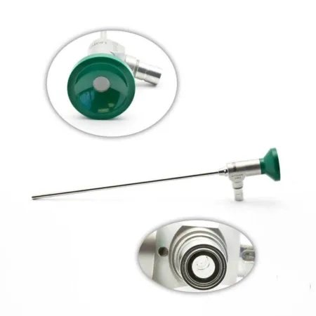
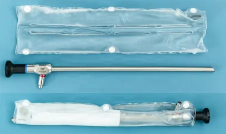
A rigid telescope holds a chain of lenses in a steel shaft. The light cord attaches at a light post near the eyepiece, and the light travels down the shaft to light up the tip. Chapter 24 names each part.
Three numbers describe a telescope:
- Optical angle: the direction the lens looks, in degrees off straight ahead. The usual angles are 0, 30, 45 and 70 degrees.
- Diameter: the width of the shaft, in millimeters, such as 2, 5 or 10 mm.
- Length: the length of the shaft, in centimeters.
A 0-degree scope looks straight ahead. An angled scope looks off to one side, so turning it on its own axis sweeps the view around and shows much more of the space. Very angled scopes, such as 70 degrees, let the surgeon see around corners, but they make it harder to stay oriented. Some special scopes are angled as far as 120 degrees.
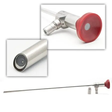
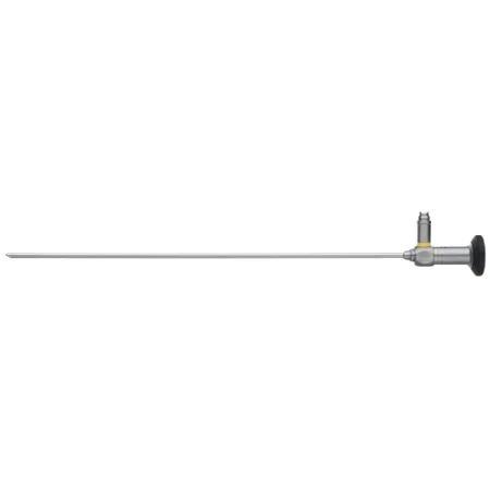
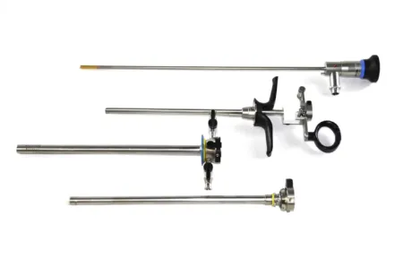
Specialty telescopes are built for the work of each specialty, with their own lengths and widths, channels for continuous irrigationeer-ih-GAY-shunWashing out a wound, body cavity, tube or eye with a steady flow of liquid.Full entry , or a way to carry electrosurgeryee-LEK-troh-SUR-jer-eeUsing high-frequency electric current through a handheld tool to cut tissue or seal bleeding vessels during surgery.Full entry . A resectoscope is a rigid telescope inside a cutting and coagulationkoh-ag-yuh-LAY-shunThe process by which blood changes from a liquid into a gel, forming a clot.coagul clot-ation processFull entry instrument: its laser or electrosurgical tip shaves tissue away piece by piece, as when tumors of the bladder or uterus, or the prostatePROS-taytA walnut-sized gland below the bladder in males that surrounds the urethra and makes part of the fluid in semen.Full entry , are resectionrih-SEK-shunSurgery to remove part of an organ or tissue.re- back, againsect to cut-ion processFull entry . The urology and gynecologygy-nuh-KOL-uh-jeeThe branch of medicine that cares for the female reproductive organs and breasts.gynec/o woman-logy study ofFull entry chapters teach it.
In some newer scopes the camera chip sits right at the tip, with no separate camera head or coupler. Fewer links in the chain means less fogging and fewer loose connections. Some scopes are hybrids, with a reusable eyepiece and a disposable shaft and tip, which must be lined up and focused carefully when they are put together.
Handling a telescope
The lenses are delicate and the scope is expensive, so handle it the same careful way every time.
- Hold it by the heavy head end, never by the tip or the shaft. Held by the light end, the weight of the head can bend the shaft.
- Keep it away from heavy instruments and sharps, which scratch and dent the shaft and its optics.
- Wipe it only with soft, lint-free material. Woven cloth leaves tiny scratches that blur the picture, and oils on the lens distort it.
- Never assume someone else checked it. Everyone who handles a scope, from sterile processing to the scrub, is responsible for checking it.
- Between uses, lay it on a soft towel that sheds no lint, or keep it in its warm water bath, and leave slack in its cables so a tug cannot pull it off the table.
To check a scope during setup, look at the distal lens and the eyepiece under indirect light for debris, scratches, chips and fingerprints. Look through it for a clear view, and turn the shaft as you look. Sight down its length to see that it is straight. Fog trapped inside, between the lens and its seal, means the seal leaks: report it, and take the scope out of service.
Flexible endoscopes
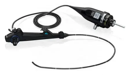
A flexible endoscopeEN-duh-skohpA thin tube with a light and a camera that is passed into the body to look inside an organ or cavity.endo- within-scope instrument for viewingFull entry has two main sections. The control head is the part you hold: it has the dials that bend the tip, the suction and air-water valves, and the connections for the camera, light, suction and irrigation. The insertion tube is the long, narrow part that goes into the patient, ending in a bending section at the tip. A cord from the control head carries the light and connects to the video processor.
Inside the insertion tube run the light fibers, the optics and several channels:
- The instrument channel, the largest, carries biopsy forcepsFOR-sepsA hand instrument with two blades or arms used to grasp, hold or pull tissue or objects.Full entry , brushes and other tools. One reference also calls it the biopsy or elevator channel. Its port sits where the control head meets the insertion tube.
- The auxiliary water channel sprays water at the tip to wash blood and debris off the lens.
- The air channel blows air into the bowel to open it up, much as gas opens the abdomen.
There are two kinds. A fiberoptic scope carries the image back through a bundle of tiny glass fibers to an eyepiece. A videoscope has a video chip at its tip and sends the picture straight to the monitor, with no eyepiece.
Two special kinds are worth knowing. An echoendoscope carries an ultrasound transducer at its tip, so it can image tissue beyond the wall of the organ. A capsule endoscope is a camera with a light and a batteryBAT-uh-reeIn law, touching a person on purpose without their consent.Full entry , about the size of a large vitamin: the patient swallows it, sensors on a belt pick up its pictures, and it passes out of the body on its own.
Assisting with flexible endoscopy
Most flexible endoscopyen-DOS-kuh-peeA procedure that uses a thin, flexible tube with a light and camera to look inside the body.endo- within-scopy looking with an instrumentFull entry is done in an outpatient endoscopy unit near the OR. The cases are short, the patient is usually sedationsih-DAY-shunA calm, relaxed or sleepy state brought on by drugs, or the use of drugs to cause it.sed calm, settle-ation processFull entry , and most go home once the sedation wears off.
The surgical technologistSUR-jih-kul tek-NOL-uh-jistA member of the operating room team who prepares the sterile field and instruments and hands them to the surgeon.Full entry helps position the patient, prepares the equipment and instruments, runs the suction and irrigation, and helps pass biopsy instruments down the channel. Tissue samples from a scope are tiny and easy to lose, so have labeled specimen containers ready before the first biopsy.
- To free tissue from biopsy forceps, dip the jaws in saline (or whatever the surgeon prefers) and move them gently. If the tissue stays stuck, lift it off with a needle.
- For a brush biopsy, the brush is smeared onto a glass slide and a fixative applied, or the brush itself is clipped off and sent wet, as pathologypuh-THOL-uh-jeeThe study of disease and its causes and effects, or the disease changes found in a person or tissue.path/o disease-logy study ofFull entry requires.
Flexible and rigid scopes can also work together: a separate team may run a flexible endoscope during a laparoscopylap-uh-ROS-kuh-peeSurgery or an exam done through small cuts in the belly, using a thin, lighted tube with a camera.lapar/o abdomen-scopy looking with an instrumentFull entry to get a better view of the bowel.
A scope is a chain of delicate parts. Hold it by the head, keep sharps away from it, and check it yourself every time, because a scratch or a cracked fiber shows up as a poor picture in the middle of the case.
4The video chain
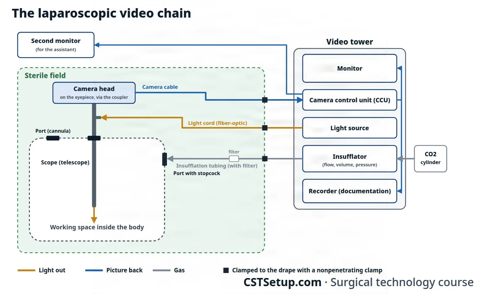
The surgeon sees only what the video chain (the imaging system) shows, so every link in it matters. Light from the light source runs down the light cord to the scope and lights up the inside of the body. The scope carries the picture back to the camera head, which turns it into an electrical signal. The camera cable carries that signal to the camera control unit (CCU), which processes it and sends it to the monitors and the recorder.
The chain is only as good as its weakest link. A sharp camera on a poor monitor gives a poor picture, and so does a perfect monitor fed by a cord full of broken fibers.
The light source and light cord
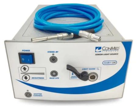
The light source makes the intense white light that high-definition video needs, usually from a xenon or LED lamp. Its panel sets the mode and the brightness. In automatic mode it adjusts the brightness to the picture by itself, so nobody has to keep turning it up and down.
If the picture still looks too dark, the usual fix is to raise the gain, the strength of the camera's signal, rather than the light.
Light at the tip of a connected scope is cool, because the heat stays behind in the light source. The loose end of a lit cord is a different matter: it is hot enough to burnBURNAn injury to the skin or deeper tissues caused by heat, chemicals, electricity, radiation or friction.Full entry skin or set drapes and sponges on fire, all the more near alcohol or another liquid that burns.
Never leave a lit light cord on the drapes. Connect the cord before the light goes on, and put the light source on standby before you disconnect it and whenever it is not in use. If the cord must come off the scope during the case, keep its end off the drapes, or rest it on a moist towel.
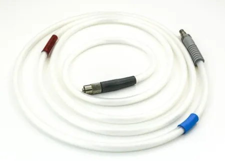
The light cord, also called the light cable or light guide, is a bundle of many thousands of fine glass or plastic fibers. A fiber that breaks no longer carries light, and breaks inside the cord do not show from the outside: you see them as a dim or distorted picture.
- Coil the cord loosely. Never kink it, hang it or bend it tightly, and store it lying flat.
- Never drop it or let it strike a hard surface.
- Look along its cover for cuts, nicks and dents before every use.
- To check the fibers, point one end at a bright room light and look at the other end. Dark spots ("peppering") are broken fibers, and once about 20 percent are broken, the cord is replaced. Never look into the end of a cord that is plugged into the light source: that light can harm your eyes.
- Match the cord to the scope. A cord too large for a small scope can overheat or damage its optics; one reference pairs a 2.5 mm cord with scopes under 4 mm and a 4.5 mm cord with a 10 mm scope. Some cords need an adapter to fit the light source or the scope, so have it ready before the case.
- Never touch a lamp with bare skin: it may be hot, and oil from your fingers shortens its life. Keep a spare lamp at hand.
The camera
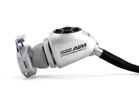
The camera head attaches to the scope's eyepiece through a coupler, called an endocoupler, that is made for that camera and scope. A lock ring holds them together, and a focus ring sharpens the picture.
Inside the camera head, a sensor chip turns light into a signal. Each light-sensing element on the chip is one pixel, and the more pixels, the sharper the picture. A three-chip camera gives each primary color (red, green and blue) its own chip, for natural color, which helps the surgeon tell healthy tissue from diseased tissue. Newer single-chip cameras boost their sharpness with digital processing in the control unit.
Modern systems use high-definition (HD) video, a widescreen format much sharper than the older standard definition. Each part must match the others, so check that the camera suits the rest of the system when you connect it.
Buttons on the camera head let the surgeon white balance, brighten a dark picture, start and stop recording, take still images, and on some heads zoom and focus, without asking the circulator each time.
The camera head is delicate. Treat it this way:
- Hold it firmly when you carry it.
- Open the lock ring before you connect or disconnect the scope, and check that the scope is firmly locked afterward. Never force or twist a connection.
- Plug and unplug the camera and video connectors only with the power off.
- Keep every connection dry.
- Unplug by the plug, never by pulling the cable.
- Never twist, crimp or kink the cable, and give it enough length to reach between the field and the tower. Most camera failures turn out to be in the cable, not the camera.
- Focus before surgery, with the mount locked, and check that the light comes through clearly, with no flicker.
The camera control unit, cables and recording
The CCU is the box on the tower where the camera cable plugs in. It holds the controls for light intensity, white balance and resolution, and it processes the camera's signal into the picture on the monitor. A good CCU also converts between standard and high definition, so pictures from one format can be shown in the other.
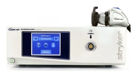
Video cables carry the signal between the units. HD signals need fiber-optic cables, so treat them as gently as light cords. They plug into marked sockets on the back of the CCU.
The documentation system records still pictures and video of the case, which can become part of the permanent record; chapter 19 teaches records and chapter 15 privacy.
The monitors are flat-panel, widescreen screens matched to the camera's resolution. For abdominal and chest cases there are usually two, one on each side of the patient, so the surgeon and the assistant can both see. One is enough when it can sit where everyone sees it.
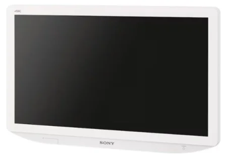
All of these units ride on the video tower, whose power strip usually has its main switch near the base, or they hang from ceiling booms in an integrated OR, where they may be run by remote control or by voice and can send video out of the room. Chapter 23 covers the tower as room equipment.
5Getting a clear picture
White balance
Light sources do not all give the same color of light. White balancing teaches the camera what white looks like through this scope, cord and coupler, so it shows every other color correctly, with no tint over the picture.
Some cameras must be white balanced before every case; others do it on their own. To white balance:
- Connect the light cord and the camera to the scope.
- Turn the light source on, at high or at the setting the maker gives.
- Aim the scope a few inches away, about 2 to 5, at a solid white surface such as white gauze or a white drape, never a woven sponge, which casts shadows on the picture.
- Press the white balance button, usually on the camera head or the CCU, and the system registers the white.
On the common multi-arm robot, the scrub does this with a sterile piece of white paper, from the camera head or the robot's touchscreen; newer robotic endoscopeEN-duh-skohpA thin tube with a light and a camera that is passed into the body to look inside an organ or cavity.endo- within-scope instrument for viewingFull entry may need no manual white balance.
Focus and brightness
Focus the camera before surgery with its mount locked. Most cameras also adjust to the light by themselves, which cuts the glare off shiny instruments and moist tissue.
Fogging
A cool scope fogs as soon as it enters the warm, moist body. Cold carbon dioxideKAR-bun dy-OK-sideA gas made by the body's cells as waste, carried in the blood to the lungs and breathed out.Full entry streaming past the scope at its port adds to the problem.
- Warm the scope first: keep its tip in a basin of sterile water at body temperature, or in a scope warmer (a warm-water thermos, a sleeve or a wrap), or wrapped in laps soaked in warm sterile water.
- Antifog solution: wipe it onto the lens as its maker directs, and dry the lens before you hand the scope up, so leftover solution does not blur the view.
- Warm gas: an insufflationin-suh-FLAY-shunBlowing a gas into a body cavity, such as carbon dioxide into the abdomen during laparoscopic surgery.Full entry that warms the carbon dioxide helps keep the lens clear.
- Another port: moving the gas line to a different port keeps cold gas from flowing past the scope.
- Touching tissue: the surgeon can warm and clear the lens by touching it gently to an organ, without pulling the scope out.
Look at the monitor during the case and clean or warm the scope as soon as the view clouds. Fog inside the scope that will not wipe away means its seal leaks.

Light for you
The room lights are usually dimmed so the monitor is easier to see. Keep one surgical light or another light on your back table, so you can find and set up instruments without mistakes.
For a clear picture: white balance, focus, and keep the lens warm and clean. When the view goes bad, check the simple things first: fog or smudge on the lens, a loose coupler, the light cord, the camera cable.
6Making room: insufflation
Inside the abdomen the organs lie snugly against the wall, with no empty space to work in. insufflationin-suh-FLAY-shunBlowing a gas into a body cavity, such as carbon dioxide into the abdomen during laparoscopic surgery.Full entry fills the abdomen, and sometimes the chest, with carbon dioxideKAR-bun dy-OK-sideA gas made by the body's cells as waste, carried in the blood to the lungs and breathed out.Full entry gas. The abdomen swollen with gas is called a pneumoperitoneum. The gas lifts the wall away from the organs, which gives the surgeon room to see and work, and lets the trocarTROH-karA sharp or bladed instrument inside a tube (cannula), used to pierce a body wall and leave the tube in place as a port.Full entry go in with less risk of hitting an organ.
Carbon dioxide is used because it is nontoxic, it does not burnBURNAn injury to the skin or deeper tissues caused by heat, chemicals, electricity, radiation or friction.Full entry or support burning, it is cheap, and the body absorptionub-ZORP-shunThe taking in of a substance, such as a drug or a nutrient, into the bloodstream.Full entry it quickly and breathes it out through the lungs.
The insufflator
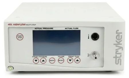
The insufflator takes gas from a carbon dioxide cylinder and feeds it to the patient through sterile tubing. Its panel shows three numbers: the flow, the volume of gas given and the pressure inside the abdomen.
- Flow is how fast the gas goes in, in liters per minute. It starts low, at about 1 to 2 L/min, and is raised once the needle is safely in place. A high flow refills the abdomen quickly when gas is lost through instrument changes or smoke evacuation.
- Pressure is how tight the abdomen is, in mm Hg, and it is the number to watch. In adults it is usually held at 12 to 15 mm Hg, with the insufflator set to stop at 15.
The insufflator stops filling when it reaches the set pressure, lets gas out if the pressure climbs too high, and sounds an alarm when something is wrong, such as a low gas supply or too much pressure. Pressure can also rise from other sources: someone leaning on the abdomen, or gas from an argon beam coagulator or a laser.
Keeping the gas safe
Most of these steps belong to the circulator, but the whole team watches them.
- Use only medical-grade carbon dioxide, from a labeled cylinder. Check the level before the case, keep a spare in the room, and change the cylinder before it runs low, so particles from the tank are not carried into the tubing.
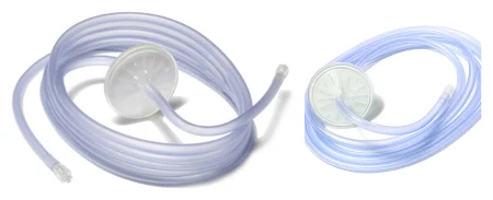
- Put a disposable hydrophobic (water-repelling) filter in the tubing. It stops particles from the tank and germs that grow inside the insufflator from being blown into the patient.
- Purge the tubing of air with carbon dioxide before it is connected to the patient. Air left in the tubing can cause a fatal air embolismEM-buh-liz-umThe blocking of a blood vessel by a mass carried there in the blood (an embolus), such as a clot, fat or an air bubble.embol/o plug-ism conditionFull entry .
- Keep the insufflator above the level of the patient's abdomen, so fluid cannot flow back into it.
- Watch the pressure for the whole case, and never lean on the abdomen.
- Put compression stockingkum-PRESH-un STOK-ingA tight elastic stocking that squeezes the leg, most firmly at the ankle, to help blood flow back toward the heart.Full entry or a sequential compression devicesih-KWEN-shul kum-PRESH-un dih-VYSInflatable leg sleeves that fill with air and squeeze the legs in turns to keep blood moving in the veins.Full entry on the patient before surgery.
- At the end, unhook the tubing from the machine first, and only then switch the insufflator off.
Cylinder carbon dioxide starts out cold: it is stored as a liquid and chills as it expands into a gas, and the faster it flows, the colder it is. The tubing warms it only a little, so many insufflators warm the gas, and the warmer must be watched.
As the scrub, you receive the sterile tubing onto the field, secure it to the drapes with a nonpenetrating clamp, and pass the other end off the field for the circulator to connect to the insufflator. Its patient end is attached to the Veress needle once the needle's position is confirmed, or to the Hasson cannulaKAN-yoo-luhA thin tube placed into the body or a body opening to deliver or remove fluid or air.Full entry or another trocar. Checking the insufflator and its cylinder before the case is part of testing the equipment (chapter 23).
Letting the gas out
At the end of the case the gas is removed through a filtered system, such as a smoke evacuator, never vented into the room. The gas can carry germs, viruses, cancer cells and anesthetican-us-THET-ikA drug that causes loss of feeling, with or without loss of consciousness.an- withoutesthesi/o feeling, sensation-ic relating toFull entry gases, so nobody opens a valve to let it escape into the air, and nobody presses on the abdomen to push out the last of it before the final port comes out.
7What the gas does to the patient
carbon dioxideKAR-bun dy-OK-sideA gas made by the body's cells as waste, carried in the blood to the lungs and breathed out.Full entry is safe in the right amount at the right pressure. Too much, or gas in the wrong place, can do serious harm. Watch the pressure, and speak up when you see a problem.
| Problem | What happens |
|---|---|
| Too much carbon dioxide in the blood (hypercapniahy-per-KAP-nee-uhToo much carbon dioxide in the blood, a PaCO2 above 45 mm Hg.hyper- abovecapn/o carbon dioxide-ia conditionFull entry , also called hypercarbia) | The gas crosses into the blood, more so when the pressure is high, and turns it acidic (respiratory acidosisRES-pur-uh-tor-ee as-ih-DOH-sisA state in which the blood becomes too acidic because the lungs do not remove enough carbon dioxide.Full entry ). The anesthesia provideran-us-THEE-zhuh pruh-VY-derThe licensed professional who gives anesthesia and watches over the client's breathing and vital functions during a procedure.Full entry watches the end-tidal carbon dioxide to catch it early. |
| Breathing and circulation | The swollen abdomen pushes the diaphragmDY-uh-framThe dome-shaped sheet of muscle under the lungs that is the main muscle of breathing; also the flat side of a stethoscope.Full entry up, so the lungs hold less air and breathing and the heart's output fall. With the head down, oxygen can drop; with the head up, less blood returns to the heart and the blood pressure falls. |
| Gas embolismEM-buh-liz-umThe blocking of a blood vessel by a mass carried there in the blood (an embolus), such as a clot, fat or an air bubble.embol/o plug-ism conditionFull entry | Gas forced into an open vein can block the circulation through the heart and lungs, causing collapse, cardiac arrestKAR-dee-ak uh-RESTThe sudden stop of the heart's pumping, so blood no longer reaches the brain and body.Full entry and death. Free gas can also block blood flow to the brain and cause a stroke. A pressure above 15 mm Hg can push gas into an exposed vessel. |
| Gas where it does not belong | Gas can track under the skin (subcutaneoussub-kyoo-TAY-nee-usUnder the skin; in the fatty layer between the skin and the muscle.sub- undercutane/o skin-ous relating toFull entry emphysemaem-fuh-SEE-muhA long-term lung disease in which the walls of the tiny air sacs are damaged and lose their stretch.Full entry ), into the chest (pneumothoraxnoo-moh-THOR-aksAir in the space between a lung and the chest wall, which lets part or all of that lung collapse.pneum/o air, lungthorac/o chestFull entry ), into the middle of the chest around the heart (mediastinal emphysema) or into the scrotumSKROH-tumThe pouch of skin and muscle that holds the testicles.Full entry . |
| Heart rhythm | The heart can beat irregularly, and too much pressure can slow it (bradycardiabray-dee-KAR-dee-uhA heart rate slower than normal; in an adult, fewer than 60 beats per minute.brady- slowcardi/o heart-ia conditionFull entry ) or change the blood pressure. |
| Stomach contents | Pressure on the diaphragm can push stomach contents up into the throat, where they can be breathed in. A tube into the stomach may be placed after the patient is asleep. |
| hypothermiahy-poh-THER-mee-uhA body temperature that has fallen below the normal range.hypo- belowtherm/o heat-ia conditionFull entry | Cold gas and cold fluids, on top of the cool room and a long case, chill the patient. |
| Infection | Germs or particles from the tank or the insufflationin-suh-FLAY-shunBlowing a gas into a body cavity, such as carbon dioxide into the abdomen during laparoscopic surgery.Full entry can be blown into the patient without a filter. |
| Shoulder pain afterward | Leftover gas irritates the phrenic nerve, which serves the diaphragm, and the patient feels it as pain in the shoulder and neck. |
When the gas causes trouble, the team may be asked to change the table position quickly, lower the gas flow, and help the anesthesia provider with drugs and supplies. Chapter 13 teaches the monitors the anesthesia provider watches, such as end-tidal carbon dioxide.
8Getting in: access and trocars
Surgical access means the way into the body. An incision is one form; a port made with a trocarTROH-karA sharp or bladed instrument inside a tube (cannula), used to pierce a body wall and leave the tube in place as a port.Full entry and cannulaKAN-yoo-luhA thin tube placed into the body or a body opening to deliver or remove fluid or air.Full entry is another. The first way into the abdomen is usually made one of three ways: with a Veress needle, by the open (Hasson) technique, or with an optical trocar.
Before any of them, the bladder must be empty, so the needle cannot puncture it: the patient voids before surgery, or a catheter drains the bladder in the OR.
The Veress needle (closed entry)
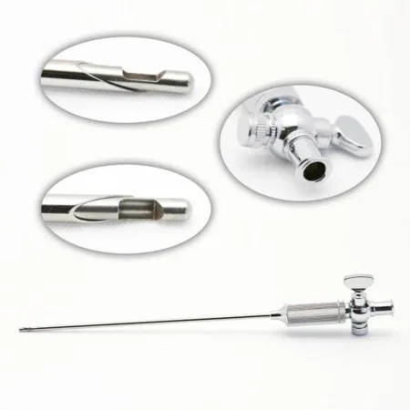
A Veress needle is a hollow, sharp needle with a blunt, spring-loaded stylet inside it. As the needle is pushed through the wall, each layer presses the stylet back and the sharp tip cuts through. As soon as the tip enters the open cavity, the spring snaps the blunt stylet forward again, ahead of the point, to shield the organs.
Before you pass it, test the spring: press lightly on the tip and watch the stylet pull back, then spring out again.
- The surgeon lifts the abdominal wall, by hand or with towel clamps, and nicks the skin with a #11 blade, near the umbilicus.
- The needle goes through the wall, at about 45 degrees, into the peritonealper-ih-toh-NEE-ulHaving to do with the peritoneum, the thin lining of the belly cavity and the organs inside it.Full entry cavity. It goes in blind, because no scope can go in until there is gas.
- Its position is checked. Drawing back on a syringe should bring up no bowel contents and no blood. Then several drops of saline are placed in the hub from a 10 mL syringe: the cavity is normally under negative pressureNEG-uh-tiv PRESH-erPressure that is lower than the pressure around it, so air or fluid is drawn in.Full entry , so if the tip is free inside it, the saline is drawn in on its own. If the tip is stuck in tissue, the saline does not flow.
- The insufflationin-suh-FLAY-shunBlowing a gas into a body cavity, such as carbon dioxide into the abdomen during laparoscopic surgery.Full entry tubing is attached and the gas started at a low flow. A low opening pressure, about 8 to 10 mm Hg, suggests the needle is free in the cavity. A higher one may mean the tip is in fat, in the omentum or inside the bowel.
- Once the abdomen is inflated, the needle comes out and the first trocar and cannula go in, usually for the scope, and the gas tubing moves to its port.
The open (Hasson) technique
The surgeon makes a short incision and cuts down through each layer to the fasciaFASH-ee-uhA sheet or band of tough connective tissue that covers and separates muscles and organs.fasci/o bandFull entry , which is opened. A stay sutureSOO-cherA stitch, or the thread used to stitch, that holds the edges of a wound or incision together.Full entry (size 0) goes in at each end of the fascial incision to hold up the wall. The peritoneumpair-ih-toh-NEE-umThe thin membrane that lines the belly cavity and covers most of the organs inside it.peri- aroundton stretched-eum a structureFull entry is opened under direct view, then the blunt Hasson cannula, with its obturatorOB-too-ray-terA smooth-tipped rod that fits inside a tube, such as a tracheostomy tube or cannula, to guide it in and then is taken out.Full entry , goes in and is tied to the stay sutures to hold it in place. The gas goes in through the cannula.
Because nothing is pushed in blind, the open technique is often chosen for patients with scars from earlier surgery near the umbilicus, likely adhesionad-HEE-zhunA band of scar tissue that joins two surfaces inside the body that are normally separate.ad- towardhes stick-ion conditionFull entry , many past operations, and for children.
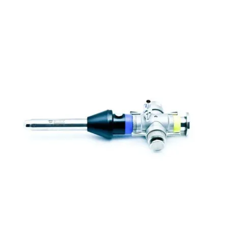
| Veress needle (closed) | Hasson (open) | |
|---|---|---|
| How the wall is entered | A spring-loaded needle punctures it through a skin nick, without sight | The surgeon cuts down through each layer and opens the peritoneum while looking |
| What goes in first | The needle; the first trocar follows once the abdomen is inflated | A blunt cannula, tied to stay sutures in the fascia |
| Where the gas goes in | Through the needle | Through the cannula |
| How placement is checked | aspirationas-puh-RAY-shunBreathing food, liquid, saliva or stomach contents into the airway and lungs; also, drawing fluid out with suction or a needle.Full entry , the saline drop test and the opening pressure | The surgeon sees into the cavity |
The optical trocar
An optical trocar is bladeless and clear at its tip. The scope goes inside it, so the surgeon watches each layer of the wall separate as the trocar is twisted and pushed through. The gas may be started before or after it goes in.
The other ports
Once the scope is in, the surgeon looks around to make sure the case can go ahead as planned, and every other trocar goes in under direct view, so the surgeon can see the tip come through and keep it off the organs.
The number, size and place of the ports depend on the operation. Most abdominal cases need at least three ports, unless the case is done through a single port. The surgeon may measure the sites with a ruler. Ports are placed to fit the instruments and the specimen: staplers, for example, pass only through 11 or 12 mm ports, and ports may be planned so two can be joined to take out a large specimen.
Trocars and cannulas
A trocar is a solid rod with a sharp, blunt or bladeless tip that fits inside a hollow tube, the cannula. You put them together before they go in; assembled, the tip of the trocar sticks out just past the end of the cannula. Once it is through the wall, the trocar (also called the obturator) comes out and goes back to the Mayo standMAY-oh standA small tray on an adjustable stand placed over or beside the patient during surgery to hold the instruments used most often.Full entry , and the cannula stays as the port for the scope and instruments.
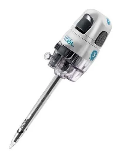
Cannulas come in sizes to fit the instruments. One reference lists:
- Microsurgical: 2 and 3 mm.
- Pediatric: 5 to 8 mm.
- Adult: 5 to 10 mm.
- Large and special purpose, such as for taking out a specimen: 10 to 15 mm, and up to 18 mm for a very large one.
Designs differ in how they enter, seal and stay put:
- A shield that slides over the sharp tip, or a tip that pulls back, once the wall is entered.
- Blunt or bladeless tips that push tissue apart instead of cutting it, and dilating tips that stretch the opening, leaving a smaller defect in the wall.
- Grippers, flanges for anchor sutures, or a foam or fabric collar that hold the cannula steady in the wall while instruments go in and out, and threads that guide it in.
- Valves that seal around an instrument, or close when it comes out, so the gas stays in, and a stopcock where the gas tubing connects.
- A reducer cap or built-in seal that lets a smaller instrument work through a larger cannula without leaking gas.
- Radiolucent cannulas that do not block the view on x-ray, for example during a cholangiogram.
Trocars come disposable, reusable or reposable (reprocessingree-PROS-es-ingThe steps that make a used reusable medical device safe to use on the next patient.Full entry a limited number of times). Disposable obturators are always sharp, and one obturator can be used for several ports of the same size on the same patient. A reusable set must have its tip sharpened routinely, its stopcocks and valves checked before and after each use, its gaskets replaced now and then, and its parts kept together, because an obturator and a sleeve may not fit another set.
Balloon dissection
To work outside the peritoneum, as in some herniaHER-nee-uhA bulge of an organ or tissue through a weak spot in the muscle or tissue wall that normally holds it in.Full entry repairs, the surgeon can open a space with a balloon dissector: a soft balloon on a tube, pushed into the tissue plane and inflated to push the layers apart without tearing them.
Closing the ports
At the end, each port comes out while the surgeon watches through the scope, and the scope and its own port come out last. The fascia is closed at the larger ports, usually 10 mm and up (one reference closes any port over 8 mm), to prevent a hernia at the port site; 5 mm sites usually need only the skin closed. Port closure devices are in chapter 25.
9Fluid instead of gas: distention
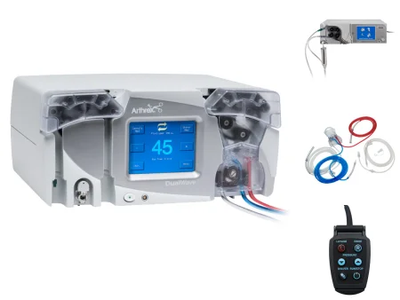
Joints, the bladder and the uterus are opened up with fluid instead of gas. In arthroscopy, cystoscopy and hysteroscopy, fluid runs in continuously to stretch the space open and to wash out the blood and bits of tissue that would cloud the view.
The fluid runs in from a pump or by gravity. A pump holds a set pressure, and some watch the pressure in the joint and adjust themselves. Fluid also runs out through an outflowOWT-flohThe flow of a fluid out of an organ, space or vessel.Full entry channel, so the space never overfills.
Choosing the fluid
The fluid has to suit the energy device. A fluid that contains electrolytes, such as normal saline or lactated Ringer's solutionLAK-tay-tid RING-erz suh-LOO-shunAn isotonic IV fluid containing water, sodium, chloride, potassium, calcium and lactate.Full entry , spreads electric current through itself, so a monopolar electrode cuts and coagulationkoh-ag-yuh-LAY-shunThe process by which blood changes from a liquid into a gel, forming a clot.coagul clot-ation processFull entry poorly in it.
- Nonelectrolyte (nonconductive) fluids, such as glycine, sorbitol and mannitolMAN-ih-tolAn osmotic diuretic, given by IV, that draws water out of tissues into the blood so the kidneys can remove it.Full entry , are used with older monopolar cutting systems. Sterile water is used for a cystoscopy with no electrosurgeryee-LEK-troh-SUR-jer-eeUsing high-frequency electric current through a handheld tool to cut tissue or seal bleeding vessels during surgery.Full entry .
- Electrolyte fluids, such as normal saline and lactated Ringer's, are used for arthroscopy and with newer systems that can work in saline, such as bipolarby-POH-lerHaving two poles or two opposite ends; most often used as a short form of bipolar disorder.bi- twopolar having polesFull entry ones.
- In hysteroscopy, the uterus may be distendeddih-STEN-didStretched out and enlarged by pressure from inside, such as from gas, fluid or urine.Full entry with normal saline, lactated Ringer's or 1.5% glycine, depending on the device.
It is your job to make sure the right solution is used for the electrode in use, as its maker directs. Warm the fluid to body temperature, 37°C, and no warmer: cold fluid running into a joint or the bladder chills the patient just as cold gas does.
AST's 2019 normothermia guideline, going by the temperatures used in studies, warms irrigationeer-ih-GAY-shunWashing out a wound, body cavity, tube or eye with a steady flow of liquid.Full entry fluid to 33 to 40°C as the fluid's maker allows, and one reference's care plan keeps it below 40.5°C (105°F).
When the fluid gets into the blood
Some of the fluid can be absorptionub-ZORP-shunThe taking in of a substance, such as a drug or a nutrient, into the bloodstream.Full entry through open blood vessels into the circulation. This is called intravasation. It can raise the blood pressure and upset the body's electrolytes.
- A nonelectrolyte fluid dilutes the blood's sodium (hyponatremiahy-poh-nuh-TREE-mee-uhA blood sodium level below normal, under 135 mEq/L.hypo- belownatr sodium (from its Latin name, natrium)-emia in the bloodFull entry ), and the brain can swell. Signs include a slow pulse, a blood pressure that rises and then falls, nausea and vomiting, headache, changes in vision, agitation, confusion and drowsiness.
- Saline is safer, but too much of it can still overloadOH-ver-lohdMore of something than the body can handle; in nursing, most often too much fluid in the circulation.Full entry the circulation (fluid overloadFLOO-id OH-ver-lohdToo much fluid in the body, in the blood vessels and the tissues.Full entry ) and cause heart failurehart FAYL-yerA long-term condition in which the heart cannot pump enough blood to meet the body's needs.Full entry in a patient with a weak heart.
So the fluid going in and the fluid coming out are measured all through the case, by hand or by the pump. The difference, the fluid deficit, is roughly what the patient has absorbed. In a healthy patient, the procedure is stopped at a deficit of about 1,000 mL of a nonelectrolyte fluid such as glycine, or about 2,500 mL of saline; the limit is lower for an older patient or one with heart or lung disease. Tell the surgeon and the anesthesia provideran-us-THEE-zhuh pruh-VY-derThe licensed professional who gives anesthesia and watches over the client's breathing and vital functions during a procedure.Full entry when the deficit reaches the limit. One reference gives 1 to 1.5 liters for a nonelectrolyte fluid. The deficit is reported at handoff reportHAND-off ree-PORTThe passing of information and responsibility for a client's care from one nurse or team to another.Full entry and recorded.
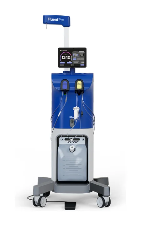
The specialty chapters teach each of these cases: hysteroscopy in chapter 33, cystoscopy in chapter 34 and arthroscopy in chapter 39.
Gas for the abdomen and chest, fluid for joints, the bladder and the uterus. The fluid must suit the energy device, it is warmed to body temperature, and every milliliter in and out is tracked, because what stays behind is in the patient's bloodstream.
10Instruments and supplies for MIS
The instruments
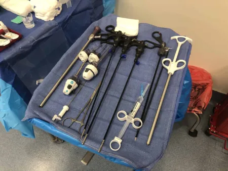
Minimally invasive instruments do precise work in a tight space from a distance. Their handles sit far from the working tip, their hinges and springs are tiny, and many shafts rotate so the tip can turn. The most common handle has two finger rings set at a right angle to the shaft. They come reusable, disposable, or reposable, where only the critical parts such as the tips are thrown away.
The main groups match open surgery: graspers (some with teeth for tissue being removed, some smooth to hold the bowel or liver gently), scissors, dissectors, retractorrih-TRAK-terA surgical instrument that holds back tissue or organs so the surgeon can see and reach the operative site.Full entry and probes, needle holders, clip appliers, staplers, suction-irrigators and specimen bags. Chapter 24 names them.
Clips are placed on vessels and ducts in place of ties, from disposable appliers that fire many clips in a row without reloading. Staplers made to pass through a port cut and staple at once. Knots are tied outside the body and pushed down, tied inside with two instruments, or made with a pre-tied loop; chapter 25 teaches these.
Energy devices (electrosurgeryee-LEK-troh-SUR-jer-eeUsing high-frequency electric current through a handheld tool to cut tissue or seal bleeding vessels during surgery.Full entry , ultrasonic shears, high-frequency bipolarby-POH-lerHaving two poles or two opposite ends; most often used as a short form of bipolar disorder.bi- twopolar having polesFull entry devices and lasers) do much of the cutting and sealing. Inside the body, energy is used close to tissue the camera cannot see, and a burnBURNAn injury to the skin or deeper tissues caused by heat, chemicals, electricity, radiation or friction.Full entry may go unnoticed until signs of a burned or perforationper-fuh-RAY-shunA hole that goes all the way through the wall of an organ or tissue.per- throughforat bored (a hole)-ion processFull entry organ appear after surgery. Such a burn can come from a break in an instrument's insulation, from the active tip touching or nearly touching another metal instrument (direct coupling), or from stray current passing through intact insulation into a cannulaKAN-yoo-luhA thin tube placed into the body or a body opening to deliver or remove fluid or air.Full entry or tissue nearby (capacitive coupling). Chapter 20 teaches these risks and how they are prevented, including active electrode monitoring, which stops the current when it senses a fault.
Caring for them during the case
- Inspect them at setup and again at the close: moving parts working, pins tight, shafts straight, surfaces without scratches, dents or nicks, and insulation unbroken.
- Keep them in a rack on the back table, apart from heavy instruments, so they are protected and easy to find.
- Pass each one with its working tip pointing down, and help guide it into the port if asked.
- Wipe the tips and shafts with a damp sponge, and flush suction tips often so they do not clog. Use sterile water, never saline, which leaves salt deposits and harms the instruments.
- Disconnect cables and tubing before you bring an instrument back from the field to the table.
Laparoscopic and robotic supplies
| Supply | What it does |
|---|---|
| Antifog solution | Keeps the lens from fogging; wiped on before the scope goes in and whenever the view clouds |
| Endo Stitch | A suturing device that passes its needle back and forth between two jaws, for stitching through a port (chapter 25) |
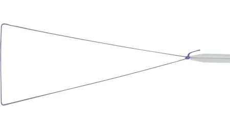
| Endoloop | A pre-tied sutureSOO-cherA stitch, or the thread used to stitch, that holds the edges of a wound or incision together.Full entry loop on a pusher that is slipped over a pediclePED-ih-kulA narrow stem or stalk that attaches a structure, such as a tumor or a skin flap, to the body.Full entry , such as the appendixuh-PEN-diksA small, finger-shaped pouch attached to the first part of the large intestine, in the lower right abdomen.Full entry , and cinched tight (chapter 25) |
| Hand-assisted port | A sealed port that lets the surgeon's hand into the abdomen without losing the gas |
| insufflationin-suh-FLAY-shunBlowing a gas into a body cavity, such as carbon dioxide into the abdomen during laparoscopic surgery.Full entry tubing | Carries carbon dioxideKAR-bun dy-OK-sideA gas made by the body's cells as waste, carried in the blood to the lungs and breathed out.Full entry from the insufflator, through a filter, to the Veress needle or a port |
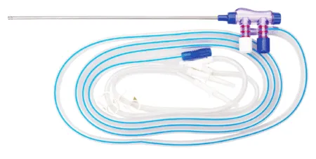
| irrigationeer-ih-GAY-shunWashing out a wound, body cavity, tube or eye with a steady flow of liquid.Full entry and aspirationas-puh-RAY-shunBreathing food, liquid, saliva or stomach contents into the airway and lungs; also, drawing fluid out with suction or a needle.Full entry device | A probe with two valves, one to suction and one to irrigate, to clear blood and fluid and wash the field |
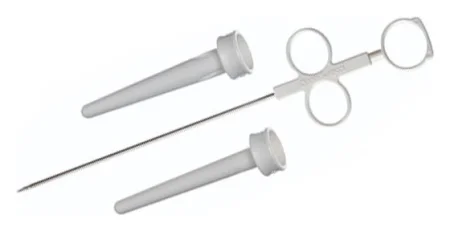
| Port closure device | A suture passer that carries suture through the fasciaFASH-ee-uhA sheet or band of tough connective tissue that covers and separates muscles and organs.fasci/o bandFull entry beside a port site, so the fascia can be closed (chapter 25) |
| Scope warmer | Keeps the scope warm on the back table so it does not fog |
| Specimen retrieval pouch | A bag passed through a port to hold tissue so it comes out without spilling |
| Stopcock | The small valve on a cannula or Veress needle where the gas tubing attaches, opened and closed to control the gas |
| trocarTROH-karA sharp or bladed instrument inside a tube (cannula), used to pierce a body wall and leave the tube in place as a port.Full entry | Make the ports the scope and instruments go through |
| Veress needle | The spring-loaded needle for closed entry and the first gas |
Taking out the specimen
A specimen is often placed in a retrieval bag passed in through a large port: the surgeon puts the tissue in, closes the bag, and draws it out, sometimes after widening the port incision. A very large specimen may need an 18 mm port.
Large, dense tissue can be cut into small pieces inside the body (morcellation), often inside a containment bag that keeps the pieces in. Some specimens come out through a natural opening instead, such as the vagina after a hysterectomyhis-tuh-REK-tuh-meeSurgery to remove the uterus.hyster/o uterus-ectomy surgical removalFull entry . You may be asked to grasp and insert the bag, as the surgeon directs. Handle the specimen as chapter 29 teaches.
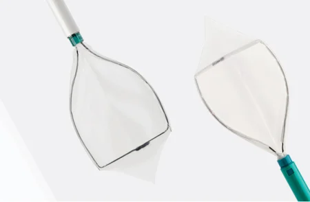
11Preparing the patient and the room
A patient having MIS is prepared much as for the open version of the same operation, with extra attention to the risks that come with the scope, the gas and the equipment.
Position
The position depends on the operation, and tilting the table lets gravity move the organs out of the way:
- Upper abdomen and lower esophagusih-SOF-uh-gusThe muscular tube that carries food and liquid from the throat to the stomach.esophag/o esophagusFull entry : reverse Trendelenburg positionree-VERS tren-DEL-un-burg puh-ZISH-unA position lying on the back on a bed or table that is tilted so the head is higher than the feet, with the body kept straight.Full entry , head up, so the bowel falls away. A padded footboard keeps the patient from sliding down.
- Pelvis and lower abdomen: Trendelenburg positiontren-DEL-en-burg puh-ZISH-unA position in which the client lies on the back with the bed tilted so the head is lower than the feet.Full entry , head down. gynecologygy-nuh-KOL-uh-jeeThe branch of medicine that cares for the female reproductive organs and breasts.gynec/o woman-logy study ofFull entry and prostatePROS-taytA walnut-sized gland below the bladder in males that surrounds the urethra and makes part of the fluid in semen.Full entry cases are often in lithotomylih-THOT-uh-meeA position lying on the back with the hips and knees bent and the legs raised and spread apart, often in stirrups.lith/o stone-tomy cutting intoFull entry . In a steep tilt the patient can slide toward the head, so an anti-skid pad or vacuum positioner is used.
- Chest (VATS): lateral, on the side.
- Shoulder arthroscopy: beach chair, with the arm free to move. Knee arthroscopy: supine, with the leg free.
When the table tilts during the case, move the Mayo standMAY-oh standA small tray on an adjustable stand placed over or beside the patient during surgery to hold the instruments used most often.Full entry and any tray over the patient so nothing presses on the patient. Chapter 31 teaches positioning in full.
Prep, drapes and warmth
The skin is prepped and draped as widely as for the open operation, so the case can convert without a new prep. The bladder is emptied before the needle goes in, and a tube may drain the stomach.
Gas and fluid both chill the patient. Use warmed gas and warmed fluids, a forced-air warming blanket, and keep the patient covered except where needed.
Setting up the field
- Keep the scope on a soft towel or in its warmer, where it cannot roll off, and set the warm water thermos where it will not be knocked over.
- Secure the light cord, camera cable, gas tubing and energy cords to the drapes with nonpenetrating clamps only. Leave enough slack, and watch them when the table moves, since a tilt can pull them tight.
- Anything that is part of the count, such as sponges and sharps, is counted as it is added, as in every case. Many facilities count during a laparoscopiclap-uh-roh-SKOP-ikDone through a few small cuts in the abdomen with a thin camera and long instruments, instead of one large opening.lapar/o abdomen-scopic viewingFull entry case just in case it converts to an open one; chapter 28 teaches when instruments are counted.
Holding the camera
In the second scrub role you may run the camera. AST's position is that a CSTsee-es-tee (said as letters)Certified Surgical Technologist, a credential for surgical technologists; in maternity care, contraction stress test.Full entry from an accredited program may advance a rigid or flexible scope, hold it steady and move it to keep the surgeon's view, under the surgeon's direction, as state law and facility policy allow. A CST who has had little chance to practice this gets more training and a documented competencyKOM-puh-tun-seeA specific skill or ability that a person has been shown to perform to an expected standard.Full entry first.
12Converting to an open operation
Any minimally invasive case can become an open one. The patient is told before surgery that this can happen, and the equipment for it is ready before the case starts.
Common reasons are bleeding that cannot be controlled through the scope, which is the most common; adhesionad-HEE-zhunA band of scar tissue that joins two surfaces inside the body that are normally separate.ad- towardhes stick-ion conditionFull entry that block the way in, anatomy too distorted to dissectdy-SEKT, or dih-SEKTTo cut apart or separate tissues carefully during surgery.Full entry safely, or heavy contamination or infection; equipment that fails; and a patient whose heart or lungs cannot tolerate the gas or the steep position.
Bleeding is harder to manage through ports. In an open case the surgeon can pack the area with laps and press, and quickly find the bleeding vessel; through small ports there is little room to press, and the source is harder to see.
Being ready
When you set up, read the surgeon's preference card for the open operation too, and collect its supplies where they can be opened within minutes. Whether the open set is opened and counted at the start is decided by policy and the case (chapter 28).
When the surgeon says "open"
The decision to convert is the surgeon's, and the team moves at once, quickly but with deliberate, careful moves.
- Free the cords and tubing from the drapes and clear them off the field, so they do not tangle in the drapes.
- Put the MIS instruments aside or off the field.
- Hand up suction, the electrosurgeryee-LEK-troh-SUR-jer-eeUsing high-frequency electric current through a handheld tool to cut tissue or seal bleeding vessels during surgery.Full entry and the scalpel first, so the incision can start without delay.
- Take the open instruments and supplies from the circulator, and count everything as it is added, as in every case.
Haste makes errors. The return electrode can shift if the patient is moved, the patient may need to be repositioned, prepped and draped again, and rushed conversions are where instruments and sponges get left behind.
13The risks of endoscopic surgery
Keep these in mind through every scope case:
- Injury at entry: the Veress needle and first trocarTROH-karA sharp or bladed instrument inside a tube (cannula), used to pierce a body wall and leave the tube in place as a port.Full entry go in partly blind and can injure the bowel, the bladder or a major blood vessel.
- Burns out of view: energy can injure tissue the camera does not show, and the injury may not be found until later.
- Bleeding: harder to find and to control than in open surgery, and the most common reason to convert.
- Gas and fluid: everything in the gas and fluid sections above, from hypercapniahy-per-KAP-nee-uhToo much carbon dioxide in the blood, a PaCO2 above 45 mm Hg.hyper- abovecapn/o carbon dioxide-ia conditionFull entry and gas embolismEM-buh-liz-umThe blocking of a blood vessel by a mass carried there in the blood (an embolus), such as a clot, fat or an air bubble.embol/o plug-ism conditionFull entry to fluid overloadFLOO-id OH-ver-lohdToo much fluid in the body, in the blood vessels and the tissues.Full entry and hypothermiahy-poh-THER-mee-uhA body temperature that has fallen below the normal range.hypo- belowtherm/o heat-ia conditionFull entry .
- Port sites: infection and a herniaHER-nee-uhA bulge of an organ or tissue through a weak spot in the muscle or tissue wall that normally holds it in.Full entry at a port site.
- Position: long cases in steep positions put pressure on nerves and skin, and can shift the patient (chapter 31).
Tell the surgeon at once if you see something wrong on the monitor or the insufflationin-suh-FLAY-shunBlowing a gas into a body cavity, such as carbon dioxide into the abdomen during laparoscopic surgery.Full entry : a pressure alarm, a falling gas supply, bleeding at a port, or an instrument out of view with the energy on. Through a scope, the team sees only what is on the screen.
14Robotic surgery
AST defines robotic surgery as surgery that puts a computer-assisted electromechanical device between the surgeon and the patient. The device improves what the surgeon can see, how finely tissue can be handled and how tissue can be sensed, and it is now a routine way to do minimally invasive surgery in most specialties.
The U.S. Food and Drug Administration (FDA) calls these devices robotic-assisted surgical (RAS) systems, a kind of computer-assisted surgery in which computer technology and software control and move the instruments.
The first systems appeared in hospitals in the mid-1980s. Their prototypes came from NASA, the Department of Defense, the Stanford Research Institute and private companies, who wanted surgery that could be done from a distance for wounded soldiers and for people in space. The first FDA-cleared device came in 1994.
A robotic-assisted system is not autonomyaw-TON-uh-meeA person's right to make their own choices about their body and their care.Full entry . A fully robotic machine, like one in a factory, carries out a programmed task on its own; a surgical robot moves only when the surgeon moves its controls, although newer systems may automate some steps.
Robotic surgery grew out of MIS and shares its basics: ports, an endoscopeEN-duh-skohpA thin tube with a light and a camera that is passed into the body to look inside an organ or cavity.endo- within-scope instrument for viewingFull entry whose picture appears on a monitor, a gas-filled working space, long instruments including staplers, and the usual energy devices. That is why learning to assist in laparoscopylap-uh-ROS-kuh-peeSurgery or an exam done through small cuts in the belly, using a thin, lighted tube with a camera.lapar/o abdomen-scopy looking with an instrumentFull entry comes first.
The words of robotic surgery
- Surgeon console: the nonsterile station where the surgeon sits or stands to work the instruments from a distance.
- Patient cart or bedside unit: the part beside the patient that holds the arms, the endoscope and the instruments, and takes its orders from the console.
- Manipulators: the technical name for the robot's arms.
- Modular system: one in which each arm sits on its own movable base, instead of all the arms on one cart.
- Hand controllers: the devices at the console that the surgeon moves by hand to work the instruments.
- Docking: moving the patient cart into place at the bedside and attaching its arms to the ports.
- Port and surgical access: the opening made by a trocarTROH-karA sharp or bladed instrument inside a tube (cannula), used to pierce a body wall and leave the tube in place as a port.Full entry and cannulaKAN-yoo-luhA thin tube placed into the body or a body opening to deliver or remove fluid or air.Full entry for the instruments and endoscope, and the method used to get into the body.
- Degrees of freedom: the number of separate ways an instrument or arm can move.
- Pitch, yaw and roll: up and down, side to side, and turning on its own axis, the way an airplane moves.
- Wristed instruments: robotic instruments whose tips bend and turn like a wrist: seven degrees of freedom plus opening and closing the jaws.
- Scaled movement: the system shrinks the surgeon's hand movements, usually 5 to 1, so a large, easy motion of the hand makes a tiny, precise one at the tip. It also filters out the natural tremorTREM-erShaking of a part of the body, such as the hands, in small rhythmic movements that the person cannot control.Full entry of the hand.
- Haptic feedbackFEED-bakInformation about a result that is sent back to change what happens next, in the body or between people.Full entry : the sense of touch and pressure passed back to the surgeon's hand.
- Reposable: an instrument that can be reprocessingree-PROS-es-ingThe steps that make a used reusable medical device safe to use on the next patient.Full entry a set number of times and is then thrown away.
- Remote data display: scans such as computed tomographykum-PYOO-tid toh-MOG-ruh-feeAn imaging test that uses x-rays and a computer to make cross-section pictures of the inside of the body.tom/o cut, slice-graphy process of recordingFull entry or magnetic resonance imagingmag-NET-ik REZ-uh-nunts IM-uh-jingAn imaging test that uses a strong magnet and radio waves to make detailed pictures of the inside of the body.Full entry images shown on the surgeon's screen during the operation.
- Registration (of images): layering separate images of the same patient into one three-dimensional picture.
- Telesurgery: operating on a patient from another place, even miles away. The delay between the surgeon's hand and the robot's movement has been a major obstacle.
15The three parts of a surgical robot
A robotic system is a set of separate parts joined by cables. The Core Curriculum names three: the surgeon console, the patient cart and the vision cart. Chapter 23 introduced them as room equipment; here is what each one does.
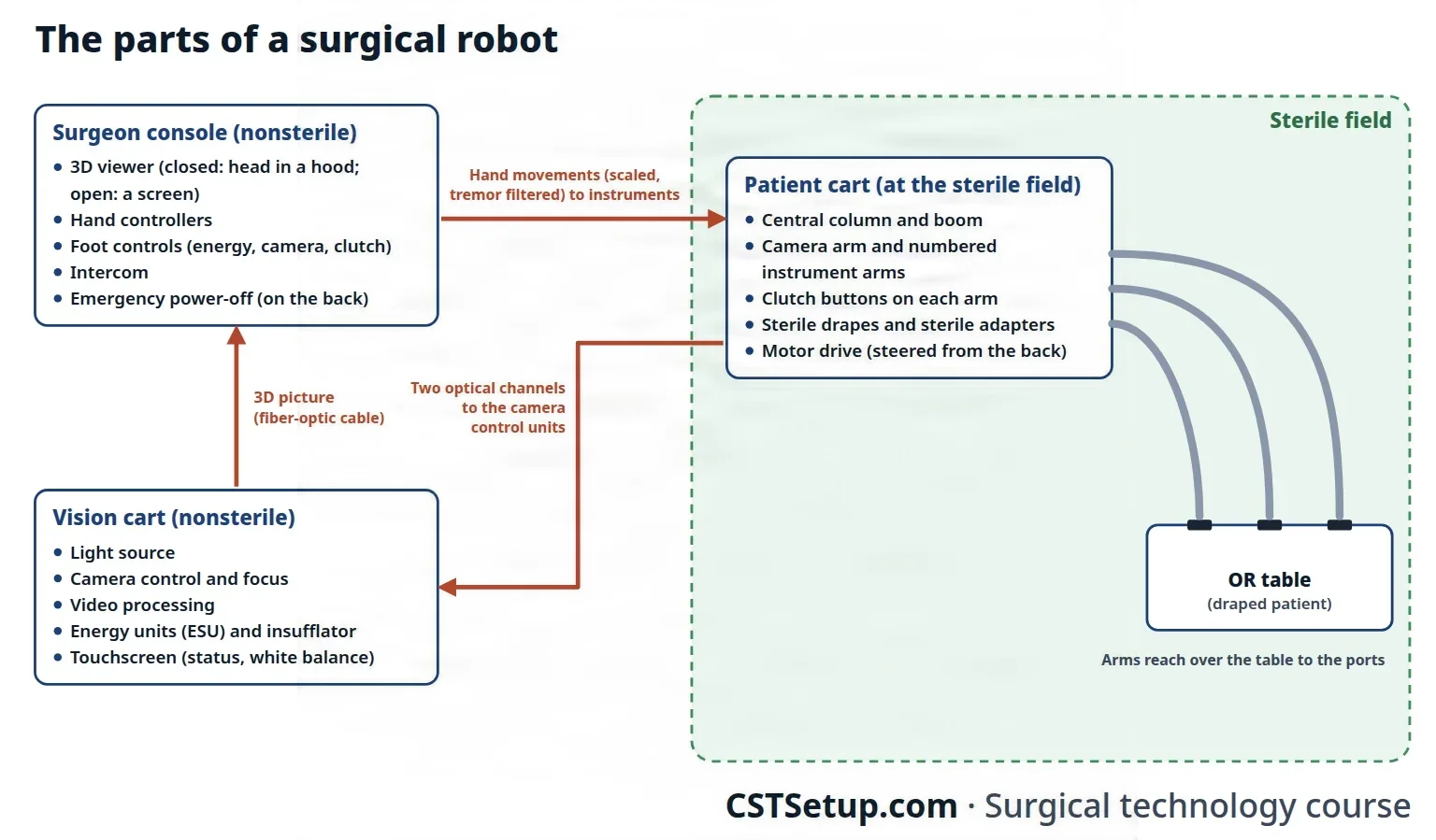
The surgeon console
The surgeon console is the nonsterile control station. From it the surgeon moves the instruments, adjusts the camera and runs much of the system. It gives the surgeon three things the Core Curriculum lists: a high-definition 3D view, filtering of hand tremorTREM-erShaking of a part of the body, such as the hands, in small rhythmic movements that the person cannot control.Full entry , and control of wristed instruments.
- Closed or open: a closed (immersive) console has a cockpit-style stereo viewer, and the surgeon puts their head into it to see. An open console has no hood: the surgeon looks at a screen as at a computer, on some systems through 3D glasses.
- Hand controllers sit below the viewer and move freely in the air, turning the surgeon's finger, hand and wrist movements into the instruments' movements. Adjustable arm rests ease strain on the shoulders and neck.
- Foot controls fire the energy instruments and, on some systems, control the camera and the clutch; newer systems build these into the hand controllers.
- An intercom lets the surgeon talk with the team without looking up from the viewer.
- An emergency power-off button on the back of the console cuts power to the whole system.
Some programs add a second console, for teaching or for two surgeons.
The patient cart
The patient cart holds the arms that hold the endoscopeEN-duh-skohpA thin tube with a light and a camera that is passed into the body to look inside an organ or cavity.endo- within-scope instrument for viewingFull entry and the instruments. On the common multi-arm system, all the arms hang from a boom that projects from a central column, usually four of them: one holds the camera and the others the instruments. The cart is heavy, so it moves on a motor drive, steered from a helm at its back.
- On that system, each arm has clutch buttons. Press one and the arm moves freely; let go and the arm resists a push and returns to where it was. A clutch at the top of each arm sets its final position and is used to slide the endoscope and instruments in and out.
- A touchscreen shows the system's status and messages, and which instrument is in which arm, for the scrub and the circulator.
- Sterile adapters fit between each draped arm and its instrument or camera; some can be reused a limited number of times, some only once.
- The cables connect at the back of the cart.
In a modular system, each arm stands on its own movable cart, one for each instrument and one for the endoscope. These carts move easily between rooms, and on one such system colored rings on the arms match the colors of the instruments on the screen.
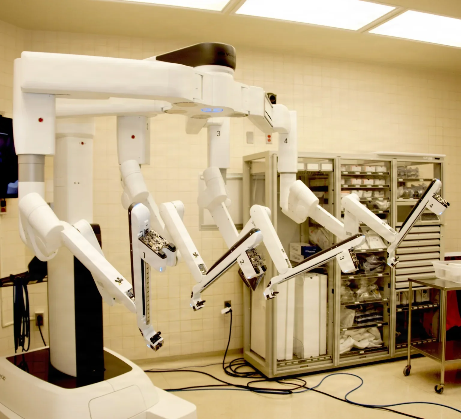
The vision cart
The vision cart, also called the equipment tower, holds the light source, the camera controls and focus control, the video processing equipment and the energy units, often the insufflationin-suh-FLAY-shunBlowing a gas into a body cavity, such as carbon dioxide into the abdomen during laparoscopic surgery.Full entry too, with a monitor or touchscreen for the team. Its signals travel by fiber-optic cables to the surgeon console.
- The two optical channels of the robotic endoscope feed their own camera control units, which build the 3D picture the surgeon sees.
- A digital zoom lets the surgeon magnify the tissue without moving the endoscope.
- White balance is done from its screen or the camera head.
- On the common multi-arm system, the vision cart's wheel locks are on its rear wheels.
Some systems connect to the maker's engineers over the internet. When a fault occurs, the circulator or robotics coordinator calls the maker's hotline with the system's identification number, and an engineer can read the same messages the team sees and help solve the problem.
Systems you may meet
Many robotic systems now exist, and more are coming since the patents on the first widely used one expired. The references describe these as examples, without ranking them:
- One cart, many arms: the da Vinci system, the most widely used in the United States, with a closed console, 3D viewing, wristed reposable instruments and no haptic feedbackFEED-bakInformation about a result that is sent back to change what happens next, in the body or between people.Full entry .
- Modular carts: the Versius, with wristed instruments and an open console where the surgeon can sit or stand, and the Senhance, which uses standard reusable laparoscopiclap-uh-roh-SKOP-ikDone through a few small cuts in the abdomen with a thin camera and long instruments, instead of one large opening.lapar/o abdomen-scopic viewingFull entry instruments, gives haptic feedback, and moves the camera by following the surgeon's eyes.
- Joint replacement systems, such as the MAKO, NAVIO and iBlock systems: they use the patient's imaging to plan the bone cuts and then help make them.
- Flexible robotic systems, such as the Flex, which drives a flexible scope and instruments for surgery through the mouth or anus, and the Monarch, a robotically steered bronchoscope for biopsies deep in the lungs.
- Spine systems, such as the Mazor X and Renaissance, which help place spinal implants precisely; chapter 42 teaches spine surgery.
Joint replacement robots fall into three groups. A passive robot holds a cutting guide in the planned place while the surgeon cuts by hand. A semi-active robot lets the surgeon make the cuts but uses haptic limits to keep the cut inside the plan. An active robot makes the cut itself, and the surgeon can stop it at any time.
16Robotic instruments and the 3D view
The instruments
A robotic instrument has a long shaft, a working tip called the end effector, and a housing that locks into the arm. Instruments range from about 3 to 10 mm across.
A standard laparoscopiclap-uh-roh-SKOP-ikDone through a few small cuts in the abdomen with a thin camera and long instruments, instead of one large opening.lapar/o abdomen-scopic viewingFull entry instrument has four degrees of freedom, plus opening and closing the jaws. A wristed robotic instrument has seven, plus the jaws, with a tip that can bend and turn even more than a human wrist. That freedom makes delicate work such as suturing in a tight space possible. Not every system is wristed: some use standard laparoscopic instruments.
Fewer instruments are needed than in open surgery: many laparoscopic-style robotic cases use 6 to 10 instruments, plus staplers and energy devices. The common groups are graspers, scissors, needle drivers, forcepsFOR-sepsA hand instrument with two blades or arms used to grasp, hold or pull tissue or objects.Full entry and dissectors. A set for one common system may include Cadiere and ProGrasp forceps, Maryland bipolarby-POH-lerHaving two poles or two opposite ends; most often used as a short form of bipolar disorder.bi- twopolar having polesFull entry forceps, monopolar curved scissors, a hook, needle drivers, a clip applier and a stapler.
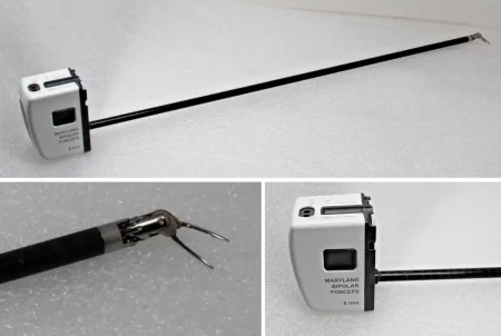
Robotic instruments have a limited life:
- Each can be used only a set number of times, for most up to 10 depending on the system, and then it is discarded like a sharp. This is a safety feature, since its electronics wear out with use.
- On the common multi-arm system, the system counts the uses left and shows them on screen, and an arm will not work with an instrument that has used them all, so keep a spare of each in the room.
- Instruments for one model may not fit another, so confirm the system before you open them.
- A designated team member keeps the inventory and the reorders.
Inspect each robotic instrument for cracks, chips and wear when you set up. reprocessingree-PROS-es-ingThe steps that make a used reusable medical device safe to use on the next patient.Full entry follows the maker's instructions, most often ending in steam sterilizationstair-uh-luh-ZAY-shunA process that destroys all microorganisms, including spores, on an item; also a permanent birth control procedure.Full entry (chapter 27).
Some systems give no haptic feedbackFEED-bakInformation about a result that is sent back to change what happens next, in the body or between people.Full entry : the surgeon cannot feel the tissue and judges pressure by what the instruments look like on the screen. Other systems do pass the sense of touch back.
The robotic camera
The robotic endoscopeEN-duh-skohpA thin tube with a light and a camera that is passed into the body to look inside an organ or cavity.endo- within-scope instrument for viewingFull entry is a rigid scope like a laparoscope, in different lengths, widths and angles; the Core Curriculum's robotic setup lists 0- and 30-degree scopes.
Its picture is three-dimensional. Like two eyes, it captures the field from two angles, and the system merges them, so the surgeon sees depth that a flat high-definition picture cannot give. The surgeon can zoom in and out without losing light or sharpness, and see past the point of focus without moving the scope. Because the surgeon controls the camera, the view is steady.
On the common multi-arm system, each robotic endoscope is calibrated before use (newer endoscopes may not need it), and fogging is still your job: warm the scope, use a defogging agent, and clean and defog it each time it comes out.
Fluorescence imaging and Firefly
Near-infrared imaging lights up structures that the eye cannot pick out. The anesthesia provideran-us-THEE-zhuh pruh-VY-derThe licensed professional who gives anesthesia and watches over the client's breathing and vital functions during a procedure.Full entry injects a fluorescent dye, indocyanine green (ICG), into a vein, and a special near-infrared endoscope shows the dye glowing on the monitor. Its most common use is to check the blood supply (perfusionper-FYOO-zhunThe flow of blood through the body's tissues, bringing them oxygen and nutrients.per- throughfus pour-ion processFull entry ) of tissue, such as a new bowel anastomosisuh-nas-tuh-MOH-sisA connection between two tube-shaped structures, such as two ends of bowel or two blood vessels, made by surgery or found naturally.Full entry , before the surgeon trusts it.
On the da Vinci system this imaging is called Firefly, which the Core Curriculum lists as an emerging technology. It is used, for example, to check bowel perfusion and to show which part of a kidney still has blood flow while the surgeon clamps a vessel. It has become a common addition to intestinal surgery, though it has not yet been proven to prevent complications.
When the preference card calls for fluorescence, make sure a near-infrared endoscope is ready on the field.
17Before the robotic case
Training and the team
AST asks every CSTsee-es-tee (said as letters)Certified Surgical Technologist, a credential for surgical technologists; in maternity care, contraction stress test.Full entry to train on the specific robot before working with it: classroom and hands-on training from the maker and the facility, starting with watching and moving to a skills lab or simulator, and continuing education whenever the facility buys a new system, instrument or accessory. The facility sets the competencyKOM-puh-tun-seeA specific skill or ability that a person has been shown to perform to an expected standard.Full entry requirements.
That training covers moving and connecting the parts, draping, white balance and calibration, inspecting instruments, putting instruments into the arms and changing them, handling the endoscopeEN-duh-skohpA thin tube with a light and a camera that is passed into the body to look inside an organ or cavity.endo- within-scope instrument for viewingFull entry , troubleshooting, and moving the system fast in an emergency. AST recommends training three or four complete teams of a surgeon, a CST and an RN, for continuity of carekon-tih-NOO-ih-tee uv KAIRCare that flows smoothly from one provider, shift or setting to the next, without gaps or repeated steps.Full entry .
As a student you learn the words and the parts in school, and you may watch or scrub robotic cases during your clinical rotation if the facility has a robot and allows it.
Training comes from many places after that: the job, the makers' online and on-site courses, and courses run by other schools and companies. Before you take a course outside your program or facility, check what it says you will be able to do at the end and how it teaches. Reading is no substitute for hands-on practice.
A robotics program has a leader who schedules cases, buys and stocks the instruments and supplies, arranges training and documents each member's competency. AST calls this person the Robotics Team Leader and says a trained, experienced CST can fill the role; one reference describes a robotics coordinator who is an experienced registered nurse. The robotic policies are reviewed every year.
Who does what
- The surgeon leads the case. At the bedside the surgeon is scrubbed; at the console the surgeon is nonsterile, directingdih-REK-tingGuiding and instructing others, such as staff members, so that work gets done as planned.Full entry the scrubbed team to reposition, add or change instruments and assignuh-SINETo give a staff member the care of a client, or tasks that are already part of that person's usual job and allowed by their license.Full entry them to arms.
- The assistant surgeon stays at the bedside for the whole case and works through an extra assistant's port: suction, retractionrih-TRAK-shunA drawing back or pulling in of tissue; in breathing, the skin sinking in between, below or above the ribs with each breath.Full entry , clips, passing sutureSOO-cherA stitch, or the thread used to stitch, that holds the edges of a wound or incision together.Full entry . The assistant and the scrub change robotic instruments together and solve equipment problems.
- Advanced practice providers, such as physician assistants and nurse practitioners licensed by their state, may be trained to do the assistant surgeon's tasks, under a surgeon's superviseSOO-per-vyzeTo guide, watch over and check the work of another staff member who is doing a task.Full entry . Each state sets their education and legal requirements.
- The circulator keeps the usual role: patient care, guardingGAR-dingTightening of muscles, or protecting a body part with the hands or posture, to avoid pain when it is touched or moved.Full entry the sterile fieldSTAIR-ul feeldAn area set up with sterile drapes and supplies, kept free of all germs, for a sterile procedure.Full entry , documentation and getting supplies. The circulator also helps set up and test the robot, drives the patient cart into place under the scrub's guidance, and moves the robot away at the end.
- The scrub usually works in the scrubbed role, doing the usual tasks plus the robotic ones: draping the robot, helping with ports and docking, putting instruments into the arms and changing them, and troubleshooting faults. A highly trained CST may take other roles as state law and facility policy allow.
Room layout
Each type of robotic case has a planned map of where every part and every cable goes. The plan considers:
- the anesthesia provideran-us-THEE-zhuh pruh-VY-derThe licensed professional who gives anesthesia and watches over the client's breathing and vital functions during a procedure.Full entry 's access to the airway, the monitors and the lines;
- which parts are sterile and which are not, the distances between them, and the sterile field's place in relation to the doors;
- a separate dedicated circuit for each part, with no extension cords;
- cables laid where nobody trips on them and no equipment rolls over them.
The surgeon console goes in the nonsterile area, where the surgeon can see the patient and the field, and watch for arms colliding. A clear path runs from it to the main door, so the surgeon can scrub and reach the patient fast in an emergency.
The patient cart is placed for the operation with room to back it away in an emergency, and the anesthesiaan-us-THEE-zhuhLoss of feeling or sensation, often brought on by drugs so that surgery or a procedure can be done without pain.an- withoutesthesi/o feeling, sensation-ia conditionFull entry team helps plan it so it does not block the airway or the lines. It is draped away from the table and moved in only after the patient is positioned and draped.
The vision cart stands in the nonsterile area, where the circulator can reach it and the scrub can see it. Its cables are never tangled or knotted: coil the extra length neatly and secure it, and keep every connection free of tension.
Crowding is a real hazard. In a conversion the team needs space and fast access to supplies, and the robot must be moved back out of the way.
Move the parts slowly and carefully, the patient cart on its motor drive with its arms stowed so they cannot hit a wall or a door. Allow enough time before the case to set up the parts and let the camera warm.
Pulling the case and setting up
With the circulator, read the preference card, pull everything for the case, check the robotic equipment together, and check the room layout and the cables. Set apart the supplies held back for later in the case or for an emergency, such as the open conversion set.
On the common multi-arm system, AST's guideline lists the endoscopes and their sterile adapters, the arm drapes, the light cable, a Veress needle and the trocarTROH-karA sharp or bladed instrument inside a tube (cannula), used to pierce a body wall and leave the tube in place as a port.Full entry and cannulaKAN-yoo-luhA thin tube placed into the body or a body opening to deliver or remove fluid or air.Full entry , two 12 mm and three 5 or 8 mm. Your surgeon's card sets the real list.
Then do the usual: scrub, gown and glove, set up the back table, and inspect every instrument, the wristed ones above all.
Draping the robot
The robot's drapes are single-use, made by its maker to fit each part, and clearly marked. The scrub and the circulator drape it as a team, following the maker's steps; it is a complex job, so practice it together before your first case.
In outline, for the common multi-arm system:
- The cables are connected and the system is turned on. It tests itself; nobody touches the arms during the test, or it will register a fault.
- The circulator moves the arms out of their stowed position and into place for draping, and the system runs its homing sequence. Both of you confirm out loud that it has finished.
- Each instrument arm is draped on its own, fully extended. Standing in front of the arm, the scrub slides the drape over it while the circulator reaches inside the drape to pull it through. Each draped arm is moved a little to the side so it cannot touch an undraped one. Never drape an arm so tightly that the drape limits its movement.
- The camera arm is draped the same way, and the touchscreen is draped.
- The endoscope and camera head are joined through a sterile adapter and covered by their drape, and the sterile light cable is connected.
- The scrub white balances the camera with a sterile white paper and calibrates each endoscope that will be used, with its alignment target.
- The camera arm is set at the point the maker's guide marks, which gives all the arms the most room to move without hitting each other. The draped arms are then folded in close until docking.
Newer robotic endoscopes may need fewer of these steps, with no manual white balance, calibration or drape, so always follow your system's own instructions.
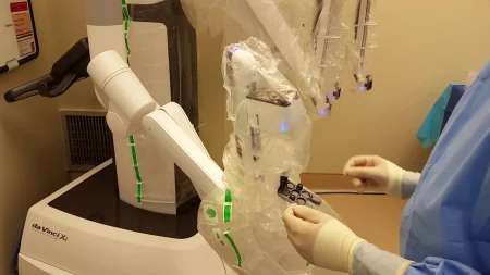
Put the Mayo standMAY-oh standA small tray on an adjustable stand placed over or beside the patient during surgery to hold the instruments used most often.Full entry at the side or the foot of the table, and keep every device that stays on the field in a drape pocket so it cannot fall.
The patient
Robotic cases often use exaggerated positions, such as steep Trendelenburg positiontren-DEL-en-burg puh-ZISH-unA position in which the client lies on the back with the bed tilted so the head is lower than the feet.Full entry with lithotomylih-THOT-uh-meeA position lying on the back with the hips and knees bent and the legs raised and spread apart, often in stirrups.lith/o stone-tomy cutting intoFull entry for pelvic surgery, and these magnify the usual risks. An anti-skid device is used whenever the patient could slide. A split-leg position is used when a part of the robot, or a team member, works between the legs.
Once the robot is docked, the table must not be moved, or the fixed arms could injure the patient. The exception is a table built to move together with the robot, which one system offers. Positioning is chapter 31.
The skin prep may go beyond the usual borders, to take in every port site, and hair is clipped in the preoperative area when needed. The patient drapes are like those for laparoscopylap-uh-ROS-kuh-peeSurgery or an exam done through small cuts in the belly, using a thin, lighted tube with a camera.lapar/o abdomen-scopy looking with an instrumentFull entry , with side pockets for the energy devices.
Robotic cases can last for hours, so watch for hypothermiahy-poh-THER-mee-uhA body temperature that has fallen below the normal range.hypo- belowtherm/o heat-ia conditionFull entry , pressure injuryPRESH-er IN-juh-reeDamage to the skin and the tissue beneath it from pressure, or pressure with shear, usually over a bony area or under a device.Full entry and other position-related injuries, and the risks of a long case such as blood clots. A steep head-down tilt can also swell the face and upper body and make breathing harder.
18Ports and docking
Ports
The ports go in first, before the robot is docked. The surgeon's preference card gives their number, sizes and layout, and robot makers supply port placement diagrams. You help the surgeon and the assistant place them.
The gas goes in by the Veress or the open technique, as for any laparoscopylap-uh-ROS-kuh-peeSurgery or an exam done through small cuts in the belly, using a thin, lighted tube with a camera.lapar/o abdomen-scopy looking with an instrumentFull entry , then the camera port and, under its view, the other ports. Robotic instrument cannulaKAN-yoo-luhA thin tube placed into the body or a body opening to deliver or remove fluid or air.Full entry must fit the system in use, and most cases add an assistant's port for standard instruments.
Docking
Before docking, the patient is placed in the final position, such as steep Trendelenburg positiontren-DEL-en-burg puh-ZISH-unA position in which the client lies on the back with the bed tilted so the head is lower than the feet.Full entry for the pelvis, the overhead lights are moved out of the way, and there must be room between the arms and the patient. On some systems the cart is first aimed at the target anatomy, and the arms move into the best positions for the procedure. On the common multi-arm system, AST describes docking this way:
- The circulator drives the patient cart in with its motor drive, guided by the scrub. Everyone watches that it does not hit, pinch or press on the patient's arms, body or legs.
- The camera arm is attached first, to the camera cannula. Move it with its positioning joints, not the camera clutch, which would limit how far the camera can move.
- The instrument arms are attached to their cannulas next, moved with their port clutch, with the final angle set using the instrument clutch.
- Once every arm is attached, the team checks that each one has room to work and is not pressing on the patient, and says both checks out loud.
On that system, once the first instrument arm is attached, the cart's drive locks so the cart cannot roll.
Putting in the endoscope and instruments
You put the endoscopeEN-duh-skohpA thin tube with a light and a camera that is passed into the body to look inside an organ or cavity.endo- within-scope instrument for viewingFull entry and each instrument in while the surgeon watches the ends of the cannulas from the console. Take care not to puncture the arm drapes. On the common multi-arm system, AST describes the steps this way:
- The endoscope: slide the lens into its cannula, lock the scope into the camera mount, and advance it with the camera clutch into the surgeon's view.
- Each instrument: confirm out loud with the surgeon that its wrist is straight, slide its tip into the cannula and its housing into the sterile adapter, and advance it with the clutch until the tip just shows on the screen.
The surgeon then leaves the field for the console, and the scrub and the assistant stay at the bedside.
19During the robotic case
Your jobs
- Changing instruments: confirm with the surgeon that the instrument's wrist is straight and its jaws are not holding tissue, squeeze its release levers and draw it out while watching the screen, then put in the next one the same way. Some systems guide the new instrument back to just short of where the last one worked.
- Cleaning: wipe instrument tips with instrument wipes at each change. Clean the endoscopeEN-duh-skohpA thin tube with a light and a camera that is passed into the body to look inside an organ or cavity.endo- within-scope instrument for viewingFull entry and defog it each time it comes out.
- Watching: follow the case on the bedside monitor, so you are ready with the next instrument or sutureSOO-cherA stitch, or the thread used to stitch, that holds the edges of a wound or incision together.Full entry .
- Specialty tasks as the surgeon directs: moving the uterus in gynecologygy-nuh-KOL-uh-jeeThe branch of medicine that cares for the female reproductive organs and breasts.gynec/o woman-logy study ofFull entry cases, pulling on or changing the indwelling urinary catheterin-DWEL-ing YUR-ih-nair-ee KATH-uh-terA tube passed through the urethra into the bladder and left in place to drain urine into a bag.Full entry in urologic cases, and passing in the specimen bag.
- guardingGAR-dingTightening of muscles, or protecting a body part with the hands or posture, to avoid pain when it is touched or moved.Full entry the field: watch for arms crowding or colliding, keep cords and tubing in order, keep the field sterile and keep the patient safe.
Communication
Talking clearly matters even more in a robotic case. The surgeon sits away from the field and may not be able to see the patient or the anesthesia provideran-us-THEE-zhuh pruh-VY-derThe licensed professional who gives anesthesia and watches over the client's breathing and vital functions during a procedure.Full entry . In a closed console the surgeon's voice comes through speakers, facial expression and gesture are lost, and the noise of the room can drown out words. Open consoles were designed partly so the surgeon can keep eye contact with the team.
Say each step out loud as you insert or change an instrument or the endoscope, and confirm what you heard, so no movement damages an instrument or injures the patient. Chapter 16 teaches closed-loop communication.
The end of the case
On the common multi-arm system, AST describes the end of the case this way:
- The instruments come out first, then the endoscope.
- The arms are released from the cannulaKAN-yoo-luhA thin tube placed into the body or a body opening to deliver or remove fluid or air.Full entry and the cart is undocked. Its drive will not run until everything is out and every arm is free.
- The surgeon returns to the field and widens a port incision to bring out the specimen bag.
- Have suture ready to close the fasciaFASH-ee-uhA sheet or band of tough connective tissue that covers and separates muscles and organs.fasci/o bandFull entry of the widened incision and the 12 mm port sites; 5 and 8 mm sites usually need no fascial closure.
- The circulator moves the robotic equipment away from the bedside. If another robotic case follows, the system can stay on.
The robotic instruments get point-of-use care by their maker's instructions, so soil does not dry on them, and are handled gently; then they are handed to sterile processing, which chapter 27 teaches.
20Troubleshooting and emergencies
When the system has a problem
Complex systems can fail despite their safety features. Losing the picture is especially dangerous, above all during dissectiondy-SEK-shunA tear in the inner layer of an artery wall that lets blood split the layers apart; also, the cutting apart of tissue in surgery.Full entry near large vessels or during suturing, and a failed instrument or a system error may lead to conversion.
When a fault occurs, follow the instructions on the screens. The system stores its past errors and sorts faults into two kinds:
- A recoverable fault: the system sounds an alarm, shows a message and flashes lights on the arms, and locks until the team finds and fixes the problem. Then the case goes on.
- A nonrecoverable fault cannot be fixed. The team shuts the system down and converts to an open operation.
The maker's engineers can help from a distance, as described above. You and the assistant handle the bedside problems, such as arms crowding or colliding, by repositioning as the surgeon directs.
If an instrument fails or sticks, systems provide a way to remove it by hand without the clutch, such as a sterile release tool (a "wrench") kept ready, peel-packed, in the room.
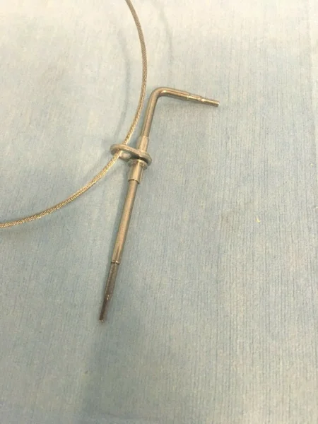
When the patient has a problem
Bleeding is the most common and most dangerous emergency in robotic surgery, and the most common reason to convert.
The robot also gets in the way of emergency care. A large patient cart can block the airway, the monitoring lines, the crash cart and the defibrillator, and a cart near the head or shoulder can block the chest completely, so the robot may have to be undocked before chest compressions can start.
Undocking in an emergency
Plan for emergencies before they happen. Each facility sets every team member's job for an emergency conversion, writes it into its robotic policies, teaches it in orientation and practices it in drills. At the time-outTYM-owtA pause just before a procedure when the whole team confirms the right client, procedure and site; also, a short break to calm a child.Full entry , each person can say their emergency role. Keep ready:
- a checklist for fast conversion;
- an emergency tray or cart with the open instruments, just outside the room;
- the sterile release tool;
- a clear path to the doors.
When an emergency happens:
- The type of emergency is announced (bleeding, a code, a technical failure), and help is called.
- The room lights go on.
- The scrub takes the instruments and the endoscopeEN-duh-skohpA thin tube with a light and a camera that is passed into the body to look inside an organ or cavity.endo- within-scope instrument for viewingFull entry out of the patient and sets them on the back table.
- The arms are released from the cannulaKAN-yoo-luhA thin tube placed into the body or a body opening to deliver or remove fluid or air.Full entry , and the circulator undocks the cart and moves it away.
- The open instruments and supplies are opened and the operation is converted. Counts are done if there is time; if they are waived, imaging may follow by facility protocol to make sure nothing is left behind (chapter 28).
AST says a well-trained team should get the instruments out and the robot undocked in 2 to 4 minutes.
During the conversion, take the sterile instruments and supplies quickly, set apart what is needed first, and prepare them as usual. Depending on your facility's plan, you or the assistant surgeon removes the robotic instruments. Move deliberately: in an emergency, a rushed mistake costs more time than it saves.
In a robotic emergency, everyone already knows their job. Instruments and the endoscope out, arms off, cart away, open set up, and the lights on. Practice it before you ever need it.
21Robotic applications, advantages and risks
Where robots are used
Robotic surgery is used in general, colorectal, urologic, gynecologygy-nuh-KOL-uh-jeeThe branch of medicine that cares for the female reproductive organs and breasts.gynec/o woman-logy study ofFull entry , chest, heart and ear, nose and throat surgery. Common operations include prostatectomy, hysterectomyhis-tuh-REK-tuh-meeSurgery to remove the uterus.hyster/o uterus-ectomy surgical removalFull entry and colorectal resectionrih-SEK-shunSurgery to remove part of an organ or tissue.re- back, againsect to cut-ion processFull entry , as well as fundoplication, gastric bypass, mitral valveMY-trul valvThe heart valve between the left atrium and the left ventricle.Full entry repair and many herniaHER-nee-uhA bulge of an organ or tissue through a weak spot in the muscle or tissue wall that normally holds it in.Full entry repairs. It can help in very obese patients for whom regular laparoscopylap-uh-ROS-kuh-peeSurgery or an exam done through small cuts in the belly, using a thin, lighted tube with a camera.lapar/o abdomen-scopy looking with an instrumentFull entry would be hard, and newer systems work through a single incision. Robots also guide bone cuts in joint replacement and implants in the spine.
Complex, high-risk operations are still usually done open, to avoid the risk of an emergency conversion.
Advantages
- A high-definition 3D view, with a steady camera the surgeon controls.
- Scaled, tremorTREM-erShaking of a part of the body, such as the hands, in small rhythmic movements that the person cannot control.Full entry -free movement and wristed instruments, for fine dissectiondy-SEK-shunA tear in the inner layer of an artery wall that lets blood split the layers apart; also, the cutting apart of tissue in surgery.Full entry and suturing.
- A seated surgeon with comfortable controls, and less physical strain and fatigue.
- Several arms, and an assistant at the bedside with a port of their own.
- For the patient, compared with open surgery: fewer complications, less blood loss, a shorter stay and a faster recovery.
Disadvantages and risks
- Large, bulky equipment, a narrower field of view, and a surgeon away from the field who depends on others to insert the instruments.
- High costs: buying the system, yearly maintenance and upgrades, and limited-use instruments.
- Longer setup and often longer operations, so longer anesthesiaan-us-THEE-zhuhLoss of feeling or sensation, often brought on by drugs so that surgery or a procedure can be done without pain.an- withoutesthesi/o feeling, sensation-ia conditionFull entry .
- A steep learning curve that needs special training and a dedicated team.
- Slower emergency access to the abdomen, because the robot must be undocked first.
- All the risks of laparoscopy, including the effects of the gas, plus failure of the system itself, which can injure the patient or force a conversion.
Robotic features do not by themselves make the outcome better. A skilled surgeon is skilled with a robot, with a laparoscope or in open surgery, and so far the results of robotic surgery are comparable with those of regular laparoscopy.
22Navigation: image-guided surgery
Computer-assisted surgery, also called image-guided surgery or surgical navigation, uses three-dimensional images and real-time tracking to show the surgeon exactly where an instrument is inside the patient. The Core Curriculum teaches it with MIS, although it is used in open operations too, such as joint replacement.
How it works
The patient's scans (computed tomographykum-PYOO-tid toh-MOG-ruh-feeAn imaging test that uses x-rays and a computer to make cross-section pictures of the inside of the body.tom/o cut, slice-graphy process of recordingFull entry , magnetic resonance imagingmag-NET-ik REZ-uh-nunts IM-uh-jingAn imaging test that uses a strong magnet and radio waves to make detailed pictures of the inside of the body.Full entry , ultrasound) are layered by the computer into one 3D map, a step called image registration. Stereotactic surgery, from the Greek for "touching in space," then uses a 3D coordinate system on that map to guide the surgeon to a precise target, such as a tumor.
The map needs fixed reference points on the patient. In frame-based surgery, a light frame is fixed to the patient's head before the scans. In frameless surgery, markerMAR-kerSomething that can be measured, such as a substance in the blood, that shows a disease or body process is present.Full entry called fiducials are fixed to the skin or the bone instead.
In the OR, a tracking camera or an electromagnetic field follows markers on a reference frame fixed to the patient, or to the head holder, and on the surgeon's probe or instruments. The computer then shows where the tip of the probe is on the patient's own scans, in three planes (axial, coronal and sagittal), so the surgeon can plan the approach and follow it. The tracked instruments must stay inside the camera's working space.
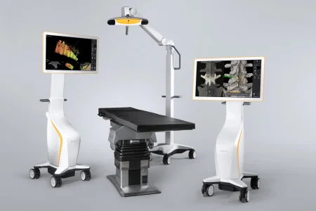
The images it uses
The Core Curriculum lists four kinds of imaging behind navigation:
- CT (computed tomography) combines x-rays and a computer to make cross-sectional pictures that show tissues and their sizes precisely; contrastKON-trastA dye or other substance given before an imaging test so body parts show up more clearly.Full entry can make vessels and ducts stand out.
- MRI (magnetic resonance imaging) uses strong magnetic fields and radio waves to make detailed pictures, mainly to find tumors and other structural problems.
- O-arm: an x-ray unit built as an open ring (the gantry) around the patient, which slides along the table and images all the way around. It builds 3D images in real time during the operation. It has become the standard image-guided system in spine surgery, where it makes screw placement more accurate and lowers the patient's x-ray dose, which matters most in children. It is bulky, needs room to move, and shows soft tissue poorly.
- Ultrasound can update the picture during surgery at low cost, though its image is often too poor to rely on.
radiationray-dee-AY-shunEnergy that travels as waves or particles, such as X-rays, used to make images or treat cancer; also pain that spreads from its starting point.radi/o rays, radiation-ation processFull entry safety and the danger the MRI magnet poses to metal are chapter 22.
Fiducial markers
Fiducials are small markers placed on the patient before the scan, often adhesive stickers on the scalp, sometimes small screws in the bone. They show up in the images and give the computer fixed points to match to the patient.
- They work best when there are several, spread widely, not in a straight line, and on skin that does not slide. Markers fixed into bone are the most accurate.
- They are left in place from the scan until they are registered in the OR.
- Registration: in the OR, the surgeon touches each fiducial with the tracked pointer so the computer can line up the images with the patient. After registration the fiducials may be removed, and the area is prepped and draped.
In robotic joint replacement, the bone itself is registered: the surgeon touches many points on it with a tracked pointer to match it to the model built from the CT scan. Chapter 39 teaches joint replacement.
Advantages
- Precise localization: the surgeon can find a deep or small target and choose the least damaging path to it.
- Smaller openings: in brain surgery, smaller, better-placed openings, with shorter operations, fewer wound infections and shorter stays; chapter 42 teaches the brain and spine operations.
- A check on what has been removed, and, in joint replacement, on the accuracy of cuts in places the surgeon cannot see well, with less blood loss.
- Reports suggest navigation may lower surgical complications and cost.
Risks and limits
- A moved reference: once the system is registered, the reference frame must not move. A bump to the navigation arm or the frame throws the map off and can make it useless, so keep it out of the team's way.
- Old pictures: navigation usually relies on scans taken before surgery, but in brain surgery the brain shifts as the operation goes on, so the map drifts from reality and may need scans during surgery to update it.
- Line of sight: an infrared camera must see the markers on the pointer and the reference frame at all times, which can be hard to keep, for example when an operating microscope is in use.
- Radiation: scanning during surgery with CT or the O-arm exposes the team to ionizing radiation.
- Cost and time: the equipment, software and scans are expensive, and setting up takes time.
Navigation is only as accurate as its registration. Leave the fiducials in place until registration, keep the reference frame and the camera's view clear after it, and never bump the navigation arm.
What to take from this chapter
- Minimally invasive surgery works through small incisions or natural openings, watching a monitor. Patients have less pain, less blood loss, fewer wound problems and faster recovery, but every case can convert, so the open setup is always ready.
- The video chain runs light source, light cord, scope, camera head, camera control unit and monitor, and it is only as good as its weakest link. White balance, focus and a warm, clean lens give a clear picture.
- A lit light cord off the scope can start a fire: standby whenever it is disconnected, never left on the drapes.
- Carbon dioxide makes the working space because it is nontoxic, does not burn and is quickly absorbed. Watch the pressure (usually 12 to 15 mm Hg, stopping at 15). The gas can cause hypercapnia, gas embolism, gas under the skin or in the chest, low blood pressure, hypothermia and shoulder pain.
- Closed (Veress) entry is a blind puncture checked by the saline drop test and a low opening pressure; open (Hasson) entry is cut down under direct view and suits scarred abdomens. The other ports go in under the scope's view.
- Joints, the bladder and the uterus are distended with fluid that must suit the energy device and be warmed to body temperature, and the fluid in and out is tracked because absorbed fluid can cause fluid overload and hyponatremia.
- A surgical robot has three parts: the nonsterile surgeon console, the patient cart with its arms, and the vision cart. The surgeon controls the robot, which scales movement, filters tremor, gives a 3D view and, on many systems, moves wristed instruments with seven degrees of freedom.
- The scrub drapes the robot with the circulator, helps place ports, guides docking, inserts and exchanges instruments with spoken checks, and must be able to get the instruments out and the robot undocked in minutes for an emergency. Navigation guides instruments on the patient's own scans through fiducials and registration, and is only as good as that registration.
Check yourself5 quick questions
Free for everyone. Pick an answer for each, then check them all at once.
In the ORWhere you'll use this
The chapter's whole question pool in sets of 10, with the answer explained after each one.
Part of the educators programThe chapter's words on spaced cards: the ones you miss come back sooner.
Part of the educators programGiven by your class's teacher. Each class draws its own 25 from the chapter's pool.
Part of the educators program
Suggest a fix
Something wrong, unclear or out of date in this chapter? Tell us and we will fix it for everyone. Your email is only used to reply.