Electricity, electrosurgery and lasers
How electricity and laser light cut and seal tissue, the shocks, burns, fires and smoke they can cause, and what you do so they never hurt the patient or the team.
1Why it matters
electrosurgeryee-LEK-troh-SUR-jer-eeUsing high-frequency electric current through a handheld tool to cut tissue or seal bleeding vessels during surgery.Full entry is used in nearly every kind of operation, mostly to cut tissue and to seal small bleeding vessels, part of stopping bleeding (hemostasishee-moh-STAY-sisThe stopping of bleeding, either by the body's own clotting process or by medical or surgical methods.hem/o blood-stasis stopping, standing stillFull entry ). Lasers are used in many specialties too. Both put a great deal of energy into one small spot, which is what makes them useful and what makes them dangerous.
The electrosurgical unit is the most common cause of electrical injury to surgical patients. Energy at the field can also start a fire on the patient, and burning tissue gives off smoke the whole team breathes. Safety practices prevent these injuries, and the surgical technologistSUR-jih-kul tek-NOL-uh-jistA member of the operating room team who prepares the sterile field and instruments and hands them to the surgeon.Full entry is one of the people who carries them out.
A rule you understand is a rule you keep. AST expects CSTs to know the principles of electrosurgery and its safety measures, to be trained on each unit they use, and to keep learning as the technology changes. When you know why current burns a patient through a loose pad, or why a laser beam bounces off a shiny retractorrih-TRAK-terA surgical instrument that holds back tissue or organs so the surgeon can see and reach the operative site.Full entry , you will spot the danger before it happens.
This chapter covers how electricity works, how it can hurt people, electrosurgery and the return electrode, fire at the surgical field, surgical smoke and lasers. Other chapters hold the pieces around it:
- the facility's fire plan (RACE, PASS and the extinguishers, the zone valves and evacuation), oxygen as a medical gas, the red outlets and the room's power: chapter 17
- oxygen and the other gases during anesthesiaan-us-THEE-zhuhLoss of feeling or sensation, often brought on by drugs so that surgery or a procedure can be done without pain.an- withoutesthesi/o feeling, sensation-ia conditionFull entry : chapter 13
- the electrosurgical unit and the smoke evacuator as room equipment, and readying and testing them: chapter 23
- placing the patient return electrode, as part of positioning and the skin prep: chapter 31
- energy devices for stopping bleeding (vessel sealers, ultrasonic shears, the argon beam) and when to choose each: chapter 29
- energy at the trocarTROH-karA sharp or bladed instrument inside a tube (cannula), used to pierce a body wall and leave the tube in place as a port.Full entry and the scope in laparoscopylap-uh-ROS-kuh-peeSurgery or an exam done through small cuts in the belly, using a thin, lighted tube with a camera.lapar/o abdomen-scopy looking with an instrumentFull entry : chapter 21
- ionizing radiationray-dee-AY-shunEnergy that travels as waves or particles, such as X-rays, used to make images or treat cancer; also pain that spreads from its starting point.radi/o rays, radiation-ation processFull entry from imaging: chapter 22
2How electricity works
Electricity is the flow of electrons. In metals and other conductive materials, some electrons are held so loosely that they drift from atom to atom. When they are pushed in one direction along a continuous path, they carry energy with them. That path is a circuit, and understanding it is one of the most important things you will learn in this chapter.
The words
- Circuit: the complete, unbroken loop electrons travel, from the power source to the device that uses the power and back to the source. Break the loop anywhere and the current stops, the way a flashlight goes dark when its switch is open.
- Conductor: a material that lets electrons flow easily. Metals such as copper and silver are good conductors, and so are water, salt water and the human body. Copper is the most common conductor in electrical wiring.
- Insulator: a material that resists the flow of electrons, such as glass, rubber and plastic. Insulation is wrapped around conductors to keep current inside them, like the plastic covering an electrosurgeryee-LEK-troh-SUR-jer-eeUsing high-frequency electric current through a handheld tool to cut tissue or seal bleeding vessels during surgery.Full entry cord. Insulate means to cover a conductor with such a material.
- Current: the flow of electrons, measured in amperes (amps).
- Voltage: the force or pressure that pushes electrons through the circuit, measured in volts. The higher the voltage, the harder the push.
- Resistance, or impedance: how much a material opposes the flow of current, measured in ohms. Where electrons meet resistance, their energy turns to heat.
- Frequency: the number of times alternating current changes direction each second, measured in hertz (Hz), or cycles per second.
- Radio frequency (RF): electromagnetic energy at frequencies in the range used for radio. Electrosurgery runs at radio frequencies, from about 200,000 to 3.3 million cycles a second.
- Ground: the earth, or a connection to it, which has zero electrical potential. Grounding gives stray current a path into the ground, where it spreads out and does no harm.
- Isolated circuit: a circuit with no ground reference at the point of use. A transformer separates it from the grounded supply, so its current returns only to its own source and does not look for a way into the ground.
Alternating and direct current
Direct current (DC) flows in one direction only. It is low voltage, and it comes from a batteryBAT-uh-reeIn law, touching a person on purpose without their consent.Full entry . Alternating current (AC) flows back and forth as its polarity reverses. It comes from the wall outlet, at 110 or 220 volts, and carries far more power than a battery.
In the United States, wall current changes direction 60 times a second (60 Hz). AC can also be stepped up or down to a different voltage by a transformer, which is why power companies can send it at very high voltage and the hospital can step it down for use.
How electricity makes heat
Electricity behaves a little like water in a hose. It can be started, stopped and controlled, and it always takes the easiest path open to it, the path of least resistance. When the flow meets a material that resists it and no easier path is available, the energy turns to heat. A light bulb works this way: its thin wire resists the current, heats up and glows.
The same thing happens in tissue. Push a large number of electrons through a small area and you get a lot of heat in that area. How concentrated the current is, called the current density, decides how hot a spot gets. This one idea explains how electrosurgery cuts and why a bad return electrode burns.
3How electricity hurts people
An electric shockshokA life-threatening state in which blood flow is too low to bring enough oxygen to the body's tissues.Full entry is injury from current passing through the body. It can range from a small burnBURNAn injury to the skin or deeper tissues caused by heat, chemicals, electricity, radiation or friction.Full entry to charred skin, damage to internal organs and death. Whether a shock kills depends on the path the current takes through the body, how much current flows and how much the skin resists it. Even a very small current that reaches the heart directly can stop it or throw it into ventricular fibrillationven-TRIK-yuh-ler fib-ruh-LAY-shunA heart rhythm in which the lower chambers quiver in a fast, chaotic way instead of pumping.ventricul/o ventricle-ar relating tofibrill small fiber, twitching-ation processFull entry , a rhythm in which the ventricleVEN-trih-kulOne of the two lower chambers of the heart, or one of the fluid-filled spaces inside the brain.Full entry quiver instead of contracting, so the heart pumps no blood.
Electrocution means severe burns, heart rhythm problems or death from current discharged into the body. Because the body conducts electricity, current that enters it and is not led safely back to its source can cause severe burns and cardiac arrestKAR-dee-ak uh-RESTThe sudden stop of the heart's pumping, so blood no longer reaches the brain and body.Full entry .
Electrical burns happen where current leaves the body through a small area, because the current crowds together there. Electrical hazards in the OR include shock, burns, fire and explosion, and a burn can come from touching an uninsulated wire, a damaged plug or a piece of metal equipment that is not grounded.
Electrical safety in the OR
In the United States, electrical faults are one of the main causes of fires in hospitals. Accredited hospitals must meet building codes for their wiring and outlets, but the equipment plugged into them must be looked after too, and the more complex a device is, the more ways it can fail.
- Grounded plugs: hospital equipment has a three-prong plug. Two prongs carry the current to the device, and the third connects it to the ground. If a short happens inside the device, the current runs out through the ground prong instead of through you or the patient. Never remove that prong to make a plug fit, and a plug with a bent prong comes out of service.
- Approved equipment: equipment used in the OR must be inspected and approved by Underwriters Laboratories (UL), a nonprofit agency that tests electrical products for safety. Equipment without UL approval is not used.
- Inspected equipment: biomedical engineering inspects equipment on a schedule, and its inspection sticker should be current.
- Cords: never use equipment with a frayed cord or exposed wires. Cords are never spliced or threaded through solid objects, and the electrosurgeryee-LEK-troh-SUR-jer-eeUsing high-frequency electric current through a handheld tool to cut tissue or seal bleeding vessels during surgery.Full entry 's cord never goes into an extension cord.
- Dry: keep liquids away from outlets, cords and switches, and use only devices made for use around fluids near them. Wipe up fluid on the floor near a cord at once.
- Off before unplugging: switch a device off before you pull its plug, and pull by the plug, never the cord.
- Smoke or sparks: equipment that smokes or sparks comes out of service at once, because a spark can light a fire on the field.
- Repairs: no one on the surgical team repairs electrical equipment. A faulty unit goes to biomedical engineering or its maker, and the department follows Occupational Safety and Health AdministrationOH-shuh (said as a word)The US federal agency, part of the Department of Labor, that sets and enforces rules to keep workplaces safe.Full entry 's lockout/tagout standard for controlling hazardous energy.
Chapter 23 shows how to check the room's equipment, cords and plugs before the case, and chapter 17 covers the red outlets on the emergency power supply.
Protecting yourself
Your own body conducts electricity, so you can become part of a circuit. A surgeon who leans against the patient during monopolar electrosurgery can become part of the circuit and be burned. Keep your gloves dry when you work near an active electrode, never touch an uninsulated wire or a damaged plug, and report any equipment that sparks or smokes so it comes out of service.
Static electricity is a hazard too: in dry air, static builds into sparks that can light a fire, one reason the room's humidity is kept in range (chapter 17).
4Electrosurgery
electrosurgeryee-LEK-troh-SUR-jer-eeUsing high-frequency electric current through a handheld tool to cut tissue or seal bleeding vessels during surgery.Full entry is the use of electricity to heat tissue so that it cuts, seals or is destroyed. A high-frequency alternating current passes through the tissue itself, and the tissue's resistance turns the current into heat inside the cells.
Electrocautery is something different, although people often use the word when they mean electrosurgery. A cautery runs direct current, usually from a batteryBAT-uh-reeIn law, touching a person on purpose without their consent.Full entry , through a wire that resists it and gets hot, and the hot wire seals whatever it touches. The tissue is heated by the hot wire itself, not by current passing through it. Small battery-powered cautery units seal tiny vessels, as in eye surgery.
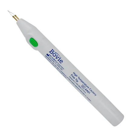
You will also hear the ESU called the "Bovie." The physicist W. T. Bovie built the first electrosurgical generator in the 1920s, and many ORs still use that name for the unit and for the pencil.
Why the current does not shock the patient
Current from the wall alternates 60 times a second. At that frequency it stimulates nerves and muscles, which is why a shockshokA life-threatening state in which blood flow is too low to bring enough oxygen to the body's tissues.Full entry from an outlet makes muscles clench and can stop the heart. Nerve and muscle stimulation stops above about 100,000 Hz. The ESU raises the frequency to more than 200,000 Hz, so its current can pass through the patient without stimulating muscle and without electrocuting the patient.
The electrosurgical unit
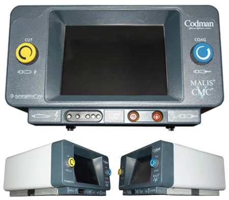
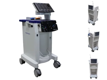
The electrosurgical unit (ESU) is the generator and control center of the system. It makes the radio-frequency current, and its panel sets the power for cutting, coagulationkoh-ag-yuh-LAY-shunThe process by which blood changes from a liquid into a gel, forming a clot.coagul clot-ation processFull entry and bipolarby-POH-lerHaving two poles or two opposite ends; most often used as a short form of bipolar disorder.bi- twopolar having polesFull entry work. Most modern units also sense how much the tissue resists and adjust their current and voltage to it, so the effect stays the same from one kind of tissue to the next.
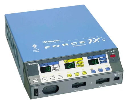
Chapter 23 covers the ESU as room equipment: where it sits, keeping fluids off it, its self-check and its alarms.
What electrosurgery does to tissue
What happens to the tissue depends on the waveform, the power setting, how long the current is applied, the size of the active electrode, the type of tissue and whether charred tissue is stuck to the tip.
| Effect | How it is done | What happens in the tissue |
|---|---|---|
| Cut | A continuous current at lower voltage, with the electrode held just off the tissue so the current jumps a tiny gap | The cells heat so fast that their water flashes into steam and they burst, and the tissue divides |
| Coagulation | An interrupted current, on only about 6 percent of the time, pushed at much higher voltage | The cells heat and cool in turn instead of bursting, which seals small vessels |
| Desiccation | Coagulation with the electrode touching the tissue, with either waveform | The tissue dries out, and the effect can reach deeper than fulguration |
| Fulguration, or spray coagulation | Pulsed, high-voltage current sparking from an electrode held just above the tissue | The sparks spread over a wide area and destroy only the surface, leaving a coagulated, sometimes charred layer |
| Blend | A cutting current that is switched off for part of the time | It cuts and seals small vessels at once; the more it is interrupted, the more it coagulates |
Coagulation is the general word for both desiccation and fulguration. The burned tissue that sticks to the tip is escharES-karThick, dry, dark dead tissue that covers part or all of a wound.Full entry : it holds heat like charcoal, it can spark, and it can catch fire. The sticky, semiliquid material that forms when energy alters tissue is called coagulum.
A few more rules of how tissue responds:
- Higher power and longer activation spread heat further into the tissue around the target.
- A small electrode, such as a needle, concentrates the current and needs less power; a large one spreads it and needs more.
- Fat (adipose tissueAD-ih-pohs TISH-ooBody fat: connective tissue made mostly of fat cells that store energy.adip/o fat-ose full ofFull entry ) holds little water and resists current, so it takes more power; well-supplied tissue such as muscle conducts easily and takes less.
- Eschar on the tip blocks the current, so the surgeon needs more power for the same effect.
Cut mode pushes its current with the lowest voltage, so it is considered safer than the other modes, and the safety guidance asks the team to use cut or blend in place of coagulation when either will do the job. High voltage pushes current harder through weak insulation and into unintended paths.
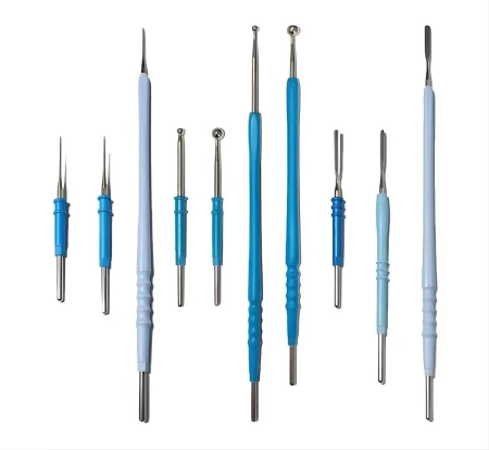
The tip of the active electrode comes in many shapes. A flat blade (spatula) tip is used for most cutting and coagulation, a fine needle for precise work, a ball tip for fulguration, a loop for biopsy, and extended tips for deep wounds.
Monopolar electrosurgery
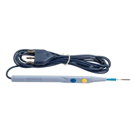
In monopolar electrosurgery, the patient is part of the circuit. Current flows from the generator, through the active electrode cord to the active electrode, into the patient's tissue, across the body to the patient return electrode, and home to the generator through the return cable. It is the most common kind of electrosurgery. It is also powerful: it can cut any tissue, bone included.
The monopolar system has three main parts besides the patient:
- The generator (the ESU), which makes the current.
- The active electrode: the metal tip, in a handheld, insulated pencil or handpiece, where current enters the tissue and turns into heat. The surgeon fires it with buttons on the pencil, one for cut and one for coagulation, or with a foot pedal.
- The patient return electrode, which collects the current from the patient and carries it back to the generator.
Bipolar electrosurgery
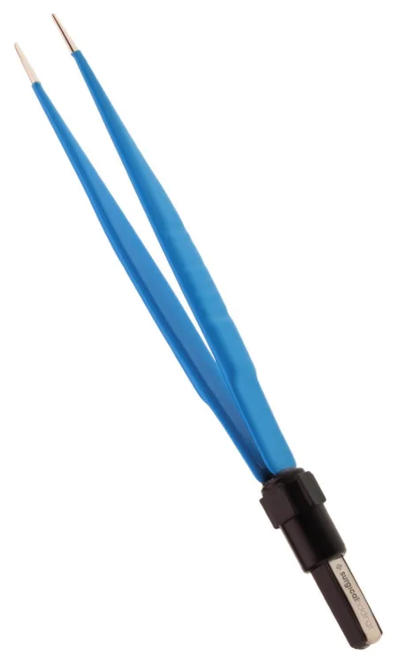
In bipolar electrosurgery, both electrodes are in the instrument itself, usually the two tines of a forcepsFOR-sepsA hand instrument with two blades or arms used to grasp, hold or pull tissue or objects.Full entry . Current flows from the generator to one tine, through only the tissue held between the tines, to the other tine and back to the generator. It never travels through the rest of the body, so no return electrode is needed.
Compared with monopolar:
- Bipolar runs at lower voltage, which makes it the safer technique, with fewer ways to injure the patient.
- That lower voltage is not strong enough for dense tissue such as bone, so bipolar is used mainly on tissue that conducts easily.
- Little heat spreads beyond the tips, so it is used on delicate tissue such as the brain, nerves and tiny vessels.
- Because no current crosses the body, it is the safer choice for a patient with an implanted electronic device such as a pacemakerPAYS-may-kerA small device that sends electrical pulses to the heart to keep it beating at a set rate; also the heart's own natural rhythm center.Full entry .
- Many bipolar generators stop the current once the tissue between the tips has dried enough to reach a set resistance.
The surgeon usually fires the bipolar forceps with a foot pedal. Bipolar forceps and cords are usually reusable, so inspect the cord and check that the tips line up and are not bent. Never plug a bipolar cord into a monopolar jack: the monopolar output is far higher and can burnBURNAn injury to the skin or deeper tissues caused by heat, chemicals, electricity, radiation or friction.Full entry tissue.
A return electrode is not used when only bipolar will be used. When a case needs both, the pad goes on for the monopolar work, and any mode that is not in use is set to zero or its lowest setting.
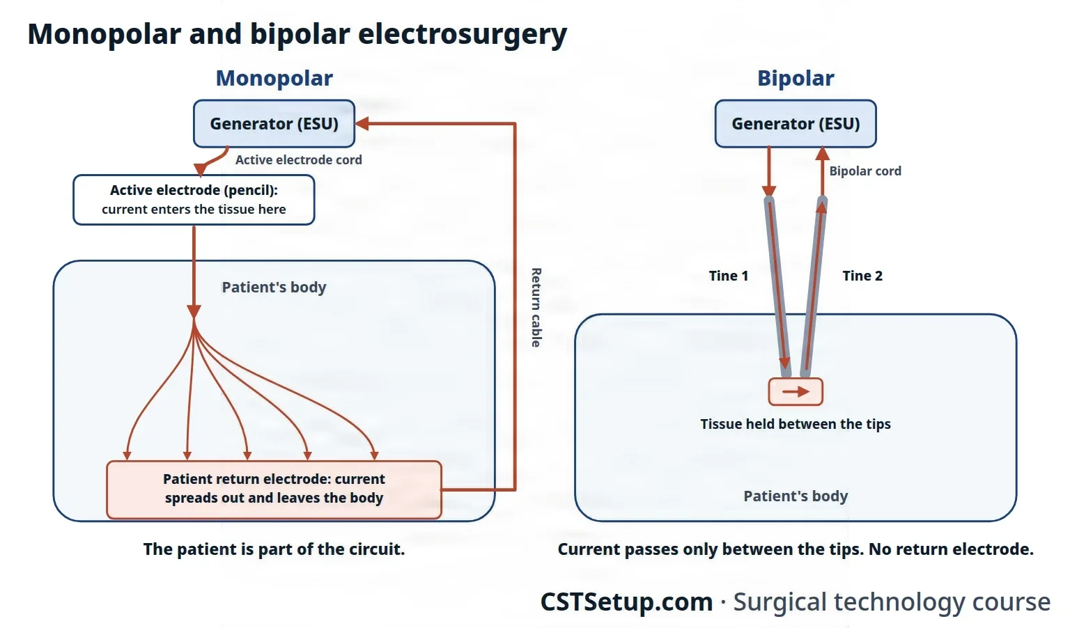
Monopolar current travels through the patient to a return electrode. Bipolar current travels only through the tissue between the two tips, so it needs no return electrode.
5The patient return electrode
The active electrode is small, so the current going into the tissue is concentrated and makes heat. The patient return electrode (PRE) is large, so the same current coming out spreads over a wide area of skin and stays far too weak to heat it. From there the current goes back to the generator, and the circuit is complete.
A return electrode is an adhesive pad, or a metal plate, coated with conductive gel and joined to the generator by a cable. It is also called the dispersive electrode. Many people call it the grounding pad, but that name is misleading: the pad does not ground the patient the way a third prong grounds an appliance; it brings the current back to the generator.
How a return electrode burns
- Poor contact. If the pad is tented, wrinkled, lifting at an edge or sitting over a gap or pooled fluid, only part of it touches the skin. All the current crowds into that smaller area and can burnBURNAn injury to the skin or deeper tissues caused by heat, chemicals, electricity, radiation or friction.Full entry the skin under the pad.
- No return path. If the pad is not connected to both the patient and the generator, or its cable comes loose, the current keeps looking for a way out of the body. Wherever the patient touches a conductor, such as the metal table frame, a stirrup or the metal in an electrocardiogramih-lek-troh-KAR-dee-oh-gramA test that records the heart's electrical activity as waves on paper or a screen.electr/o electricitycardi/o heart-gram recordFull entry (ECG) electrode, the current leaves there. A small exit point means a deep, full-thickness burnFULL THIK-nis BERNA burn that destroys both layers of the skin, the epidermis and all of the dermis, and may reach the tissue below.Full entry . Failing to attach the pad to the patient and the generator is a common cause of electrosurgeryee-LEK-troh-SUR-jer-eeUsing high-frequency electric current through a handheld tool to cut tissue or seal bleeding vessels during surgery.Full entry burns.
- Alternate paths. Current divides and follows the paths of least resistance. With older grounded units, patients were burned where current left through ECG pads or an IV pole that touched them, and those burns tend to be serious.
Safety built into the unit
Most modern generators are isolated: a transformer lets their current return only to the generator, so it cannot leave the patient through a grounded object such as an ECG pad, as it could with the older grounded units. Isolation does not prevent a burn under a poorly placed pad, so the unit also monitors the return electrode: with a split pad, it keeps measuring the contact under the pad, and if the contact becomes unsafe it alarms and stops the current.
Some ORs use a large reusable capacitive pad that lies on the OR bed under the sheets in place of an adhesive pad, used according to its maker's instructions.
Where it goes
The return electrode goes on clean, dry skin over a large, well-supplied muscle, as close to the surgical site as possible, after the patient is positioned. Muscle holds a lot of water and carries current well, while fat resists it. The pad stays off bony areas, scars, tattoos, metal implants and any limb below a tourniquet, and hair is clipped if it would keep the pad from lying flat. It must touch the skin fully, it is never cut to fit, and it is replaced if the patient is repositioned. The patient must touch no metal, and ECG electrodes go as far from the surgical site as possible. Chapter 31 gives the steps for placing it.
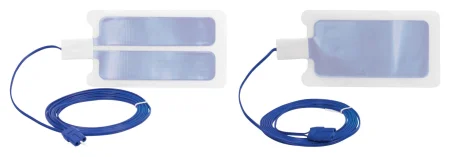
tape down a loose return electrode or cut one to fit. If a pad comes loose, the surgeon is told, the ESU is turned off and a new pad goes on.
6Using the active electrode safely
One study of injuries voluntarily reported to the FDA found direct application the most common way energy devices burned patients, in 84 percent of the reports: an instrument fired by accident while it touched tissue, for example a monopolar pencil set off by a surgeon leaning against the patient. Many of the safeguards are in the scrub's hands. AST holds the CSTsee-es-tee (said as letters)Certified Surgical Technologist, a credential for surgical technologists; in maternity care, contraction stress test.Full entry responsible for controlling the active electrode whenever it is not in use, to prevent burns, fires and contamination.
Setting it up
- Inspect it. When you set up, check the pencil, its cord and its tips, including the insulation along the cord. Single-use items can be faulty too, so check every one. A damaged electrode goes off the field and is replaced.
- Seat the tip. Push the tip fully into the pencil and check now and then during the case that it has not worked loose. Never bend a tip, and never slide a piece of rubber catheter over it as extra insulation: the rubber burns.
- Holster it. The pencil lives in a dry, insulated safety holster whenever it is not in use, clipped to the drapes, preferably with a non-metal clamp, in plain sight and within the surgeon's reach. It goes back in after every use and is never left lying on the patient or the drapes, where someone can lean on it and fire it, or it can slide off the table.
- Long electrodes: a laparoscopiclap-uh-roh-SKOP-ikDone through a few small cuts in the abdomen with a thin camera and long instruments, instead of one large opening.lapar/o abdomen-scopic viewingFull entry electrode is usually too long for the holster, so it goes on the Mayo standMAY-oh standA small tray on an adjustable stand placed over or beside the patient during surgery to hold the instruments used most often.Full entry . If the surgeon insists it stays on the drapes, that is recorded in the operative record.
- Lay the cord out straight. Fasten the cord to the drapes without knots, coils or sharp bends, and never wrap it around a metal clamp or thread it through a clamp's handles. Kinks damage the wires and insulation, and current can leak into the metal.
- Keep it apart from other cords. Run the monopolar cord as far as you can from camera, light and monitoring cords, or across them at a right angle, because its energy can pass through intact insulation into a nearby cord and burnBURNAn injury to the skin or deeper tissues caused by heat, chemicals, electricity, radiation or friction.Full entry the patient away from the surgical site (antenna coupling).
Before and during the case
- Test it. Just before the incision, check that the cord is in the correct jack on the unit, and briefly fire the pencil to test it.
- Confirm the settings. The circulator sets the power the surgeon asks for, and it is said aloud and confirmed before the incision. The lowest power that does the job is used.
- One person fires it. Only the person using the active electrode activates it, by hand switch or foot pedal. Where state law and facility policy allow, a CST may activate it under the surgeon's direction. Tell the surgeon where the foot pedal is, and keep it in a clear, waterproof cover.
- Fire it only at the target. The electrode is activated only when it is touching or close to the tissue to be treated, in view, and away from other metal that could carry the current or make it arc.
- Listen for it. The electrosurgeryee-LEK-troh-SUR-jer-eeUsing high-frequency electric current through a handheld tool to cut tissue or seal bleeding vessels during surgery.Full entry 's activation tone tells everyone the electrode is live, including when someone has leaned on a pencil by mistake. Alarms and the tone must be audible for the whole case, and music never drowns them out.
Keeping the tip clean
Help the surgeon keep the tip free of escharES-karThick, dry, dark dead tissue that covers part or all of a wound.Full entry . Eschar resists the current, so the tip heats more and the surgeon needs more power; the hot eschar can also catch fire, and small burning bits can fall into the wound.
- Use a sterile abrasive scratch pad, fixed where the surgeon can reach it but away from the wound so it cannot fall in.
- Clean a nonstick tip with a wet sponge instead, because the scratch pad ruins the coating.
- Never scrape a tip with a knife blade. The blade scratches and pits the tip, which makes more eschar and more sparking, and you risk cutting yourself.
The tip is a sharp. It is counted with the sharps, passed through the neutral zone, and thrown away in the sharps container. At the end of the case, take the batteries out of a batteryBAT-uh-reeIn law, touching a person on purpose without their consent.Full entry -powered disposable pencil before you discard it: improperly discarded pencils have started trash fires.
Coagulating a clamp
Often the surgeon clamps a bleeding vessel and seals it by touching the active electrode to the clamp, which carries the current down to the vessel. This is called buzzing the clamp. Either you hold the clamp while the surgeon buzzes it, or the surgeon holds it and asks you to buzz it.
- Holding the clamp: hold it upright, and check with the surgeon that it touches nothing but the vessel, especially not the skin edges.
- Buzzing it: touch the tip to the clamp below the fingers of whoever is holding it.
- Fire only when the tip is in full contact with the clamp and you can see the contact, so the current does not arc.
- Keep both your gloves and the surgeon's dry.
Why below the fingers? The glove normally insulates the hand from the current in the clamp. But if the glove is thin, current can pass through it, and high voltage can break the glove material down and punch a tiny hole in it. Either way the holder gets a painful burn.
When the surgeon keeps asking for more power
Repeated requests to turn the ESU up can mean the unit is failing or something in the circuit is resisting the current, which can overheat and burn the patient. AST's steps:
- Tell the surgeon it may be a malfunction, and stop using the ESU until the problem is found.
- Check every cord connection, and check the cords for damage.
- Check that the return electrode is still in full contact with the skin.
- Check the tip for eschar.
- Confirm with the surgeon the type and amount of irrigationeer-ih-GAY-shunWashing out a wound, body cavity, tube or eye with a steady flow of liquid.Full entry fluid in the wound; the surgeon may change it, because the fluid changes how easily current flows.
- If none of this fixes it, the ESU is taken out of the room and replaced, with a new return electrode on the patient and a new active electrode. The faulty unit goes to biomedical engineering.
Jewelry and monitoring electrodes
Metal jewelry and body piercings can burn the patient, so they come off before surgery, as chapter 31 teaches; if a piercing cannot come out, the team uses another kind of energy device. Needle monitoring electrodes also give the current a way out of the body, so bipolarby-POH-lerHaving two poles or two opposite ends; most often used as a short form of bipolar disorder.bi- twopolar having polesFull entry electrosurgery or a laser is used with them.
7Stray current in laparoscopy
In laparoscopylap-uh-ROS-kuh-peeSurgery or an exam done through small cuts in the belly, using a thin, lighted tube with a camera.lapar/o abdomen-scopy looking with an instrumentFull entry the active electrode works on a long, insulated shaft inside the abdomen, and the surgeon sees only what is in front of the scope. Three hazards come with that, and the burns they cause often happen out of sight. A burned bowel may show nothing for several days after surgery, then break down into a perforationper-fuh-RAY-shunA hole that goes all the way through the wall of an organ or tissue.per- throughforat bored (a hole)-ion processFull entry and peritonitispair-ih-tuh-NY-tisInflammation of the peritoneum, the thin membrane that lines the inside of the belly and covers the organs there.peritone/o peritoneum-itis inflammationFull entry , which can be life-threatening.
- Insulation failure: a crack or hole in an instrument's insulation lets current escape at that point and burnBURNAn injury to the skin or deeper tissues caused by heat, chemicals, electricity, radiation or friction.Full entry whatever it touches, at full power. AST calls it the main cause of laparoscopiclap-uh-roh-SKOP-ikDone through a few small cuts in the abdomen with a thin camera and long instruments, instead of one large opening.lapar/o abdomen-scopic viewingFull entry electrosurgeryee-LEK-troh-SUR-jer-eeUsing high-frequency electric current through a handheld tool to cut tissue or seal bleeding vessels during surgery.Full entry injuries. The power at the tip does not drop to warn anyone, and one reference puts the share of insulation defects you can see with the naked eye at only about 10 percent. High-voltage coagulationkoh-ag-yuh-LAY-shunThe process by which blood changes from a liquid into a gel, forming a clot.coagul clot-ation processFull entry current can push through even weakened insulation.
- Direct coupling: the active electrode touches, or comes very close to, another metal object, such as the scope, a metal cannulaKAN-yoo-luhA thin tube placed into the body or a body opening to deliver or remove fluid or air.Full entry , a suction tube or a clip, and the current flows into it and burns the tissue it touches. It is within the surgeon's control: fire only when the tip is in view and on the target, not on metal. In open surgery it can happen when an active electrode is passed through a metal suction tube.
- Capacitive coupling: current passes through intact insulation into a nearby conductor, such as a metal trocarTROH-karA sharp or bladed instrument inside a tube (cannula), used to pierce a body wall and leave the tube in place as a port.Full entry sleeve, a suction-irrigator or the scope, and charges it. If that charge cannot spread out safely through the abdominal wall, the conductor burns the tissue it touches. The surgeon can neither see nor control it. It is worse with hybrid trocars (part plastic, part metal), with long, thin instruments and thin insulation, and with high voltage.
The protections:
- Inspect the insulation every time. Sterile processing checks it by eye, under magnification and with an insulation scanner. The first scrub scans the instruments again while setting up, because sterilizationstair-uh-luh-ZAY-shunA process that destroys all microorganisms, including spores, on an item; also a permanent birth control procedure.Full entry can damage insulation, and once more before the end of the case. A faulty instrument comes off the field at once; if one is found faulty at the end, the surgeon is told and may decide to inspect the abdomen.
- Two-layer insulation: some instruments have a bright layer under the black outer one, so a break in the black shows up in color.
- Active electrode monitoring (AEM): shielded, monitored instruments that catch stray current from insulation failure and capacitive coupling and send it safely back to the generator.
- No hybrid trocars: conductive (all-metal) cannulas let any induced current spread safely into the abdominal wall.
- Low power, close to the target: the lowest settings that work, and firing only when the tip is at the tissue, cut the risk of all three.
Chapter 21 shows how this works with the trocars, scopes and instruments in a laparoscopic case.
If the insulation on a laparoscopic instrument looks scratched, thin or cracked, take the instrument off the field and replace it. Most insulation defects cannot be seen at all, so scan every instrument too.
8Patients with implanted electronic devices
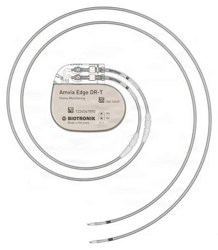
An implanted electronic device (IED) monitors or corrects a body function. Examples are a pacemakerPAYS-may-kerA small device that sends electrical pulses to the heart to keep it beating at a set rate; also the heart's own natural rhythm center.Full entry , an implanted defibrillator, a deep brain stimulator, a ventricleVEN-trih-kulOne of the two lower chambers of the heart, or one of the fluid-filled spaces inside the brain.Full entry assist device, a vagal nerve stimulator and a cochlear implant.
Monopolar electrosurgeryee-LEK-troh-SUR-jer-eeUsing high-frequency electric current through a handheld tool to cut tissue or seal bleeding vessels during surgery.Full entry works at radio frequencies that can interfere with these devices. A device can misread the electrical noise, stop working properly, or act on it; an implanted defibrillator, for example, may sense the noise as a dangerous rhythm and deliver a shockshokA life-threatening state in which blood flow is too low to bring enough oxygen to the body's tissues.Full entry .
What the team does:
- Find out what the device is, follow its maker's instructions, and ask the maker or its representative for advice before monopolar current is used.
- Prefer energy that sends no current through the body, such as bipolarby-POH-lerHaving two poles or two opposite ends; most often used as a short form of bipolar disorder.bi- twopolar having polesFull entry or ultrasonic devices, which interfere least.
- If monopolar must be used, keep the power low, and keep its cords and the path of its current away from the device and its leads, never running alongside them.
- Monitor the patient's heart rhythm, and have a defibrillator ready.
AST's 2012 electrosurgery standard names coblation (a plasmaPLAZ-muhThe pale yellow liquid part of the blood, which carries the blood cells, proteins, nutrients and wastes.Full entry -based technology) as the first choice for a patient with an IED, and bipolar electrosurgery, kept more than 1/2 inch (1 cm) from the device, as the next.
9Other energy at the field
Several other devices use electricity or other energy to cut, seal or destroy tissue. Chapter 29 teaches when each is chosen to control bleeding and how the scrub handles it; here is how each works.
- Argon-enhanced electrosurgeryee-LEK-troh-SUR-jer-eeUsing high-frequency electric current through a handheld tool to cut tissue or seal bleeding vessels during surgery.Full entry (the argon beam coagulator): a stream of argon gas, which is inert and does not burnBURNAn injury to the skin or deeper tissues caused by heat, chemicals, electricity, radiation or friction.Full entry , flows around the active electrode. The gas carries and focuses the current, prevents sparking, pushes oxygen away and makes less smoke, and it coagulationkoh-ag-yuh-LAY-shunThe process by which blood changes from a liquid into a gel, forming a clot.coagul clot-ation processFull entry oozing surfaces quickly without touching them. It is still monopolar, so the patient needs a return electrode and the same precautionprih-KAW-shunA step taken ahead of time to prevent harm, such as the infection-control practices used with every client.Full entry as the ESU, holster included. The gas can enter a vein and cause an embolismEM-buh-liz-umThe blocking of a blood vessel by a mass carried there in the blood (an embolus), such as a clot, fat or an air bubble.embol/o plug-ism conditionFull entry , so the line is purged before use and after delays, the flow is kept at the lowest setting that works, the tip is held several millimeters from the tissue at an angle, and the abdomen is not overfilled with gas.
- bipolarby-POH-lerHaving two poles or two opposite ends; most often used as a short form of bipolar disorder.bi- twopolar having polesFull entry vessel sealing: bipolar current at low voltage, combined with firm pressure from the jaws, welds a vessel wall shut. The generator senses the tissue and stops automatically when the seal is complete, with little heat spread.
- Radiofrequency ablationab-LAY-shunA procedure that destroys a small area of tissue, often with heat or cold, to stop it from causing a problem.Full entry : ablation is the complete destruction of tissue. An electrode is placed into a tumor, and radio-frequency energy heats the tissue enough to destroy it. A bipolar version works inside a fluid-filled organ, such as the bladder or uterus.
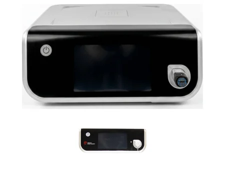
- Ultrasonic energy: electricity drives a blade that vibrates about 55,000 times a second. The vibration breaks protein bonds and forms a sticky coagulum that seals vessels as the blade cuts. It damages less of the tissue around the cut than an ESU or a laser, and no current passes through the patient, so no return electrode is needed. It makes far less plume than electrosurgery, mostly a little water vapor (an aerosol), and AST still says to evacuate it, with a smoke evacuator or wall suction with an in-line ULPA filter.
- Hot blades after use: even so, frictionFRIK-shunThe rubbing of one surface against another, such as skin against bed sheets.Full entry makes the blade itself very hot, and it stays hot after it stops, longer than electrosurgical tips do. It is held away from tissue while it cools, and the scrub keeps a moist towel on the field to rest it on between uses.
- Ultrasonic aspirationas-puh-RAY-shunBreathing food, liquid, saliva or stomach contents into the airway and lungs; also, drawing fluid out with suction or a needle.Full entry : the Cavitron Ultrasonic Surgical Aspirator (CUSA) destroys tissue with high-frequency sound waves and suctions it away, as in tumor surgery. Phacoemulsification uses ultrasound the same way to break up a cataractKAT-uh-raktClouding of the lens of the eye.Full entry .
- Cryosurgery: extreme cold destroys tissue. Liquid nitrogen removes small skin lesionLEE-zhunAn area of tissue that has been damaged or has changed from normal, such as a sore, wound, lump or spot.Full entry , and in cryoablation a probe placed in a tumor is cooled with high-pressure argon gas, freezing the tissue around it, which dies and is absorptionub-ZORP-shunThe taking in of a substance, such as a drug or a nutrient, into the bloodstream.Full entry .
10Fire at the surgical field
A surgical fire is a fire on or in the patient during an operation. No national system counts them, but ECRI estimates 90 to 100 surgical fires a year in the United States, as The Joint Commissionthuh JOYNT kuh-MISH-unAn independent US organization that inspects and accredits hospitals and other health care organizations against safety and quality standards.Full entry reported in 2023, and electrosurgeryee-LEK-troh-SUR-jer-eeUsing high-frequency electric current through a handheld tool to cut tissue or seal bleeding vessels during surgery.Full entry devices start about 70 percent of them. Older references give ECRI's earlier estimate of 550 to 650 a year. A flash fire can engulf a patient in moments. One report found about 21 percent of patient fires in the airway, 44 percent on the face, 26 percent on the skin and 8 percent inside the body.
The Joint Commission's sentinel eventSEN-tih-nul ee-VENTAn unexpected event in health care that causes a client's death, permanent harm or severe temporary harm.Full entry Policy lists a fire, flame, or unexpected smoke, heat or flash during patient care, caused by equipment in use, as a sentinel event even when no one is hurt; chapter 15 explains sentinel events.
This section covers fire at the field: what feeds it, how to prevent it, and what the team does when one starts on the patient.
The fire triangle
A fire needs three things: an oxidizer, a fuel and an ignition source. This is the fire triangle. All three are present in every operation. Take any one away and there is no fire, so fire prevention is the work of keeping the three apart in space and in time.
AST sets three goals for fire safety: handle fuels safely, handle ignition sources safely, and avoid or minimize an oxygen-enriched atmosphere near the surgical site.
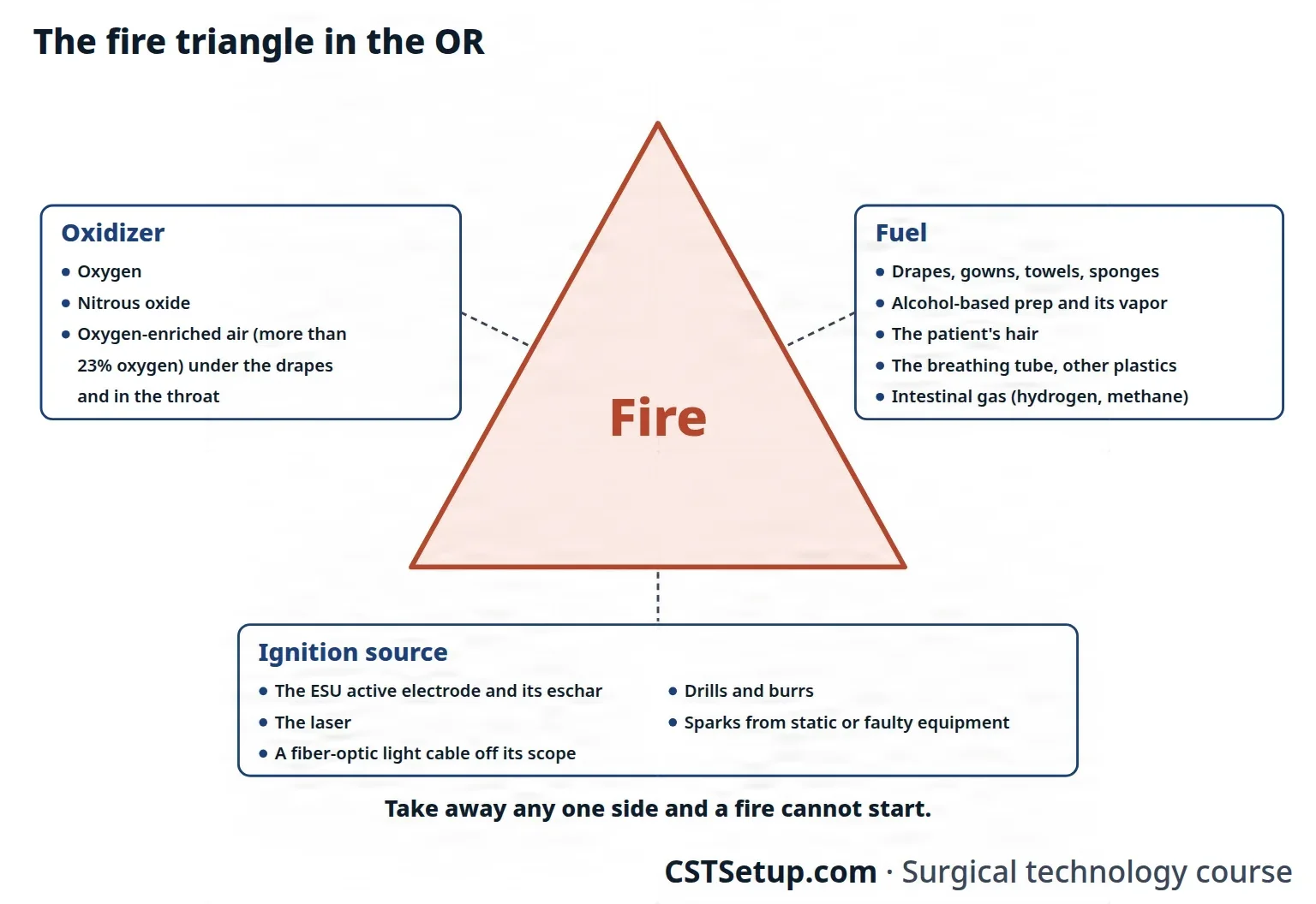
The oxidizer. An oxidizer is a substance that supports burning. In the OR the oxidizers are oxygen and nitrous oxide. Room air is about 21 percent oxygen; air with more oxygen than normal is an oxygen-enriched atmosphere (OEA), which AST defines as more than 23 percent oxygen. Chapter 17 covers oxygen as a medical gas and chapter 13 its use in anesthesiaan-us-THEE-zhuhLoss of feeling or sensation, often brought on by drugs so that surgery or a procedure can be done without pain.an- withoutesthesi/o feeling, sensation-ia conditionFull entry .
- The more oxygen, the faster things catch fire and the hotter and longer they burnBURNAn injury to the skin or deeper tissues caused by heat, chemicals, electricity, radiation or friction.Full entry . Materials that would barely burn in room air burn easily in an OEA.
- Oxygen is heavier than air, so it settles and collects under drapes and in body cavities.
- Nitrous oxide breaks down into oxygen when it is heated, adding to the oxygen that has built up.
- During airway surgery, oxygen and nitrous oxide can leak around the breathing tube and build up in the throat.
- ECRI estimates that about 75 percent of surgical fires happen in an oxygen-enriched atmosphere, which is why fires cluster around the face, head and neck.
The fuel. Anything that can burn is fuel. Flammable means able to burn. Volatile describes a substance, such as alcohol, that turns to vapor at a low temperature.
- Fabrics and paper: drapes, gowns, towels, sponges, dressings, caps, masks and shoe covers. Items labeled flame resistant can still catch fire and keep burning in an OEA.
- Alcohol-based prep solutions: alcohol above 20 percent is flammable, and most prep solutions are about 70 percent alcohol. Their vapor can collect under the drapes, where a fire starts out of sight, and most prep fires are lit by a spark from the ESU. Alcohol soaked into hair can take up to an hour to dry.
- Other flammable chemicals: tinctures, collodion, skin degreasers, petroleum-based ointments and lubricants, tissue glue (cyanoacrylate) and bone cement (methyl methacrylate).
- Rubber and plastic: the endotracheal tubeen-doh-TRAY-kee-ul toobA flexible plastic tube passed through the mouth or nose into the windpipe to keep the airway open and allow breathing support.endo- withintrache/o windpipe-al relating toFull entry , masks, airways, oxygen tubing, breathing circuits and gloves.
- The patient: hair, including the fine body hair called lanugoluh-NOO-gohFine, soft, downy hair that covers the body of a fetus.Full entry , and the gases in the intestine, hydrogen and methane, which burn; methane explodes at concentrations of 5 to 15 percent.
- Smoke: surgical smoke itself can burn if it builds up in a small space, such as the back of the throat.
- Positioning equipment: foam pads, the table mattress and gel pads, which give off toxicitytok-SIS-ih-teeThe harm a substance does to the body, or a state in which a drug has built up to a harmful level.tox/o poison-icity state or quality ofFull entry gases as they burn.
The ignition source. Anything that makes heat can light a fire.
- The active electrode: its tip can reach 1292°F (700°C), hot enough to light drapes. escharES-karThick, dry, dark dead tissue that covers part or all of a wound.Full entry on the tip holds heat like charcoal and sparks, and it can drop burning bits into the wound. Sparks also fly when the tip touches metal.
- Lasers: ECRI has reported that roughly 10 to 13 percent of surgical fires involve a laser.
- Fiber-optic light cables: the light from a cable that is disconnected from its scope is intense enough to set drapes alight.
- Power instruments: drills, burrs and saws heat up from frictionFRIK-shunThe rubbing of one surface against another, such as skin against bed sheets.Full entry , and so can an ultrasonic blade.
- Other devices: the argon beam coagulator, a batteryBAT-uh-reeIn law, touching a person on purpose without their consent.Full entry -powered cautery and defibrillator paddles.
- Sparks: static electricity, two metal instruments striking together, and an electrical short or other fault in a device.
The fire risk assessment
AORN has the team assess the fire risk before every procedure and repeat the assessment during the time-outTYM-owtA pause just before a procedure when the whole team confirms the right client, procedure and site; also, a short break to calm a child.Full entry . Its Fire Prevention Assessment Tool walks through four questions, and every yes adds steps to take:
- Will the skin be prepped with an alcohol-based antiseptic, or will any other flammable solution be used?
- Is the procedure above the xiphoid processZY-foyd PROS-esThe small, pointed tip at the lower end of the breastbone.Full entry (the lower tip of the breastbone) or in the mouth and throat?
- Is open oxygen or nitrous oxide being given, such as by mask or nasal prongs?
- Will an ESU, a laser or a fiber-optic light be used?
When all three sides of the triangle come close together, such as an ignition source used near the face of a patient breathing open oxygen, the risk is highest. Your facility's policy sets how the assessment is done and recorded.
Preventing a fire
Keep fuel under control.
- Read the safety data sheet for the prep solution to see whether it is flammable. Tuck dripDRIPA continuous IV infusion of fluid or a drug, given slowly over time by gravity or a pump.Full entry towels to catch excess solution, and take out anything soaked with it before the drapes go on.
- Let a flammable prep dry completely and its fumes clear before the drapes go on and before any ignition source is used. Follow the maker's dry time, which is longer for hair, and never let prep pool under the patient or around the return electrode. Chapter 31 covers the skin prep itself.
- Use a nonflammable agent when one will do.
- Wet the towels, sponges and absorbent drapes near the active electrode, and keep them wet. Fires have started from dry sponges around and inside the wound.
- Coat hair near the surgical site with water-soluble gel.
- When you set up, put sutureSOO-cherA stitch, or the thread used to stitch, that holds the edges of a wound or incision together.Full entry packets that contain alcohol into the sterile waste bag on the back table, away from the active electrode.
- Mix bone cement in a closed system that suctions away its fumes.
Keep oxidizers away from the site. The anesthesia provideran-us-THEE-zhuh pruh-VY-derThe licensed professional who gives anesthesia and watches over the client's breathing and vital functions during a procedure.Full entry controls the gases, and the whole team helps keep oxygen from building up at the field.
- The patient gets the lowest concentration of oxygen that keeps the blood oxygen level adequate.
- For surgery above the xiphoid, a patient who needs more than 30 percent oxygen gets a larynxLAIR-inksThe voice box: the part of the airway in the neck, above the windpipe, that holds the vocal cords.laryng/o larynxFull entry mask airway or an endotracheal tube, which keeps the gas inside the airway, rather than open oxygen by mask or prongs.
- Before an ignition source is used on the head, neck or upper chest, the surgeon tells the anesthesia provider, who stops or turns down the oxygen and nitrous oxide for 1 minute and says when it is safe to go ahead.
- Drapes are arranged so oxygen can flow away from the patient instead of collecting underneath. An adhesive incise drape placed to separate the oxygen source from the surgical site helps, and a flow of medical air may be run under the drapes to flush out extra oxygen.
- The anesthesia circuits are checked for leaks, and gas that has built up in the mouth or throat is suctioningSUK-shun-ingRemoving mucus, saliva or other secretions from the mouth, nose or airway with a tube connected to a suction source.Full entry out before an ignition source is used there.
In the past, oxygen was often given openly at 100 percent during head, neck and chest surgery, where it collected under the drapes. ECRI's 2009 surgical fire guide recommends stopping that practice.
Keep ignition sources under control.
- Holster the active electrode, keep its tip clean, use the lowest power, and fire it only at the target, away from oxygen sources.
- Never use the ESU or a laser to enter the airway: the surgeon cuts into the tracheaTRAY-kee-uhThe windpipe: the tube that carries air from the throat to the lungs.Full entry with a knife or scissors.
- Never open the bowel with the ESU or a laser. Use either one with caution whenever intestinal gas is present, and suction the gas away from the site.
AST's 2012 electrosurgery standard says the ESU should not be used at all once the gastrointestinal tract is entered, as in a colon resectionrih-SEK-shunSurgery to remove part of an organ or tissue.re- back, againsect to cut-ion processFull entry .
- Put the light source on standby, or turn it off, whenever its cable is not connected to the scope. Never lay a lit cable on drapes or sponges, and never let one hang over the side of the field.
- Cool drills and burrs with a slow drip of saline, and never rest them on the patient.
- Use defibrillator paddles of the right size, with the gel their maker names and full contact with the skin.
- Use only equipment that has been inspected and has sound cords, and take any equipment that smokes out of service.
Be ready. Always have sterile water or saline on the back table to put out a fire, and know where the fire alarm, the extinguishers and the room's gas shutoff valves are.
A fire needs an oxidizer, a fuel and an ignition source. Every safety step in this section takes one of them away from the other two.
When a fire starts on the patient
Time is critical. The team acts at once, together:
- Call it out. Announce the fire and stop the procedure.
- Stop the gases. The anesthesia provider shuts off every gas flowing to the patient's airway.
- Remove what is burning. At the same moment, burning drapes and other materials come off the patient, as safely as possible.
- Put it out. Smother a small fire with a wet towel or sponge, or douse it with saline or water. As the scrub, you hand the water or saline over at once and stand by for the surgeon's direction.
- Care for the patient. The team checks the patient for burns and treats them.
An extinguisher is not used to put out a burning drape, and it is not the first choice for a fire at the field, but it may be needed if the fire is extensive and keeps burning the patient or threatens the team. A carbon dioxideKAR-bun dy-OK-sideA gas made by the body's cells as waste, carried in the blood to the lungs and breathed out.Full entry (Class C) extinguisher is the type preferred for electrical and laser fires. Fire blankets are not used in the OR: they can catch fire themselves, they can trap the fire against the patient, and they contaminate the wound.
If the fire spreads beyond the patient, the team switches to the facility's fire plan in chapter 17: RACE, PASS and the extinguishers, the zone valves and evacuation.
let a fire on the field burn while you look for help. Smother it or douse it with the saline or water already on your back table, and say "fire" loudly.
An airway fire
An airway fire can be deadly. A burning breathing tube acts like a blowtorch: in a few seconds its flames can shoot out 5 to 10 inches (12.5 to 25 cm), and hot gases can be driven down into the lungs. The steps:
- The anesthesia provider takes out the breathing tube, the whole tube, and stops the gases.
- The team removes anything burning from the airway. Hand the surgeon a clamp or forcepsFOR-sepsA hand instrument with two blades or arms used to grasp, hold or pull tissue or objects.Full entry to pull out the sponges packed around the tube.
- Hand over water or saline to pour into the airway.
- The anesthesia provider restores breathing, by mask or with a new tube.
- The surgeon checks the airway for burns and pieces of the tube; you may need to set up for a bronchoscopybrong-KOS-kuh-peeA procedure in which a thin, lighted tube with a camera is passed through the nose or mouth to look inside the airways.bronch/o airway (bronchus)-scopy looking with an instrumentFull entry .
- The surgeon and anesthesia provider decide whether the operation can go on.
- Everything involved is bagged and labeled so the cause can be found.
After a fire or a burn
- Keep the evidence. The equipment involved is tagged, taken out of service and kept as it was, with its settings untouched, and sent to biomedical engineering so they can work out what went wrong.
- Report it. The event goes through your facility's reporting process, and the CSTsee-es-tee (said as letters)Certified Surgical Technologist, a credential for surgical technologists; in maternity care, contraction stress test.Full entry should know that process. Under the Safe Medical Devices Act, a device-related death is reported to the FDA and the maker within 10 days, and a serious injury to the maker within 10 days, or to the FDA if the maker is not known. Chapter 15 covers incident reportIN-sih-dent rih-PORTA written record of an unexpected event in a health care setting that caused or could have caused harm.Full entry .
11Surgical smoke
When the electrosurgeryee-LEK-troh-SUR-jer-eeUsing high-frequency electric current through a handheld tool to cut tissue or seal bleeding vessels during surgery.Full entry or a laser heats tissue, the water in the cells boils, the cells burst, and their contents spray into the air as smoke. This surgical smoke is also called plume or smoke plume. Occupational Safety and Health AdministrationOH-shuh (said as a word)The US federal agency, part of the Department of Labor, that sets and enforces rules to keep workplaces safe.Full entry estimates that about half a million health care workers, surgical technologistSUR-jih-kul tek-NOL-uh-jistA member of the operating room team who prepares the sterile field and instruments and hands them to the surgeon.Full entry among them, are exposed to it every year.
What is in it
About 95 percent of surgical smoke is water. The other 5 percent is what makes it harmful:
- toxicitytok-SIS-ih-teeThe harm a substance does to the body, or a state in which a drug has built up to a harmful level.tox/o poison-icity state or quality ofFull entry chemicals: benzene, formaldehyde, hydrogen cyanide, toluene, acrolein, carbon monoxide and many others, some at levels that can exceed OSHA's limits.
- Particles: blood, bits of tissue, and living and dead cells. Many particles are tiny, from 0.1 to 0.8 microns across. They can carry much smaller viruses and bacteria, and they are easily breathed into the lungs.
- Germs: whole and broken bacteria and viruses. One report described two laser surgeons with human papillomavirusHYOO-mun pap-ih-LOH-muh-vy-rusA common group of viruses spread by close skin-to-skin contact, most often during sex.Full entry (HPV) in the throat, and a study found more warts in the nose and throat among laser surgeons than in a comparison group; together they suggest HPV can travel in laser plume. OSHA notes that no infection has yet been proven to pass through surgical smoke, but the risk may exist, most of all after treating genital wartJEN-ih-tul WORTA small, soft growth on or around the genitals or anus caused by certain types of human papillomavirus (HPV).Full entry .
- Cancer cells: researchers suggest the smoke may also carry cancer cells.
The smoke irritates the eyes, nose and throat. It can damage genes, and its chemicals include some that may cause cancer; a substance that causes cancer is a carcinogenkar-SIN-uh-jenAnything that can cause cancer, such as tobacco smoke, asbestos, UV light or certain chemicals and viruses.carcin/o cancer-gen something that producesFull entry . It also smells, it blocks the surgeon's view, and in a small space it can even feed a fire.
Getting rid of it
The room's normal ventilationven-tuh-LAY-shunAir moving in and out of the lungs as a person breathes.Full entry cannot capture surgical smoke. It must be removed right where it is made.
- Evacuate all of it, every time. Remove the smoke in every procedure, open and laparoscopiclap-uh-roh-SKOP-ikDone through a few small cuts in the abdomen with a thin camera and long instruments, instead of one large opening.lapar/o abdomen-scopic viewingFull entry , in which the ESU or a laser is used, however little smoke there seems to be. Every OR where plume is made needs a smoke evacuator, kept running whenever smoke is produced.
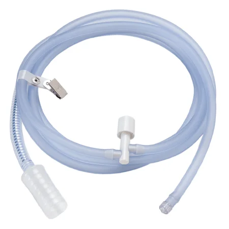
- Small amounts: wall suction with an in-line filter between the suction canister and the wall connection. Room suction is built to carry fluid and pulls air slowly, so it handles only light smoke, and it needs the filter to be safe.
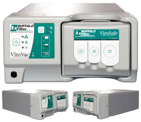
- Larger amounts: a smoke evacuator, a unit with its own vacuum pump, a charcoal filter that takes out gases and odor, and a high-efficiency (ULPA or HEPA) filter that catches the tiny particles.
- The wand: hold the evacuator wand or suction tip as close as possible to where the smoke is made, always within 2 inches (5 cm), without blocking the surgeon's view. Farther away, it captures less, and the smoke builds up.
AST's 2012 electrosurgery standard says to hold the wand less than 1 centimeter from where the electrode touches the tissue.
- With a laser: keep the wand out of the beam's path, where its surface could reflect the beam.
- Other ways: some ESU pencils have smoke evacuation built in, and some departments have a central system serving several rooms.
- In laparoscopylap-uh-ROS-kuh-peeSurgery or an exam done through small cuts in the belly, using a thin, lighted tube with a camera.lapar/o abdomen-scopy looking with an instrumentFull entry : a filtered, low-pressure suction removes the smoke without collapsing the gas-filled abdomen, and at the end the gas goes out through a closed, filtered system, so the team is not exposed to the gas and smoke.
- The foot pedal: if the evacuator has one, it goes where the CSTsee-es-tee (said as letters)Certified Surgical Technologist, a credential for surgical technologists; in maternity care, contraction stress test.Full entry or surgeon can work it.
- Filters and tubing: use new tubing and filters for each patient. Used filters, tubing and wands are biohazardous waste, and filters are changed as the maker directs by trained staff wearing gloves and respiratory protection.
Chapter 23 covers setting up the smoke evacuator as room equipment.
Protecting your lungs
Smoke evacuation is the only practice that truly protects the team from surgical smoke. Respiratory protection is the backup, for whatever smoke escapes: the current recommendation for everyone in the room is a fit-tested surgical N95en-NINE-tee-fiveA close-fitting respirator mask that filters at least 95% of tiny particles from the air the wearer breathes.Full entry , because surgical masks, even high-filtrationfil-TRAY-shunThe passing of fluid through a filter or membrane, which holds back larger particles; in the kidneys, the first step in making urine.filtr strain through-ation processFull entry ones, do not reliably keep the smoke out.
AST's 2012 electrosurgery standard has the whole team wear high-filtration masks whenever the ESU is used, never a regular surgical mask and never two masks at once. AST's 2019 laser guideline still names high-filtration laser masks as the second line of defense.
Evacuate the smoke first, at the source, within 2 inches. The respiratorRES-pih-ray-terA close-fitting mask or hood that filters the air a person breathes to protect them from germs or harmful particles.Full entry is only the backup.
12How a laser works
Laser stands for light amplification by stimulated emission of radiationray-dee-AY-shunEnergy that travels as waves or particles, such as X-rays, used to make images or treat cancer; also pain that spreads from its starting point.radi/o rays, radiation-ation processFull entry . The word means both the process and the machine that does it.
Light is electromagnetic energy, and it travels in waves. The distance from one wave's peak to the next is its wavelength, measured in nanometers (nm), and the wavelength decides the light's color. The spectrum runs from short ultraviolet waves, through visible light, to long infrared waves. Surgical lasers give off nonionizing radiation: unlike x-rays, it does not damage the DNAdee-en-AY (said as letters)Deoxyribonucleic acid: the molecule in cells that carries the genetic instructions for growth and function.Full entry in cells with repeated exposure, so pregnant team members can work with lasers.
Making laser light
- An atom at rest has its electrons in their lowest energy orbits. Energy from outside, such as electricity, a flash lamp or another laser, excites the atom and bumps an electron up to a higher, unstableun-STAY-bulDescribes a client whose condition is changing quickly or is hard to predict.Full entry orbit.
- Almost at once, the electron drops back and gives off a tiny packet of light, a photon.
- If that photon meets another excited atom, it triggers that atom to release a second photon exactly like the first, traveling with it. This is stimulated emission.
- Mirrors at both ends of the chamber bounce the photons back and forth through the material, so each photon triggers more. This is the amplification.
- One mirror is only partly reflective. When the laser fires, a stream of photons escapes through it as the beam and enters the delivery system.
The material inside the chamber is the active medium, or lasing medium: a gas, a solid crystal, a liquid dye or a semiconductor. Each laser is named for its medium.
The parts of a laser system
- Laser head (the optical resonator or resonating chamber): holds the active medium and the mirrors, where the light is made and amplified.
- Excitation source: supplies the energy that excites the medium, such as flash lamps, electricity or radio waves. Solid lasers are usually excited by flash lamps and gas lasers by electricity.
- Ancillary components: a cooling system, by air or water, keeps the laser head from overheating, and some lasers need a vacuum pump.
- Control panel: sets the power, the mode and how long the beam fires. A key must be in place before the laser will run.
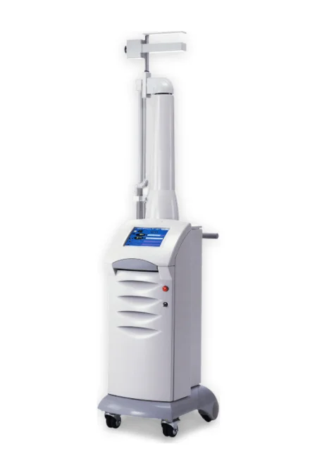
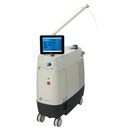
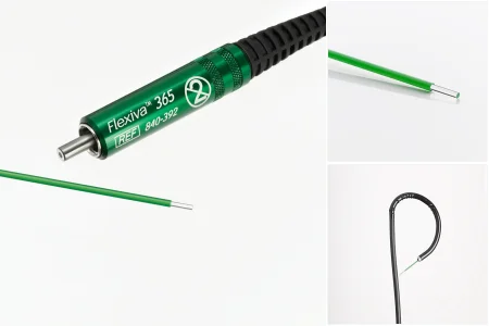
- Delivery system: carries the beam to the tissue. The carbon dioxideKAR-bun dy-OK-sideA gas made by the body's cells as waste, carried in the blood to the lungs and breathed out.Full entry laser uses an articulated (jointed) arm with mirrors at its joints, or a hollow waveguide, ending in a handpiece, a microscope or a scanner. Most other surgical lasers use a thin quartz fiber, through a handpiece, a scope or a microscope. Most fibers work without touching tissue (noncontact), while contact tips are held against it.
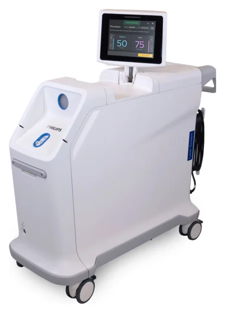
The surgeon fires the laser with a single foot pedal, and a nurse or surgical technologistSUR-jih-kul tek-NOL-uh-jistA member of the operating room team who prepares the sterile field and instruments and hands them to the surgeon.Full entry trained on the laser runs its console.
What makes laser light different
Ordinary light, such as a flashlight's, is made of many wavelengths, and its waves spread out in every direction and lose strength as they travel. Laser light has three properties:
- Monochromatic: all its waves have the same wavelength, so it is one color.
- Collimated: its waves travel parallel to one another and hardly spread, so a lens can focus the beam into a tiny, intense spot.
- Coherent: its waves are in step, peaks lined up with peaks, so their power adds together.
Together these let a laser pack a tremendous amount of energy into a very small area. That is what makes it precise, and what makes it dangerous.
How the beam acts on tissue
When a laser beam strikes something, four things can happen:
- Reflection: the beam bounces off. A mirror can aim it into a hard-to-reach spot on purpose, but a beam that bounces off a shiny instrument can strike the wrong tissue, an eye, or a drape or mask and set it alight.
- Scattering: the beam spreads out as it passes into tissue and grows weaker. The Nd:YAG beam can even scatter back up a scope.
- Transmission: the beam passes through clear fluid or tissue without heating it. The argon beam passes through the clear front of the eye to the retinaRET-ih-nuhThe thin layer of light-sensing tissue at the back of the inside of the eye.Full entry , and the Nd:YAG beam passes through fluid in the bladder.
- absorptionub-ZORP-shunThe taking in of a substance, such as a drug or a nutrient, into the bloodstream.Full entry : the tissue takes in the energy, and this is where the surgical effect happens.
Most surgical lasers work by heating tissue (a photothermal effect). As tissue heats, it first blanches as it coagulationkoh-ag-yuh-LAY-shunThe process by which blood changes from a liquid into a gel, forming a clot.coagul clot-ation processFull entry , then turns white or gray as its proteins change, then dries and puckers; above 212°F (100°C), the water in its cells boils, the cells burst and the tissue vaporizes and chars, giving off smoke. Some lasers work in other ways: by a chemical reaction in tissue that holds a light-sensitive dye (photochemical) or by a burst of mechanical energy, heard as a snap, that breaks tissue apart without much heat (photoacoustic).
How much energy a tissue absorbs depends on the laser's wavelength and power and on the tissue's color, density and water content. This selective absorption is why each laser has its uses. The argon, KTP and Nd:YAG beams are taken up by dark pigments such as hemoglobin and melaninMEL-uh-ninThe dark pigment that gives color to the skin, hair and eyes.melan/o black, dark-in substanceFull entry , while the CO2 beam is taken up by water, whatever the tissue's color.
The surgeon controls the effect with the power, the spot size and the time:
- Power density is the power in watts divided by the area of the spot. Focus the beam to a small spot and it becomes more intense and cuts deeper; widen it and the same power spreads out and coagulates.
- Total energy, in joules, is the power multiplied by the time. Fluence (radiant exposure) is the energy delivered per area of tissue.
- The same total energy does more damage around the target when it is given at low power over a long time than at high power briefly, so surgeons use the shortest exposure that does the job.
A laser can fire as a steady beam (continuous wave) or in bursts (pulsed, also called Q-switched).
Why surgeons use lasers
Depending on the procedure, a laser can:
- seal small blood vessels as it cuts, so there is less bleeding during and after surgery
- seal lymphaticlim-FAT-ikRelating to lymph, the clear fluid that drains from body tissues, or to the vessels and nodes that carry it.lymph clear body fluid-atic relating toFull entry vessels, which means less swelling afterward and less chance of spreading cancer cells through the lymphLIMFThe clear fluid that flows through the lymphatic vessels and carries white blood cells.Full entry
- seal nerve endings, which means less pain after some procedures
- sterilizationstair-uh-luh-ZAY-shunA process that destroys all microorganisms, including spores, on an item; also a permanent birth control procedure.Full entry the tissue it touches with its heat, and lower the chance of a surgical site infection
- cut precisely, with little damage to the tissue nearby and less scarring that could narrow a passage
- shorten the operation and the time under anesthesiaan-us-THEE-zhuhLoss of feeling or sensation, often brought on by drugs so that surgery or a procedure can be done without pain.an- withoutesthesi/o feeling, sensation-ia conditionFull entry , allow more procedures as outpatient surgery under local anesthesiaLOH-kul an-es-THEE-zhuhNumbing medicine applied to or injected into one small area, so a procedure there is not felt while the person stays awake.Full entry , and speed recovery
Lasers have also made many minimally invasive, endoscopicen-doh-SKOP-ikDone with an endoscope, a thin tube with a light and camera passed into the body.endo- within-scopic viewingFull entry , eye, skin and robotic procedures possible.
13The laser systems
The surgeon chooses the laser for the tissue and the procedure. These are the main ones.
| Laser | Medium and beam | What it does | Common uses |
|---|---|---|---|
| carbon dioxideKAR-bun dy-OK-sideA gas made by the body's cells as waste, carried in the blood to the lungs and breathed out.Full entry (carbon dioxide) | A gas; invisible infrared, 10,600 nm, so a red helium-neon aiming beam shows where it will strike; delivered by articulated arm, waveguide or microscope | absorptionub-ZORP-shunThe taking in of a substance, such as a drug or a nutrient, into the bloodstream.Full entry by water, whatever the tissue's color, to a very shallow depth (about 0.1 to 0.2 mm); does not scatter, and cannot work through clear fluid | Cutting, vaporizing and coagulationkoh-ag-yuh-LAY-shunThe process by which blood changes from a liquid into a gel, forming a clot.coagul clot-ation processFull entry soft tissue in many specialties, microsurgery included; airway work such as laser bronchoscopybrong-KOS-kuh-peeA procedure in which a thin, lighted tube with a camera is passed through the nose or mouth to look inside the airways.bronch/o airway (bronchus)-scopy looking with an instrumentFull entry ; with a scanner, skin ablationab-LAY-shunA procedure that destroys a small area of tissue, often with heat or cold, to stop it from causing a problem.Full entry and gynecologygy-nuh-KOL-uh-jeeThe branch of medicine that cares for the female reproductive organs and breasts.gynec/o woman-logy study ofFull entry vaporization |
| Argon | A gas; visible blue-green light, delivered by fiber | Absorbed by red and brown pigment such as hemoglobin; passes through clear tissue and fluid; coagulates and seals | Eye surgery (retinalRET-ih-nulRelating to the retina, the thin layer at the back of the eye that senses light and sends images to the brain.retin/o retina-al relating toFull entry tears, diabetic retinopathydy-uh-BET-ik ret-ih-NOP-uh-theeDamage to the small blood vessels of the retina caused by diabetes.retin/o retina-pathy diseaseFull entry , glaucomaglaw-KOH-muhAn eye disease in which the optic nerve is damaged, usually with raised pressure inside the eye, leading to loss of vision.Full entry , macular degenerationMAK-yuh-ler dih-jen-uh-RAY-shunAn eye disease in which the macula, the center of the retina used for sharp, straight-ahead vision, slowly breaks down.Full entry ) and pigmented skin lesionLEE-zhunAn area of tissue that has been damaged or has changed from normal, such as a sore, wound, lump or spot.Full entry |
| Krypton | A gas | Removes superficial lesions | Dermatology |
| Diode | A semiconductor crystal; compact and reliable; wavelengths vary | Depends on its wavelength | Eye and skin procedures |
| Tunable dye | A liquid dye excited by another laser; the wavelength can be tuned within a range of visible light | Some wavelengths are taken up strongly by pigment | Skin and eye procedures, and breaking up urinary stones |
| Nd:YAG (neodymium: yttrium aluminum garnet) | A solid crystal; invisible near-infrared, 1064 nm, with an aiming beam; fiber, with or without a contact tip | Penetrates deepest of these lasers (about 3 to 5 mm; one reference gives up to 7 mm) and coagulates best, sealing vessels up to about 4 mm; passes through clear fluid | Coagulating and vaporizing tumors and bleeding vessels through scopes, as in the bladder, the bowel, the airway and the uterus |
| KTP (potassium titanyl phosphate) | An Nd:YAG beam passed through a KTP crystal, which halves its wavelength to visible green, 532 nm | Absorbed by pigmented tissue; passes through clear fluid; a very fine beam suited to microsurgery | Ear, nose and throat, urology, gynecology, general surgery and dermatology |
| Holmium:YAG | A solid crystal; infrared, 2140 nm, invisible, with an aiming beam; delivered by fiber | Very shallow (well under a millimeter) and works in fluid; cuts, shaves, ablates and coagulates soft tissue | Breaking up kidney, ureterYOOR-uh-terOne of the two tubes that carry urine from the kidneys down to the bladder.Full entry and bile ductbyle duktOne of the tubes that carry bile from the liver and gallbladder to the small intestine.Full entry stones; also urology, orthopedicor-thuh-PEE-dikHaving to do with the bones, joints, muscles, ligaments and tendons, and the branch of medicine that treats them.orth/o straightped/o child (the field began with straightening children's bones)-ic relating toFull entry , ear, nose and throat, gynecology and general surgery, often through a scope |
| Erbium:YAG | A solid crystal; infrared, 2940 nm, pulsed | Strongly absorbed by water, so it works only at the surface | Skin resurfacing and dentistry |
| Excimer | A gas mixture ("excited dimer"); ultraviolet; its gases are toxicitytok-SIS-ih-teeThe harm a substance does to the body, or a state in which a drug has built up to a harmful level.tox/o poison-icity state or quality ofFull entry , so the unit is heavily enclosed | Breaks the bonds holding molecules together with very little heat, so it is extremely precise and chars little | Reshaping the corneaKOR-nee-uhThe clear, dome-shaped layer at the front of the eye, over the iris and pupil.Full entry , skin conditions, and clearing plaquePLAKA deposit that builds up on a surface in the body, such as a sticky film of bacteria on teeth or a fatty buildup in an artery wall.Full entry from narrowed arteries |
| Ruby and alexandrite | Solid crystals; synthetic ruby was the first lasing medium | Removes marks at the skin's surface | Tattoos and other skin marks; alexandrite has largely replaced ruby |
14Laser safety
A laser's risks to patients and staff are eye injury, burns, fire and smoke. Eye injury is the greatest. All of them can be managed, but only when every member of the team follows every step.
Standards and the laser safety officer
- The standards: the American National Standards Institute's laser standards, ANSI Z136.1 and, for health care, ANSI Z136.3 (2018), published by the Laser Institute of America, are the main safety standards for lasers. Occupational Safety and Health AdministrationOH-shuh (said as a word)The US federal agency, part of the Department of Labor, that sets and enforces rules to keep workplaces safe.Full entry uses the ANSI standards to judge a facility's laser safety, the FDA sets performance standards for laser products, and The Joint Commissionthuh JOYNT kuh-MISH-unAn independent US organization that inspects and accredits hospitals and other health care organizations against safety and quality standards.Full entry checks that each facility has a laser safety program. AST's laser safety guideline builds on these.
- The laser safety officer (LSO): every facility that uses lasers is expected to have an LSO, a trained and certified person given the authority to run the laser safety program. The LSO classifies the lasers, sets the hazard zone for each, approves procedures and protective equipment, makes sure warning signs are posted and training is done, keeps the laser log, investigates incidents and audits the program every year.
- The laser safety committee: a group from every profession involved, a CSTsee-es-tee (said as letters)Certified Surgical Technologist, a credential for surgical technologists; in maternity care, contraction stress test.Full entry among them, that writes the policies, reviews incidents and decides who may use lasers.
- Training: everyone who works with high-powered lasers completes laser safety training when they start and every year, and only people trained in the department's laser safety may be in the room while a laser is in use. A CST with at least 3 years of documented laser experience can train and certify to become an LSO.
- Records: each laser use is documented in the patient's record: the laser, its settings, the energy used and the time it was on, the protection for the patient's eyes, the team, and the safety measures taken.
Laser classes
Lasers are classed by how much harm they can do. The ANSI classes are:
| Class | Hazard | Examples |
|---|---|---|
| 1 | No hazard | Laser printers, bar code scanners, CD players |
| 1M | No hazard to the naked eye, but can be one when viewed through optics such as a microscope | Distance-measuring instruments |
| 2 | Low-power visible lasers; the natural blink protects the eye, but staring into the beam for a long time can harm it | Bar code scanners and some laser pointers |
| 2M | Like class 2 to the naked eye, but hazardous through optics | Distance-measuring instruments |
| 3R | A lower risk of eye injury than the other class 3 lasers | Some laser pointers and laboratory lasers |
| 3B | Harms the eye viewed directly or by mirror-like reflection; generally not by scattered light, and generally not a fire hazard | Laboratory lasers and some medical lasers, such as the Nd:YAG lasers used inside the eye |
| 4 | Harms the eye directly and by any kind of reflection, and is a serious skin and fire hazard | Most surgical lasers, and industrial lasers |
Lasers once classed as class 3a are treated as class 3R; one reference still uses the old name, class 3a.
A class 3B or 4 beam can injure the retinaRET-ih-nuhThe thin layer of light-sensing tissue at the back of the inside of the eye.Full entry in an instant, and the damage may never heal. Class 4 lasers can even pass through the white of the eye to the retina. Turning your head away or closing your eyes does not protect you, because the beam scatters and reflects.
Protecting the eyes
Which part of the eye a laser injures depends on its wavelength. The carbon dioxideKAR-bun dy-OK-sideA gas made by the body's cells as waste, carried in the blood to the lungs and breathed out.Full entry beam is absorptionub-ZORP-shunThe taking in of a substance, such as a drug or a nutrient, into the bloodstream.Full entry by the water at the eye's surface and burns the corneaKOR-nee-uhThe clear, dome-shaped layer at the front of the eye, over the iris and pupil.Full entry , which hurts at once. The argon and Nd:YAG beams pass through the clear parts of the eye and are focused by its lens onto the retina, causing permanent damage that may cause no pain at all.
- Laser protective eyewear: glasses or goggles made for laser use, with side shields, and lenses rated for the exact wavelength in use. The wavelength and the optical density (how strongly the lens blocks that wavelength) are printed on the eyewear; Nd:YAG goggles, for example, might read "1064 nm, ODoh-DEE (said as letters)An abbreviation that has been used for right eye, for once daily and for overdose.Full entry 5."
- Read the label, not the color. The color or darkness of a lens tells you nothing about its protection. Check the wavelength and optical density every time, especially when the department has more than one laser; AST then recommends color-coding the eyewear to match each laser's handpiece, as a reminder.
- Fit: eyewear wraps around the eyes, covering the sides, top and bottom. Ordinary prescription glasses and contact lenses are not laser protection; wear proper laser eyewear over or instead of them.
- Inspect it before every use for cracks, pitting, scratches, discoloration or a damaged coating, and check the frame and strap. Damaged eyewear is thrown away and reported to the LSO.
- Everyone in the zone wears it while the laser is in use, including during scope procedures, when the beam seems contained. Eyewear for that laser is kept outside every entrance to the room.
- The patient's eyes: under general anesthesiaJEN-er-ul an-es-THEE-zhuhDrug-induced loss of consciousness and feeling over the whole body, so a person feels nothing and is not aware during surgery.an- withoutesthesi/o feeling, sensation-ia conditionFull entry , the closed eyes are covered with moistened eye pads; an awake patient wears laser glasses or goggles. When the laser is used near the eye, a special laser eye shield is placed on the eye itself, and a steam-sterilizationstair-uh-luh-ZAY-shunA process that destroys all microorganisms, including spores, on an item; also a permanent birth control procedure.Full entry shield must be fully cooled first.
- Scopes and microscopes: a lens filter goes over the eyepiece of a scope, and a filter or shutter on the microscope protects the surgeon looking through it.
- Eye exams: AST recommends an eye exam before working with high-powered lasers, and another within 48 hours of any incident that may have affected the eyes.
Protecting the room and the skin
- The nominal hazard zone (NHZ): the space in which the direct, reflected or scattered beam is stronger than the maximum permissible exposure, the most laser energy a person can take without harm to the eyes or skin. The LSO works it out, but because the settings change during a case, the whole room is usually treated as the hazard zone.
- Signs and doors: OSHA requires warning signs, so a laser sign goes on every door into the room while the laser is in use, and the doors stay closed. The signs come down or are covered when no laser is in use.
- Windows: the argon and Nd:YAG beams, among others, pass straight through ordinary glass, so for them the windows and door ports are always covered with a barrier that blocks that wavelength. A laser with a short hazard zone, such as a CO2 beam focused through a lens, may not need it, but many facilities cover the windows for every laser case: your facility's policy sets it.
- The key: the laser key is kept secure, given only to authorized people, and never left in the laser in storage.
- Reflection: shiny surfaces are covered or removed. Instruments used near the beam are anodized or ebonized, coated with a dark, rough, nonreflective finish, and large shiny retractorrih-TRAK-terA surgical instrument that holds back tissue or organs so the surgeon can see and reach the operative site.Full entry are covered with wet towels or sponges.
- Skin: a direct hit from the beam burns. The team's gloves and gown protect their skin, the circulator wears a flame-resistant jacket that covers the arms, and everyone takes off metal jewelry, which can reflect the beam or heat up. Wet towels go around the patient's surgical site, and body openings such as the nose or rectum may be packed with wet sponges.
- Electrical safety: a laser runs on high voltage. Never set fluids on it, keep the floor and the foot pedal dry, check that it is grounded and its cords are sound, and leave its housing to trained service staff.
- Moving it: some lasers are very heavy. Push one rather than pull it, using good body mechanics to protect your back, and never bump it or its arm into a wall or doorway, which can damage it and knock its beams out of alignment.
Preventing laser fires
A class 3B or 4 laser is always a serious ignition source, and in an oxygen-enriched atmosphere no material is safe from it. Everything in the fire section applies, plus these laser steps:
- Standby: the laser stays in standby at all times except when the surgeon is holding the handpiece and ready to fire.
- Water and wet materials: a basin of sterile water is on the field. No dry towels or sponges go near the site: the towels around the incision are moistened with sterile water, sponges are wet, and you keep checking that they stay wet. Foam positioning devices are avoided, or wetted.
- Drapes: fire-resistant drapes are recommended, but they still burnBURNAn injury to the skin or deeper tissues caused by heat, chemicals, electricity, radiation or friction.Full entry . Make sure the drapes are fixed so they cannot shift into the beam's path.
- Prep and hair: no flammable solution on the field, and the prep dry with no pooling. A tinted, iodineEYE-uh-dyneA mineral the thyroid gland uses to make thyroid hormone; it is also used to kill germs on the skin and in some contrast dyes.Full entry -based prep is wiped off where the argon or Nd:YAG beam will strike, because the color makes the skin absorb more of the beam. Hair near the site is covered with wet sponges or towels, and neither patient nor team wears hair spray or styling gel.
- Teeth: in laser surgery near the teeth, a nonflammable tooth guard protects them.
- Near the rectum: methane in the bowel can explode. The bowel is emptied before surgery, the gas is suctioningSUK-shun-ingRemoving mucus, saliva or other secretions from the mouth, nose or airway with a tube connected to a suction source.Full entry out before the laser is used, and a counted wet sponge pack may close off the rectum.
- Fibers: never lay a hot fiber tip on the drapes. Cover the tip with a wet sponge when it is not in use.
- The airway: in laser surgery of the mouth, throat or airway, the anesthesia provideran-us-THEE-zhuh pruh-VY-derThe licensed professional who gives anesthesia and watches over the client's breathing and vital functions during a procedure.Full entry uses a laser-resistant breathing tube made for that laser, within its power limits. Its cuff is filled with saline tinted with methylene blue: the saline soaks up heat, and the blue shows at once if the beam bursts the cuff. Counted wet pledgets are placed around the tube and kept wet, the oxygen is kept between 21 and 30 percent, and nitrous oxide is not used.
Some references describe wrapping a standard breathing tube in metallic foil before laser surgery of the airway, and a 2014 textbook fills the cuff with sterile water and names a halon extinguisher for laser fires.
The laser safety checklist
AST recommends a laser time-outTYM-owtA pause just before a procedure when the whole team confirms the right client, procedure and site; also, a short break to calm a child.Full entry , held in addition to the surgical time-out, before the surgeon first fires the laser. It is done on a checklist that goes into the patient's record, and it is repeated whenever a team member is replaced (the anesthesia provider, the CST or the circulator) or after a long wait with the laser in standby, such as for a frozen section result.
The checklist confirms, among other things:
- The laser: the type of laser, its mode and power in watts, and the type of fiber; the laser test-fired, and the fiber checked as intact.
- The room: laser warning signs on the doors, eyewear in the room, windows covered, the key controlled, and the fire extinguisher in the room and full.
- The patient: eye protection in place, a nonflammable breathing tube when the airway is involved, a wet rectalREK-tulRelating to the rectum, the last part of the large intestine; also, given or measured through the rectum.rect/o rectum, straight-al relating toFull entry pack when needed, and sponges and towels wet.
- The team: everyone in the room wearing the right eyewear, nonreflective instruments on the field, a basin of water open, and the plume evacuator tested and ready for the CST.
- Afterward: the laser documentation completed, the key returned, the laser and evacuator cleaned and stored, and any malfunction reported.
The laser does not fire until the checklist is complete.
The scrub's role with a laser
- Set up and test. AST makes the CST responsible for aligning the laser's optics before it is used. Before the patient comes in, align and test-fire the laser according to its maker's instructions, with the delivery device attached. With the CO2 laser, the red aiming beam must line up with the invisible working beam, or the beam will strike somewhere other than where the surgeon aims. A wet tongue depressor or special test paper makes a good target, with a wet towel under it and nothing flammable behind it. A laser that is out of alignment is not used until it is fixed.
- Eyewear: check the optical density and wavelength on the eyewear for this laser, and inspect it.
- Instruments: set up anodized or ebonized instruments. Check their coating for scratches, never etch them, and watch where they are in relation to the beam. If the surgeon needs a shiny instrument, wrap it in saline-soaked radiopaqueray-dee-oh-PAYKBlocking x-rays, so the material shows up white on an x-ray image.radi/o radiation, x-rayopaque not letting throughFull entry sponges.
- Backstops: with the CO2 laser, the beam keeps going after it passes through the target, so a backstop goes behind it. Use titanium or quartz rods (quartz lets argon and Nd:YAG beams through), never glass, which shatters, metal, which heats up, or nonstick rods, which melt and give off toxicitytok-SIS-ih-teeThe harm a substance does to the body, or a state in which a drug has built up to a harmful level.tox/o poison-icity state or quality ofFull entry fumes.
- Mirrors: use laser mirrors of rhodium or stainless steel, never glass, and check them for scratches.
- Fibers: fibers are delicate. Never bend, clamp or lean on one, because a broken fiber can leak energy and set its sheathSHEETHA covering that wraps around a structure; in procedures, a short hollow tube left in a blood vessel to pass catheters through.Full entry on fire. Check that the fiber is long enough for its catheter, protect its sharp tip in a short sheath while it passes through the scope, and make sure the tip sticks out at least 1 cm beyond the end of the scope and is in view before the laser fires (one reference asks for more than an inch). Inspect the fiber and its sheath at setup, every time they come out of the patient and at the end; if either is not intact, tell the team at once and follow the retained-item policy. Never use air or gas to cool a fiber inside the uterus: gas has caused fatal gas embolismEM-buh-liz-umThe blocking of a blood vessel by a mass carried there in the blood (an embolus), such as a clot, fat or an air bubble.embol/o plug-ism conditionFull entry .
- Smoke: hold the evacuator wand within 2 inches of the target and out of the beam's path, and work the evacuator's foot pedal.
- Foot pedals: the laser pedal belongs only to the surgeon firing the laser. Whoever places it names it, and where it is, aloud to the surgeon. Other pedals are placed away from it and named too, and the laser is disabled while other pedal-controlled equipment is used.
- If something goes wrong: the laser and any other equipment involved are kept, with their settings untouched, for the LSO and risk manager to investigate.
What to take from this chapter
- Electricity needs a complete circuit and takes the path of least resistance. Where current meets resistance it turns to heat, and the more concentrated the current, the hotter the spot.
- Electrosurgery heats tissue with high-frequency current to cut, coagulate (desiccate or fulgurate) or blend. Cut uses the lowest voltage, so use cut or blend when either will do. Eschar on the tip makes heat, sparks and fire.
- Monopolar current runs through the patient to the return electrode; bipolar current runs only between the forceps tips and needs no pad. Bipolar is the choice for delicate tissue and for patients with implanted electronic devices.
- The return electrode works by spreading the current over a wide area. Poor contact or a missing return path burns the patient. Never tape or cut a pad; chapter 31 places it.
- Control the active electrode: holster it, keep the tip clean (never with a blade), keep its cord uncoiled and away from other cords, use the lowest power, let only the user fire it, and keep the tone audible. Repeated requests for more power mean check the circuit, not turn it up.
- In laparoscopy, insulation failure, direct coupling and capacitive coupling burn out of sight. Scan the insulation, avoid hybrid trocars, and fire only at the target.
- A fire needs an oxidizer, a fuel and an ignition source. Dry preps, wet sponges, low oxygen near the field, a holstered pencil and a laser on standby keep them apart; if a fire starts, stop the gases, remove what is burning and douse it with the saline or water on your back table.
- Evacuate surgical smoke every time, within 2 inches of its source, with a fit-tested N95 respirator as the backup. With lasers, wear eyewear marked for that wavelength and optical density, keep the field wet and complete the laser safety checklist.
Check yourself5 quick questions
Free for everyone. Pick an answer for each, then check them all at once.
In the ORWhere you'll use this
The chapter's whole question pool in sets of 10, with the answer explained after each one.
Part of the educators programThe chapter's words on spaced cards: the ones you miss come back sooner.
Part of the educators programGiven by your class's teacher. Each class draws its own 25 from the chapter's pool.
Part of the educators program
Suggest a fix
Something wrong, unclear or out of date in this chapter? Tell us and we will fix it for everyone. Your email is only used to reply.