Equipment
Tables, lights, suction and the rest of the room: what each piece is for, how to ready and test it, what to do when it fails, and how to care for it after the case.
1Why it matters
Most operating rooms hold the same core equipment: the OR table, instrument tables, ring stands, an electrosurgeryee-LEK-troh-SUR-jer-eeUsing high-frequency electric current through a handheld tool to cut tissue or seal bleeding vessels during surgery.Full entry , imaging equipment, an anesthesiaan-us-THEE-zhuhLoss of feeling or sensation, often brought on by drugs so that surgery or a procedure can be done without pain.an- withoutesthesi/o feeling, sensation-ia conditionFull entry machine and patient monitors. Every piece has its own place, so anyone on the team can find it fast.
The surgeon's preference card says what else the case needs. Before the case, you check that it is in the room and test it. During the case, you fix small problems and catch big ones. After it, you clean, store and send anything broken for repair. AST expects the surgical technologistSUR-jih-kul tek-NOL-uh-jistA member of the operating room team who prepares the sterile field and instruments and hands them to the surgeon.Full entry to do this with the team.
Equipment can hurt people. Faulty electrical equipment is one of the top causes of fires in American hospitals, and complex devices need specially trained technicians to keep them working safely. A table loaded past its limit, a cracked cord or an alarm turned down low can each injure a patient.
This chapter is about the equipment itself. Room temperature, humidity and air handling are in chapter 17. How electricity and electrosurgery work is in chapter 20, scopes and the robot in use in chapter 21, the C-arm and radiationray-dee-AY-shunEnergy that travels as waves or particles, such as X-rays, used to make images or treat cancer; also pain that spreads from its starting point.radi/o rays, radiation-ation processFull entry safety in chapter 22, and power instruments in chapter 24. Positioning the patient, padding and applying the SCD and tourniquet cuff are in chapter 31, and room turnover is in chapter 30.
2The OR table
The OR table, also called the OR bed, holds the patient and puts the surgical site where the surgeon can see it and reach it. Its stainless steel frame sits on a hydraulic lift, and its top is covered with firm pads that come off.
Its parts
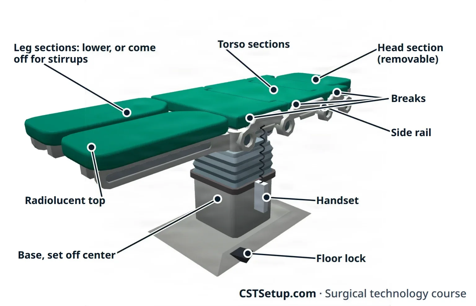
- Sections: a head section, a torso section and a leg section, each with its own removable pad. The torso section is fixed to the base, because it carries the heaviest part of the body.
- Breaks: the joints between sections, at the head, the middle and the foot. Raising or lowering a section is called breaking the table.
- Side rails: metal rails along both sides that stop at each break. Attachments and some retractorrih-TRAK-terA surgical instrument that holds back tissue or organs so the surgeon can see and reach the operative site.Full entry clamp onto them.
- Removable head and foot sections: the head section lifts off so a special headrest can slide into its place, and the foot section is lowered or removed when the legs go into stirrups.
- Kidney lift (kidney bridge): a crossbar at the middle break that rises under the patient for kidney or gallbladder surgery. Kidney rests, braces that slide onto it, hold a patient on the side.
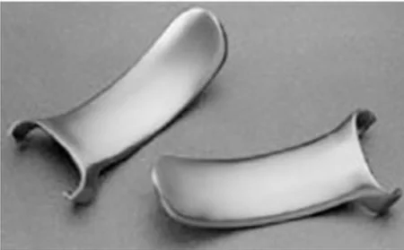
- perinealper-ih-NEE-ulHaving to do with the perineum, the area between the genitals and the anus.Full entry cutout: a curved notch at the lower end of the torso section, so the perineumpair-ih-NEE-umThe area of skin and muscle between the genitals and the anus.perine/o perineumFull entry can be reached in lithotomylih-THOT-uh-meeA position lying on the back with the hips and knees bent and the legs raised and spread apart, often in stirrups.lith/o stone-tomy cutting intoFull entry . A drainage tray can hang from it.
- Radiolucent top: a top that x-rays pass through, so images can be taken with the patient on the table.
- Base and floor locks: the base may sit off center to leave room for x-ray and C-arm equipment. The floor lock, worked by a foot pedal or the handset, holds the table in place.
- Handset: the hand control that raises, lowers, tilts and flexes the table and moves each section.
How it moves
The table rises and lowers, tilts to either side, tips head down (Trendelenburg positiontren-DEL-en-burg puh-ZISH-unA position in which the client lies on the back with the bed tilted so the head is lower than the feet.Full entry ) or head up (reverse Trendelenburg positionree-VERS tren-DEL-un-burg puh-ZISH-unA position lying on the back on a bed or table that is tilted so the head is higher than the feet, with the body kept straight.Full entry ), and flexes at its breaks. The top can also rotate.
Moving the head section to the leg end of the table gives a C-arm more room around the chest and abdomen, because that end reaches farther out from the base. Keep the patient's center over the base, where the table is most stable.
The circulator or the anesthesia provideran-us-THEE-zhuh pruh-VY-derThe licensed professional who gives anesthesia and watches over the client's breathing and vital functions during a procedure.Full entry usually works the handset. Before any section moves, confirm where the patient's hands are: fingers can be crushed in a break as it closes.
Weight limits
- Every table has a weight limit, marked on the table and obeyed. Most hold about 500 pounds (227 kg). Some newer and bariatric tables hold 1000 to 1200 pounds.
- The joints of the table have to carry the patient too, so check the table's specifications before the patient is moved onto it.
- AST is clear: if the patient weighs more than the maker's limit, that table or positioning device is not used.
- For a patient with obesity there are bariatric tables, side extensions that lock into the rails to widen the table, mattress pads rated for the weight, and longer and wider safety straps (sometimes two). The stretcher and lifting equipment must be rated for the weight as well.
Never put a patient on a table or attachmentuh-TACH-mentThe deep emotional bond between an infant and a parent or caregiver.Full entry rated for less than their weight.
Attachments
Attachments clamp onto the side rails or slide into the ends of the table. Each one is checked before the patient arrives: clean and in working order. AST also says to use each positioning device only for the position it is made for, as its maker says, and never to modify one unless its maker has approved the change.
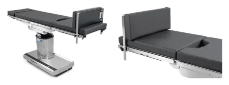
- Armboard: holds the arm out from the body at less than 90 degrees, so anesthesiaan-us-THEE-zhuhLoss of feeling or sensation, often brought on by drugs so that surgery or a procedure can be done without pain.an- withoutesthesi/o feeling, sensation-ia conditionFull entry can reach the IV and monitors. An arm strap or cradle keeps the arm on it. Lock it on a solid stretch of rail, away from a break.
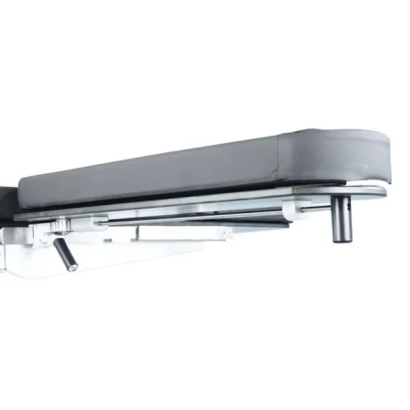
- Footboard: a platform at the foot of the table. It lengthens the table for a tall patient, or folds up, padded, to support the feet when the patient is tipped steeply head up.
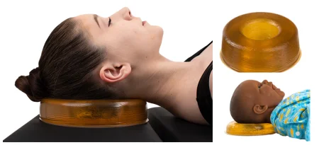
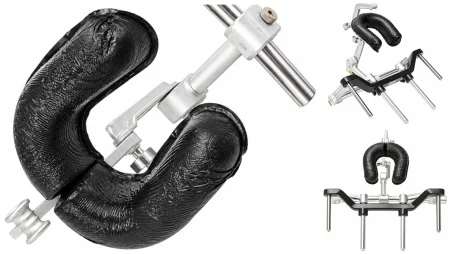
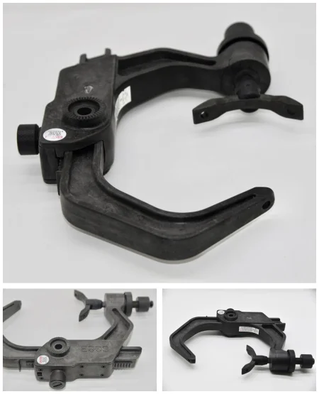
- Headrests: a donut pad supports the head in line with the body and takes pressure off the back of the skull. The horseshoe headrest is a padded U that supports the forehead in the prone position, or the head for cranialKRAY-nee-ulRelating to the skull, or toward the head end of the body.crani/o skull-al relating toFull entry surgery. Pin holders hold the head still with sterile pins fixed in the skull, for neurosurgery and cervicalSER-vih-kulRelating to the neck, or to the cervix, the lower end of the uterus.cervic/o neck, cervix-al relating toFull entry spine surgery.
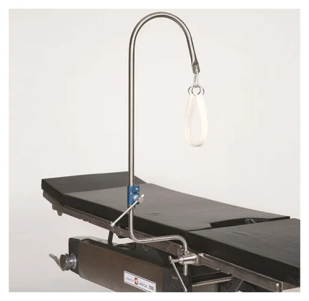
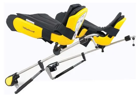
- Stirrups: lift and spread the legs so the perineum can be reached in lithotomy. Candy cane stirrups hold the feet in slings. The feet are padded, and the legs must not touch the upright posts, which can injure the peronealper-uh-NEE-ulHaving to do with the outer side of the lower leg, near the fibula bone.Full entry nerve. Boot stirrups cradle the foot and calf and spread the pressure more evenly. Both stirrups are attached at the same height.
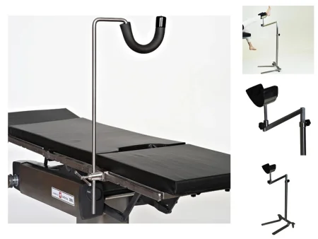
- Leg holder: supports one leg, for example the operative leg in knee arthroscopy. It is padded along its whole length.
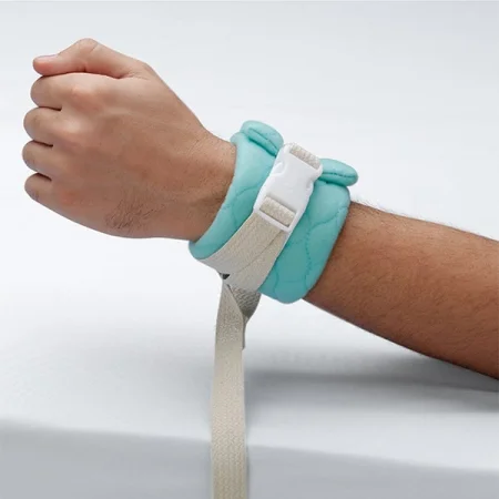
- Safety strap and restraintrih-STRAYNTAny device, drug or method used to limit a person's movement or behavior that they cannot easily remove or refuse.Full entry : the safety strap goes across the thighs about 2 inches above the knees as soon as the patient is on the table, firm but never pressing into the skin. One end is fastened to the table before the patient comes in. Arm straps and other soft restraints keep a limb on its support.
- Anesthesia screen: an attachment at the head of the table. The top of the drape is raised over it or fastened to it to form a screen, which keeps the patient's head outside the sterile fieldSTAIR-ul feeldAn area set up with sterile drapes and supplies, kept free of all germs, for a sterile procedure.Full entry while the anesthesia provider can still reach the airway.
- Shoulder braces: these were long used to keep a patient from sliding in steep head-down positions. They can injure the brachial plexusPLEK-susA network of nerves or blood vessels that branch and join together.Full entry , the network of nerves that serves the arm, so they should not be used. To stop sliding, the current recommendation is a vacuum positioner approved for that position, or a gel overlay with the patient lying directly on it, no sheet in between, so the patient does not slip.
AST's 2011 positioning standard says to avoid shoulder braces when possible, and if they must be used, to pad them well and place them on the outer part of each shoulder, away from the neck.
Positioning aids
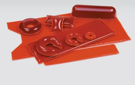
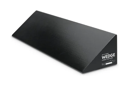
- Gel and foam pads come in many shapes: head donuts, wedges that tilt the body, arm pads, sacralSAY-krulHaving to do with the sacrum, the triangle-shaped bone at the base of the spine between the hip bones.Full entry pads that take pressure off the tailbone, and abductionab-DUK-shunMoving a limb or part away from the midline of the body, such as raising an arm out to the side.ab- away fromduct to lead-ion actionFull entry pads that keep the legs apart after hip fracturehip FRAK-churA break in the upper part of the thigh bone, near the hip joint.Full entry surgery so the hip does not dislocationdis-loh-KAY-shunAn injury in which the ends of bones that meet at a joint are forced out of their normal position.Full entry . A prone head pad is larger: it cradles the head and frames the face, and its hollow center gives access to the airway and keeps pressure off the eyes, ears and facial nerves. Anti-skid pads keep the patient from sliding on the table.
- Chest rolls lift the chest off the table in the prone position and keep the body aligned. A prone positioner raises the chest and the hips so the spine can be reached.

- Vacuum (bean bag) positioner: a sealed pouch filled with small plastic beads. It is shaped around the patient, then suction draws the air out so it sets firm and holds the position. Because it is firm, it raises pressure on the skin it touches and with it the risk of a pressure injuryPRESH-er IN-juh-reeDamage to the skin and the tissue beneath it from pressure, or pressure with shear, usually over a bony area or under a device.Full entry , so some models have a gel layer to soften it.
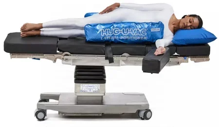
- Arm sleds and pegboards: arm sleds hold padded arms at the patient's sides. A pegboard steadies a patient in the lateral (side-lying) position, for example holding the pelvis still for hip surgery.
Specialty tables
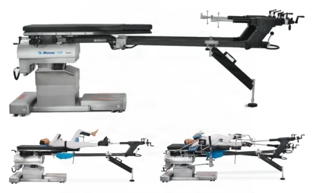
- Fracture (orthopedicor-thuh-PEE-dikHaving to do with the bones, joints, muscles, ligaments and tendons, and the branch of medicine that treats them.orth/o straightped/o child (the field began with straightening children's bones)-ic relating toFull entry ) table: the patient lies supine with the operative foot in a padded tractionTRAK-shunA steady pulling force applied to a body part with weights, ropes and pulleys.Full entry boot, so the leg can be pulled into traction, rotated or released. The other leg rests on a raised leg holder. A padded center post sits at the perineum and must never press on the genitals. The open frame lets fluoroscopyfloo-ROS-kuh-peeAn x-ray method that shows moving pictures of the inside of the body in real time.fluor/o glowing (from the fluorescent screen)-scopy looking, viewingFull entry see the hip. It is used for hip fractures and femoralFEM-er-ulRelating to the thigh or the femur, the thigh bone.Full entry nailing. Its several moving parts can injure the patient or the team if handled wrong, so you need to understand it well before you set it up.
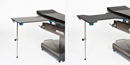
- Hand table: a padded, radiolucent extension at the side of the OR table. The arm lies on it at no more than 90 degrees, and the surgeon and assistant sit to operate. Only its draped top surface counts as sterile.
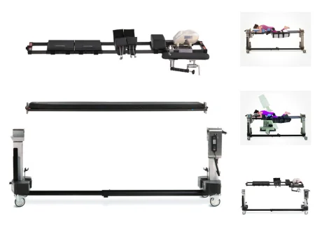
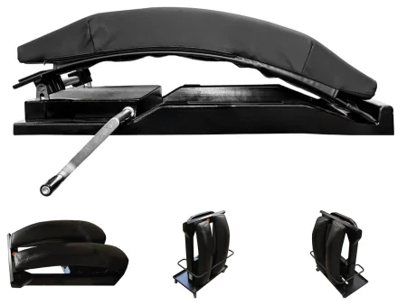
- Spine tables and frames: these hold the prone patient with the abdomen hanging free, so the big vessels and the diaphragmDY-uh-framThe dome-shaped sheet of muscle under the lungs that is the main muscle of breathing; also the flat side of a stethoscope.Full entry are not squeezed. That helps breathing and cuts bleeding. A Wilson frame is an adjustable frame set on the OR table for surgery on the back of the thoracicthuh-RAS-ikRelating to the chest, or to the middle part of the spine.thorac/o chest-ic relating toFull entry or lumbarLUM-barHaving to do with the lower back, the part of the spine between the ribs and the pelvis.Full entry spine. It is lowered all the way before the patient is turned onto it. Raising it bends the lower back forward, which opens the gaps between the laminae (the bony arches at the back of the spine) for a laminectomy or discectomy, but it is generally avoided for a lumbar fusion. On the frame, pad the bony prominences and make sure the breasts and genitals are not pinched. A Jackson table is a spine table with hinged parts for prone and semi-kneeling positions. It also leaves room for the C-arm, for fluoroscopy of the spine during surgery.
- Urology (cysto) table: has a built-in drainage tray (drain pan) and x-ray equipment, leg holders for lithotomy, and a light lower half that comes off easily. Because most cysto procedures run fluid through the bladder, the cysto room is designed with a fluid waste system, which can drain the fluid straight into the facility's waste lines.
Moving the table and the patient
Before you move the table, make sure its wheels are unlocked: pushing a table that is still locked causes a harsh jerk that can injure you. Never move one alone, and push it rather than pull it. Once it is in place, lock it.
Before a transfer, lock the wheels of both the stretcher and the table. Then use a transfer device so nobody drags the patient across. AST recommends a roller or other lifting device to move a patient onto the table.
- Transfer board: a thin, firm plastic board slid under the patient.
- Roller board: rollers inside heavy plastic fabric. Covered with a sheet, it lets the patient roll across without frictionFRIK-shunThe rubbing of one surface against another, such as skin against bed sheets.Full entry .
- Air-assisted transfer device: floats the patient on a thin cushion of air, and is inflated again after surgery to move the patient to the recovery bed.
- Lifts and friction-reducing sheets: a supine patient who weighs more than 157 pounds (71 kg) is moved with a mechanical lift, a mechanical lateral transfer device or an air-assisted device, and at least 3 or 4 caregivers.
On the stretcher, the side rails stay up and the safety strap stays on during transport. How to transfer and position the patient is chapter 31.
3Furniture
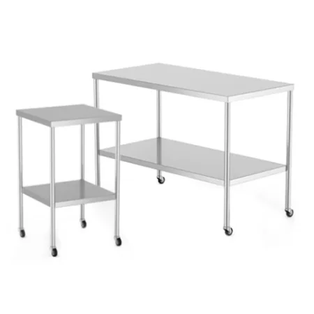
- Back table: a large stainless steel table that holds every sterile instrument and supply not in immediate use. Some cases need two.
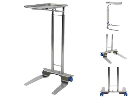
- Mayo standMAY-oh standA small tray on an adjustable stand placed over or beside the patient during surgery to hold the instruments used most often.Full entry : a smaller table with one open end, so it slides over or beside the patient. It holds what the surgeon needs next. Its height can be adjusted, for example with a foot pedal.
- Ring stand: a wheeled stand with one or two rings that hold basins for sterile water or saline. The wrapped basin sits in the ring and is opened there.
- Prep stand: a small table for the skin prep tray. Other small tables hold power equipment and supplies too bulky for the back table.
- Kick bucket: a stainless steel bucket on a low wheeled frame you can move with your foot. It is lined with an impervious biohazard bag and takes used counted sponges only, never wrappers or trash. During the case it sits near the scrub so sponges can be dropped in, not thrown.
- IV poles, hampers and stools: IV stands and sitting stools are part of the room's furniture, and the linen and trash hampers hold biohazard bags.
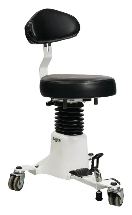
How to set up the back table and Mayo stand is chapter 28.
4Lights
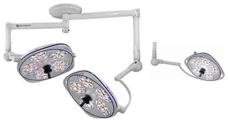
Ceiling lights
- Most rooms have two overhead surgical lights. Rooms for heart surgery, trauma and neurosurgery may have four.
- They give intense, focused light with almost no shadow and little heat, close to the color of daylight. Their brightness and the size of the beam can be adjusted.
- Most use LED or halogen lamps. LEDs are replacing halogen because they run cooler and use less energy. They are cooler than older lights but still a fire risk.
- Each light head takes a sterile handle in its center, or keeps a permanent handle under a sterile cover, so the sterile team can aim it. Putting on light handles is chapter 28.
- Turn the dimmer down before you switch a light off, which keeps its bulbs from blowing.
- Before the case, check that the lights work and swing them close to where they will shine.
Headlights and portable lights
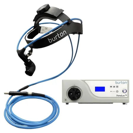
A fiber-optic headlight is worn by the surgeon to put a small, intense beam into a deep space the ceiling lights cannot reach. Its cable runs to a light source on a cart. A lighted retractorrih-TRAK-terA surgical instrument that holds back tissue or organs so the surgeon can see and reach the operative site.Full entry uses the same kind of cable, and a third light or a headlight helps when there is a second field, such as a leg where a vein is taken for a heart bypass.
Light sources and cables
- A light source sends light down a fiber-optic cable to a scope, a headlight or a lighted retractor.
- The glass fibers in the cable break if it is dropped or bent tightly. Look for cuts, nicks and dents, and never use a damaged cable.
- Turn the light source off, or to standby, before the cable is connected or disconnected, and whenever the cable is not attached.
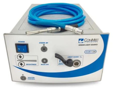
Never lay a lit cable on the drapes, the sponges or anything else that burns. Light from the loose end of a cable is hot enough to set drapes on fire. Keep the light source itself away from anything that burns.
5The video tower
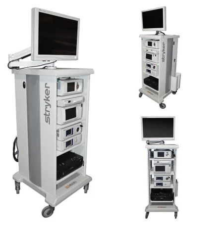
laparoscopylap-uh-ROS-kuh-peeSurgery or an exam done through small cuts in the belly, using a thin, lighted tube with a camera.lapar/o abdomen-scopy looking with an instrumentFull entry , arthroscopy and other scope cases need a video tower: a cart that carries the imaging units on shelves, with power for all of them. In an integrated OR the same units hang from ceiling booms instead.
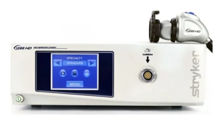
- Camera control unit (the camera box): the socket for the camera cable. It holds the controls for light intensity, white balance and resolution, and turns the camera's signal into the picture on the monitor.
- Monitors and recorders: show the picture to the whole team and can record the case or save still images.

- Light source: sends light down the cable to the scope (endoscopeEN-duh-skohpA thin tube with a light and a camera that is passed into the body to look inside an organ or cavity.endo- within-scope instrument for viewingFull entry ), as described above.
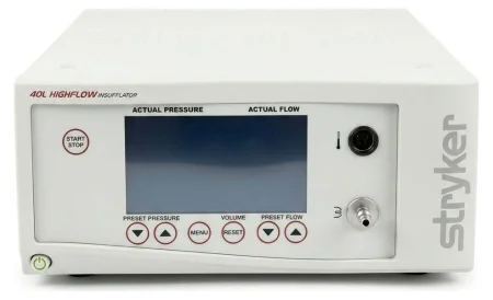
- insufflationin-suh-FLAY-shunBlowing a gas into a body cavity, such as carbon dioxide into the abdomen during laparoscopic surgery.Full entry and carbon dioxideKAR-bun dy-OK-sideA gas made by the body's cells as waste, carried in the blood to the lungs and breathed out.Full entry source: the insufflator fills the abdomen with carbon dioxide (CO2) gas to make room to see and work, a step called insufflation. The insufflator may warm the gas, which helps keep the patient warm and the lens from fogging.
- Shaver system: the console that drives the motorized shaver blades and burrs used in arthroscopy.
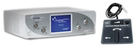
- Thermal energy source: the generator for an advanced energy device, such as a bipolarby-POH-lerHaving two poles or two opposite ends; most often used as a short form of bipolar disorder.bi- twopolar having polesFull entry vessel sealer, which uses bipolar current, low voltage and pressure to weld a vessel shut, or ultrasonic shears.
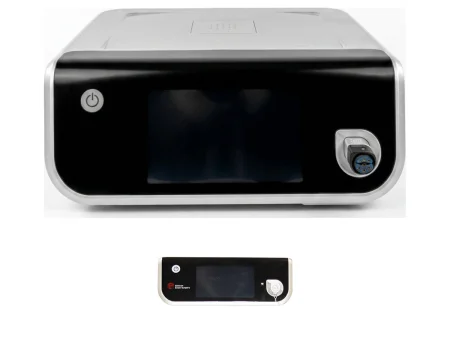
Checking the insufflator
Use only medical-grade CO2, from a labeled tank. Check the gas level before the case, change a tank before it runs low, and keep a spare in the room. Place the insufflator above the level of the abdomen and watch its pressure all through the case. The machine also watches it: it stops filling at the set pressure and lets gas out if the pressure climbs too high.
Connecting the scope, camera and light cable, and insufflating the patient, are chapter 21.
6Keeping the patient warm or cool
anesthesiaan-us-THEE-zhuhLoss of feeling or sensation, often brought on by drugs so that surgery or a procedure can be done without pain.an- withoutesthesi/o feeling, sensation-ia conditionFull entry , cold fluids and long surgery all chill a patient, and several machines help hold the body at a normal temperature. Watching the patient's temperature and malignant hyperthermiamuh-LIG-nunt hy-per-THER-mee-uhA rare inherited reaction to certain general anesthesia drugs in which the muscles' metabolism speeds up out of control.mal- badhyper- above normaltherm heat-ia conditionFull entry are chapter 13.
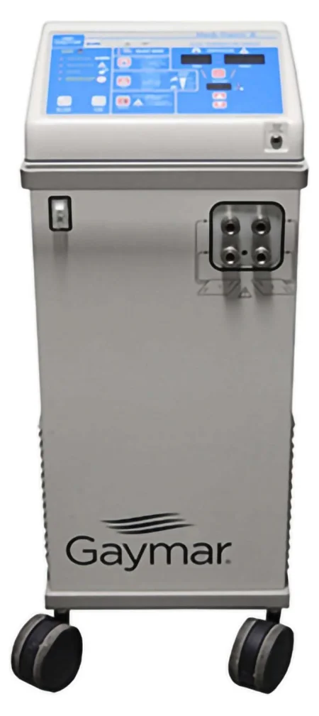
- hyperthermiahy-per-THER-mee-uhA body temperature that is higher than normal because the body takes in or makes more heat than it can lose.hyper- abovetherm/o heat-ia conditionFull entry and hypothermiahy-poh-THER-mee-uhA body temperature that has fallen below the normal range.hypo- belowtherm/o heat-ia conditionFull entry unit: pumps warmed or cooled distilled water (some use alcohol) through coils in a pad, a blanket or a mattress, usually placed on the table under the patient. The same unit can warm a cold patient or cool a hot one.
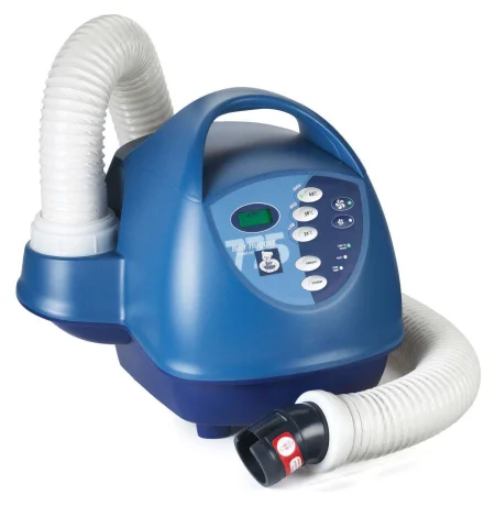
- Forced-air warming: a machine blows warm air through a hose into a paper and plastic blanket laid over the patient. Check the temperature setting and the hose-to-blanket connection before it is turned on, and again during the case: a hose that slips off blows hot air straight onto the skin and can burnBURNAn injury to the skin or deeper tissues caused by heat, chemicals, electricity, radiation or friction.Full entry it. Use it as its maker directs.
- Fluid warmers: warm IV solutions and refrigerated blood productBLUD PROD-uktAny part of donated blood prepared for transfusion, such as red cells, plasma or platelets.Full entry as they run into the patient. Blood products are warmed before a blood transfusionBLUD trans-FYOO-zhunGiving blood, or a part of blood, from a donor into a person's vein.trans- acrossfus/o pourFull entry , except plateletPLAYT-litA tiny cell fragment in the blood that sticks together at a wound to help form a clot.Full entry and cryoprecipitate, which never go through a fluid warmer; you may help the anesthesia provideran-us-THEE-zhuh pruh-VY-derThe licensed professional who gives anesthesia and watches over the client's breathing and vital functions during a procedure.Full entry set up the warmer: mount it on the IV pole, plug it in, start it and test it.
- Warm blankets: cotton blankets from a blanket warmer help keep the patient warm before and after surgery, though they warm slowly.
- Warming cabinet: usually in the substerile room, it warms IV bags. Warm a fluid or a prep solution only as its maker's instructions allow. AST notes that research has not settled on one best temperature for IV fluids; most studies warmed them to 37 to 45°C.
- Solution (basin) warmer: keeps sterile irrigationeer-ih-GAY-shunWashing out a wound, body cavity, tube or eye with a steady flow of liquid.Full entry fluid warm. Irrigation fluid is kept at body temperature, 37°C (98.6°F), and no warmer, because fluid that is too hot can burn the lining of the body cavity it is poured into. The warmer reads in degrees Celsius.
AST's 2019 normothermia guideline, going by the temperatures used in studies, says to warm irrigation fluid to 33 to 40°C, as the fluid's maker allows. One reference's care plan sets the limit at less than 40.5°C (105°F).
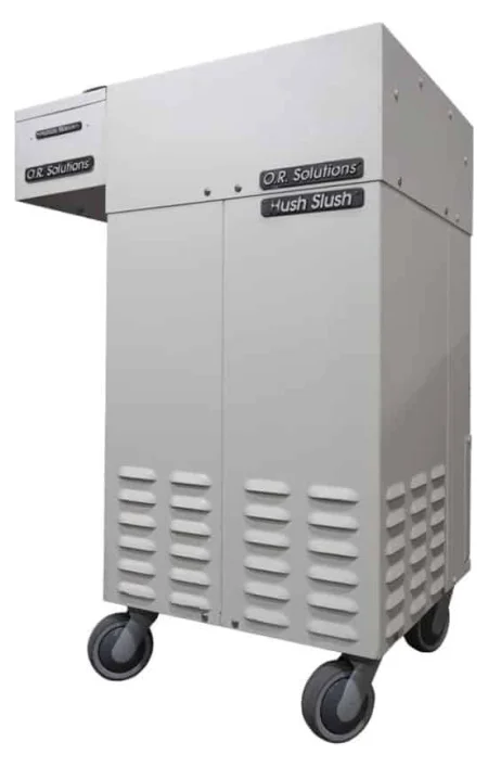
- Slush machine: freezes sterile saline into slush to cool the heart, a kidney or a transplant organ. In heart surgery, the slush is packed around the heart. Without a machine, a container of saline can be frozen to slush in a freezer and poured into a sterile basin, such as one on the ring stand.
7The electrosurgical unit
The electrosurgeryee-LEK-troh-SUR-jer-eeUsing high-frequency electric current through a handheld tool to cut tissue or seal bleeding vessels during surgery.Full entry (ESU) is the generator that powers electrosurgery. In monopolar electrosurgery, current runs from the generator to the active electrode, through the patient, and back through the patient return electrode. In bipolarby-POH-lerHaving two poles or two opposite ends; most often used as a short form of bipolar disorder.bi- twopolar having polesFull entry electrosurgery, current passes only through the tissue between the two tips of the forcepsFOR-sepsA hand instrument with two blades or arms used to grasp, hold or pull tissue or objects.Full entry , so it does not travel through the body.
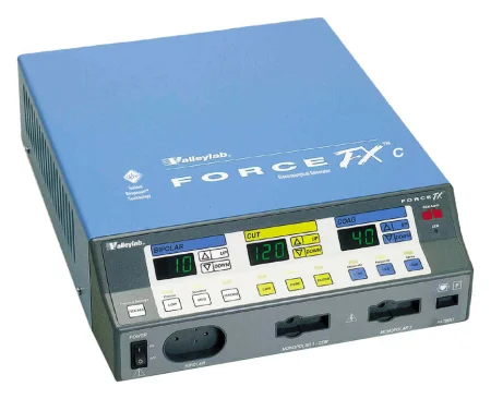
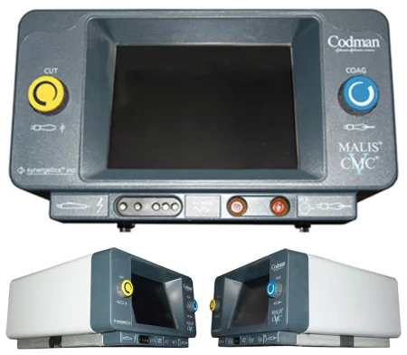
Modes, power settings and where the return electrode goes are chapter 20. Here is how the unit is handled as room equipment:
- On a cart or shelf: the cart is solid, heavier than the ESU, and big enough that no edge of the unit hangs over.
- Nothing on top: anything set on the ESU can block its cooling and make it overheat.
- Liquids away: never put fluids on or near the ESU, even sealed bottles. A spill can make it fail or cause an electrical hazard.
- The foot pedal: in use, it goes inside a clear, waterproof disposable cover against blood and irrigationeer-ih-GAY-shunWashing out a wound, body cavity, tube or eye with a steady flow of liquid.Full entry . When it is not in use, the pedal and its cord are stored where they will not be damaged.
- Self-check and alarm test: let the ESU run its self-check before any cables are connected, and test its warning alarm, which sounds when no return electrode is connected.
- Alarms stay loud: warning alarms and the activation tone must be heard through the whole case. Never turn them off, or so low that they are hard to hear.
- Where it goes: close enough to the table for its cables, then moved into its final spot after surgery starts, and away from other electronic equipment.
The ESU's buzz means someone is using the active electrode. If it buzzes while the surgeon is not using it, treat it as an alarm and tell the team at once: someone may be leaning on a pencil left on the drapes.
8Suction
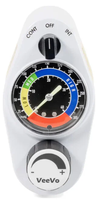
Suction uses a vacuum to pull blood and fluid from the surgical site and from the patient's airway. Every room needs at least two suction setups: one for the sterile fieldSTAIR-ul feeldAn area set up with sterile drapes and supplies, kept free of all germs, for a sterile procedure.Full entry and one for the anesthesia provideran-us-THEE-zhuh pruh-VY-derThe licensed professional who gives anesthesia and watches over the client's breathing and vital functions during a procedure.Full entry , ready for the airway at all times.
- Vacuum source: a wall or ceiling outlet, or a portable unit.
- Regulator: sets how strong the suction is.
- Canister and liner: the collection unit, on a low wheeled stand below the level of the table. It is lined with a disposable liner and marked so fluid can be measured.
- Tubing: one tube joins the canister to the wall outlet. The sterile tubing and tip come from the field and connect to the canister.
- Fluid waste management systems: some rooms drain suctioningSUK-shun-ingRemoving mucus, saliva or other secretions from the mouth, nose or airway with a tube connected to a suction source.Full entry fluid straight into the facility's waste lines, with no canisters to empty. AST recommends them because they cut staff contact with blood and the amount of medical waste.
Suction tips and tubing as supplies are in chapter 25, and using suction at the field is in chapter 29.
9The smoke evacuator
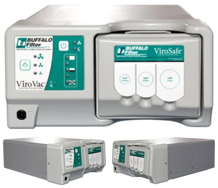
Burning or vaporizing tissue with the electrosurgeryee-LEK-troh-SUR-jer-eeUsing high-frequency electric current through a handheld tool to cut tissue or seal bleeding vessels during surgery.Full entry , a laser or an ultrasonic device makes smoke, called plume. It carries harmful chemicals and bits of tissue that may cause cancer or gene damage (mutationmyoo-TAY-shunA change in the genetic code (DNA) of a cell.Full entry ), so AST says to remove it whenever the ESU is used, in open and laparoscopiclap-uh-roh-SKOP-ikDone through a few small cuts in the abdomen with a thin camera and long instruments, instead of one large opening.lapar/o abdomen-scopic viewingFull entry cases. Smoke that builds up in a small space, such as the back of the throat, can even feed a fire.
- Small amounts of plume: an in-line filter goes between the suction canister and the wall outlet.
- Large amounts of plume: a separate smoke evacuator with an ULPA (ultra-low penetrationpen-ih-TRAY-shunPassing into or through something, such as an object entering the body through the skin.Full entry air) filter.
- The wand: hold it as close as you can to where the electrode touches the tissue, always within 2 inches (5 cm), without blocking the surgeon's view. Farther away, it captures less smoke, and the smoke builds up and blocks the view.
AST's 2012 electrosurgery standard says to hold the wand less than 1 centimeter from where the electrode touches the tissue.
- Built-in evacuation: some ESU pencils have a smoke evacuation tube built in, and some departments have a central system that serves several rooms at once.
- Filters, tubing and masks: change the filter as the maker says, wearing gloves and respiratory protection, and bag the used filter, tubing and wand as biohazard waste. Smoke evacuation comes first, and a mask is only a second line of defense. For the team, the current recommendation is a fit-tested surgical N95en-NINE-tee-fiveA close-fitting respirator mask that filters at least 95% of tiny particles from the air the wearer breathes.Full entry , because surgical masks, even high-filtrationfil-TRAY-shunThe passing of fluid through a filter or membrane, which holds back larger particles; in the kidneys, the first step in making urine.filtr strain through-ation processFull entry ones, do not reliably keep the smoke out.
AST's 2012 electrosurgery standard has the whole team wear high-filtration masks when the ESU is used, never a regular surgical mask and never two masks at once. AST's 2019 laser guideline still names high-filtration masks as the second line of defense.
10The SCD and the tourniquet
Sequential compression device
A sequential compression devicesih-KWEN-shul kum-PRESH-un dih-VYSInflatable leg sleeves that fill with air and squeeze the legs in turns to keep blood moving in the veins.Full entry (SCD) helps prevent deep vein thrombosisDEEP VAYN throm-BOH-sisA blood clot in a deep vein, most often in the leg or pelvis.thromb/o clot-osis conditionFull entry , a clot in the deep veins of the legs. Sleeves wrap the legs from ankle to thigh and connect by tubing to a pump. Each sleeve fills with air from the bottom up, squeezing blood toward the heart, then lets go so the veins refill. This keeps blood from pooling.
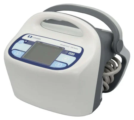
- It is used for long cases, for patients at risk of clots, for patients with obesity, and in lithotomylih-THOT-uh-meeA position lying on the back with the hips and knees bent and the legs raised and spread apart, often in stirrups.lith/o stone-tomy cutting intoFull entry .
- compression stockingkum-PRESH-un STOK-ingA tight elastic stocking that squeezes the leg, most firmly at the ankle, to help blood flow back toward the heart.Full entry (antiembolism stockings) squeeze the leg veins to help blood return, and are used instead of an SCD or along with it.
- The anesthesia provideran-us-THEE-zhuh pruh-VY-derThe licensed professional who gives anesthesia and watches over the client's breathing and vital functions during a procedure.Full entry and the circulator choose the pressure and the timing. Putting the sleeves on is chapter 31.
Pneumatic tourniquet
A pneumatic tourniquet stops blood flow to a limb, so the surgeon works in a bloodless field.
- Cuff: an inflatable bladder in a fabric or plastic cover, with one or two chambers, chosen to fit the patient's limb. The department keeps several sizes, reusable cuffs and sterile single-use ones, and contoured cuffs for tapered limbs. A wide cuff stops blood flow at a lower pressure than a narrow one.
- Tubing: joins the cuff to the control unit. Its connectors lock, so it cannot come apart by accident.
- Control unit: an air compressor with pressure controls, a pressure display and a timer. Depending on the model, it mounts on an IV pole or a portable stand, or sits on a table top. It runs on wall power, and some have a batteryBAT-uh-reeIn law, touching a person on purpose without their consent.Full entry that keeps it working for a short time if the power fails. It fills the cuff with air or nitrogen, never oxygen or nitrous oxide, which raise the risk of fire. Newer units can test the cuff for leaks and measure the limb occlusionuh-KLOO-zhunA blockage or closing off of a passage, such as a blood vessel, tube or IV line.Full entry pressure (below) on their own.
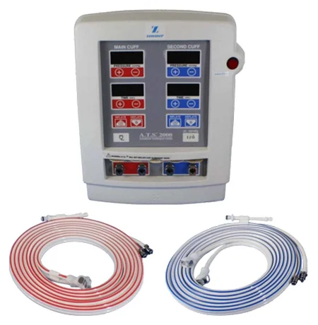
Before use, check and test the whole system as its maker's instructions say:
- The cuff and tubing are clean, with no cracks, holes or tears, and the connectors lock and are not bent, worn or loose. A unit with Luer-lock connectors is taken out of service, because its tubing could be joined to an IV line by mistake and send air into the patient.
- A battery has enough charge for the case, and the unit passes its self-test.
- Wrap the cuff around itself, then inflate and deflate it to check for leaks.
- A damaged power cord is never taped: the unit goes to biomedical engineering and a replacement comes in. Keep the tubing off the floor and free of kinks.
The surgeon and anesthesia provider decide whether a tourniquet is used and how much pressure it needs. The lowest pressure that works is the safest. AST's guideline supports working it out from the limb occlusion pressure, the lowest pressure that stops arterial blood from flowing past the cuff: 40 to 80 mm Hg is added to it, more for a higher reading, or 50 mm Hg for a child.
Some references still set the pressure from the patient's systoleSIS-tuh-leeThe part of each heartbeat when the heart muscle squeezes and pumps blood out.Full entry blood pressure, adding a set amount, more for a leg than an arm; AST's 2018 guideline also lists it for adults (50 mm Hg above systolic for an arm, 100 mm Hg for a leg). Some ORs use a standard 250 or 300 mm Hg.
AST says a CSTsee-es-tee (said as letters)Certified Surgical Technologist, a credential for surgical technologists; in maternity care, contraction stress test.Full entry working as the assistant circulator, once trained on the system, is qualified to set the pressure on the control unit. Your facility's policy decides who runs it; often it is the anesthesia provider or the RN.
During the case, help watch the pressure display and the timer, and tell the surgeon at once if the pressure changes, since that can mean the cuff is failing. The alarm sounds for a leak, a pressure that is too low or too high, a kinked tube, a machine fault or too long a time: never turn it off, or so low that the team cannot hear it. The inflation and deflation times are recorded. Applying the cuff is chapter 31.
11Specialty equipment
Some cases call for equipment brought in from storage. Learn the units your facility uses, since some, such as the cell salvage machine, need special training, and expect to help set them up, test them and drape them. How to drape equipment such as the C-arm and the microscope is chapter 28.
C-arm and other imaging
The C-arm is a portable x-ray unit shaped like a C. The x-ray tube and the image intensifier sit at opposite ends of the C, and it is used with a radiolucent table. An x-ray technologist runs it and other portable x-ray machines. Using it and radiationray-dee-AY-shunEnergy that travels as waves or particles, such as X-rays, used to make images or treat cancer; also pain that spreads from its starting point.radi/o rays, radiation-ation processFull entry safety are chapter 22.
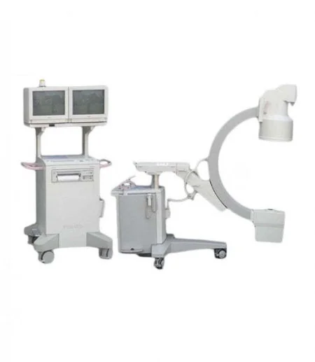
Image guidance (navigation)
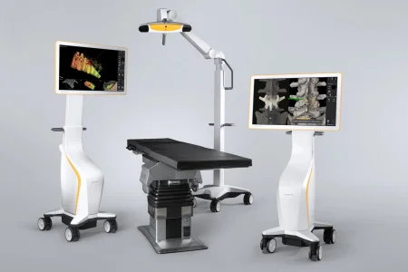
A navigation system uses cameras or an electromagnetic field to show the surgeon's instruments against the patient's own scans on a monitor. In brain surgery its arm, in a sterile sleeve, is fixed to the skull clamp, and a bump can throw off the whole system. The tracked instruments must stay in the camera's view.
Operating microscope
The operating microscope hangs from the ceiling or stands on a floor base with locking casters. It has an objective lens, whose length (100 to 400 mm) sets the working distance, eyepieces called oculars, a light, and foot controls for zoom and focus. A second set of oculars lets the assistant see.
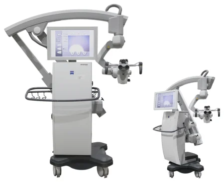
It is heavy, but its parts are delicate:
- Secure the arms before you move it, so they cannot swing into a wall, and push it with both hands on the upright column.
- Balance it, check that the brakes and controls are tight, check the cords and the bulb, and test it before it goes to the field. Keep a spare bulb in the room.
- Before the case, each user's settings go on the oculars: their angle, the distance between them (to match the user's pupils) and the focus setting that corrects the user's own vision. Each centering button (one for the side-to-side and one for the up-and-down travel) is pressed once, and the microscope head or the bed is raised or lowered until the microscope's light is roughly in focus on the field.
- The scrub drapes it in a sterile microscope drape. Keep the drape away from the light's cooling fan, or the lamp can overheat and start a fire.
- Damp-dust it before use. Never touch the lenses, and never put detergent or disinfectantdis-in-FEK-tuntA chemical used on objects and surfaces to kill most germs.Full entry on them: clean them with a lens cleaner or water and wipe them with lens paper, not cloth, which leaves lint. Cover the microscope at the end of the day to keep dust off.
Doppler unit
A Doppler ultrasoundDOP-ler UL-truh-soundAn ultrasound that uses sound waves to hear or show blood flowing through vessels or the fetal heartbeat.Full entry unit uses Doppler ultrasound: it sends sound waves into the body and plays the movement of blood as a sound, so the surgeon can hear flow through a vessel or a new connection. A hand-held probe can be placed on the skin with gel. The probes are heat sensitive, so a probe used on the sterile fieldSTAIR-ul feeldAn area set up with sterile drapes and supplies, kept free of all germs, for a sterile procedure.Full entry is sterilizationstair-uh-luh-ZAY-shunA process that destroys all microorganisms, including spores, on an item; also a permanent birth control procedure.Full entry only as its maker says, or goes in a sterile cover.
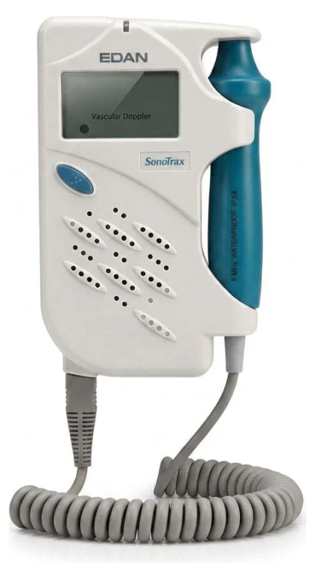
Nerve monitor
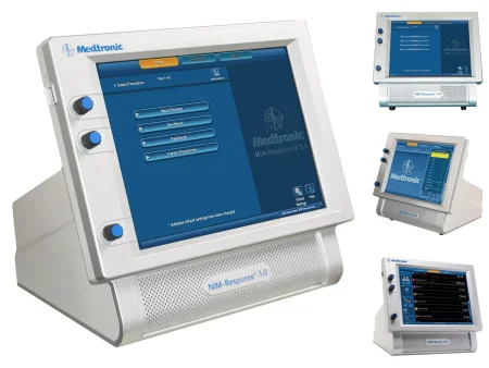
A nerve stimulator or nerve monitor helps the surgeon find a nerve and keep it safe. A small current is sent through a probe, and if a nerve is close, the muscle it controls twitches. It is used to find the facial nerveFAY-shul NERVThe seventh cranial nerve, which moves the muscles of the face and carries taste from the front of the tongue.Full entry in ear and parotid surgery.
Heart-lung machine
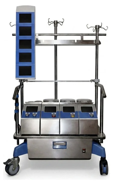
The cardiopulmonarykar-dee-oh-PUL-muh-nair-eeRelating to the heart and lungs together.cardi/o heartpulmon/o lung-ary relating toFull entry bypass (heart-lung) machine does the work of the heart and lungs so the heart can be stopped for surgery. Blood from the veins drains to the machine, passes through an oxygenator, and is pumped back into the arteries. A heat exchanger cools or warms the blood. A perfusionist runs it. The circuit is primed and every bit of air removed before bypass starts; the prime, usually 1,400 to 2,000 mL of crystalloid and colloid fluids with heparinHEP-uh-rinAn anticoagulant drug, given by injection or IV, that keeps blood clots from forming or growing.Full entry added, mixes with the patient's blood and thins it. AmSECT requires pressure monitors, a bubble detector, a level sensor on a hard reservoirREZ-er-vwarA place where germs live and multiply, such as a person, animal, water or soil.Full entry , an arterial linear-TEER-ee-ul lineA thin catheter placed in an artery, often in the wrist, to measure blood pressure all the time and draw blood.Full entry filter, a temperature alarm, a one-way valve in the vent line, hand cranks and backup gas and power on every bypass, and the perfusionist works through a checklist from setup to the end of the case.
Cell salvage
A cell salvage (autotransfusion) system collects the blood a patient loses during surgery, washes or filters it, and returns it to the same patient. It is used when heavy blood loss is expected, in trauma, and for some patients who refuse donated blood. It has its own suction tubing, separate from waste suction, and staff train on their facility's own machine.
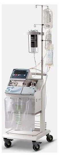
Ultrasonic units, the phacoemulsifier and the irrigating bipolar
- Ultrasonic devices turn electricity into very fast vibration. Ultrasonic shears cut and seal tissue at once, and their blades stay hot right after use, so between uses they rest on a moist towel on the field, away from the patient.
- An ultrasonic shears system has a generator, a cable, a foot switch and a handpiece. The handpiece is single use; the reusable cord and wrench that go with it are sterilized by the facility. No current passes through the patient, so no return electrode is needed. Blood and tissue that build up on the jaws are wiped off with a moist sponge.
- An ultrasonic aspirator breaks up tumor tissue and suctions it away.
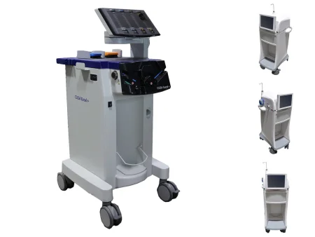
- A phacoemulsifier breaks up a cataractKAT-uh-raktClouding of the lens of the eye.Full entry (a cloudy lens of the eye) with ultrasonic vibration and suctions it out, replacing the fluid with balanced salt solution.
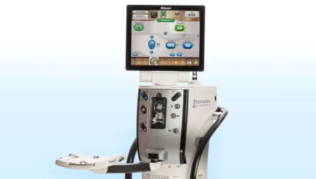
- An irrigating bipolarby-POH-lerHaving two poles or two opposite ends; most often used as a short form of bipolar disorder.bi- twopolar having polesFull entry system runs fluid to the tips of the bipolar forcepsFOR-sepsA hand instrument with two blades or arms used to grasp, hold or pull tissue or objects.Full entry , so the surgeon can coagulationkoh-ag-yuh-LAY-shunThe process by which blood changes from a liquid into a gel, forming a clot.coagul clot-ation processFull entry without tissue sticking to them.
Cryotherapy and uterine ablation
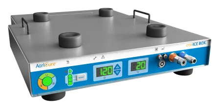
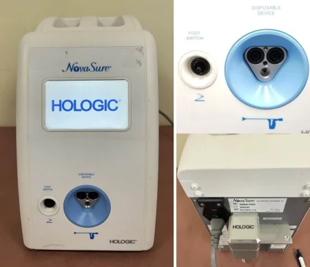
A cryotherapy unit freezes tissue through a cold probe, cooled by a compressed gas such as argon, to destroy it. It is used on tumors and on retinalRET-ih-nulRelating to the retina, the thin layer at the back of the eye that senses light and sends images to the brain.retin/o retina-al relating toFull entry detachments. A uterine ablator is a low thermal radiofrequency device: its controller sends radiofrequency energy through a device placed in the uterus to destroy the lining, to treat abnormal bleeding.
Liposuction and vacuum curettage
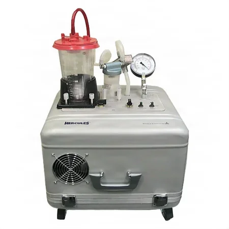
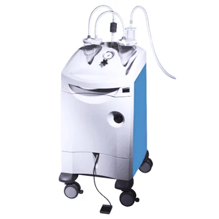
A liposuction aspirator uses high vacuum, large-bore tubing and hollow cannulaKAN-yoo-luhA thin tube placed into the body or a body opening to deliver or remove fluid or air.Full entry to remove fat. A vacuum curettage machine uses suction to empty the uterus.
Robotic systems
A da Vinci robotic system has three parts.
- Surgeon console: the nonsterile station where the surgeon sits or stands to control the instruments.
- Patient cart: carries the camera and instrument arms. It is heavy and moves on a motor drive, steered from the back.
- Vision cart: holds the light source, camera controls and video equipment. Its wheel locks are on its rear wheels.
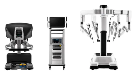
The patient cart is draped away from the table and moved in only after the patient is positioned and draped. Its size can block the airway, the chest and the crash cart, so the team plans its place ahead of time. No extension cords are used. The robot in use is chapter 21.
12Before the first case: ready the room
Planning starts the day before, when cases are usually assigned. Read the schedule and each case's preference card, and plan so the equipment each case needs is available and ready on time. A surgeon may ask ahead for special equipment; pass that on to the team. AST also asks you to look over the next day's cases for the positioning equipment they need: a piece may be out for repair, or there may not be enough to go around, and the day before leaves time to solve it.
On the day, getting ready for the first case takes a few more steps than later cases.
- Wash and damp dust. Do a basic hand wash as you enter. Then wipe the lights, furniture and fixed equipment with a clean, lint-free cloth and the hospital-grade disinfectantdis-in-FEK-tuntA chemical used on objects and surfaces to kill most germs.Full entry your facility names. Work from the highest surfaces down, so dust does not fall on what you have already cleaned. Wash your hands again when you finish.
- Check the room against the card. Everything the card lists is in the room, and anything the case does not need comes out.
- Place the table. It goes under the lights, turned the way the surgeon needs. The anesthesiaan-us-THEE-zhuhLoss of feeling or sensation, often brought on by drugs so that surgery or a procedure can be done without pain.an- withoutesthesi/o feeling, sensation-ia conditionFull entry machine goes where the anesthesia provideran-us-THEE-zhuh pruh-VY-derThe licensed professional who gives anesthesia and watches over the client's breathing and vital functions during a procedure.Full entry wants it, usually at the head. Keep the way from the door clear of sterile furniture.
- Group the sterile furniture. The back table, Mayo standMAY-oh standA small tray on an adjustable stand placed over or beside the patient during surgery to hold the instruments used most often.Full entry and ring stand go together, as far from the door and the traffic as you can. Every time a door opens, air moves and stirs up dust. Keep them at least 12 inches (about 30 cm) from the walls and from anything that is not sterile.
- Place everything else. Hampers, kick buckets and stools go out of the traffic and away from the sterile furniture. Hampers get biohazard bags, and kick buckets get impervious biohazard liners.
- Set up two suctions. Put a new liner in each canister and connect the tubing to the wall vacuum: one setup for the field and one for anesthesia.
- Bring in the electrosurgeryee-LEK-troh-SUR-jer-eeUsing high-frequency electric current through a handheld tool to cut tissue or seal bleeding vessels during surgery.Full entry . Park it close enough to the table for its cables.
- Make the table ready. Clean linen and a lift sheet, armboard covers, one end of the safety strap fastened, and the attachments and positioning aids for this procedure and this patient's size.
- Check the lights. Turn them on, make sure they work, and swing them near where they will focus.
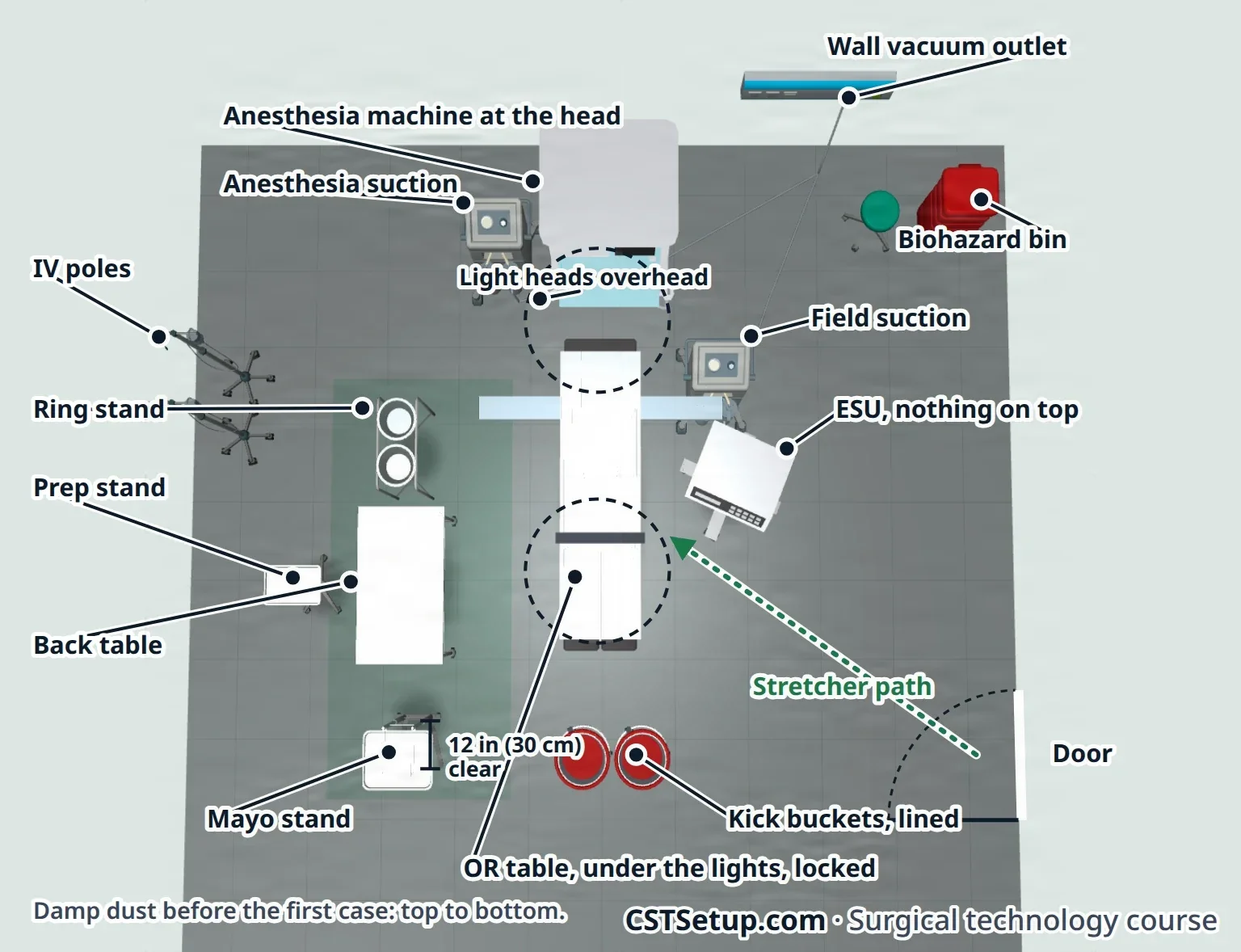
13Before the patient comes in: test everything
Every piece of equipment is tested before the case starts, while there is still time to replace it: the electrosurgeryee-LEK-troh-SUR-jer-eeUsing high-frequency electric current through a handheld tool to cut tissue or seal bleeding vessels during surgery.Full entry , monitors, suction, the tourniquet, the microscope and anything else the card lists. AST also asks the team to confirm on the day of surgery that the OR table works.
Electrical safety
- Inspection stickers: biomedical engineering inspects equipment on a schedule and puts a sticker on each unit. The sticker should be current. A unit due for its inspection goes to biomedical engineering, not into service.
- Plugs: a hospital plug has three prongs. The third is the ground, which carries stray current safely away. Check that all three are there and none is bent. Never bend a prong back, since that loosens it, and never remove the ground prong to fit an outlet.
- Cords: look along the whole cord for fraying, cracks or cuts that show the wires inside. A damaged cord is never taped or used. The unit leaves the room for repair, and a replacement comes in.
- No extension cords or added power strips: plug straight into the wall. The cord should reach without being pulled tight or lifted off the floor, and run out of the walkways so nobody trips on it.
- A video cart's own strip: most video carts have a power strip built into the cart, so fewer cords run to the wall. Its main on-off switch is usually near its base and must be on for the units plugged into it to work.
- Red outlets: these run on the hospital's backup generator during a power failure, for the anesthesiaan-us-THEE-zhuhLoss of feeling or sensation, often brought on by drugs so that surgery or a procedure can be done without pain.an- withoutesthesi/o feeling, sensation-ia conditionFull entry machine and other equipment the patient depends on.
- Handle the plug: hold the plug, never the cord, to plug in or unplug. Pulling the cord strains it and can make sparks. Switch a device off before you unplug it.
- Keep liquids away from outlets and equipment, and use only devices made for use around fluids near them.
Machine by machine
- ESU: self-check, alarm test and a covered foot pedal, as described above.
- Suction: turn on both setups and test each line for strong, safe suction. Check that every connection is tight.
- OR table: run it through its controls, check that its brakes hold and that every attachmentuh-TACH-mentThe deep emotional bond between an infant and a parent or caregiver.Full entry is there, and lock each armboard. Compare its weight limit with the patient's weight.
- Everything else: the light source on standby until its cable is connected, the carbon dioxideKAR-bun dy-OK-sideA gas made by the body's cells as waste, carried in the blood to the lungs and breathed out.Full entry level checked with a spare tank in the room, and the tourniquet and microscope checked as described above.
If an item is damaged or does not work, it is handed to the circulator, who tags it for repair, and a replacement is found.
Test it before the patient comes in. A problem found now costs a few minutes. Found after the incision, it means a delay, more time under anesthesia and more risk for the patient.
14When equipment fails during the case
- Suction stops pulling: blood clots inside the tip. Suction sterile water through the tip and tubing often to prevent it, and clear a small tip with its stylet. Use sterile water, not saline, which corrodes metal, and never poke the tip clear with the electrosurgeryee-LEK-troh-SUR-jer-eeUsing high-frequency electric current through a handheld tool to cut tissue or seal bleeding vessels during surgery.Full entry pencil.
- Suction gets weak: the canister may be full or a connection loose. The circulator changes full canisters before they overflow, and every connection should be tight.
- The surgeon keeps asking for more power: repeated requests to turn up the ESU can mean the unit is failing or the circuit has a problem. Tell the surgeon it may be a malfunction, and stop using the ESU until it is solved. If checking does not fix it, the ESU is replaced, along with a new return electrode and a new active electrode. Chapter 20 covers what to check.
- A unit sparks or smokes: stop using it and take it out of service at once. Sparks can light anything that burns on the field.
- A robot fault: the team calls the maker's hotline with the system's identification number, and the case may be converted to an open procedure.
No one on the surgical team repairs electrical equipment. A unit that fails is removed from service, a biomedical technician may be called to the room, and a replacement is brought in.
Never repair, open up or tape a failing machine, and never keep using equipment that smokes. Take it out of service and get a replacement.
15After the case: care, cleaning and repair
Once the patient has left the room, the equipment is cleaned, put away or sent for repair. Wear personal protective equipment for this work.
- Unplug: pull on the plug, never the cord.
- Cords and pedals: clean the electrosurgeryee-LEK-troh-SUR-jer-eeUsing high-frequency electric current through a handheld tool to cut tissue or seal bleeding vessels during surgery.Full entry cord as its maker says. Never use isopropyl or ethyl alcohol, which dries out the insulation until it cracks. Avoid knots and tight kinks, and store the pedal and cord where they will not be damaged.
- Equipment surfaces: wipe the ESU and other used equipment with the disinfectantdis-in-FEK-tuntA chemical used on objects and surfaces to kill most germs.Full entry your facility names, following each maker's instructions, and keep liquid out of the units.
- Suction canisters: disconnect them from the suction, wipe the outside, and dispose of them as your facility's policy says. A full canister goes in a biohazard bag. Some facilities first add a product that turns the fluid solid, and some use a fluid management system that sends the fluid straight to the sewer.
- Smoke evacuator: the used filter, tubing and wand go in a biohazard bag.
- Tourniquet: a single-use cuff is thrown away, never reprocessingree-PROS-es-ingThe steps that make a used reusable medical device safe to use on the next patient.Full entry . Clean a reusable cuff and its tubing as their maker says, with disinfectant in lukewarm water, keeping the open end of the tubing out of the solution. Dry them, connect them and test for leaks. Store the unit covered, in a clean, dust-free place, and plugged in so its batteryBAT-uh-reeIn law, touching a person on purpose without their consent.Full entry stays charged.
- OR table: take the pads off and clean both sides of each pad, the table top, the hinges, the pivot points and the casters. Unlock the table and move it to one side so the floor under it can be mopped.
- Lights: wait until they cool. Disinfectant on a hot light can crack the bulb. Spot clean any blood.
- Storage: each piece of equipment goes back to its own place in a clean storeroom, arranged so it is not damaged moving in and out. Poor storage costs operating rooms thousands of dollars in repairs every year.
Tag it for repair
Equipment that does not work is tagged for repair and leaves the department for the healthcare technology management department (formerly called biomedical engineering). If a unit harmed a patient, do not take it apart or change its settings: it goes to that department just as it is, so the technicians can find out what went wrong. Report it as your facility's policy says.
Biomedical technicians are trained to inspect, test and repair patient care equipment. They keep a record of every inspection and repair for each piece of patient care equipment; each ESU, for example, is logged by its facility identification number and its serial number. The Joint Commissionthuh JOYNT kuh-MISH-unAn independent US organization that inspects and accredits hospitals and other health care organizations against safety and quality standards.Full entry requires preventive maintenance standards for equipment. For the tourniquet, AST recommends an inspection every 6 months, and a monthly check of its pressure display against a gauge by a CSTsee-es-tee (said as letters)Certified Surgical Technologist, a credential for surgical technologists; in maternity care, contraction stress test.Full entry or RN named for the job. When a device injures or kills a patient, the facility reports it to the FDA and the maker.
Breaking down the sterile fieldSTAIR-ul feeldAn area set up with sterile drapes and supplies, kept free of all germs, for a sterile procedure.Full entry , room turnover and end-of-day cleaning are chapter 30.
What to take from this chapter
- The OR table has sections joined at breaks, side rails, removable head and foot sections, a kidney lift, a perineal cutout, a radiolucent top, a locking base and a handset.
- Every table and attachment has a weight limit. If the patient weighs more, it is not used.
- Lock armboards on solid rail away from the breaks, and use each positioning device only as it is made to be used.
- Ready the room before the first case: damp dust top to bottom, remove what the card does not need, group the sterile furniture far from the door and 12 inches from the walls, and set up two suctions.
- Test everything before the patient comes in: current inspection stickers, three-prong plugs, undamaged cords, no extension cords, the ESU self-check and alarm, both suctions and the table.
- Nothing goes on top of the ESU, liquids stay away, and alarms stay loud.
- Equipment that fails, sparks or smokes is taken out of service, tagged and sent to biomedical engineering. No one on the surgical team repairs it.
- After the case: unplug by the plug, no alcohol on cords, pads off the table, lights cool before they are wiped, and everything back in its place.
Check yourself5 quick questions
Free for everyone. Pick an answer for each, then check them all at once.
In the ORWhere you'll use this
The chapter's whole question pool in sets of 10, with the answer explained after each one.
Part of the educators programThe chapter's words on spaced cards: the ones you miss come back sooner.
Part of the educators programGiven by your class's teacher. Each class draws its own 25 from the chapter's pool.
Part of the educators program
Suggest a fix
Something wrong, unclear or out of date in this chapter? Tell us and we will fix it for everyone. Your email is only used to reply.