Oral and maxillofacial surgery
Jaws, teeth, facial fractures and clefts: the bones of the face, how the surgeon puts them back so the bite fits, the airway you share with anesthesia, and the wire cutters that never leave a patient whose jaws are wired.
1What oral and maxillofacial surgery covers
Oral and maxillofacial (OMF) surgery, also called oromaxillofacial surgery, diagnoses and treats disease, defects and injuries of the face and the mouth (the oral cavity), both the soft tissues and the bones. It repairs facial fractures, rebuilds the face after trauma, cancer surgery or a birth defect, moves the jaws to fix the bite, and removes teeth. Oral and maxillofacial surgeons do much of this work, and so do plastic surgeons and ear, nose and throat (otorhinolaryngology) surgeons.
The face takes the blows in vehicle crashes, work accidents, fast sports and assaultuh-SAWLTIn law, a threat or act that makes a person afraid they are about to be touched in a harmful or unwanted way.Full entry . By one reference's count, vehicle crashes cause about 60 percent of maxillofacial injuries, and assaults and crashes are the most common causes of a broken mandibleMAN-dih-bulThe lower jawbone, the only bone of the skull that moves.Full entry . Seat belt campaigns have cut the number of facial fractures from crashes, but crashes and violence are still the main causes in adults and children.
These injuries matter beyond the bones. They can damage skin, muscle, nerves and vessels, and they can change how a person speaks, chews and grows teeth. A changed face can also cut a person off socially and emotionally, so expect patients who are frightened about pain and about how they will look afterward.
A facial fracture is repaired as soon as the patient's general condition allows, often within the first 24 hours. It is seldom an emergency in itself, and life-threatening injuries come first. Waiting too long, though, lets the tissues swell (edema), which hides the anatomy the surgeon needs to see.
Some of this work, such as taking out teeth, is often done in an office or outpatient clinic. A patient comes to the OR when the disease is extensive, when a bleeding disorder such as hemophiliahee-muh-FIL-ee-uhAn inherited disorder in which the blood lacks a clotting factor, so it does not clot normally.hem/o blood-philia attraction, tendencyFull entry makes the office unsafe, when the patient is very frightened or cannot cooperate, or when a child needs a great deal of dental work.
Every procedure in the Core Curriculum's list for this specialty is an open approach. Cases that share instruments, positions and steps are taught together as co-related procedures: once you know how a plate goes onto a broken jaw, a broken cheekbone or an orbital rim is mostly the same steps on a different bone. Oral and maxillofacial surgery is one of the specialties in your clinical case count. Each case links to its setup card, which holds the full instrument list and every step.
Other chapters teach what this one builds on: the skull and facial bones and the chewing muscles in chapter 3; the mouth, the teeth and the salivary glandSAL-ih-vair-ee GLANDA gland that makes saliva and releases it into the mouth.Full entry in chapter 7; how a broken bone heals in chapter 3 and fractures as injury, with trauma care, in chapter 11; the airway equipment and endotracheal intubationen-doh-TRAY-kee-ul in-too-BAY-shunPutting a breathing tube through the mouth or nose into the windpipe (trachea).endo- withintrache/o windpipe-al relating toFull entry , nasal tubes included, in chapter 13; fire at the field in chapter 20; broken bones elsewhere in the skeleton, power tools, and plates and screws in general in chapter 39; the parotid gland, nasal fractures, neck dissectiondy-SEK-shunA tear in the inner layer of an artery wall that lets blood split the layers apart; also, the cutting apart of tissue in surgery.Full entry and TMJ arthroscopy in chapter 36; injuries of the eye itself in chapter 35; craniotomykray-nee-OT-uh-meeSurgery in which a piece of the skull is removed to reach the brain, and usually put back at the end.crani/o skull-tomy cutting intoFull entry in chapter 42. cleft lipkleft lipA split or opening in the upper lip that is present at birth.Full entry and palate repair also belongs to plastic surgery, and chapter 38 meets it again.
2Anatomy that matters
The bones of the face
The face is divided into three regions; an injury to all three is called a panfacial fracture.
- Upper face: the frontalFRUN-tulRelating to the front of the body, or to the forehead and the bone or brain area behind it.Full entry bone, which makes the forehead and the upper rim of each orbit and holds the frontal sinusSY-nusA hollow space in the body, most often one of the air-filled spaces in the skull bones around the nose.Full entry . The thick bone between the eyebrows, the glabella, shields the drainage passages of the frontal sinuses and the cribriform plate, the part of the skull's floor that holds the branches of the nerves of smell.
- Midface: the ethmoid, the nasal bones, the zygomas and the two maxillae. The nasal bones form the bridge of the nose. The ethmoid is a complex bone full of small sinus cavities that helps form the floor of the skull, so a fracture here can tear the duraDOOR-uhThe tough outer layer of the membranes that cover the brain and spinal cord.Full entry , the tough outer covering of the brain, and can injure the tear ducts and glands.
- Lower face: the mandibleMAN-dih-bulThe lower jawbone, the only bone of the skull that moves.Full entry , the lower jaw, the only bone of the face that moves.
The zygoma (zygomatic, or malar, bone) is the cheekbone. It forms the prominence of the cheek and part of the floor and outer wall of each orbit, and its long back process, the zygomatic arch, joins the temporal bone. Because the zygoma helps hold the eye in place, a displaced zygoma fracture often comes with an eye injury, and a zygoma left out of place can let the eye sink back.
The two maxillae join to form the upper jaw. Each one holds the upper teeth in its alveolusal-VEE-uh-lus (plural alveoli, al-VEE-uh-ly)One of the tiny air sacs at the ends of the smallest airways in the lungs.Full entry process, forms the front of the hard palateHARD PAL-itThe bony front part of the roof of the mouth.Full entry and part of the orbit, and contains a maxillary sinus, an air space lined with mucous membrane that drains into the nose. The roof of the maxillary sinus is the floor of the orbit, a shelf of bone as thin as eggshell. The infraorbital nerve runs through that floor and out of a small hole below the eye, giving feeling to the cheek, the side of the nose, the upper lip and the upper teeth and gums.
The orbit, the bony socket of the eye, is built from seven bones: the frontal, zygomatic, maxillary, lacrimal, ethmoid, sphenoid and palatine bones. Its floor and inner wall are thin, and the force of a blow to the eye passes to them, so they are the walls that break most often.
The face is not a solid block. It is a frame of thicker vertical and horizontal pillars of bone, called buttresses, with thin plates of bone and sinuses between them. The buttresses carry the force of chewing, so a midface repair puts them back in line and holds them there, and the shape of the face follows.
The mandible and the jaw joint
The mandible is the largest and strongest bone of the face, shaped like a U. It has three parts.
- Body: the horizontal part, which holds the lower teeth in its alveolar process. In the front midlineMID-lineAn imaginary line down the center of the body; also, a type of IV catheter placed in a vein of the upper arm.Full entry the two halves meet at the symphysisSIM-fih-sisA joint where two bones are joined firmly by a pad of cartilage that allows only a little movement.Full entry , and the mental protuberance below it is the chin. The mental foramen is a small opening on each side of the body.
- Ramus: the upright part on each side. At its top are two projections: the condylar process at the back, whose rounded head (the condyle) fits into the glenoid (mandibular) fossaFOS-uhA shallow hollow or dip in a bone or on the body's surface.Full entry of the temporal bone, and the coronoid process in front, where the temporalis muscle attaches. The dip between them is the mandibular notch. The mandibular foramen on the inner surface of the ramus carries the inferior alveolar nerve and its vessels. That nerve supplies the lower teeth on its side, so local anestheticLOH-kul an-es-THET-ikA drug that blocks feeling in one part of the body without making the person unconscious.Full entry for dental work is often injected near the foramen to block it.
- Angle: the corner where each ramus meets the body.
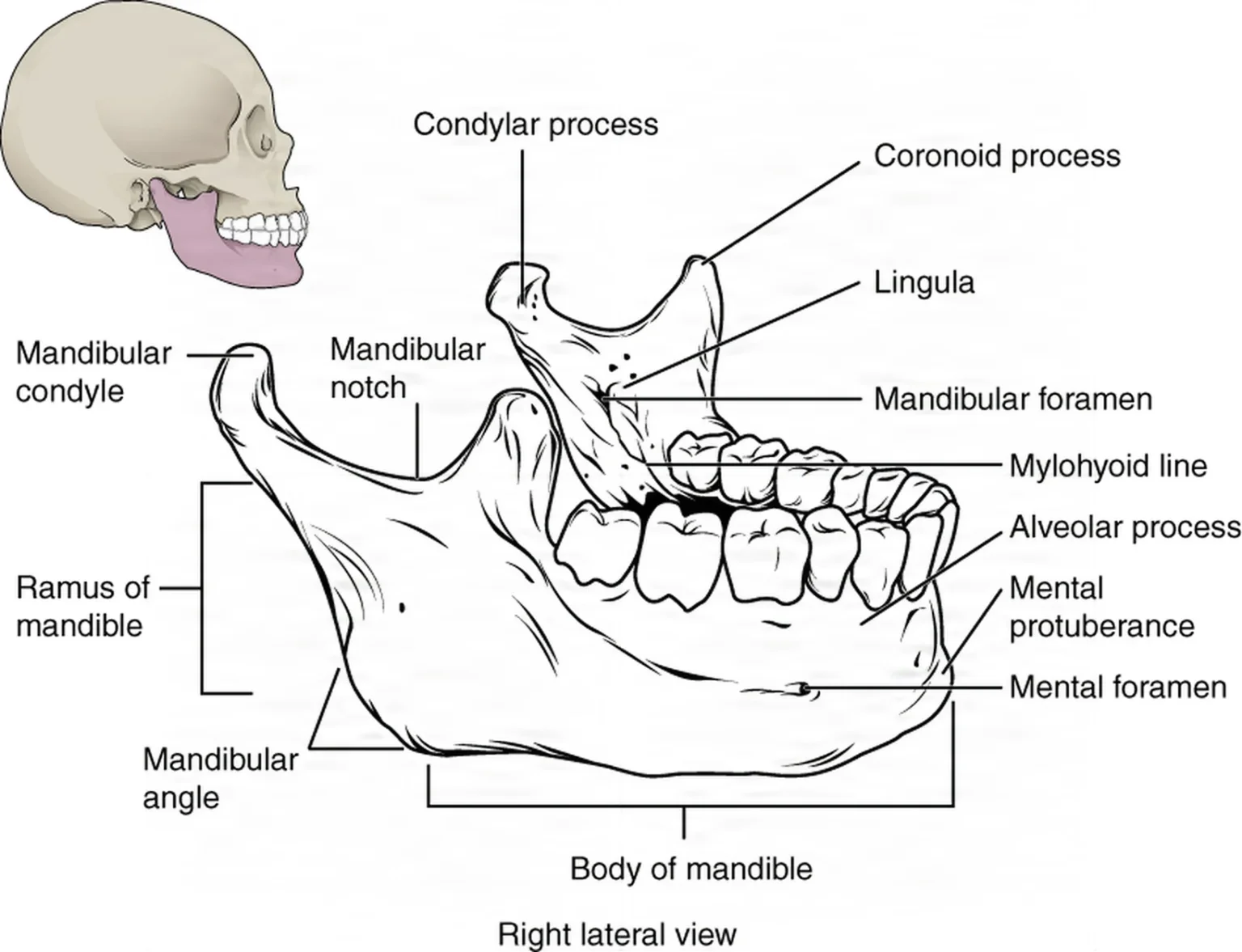
The condyle and the glenoid fossa form the temporomandibular joint (TMJ), which lets the jaw move. An avascular pad, the articular disc, glides between the bones as the jaw opens and closes.
Branches of the facial nerveFAY-shul NERVThe seventh cranial nerve, which moves the muscles of the face and carries taste from the front of the tongue.Full entry , which moves the muscles of the face, cross the surgeon's path. The marginal mandibular branch runs near the lower border of the mandible, so an incision under the jaw is made below that border and parallel to it, and the surgeon may find the branch with a nerve stimulator. The frontal (temporal) branch, to the forehead muscle, is at risk in an incision in front of the ear and when a scalp flap is lifted over the temple.
Teeth and the bite
Each tooth sits in a socket of the alveolar process. A tooth has three parts: the crown above the gumline, the root below it, and the neck where they meet. The periodontal ligament, a layer of collagenKOL-uh-jenThe main structural protein of the body, which gives strength to skin, bones, tendons, ligaments and scar tissue.coll/a glue-gen producingFull entry fibers, holds the root in its socket, and it is this ligament an elevator breaks to free a tooth. Inside, the pulp holds the tooth's vessels and nerves. The gum around the teeth is the gingiva.
People grow two sets of teeth, the primary (baby) teeth and the permanent teeth; chapter 7 counts them, names each kind and describes the layers: the enamel that covers the crown, the dentin under it and the cementum over the root. In the United States the permanent teeth are numbered 1 to 32 (the Universal, or National, system): number 1 is the upper right third molar, the count runs across the upper jaw to 16, drops to the lower left third molar as 17 and runs back to the lower right third molar as 32. The primary teeth are lettered A to T. Many other countries use a two-digit system that numbers the teeth 1 to 8 within each quarter of the mouth. The surgeon checks the images before removing a tooth so the right one comes out.
Words for the mouth that you will hear: labial means toward the lips, buccal toward the cheek and lingual toward the tongue. Dentition is the number, kind and pattern of a person's teeth. Mastication is chewing. Edentulous means without teeth. The third molars are the wisdom teeth.
occlusionuh-KLOO-zhunA blockage or closing off of a passage, such as a blood vessel, tube or IV line.Full entry is how the upper and lower teeth meet when the jaw is closed: the patient's bite. Malocclusion means the teeth do not meet as they should. A facial fracture often throws the bite off. The goal of almost every jaw and midface repair is to give the patient back their own bite from before the injury, so the teeth meet and chew as they did.
3Diagnosis and planning
Diagnosis starts with the story. A patient with a broken jaw may not be able to talk, so the history may come from paramedics, the emergency team or family, or from a pencil and paper; the type of injury and the direction of the blow hint at what broke. The physical exam is gentle, above all when the maxilla may be broken, because moving the pieces can tear the duraDOOR-uhThe tough outer layer of the membranes that cover the brain and spinal cord.Full entry , injure nerves or spread infection.
Signs that point to a fracture:
- bleeding, bruising, cuts and swelling over the bone
- malocclusion, which suggests a fracture of the teeth, the tooth-bearing bone or the jaws
- clear fluid from the nose (cerebrospinal fluidser-uh-broh-SPY-nul FLOO-idThe clear, watery fluid that bathes and cushions the brain and spinal cord.cerebr/o brainspin spine-al relating toFull entry rhinorrheary-noh-REE-uhA runny nose: thin fluid draining from the nose.rhin nose-rrhea flow, dischargeFull entry ) or the ear (otorrhea), a sign that the dura is torn
- double vision (diplopiadih-PLOH-pee-uhSeeing two images of a single object; double vision.dipl/o double-opia visionFull entry ) or an eye that has sunk back in its socket (enophthalmos), signs of an orbital fracture
- a flattened cheek, or trouble opening and closing the mouth, with a broken zygoma
- numbness of the cheek and upper lip, from an injured infraorbital nerve
Imaging:
- Plain X-rays in special views, such as the Waters and Caldwell views, show particular facial bones, and a panoramic X-ray shows on one film the tooth-bearing bone of both jaws, the whole mandibleMAN-dih-bulThe lower jawbone, the only bone of the skull that moves.Full entry , the back of the maxillary sinusSY-nusA hollow space in the body, most often one of the air-filled spaces in the skull bones around the nose.Full entry and the zygomas. Teeth to be removed are checked on a panoramic film.
- computed tomographykum-PYOO-tid toh-MOG-ruh-feeAn imaging test that uses x-rays and a computer to make cross-section pictures of the inside of the body.tom/o cut, slice-graphy process of recordingFull entry (CT) shows the facial bones in several planes. A computer can turn the scan into a three-dimensional image of the skull, which shows the full extent of a complex injury before and after the repair and helps plan a reconstruction; one reference notes that plain films are not used to diagnose a frontalFRUN-tulRelating to the front of the body, or to the forehead and the bone or brain area behind it.Full entry sinus fracture.
- magnetic resonance imagingmag-NET-ik REZ-uh-nunts IM-uh-jingAn imaging test that uses a strong magnet and radio waves to make detailed pictures of the inside of the body.Full entry shows soft tissue best, but it takes time a trauma patient may not have, so it is kept for stable patients and planned operations.
The surgeon may also take dental impressions to make models of the teeth, and may ask for photographs from before the injury and the patient's dental records, which show where the bones belong. All of these images, models and photographs come to the OR with the patient and stay where the surgeon can see them.
Case planning: facial cases vary more than most, so ask the surgeon ahead of time: open or closed reduction, a route inside the mouth or through the skin, the order of the procedures when there are several, whether X-rays will be taken during the case, and which plates and screws to have ready. For a patient injured all over the face, talking first lets you open the right sets without opening everything. If X-rays or fluoroscopyfloo-ROS-kuh-peeAn x-ray method that shows moving pictures of the inside of the body in real time.fluor/o glowing (from the fluorescent screen)-scopy looking, viewingFull entry may be needed, let radiology know before the case starts.
4Diseases and injuries the surgeon treats
Le Fort fractures of the midface
In 1901 a French physician, René Le Fort, studied cadaver skulls struck with blunt force and found lines of weakness in the midface where fractures run. Le Fort's three patterns are still the way midface fractures are named. All three cross the nasal septumNAY-zul SEP-tumThe wall of cartilage and bone that divides the inside of the nose into right and left sides.nas/o nosesept/o dividing wallFull entry and the pterygoid plates, the plates of the sphenoid bone behind the maxilla.
- Le Fort I (transversetrans-VERSLying across something, at a right angle to its length.Full entry maxillary fracture): a horizontal break straight across the maxilla above the roots of the teeth. It runs through the floor of the nose and the lower nasal septum, the front and side walls of the maxillary sinusSY-nusA hollow space in the body, most often one of the air-filled spaces in the skull bones around the nose.Full entry and the palatine bones, so the hard palateHARD PAL-itThe bony front part of the roof of the mouth.Full entry and the tooth-bearing bone, with the upper teeth, come loose from the rest of the face. It is the most common maxillary fracture, and the upper jaw may float free in the mouth.
- Le Fort II (pyramidal fracture): a pyramid-shaped break, on one side or both. Its top crosses the nasal bones; from there it runs down through the frontalFRUN-tulRelating to the front of the body, or to the forehead and the bone or brain area behind it.Full entry process of each maxilla and the lacrimal bone, across the orbital floor and out through the rim below the eye, then down across the front of the maxillary sinus to the pterygoid plates. The zygomas stay with the skull, and the break separates them from the maxilla. This fracture is also linked with leaking cerebrospinal fluidser-uh-broh-SPY-nul FLOO-idThe clear, watery fluid that bathes and cushions the brain and spinal cord.cerebr/o brainspin spine-al relating toFull entry into the nasal sinuses.
- Le Fort III (craniofacial disjunction): the highest break. It runs across the root of the nose where the nasal bones meet the frontal bone, back through the ethmoid and both orbits, out across the outer wall of each orbit where the zygoma meets the frontal bone, and through the zygomatic arches, so the whole midface, both zygomas and the nose with the maxillae, comes away from the base of the skull. The face looks flattened and longer (a dish face), with bruising and swelling around both eyes.
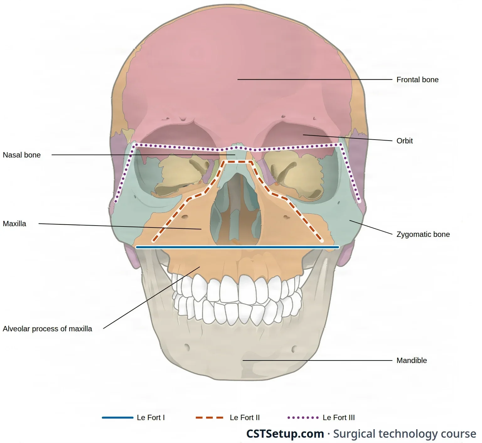
A pure Le Fort fracture is rare. Most patients have two or more levels at once, or a different level on each side, with breaks that run past the classic lines. Any maxillary fracture throws off the bite, and a severe one leaves a flattened, pushed-in look to the middle of the face.
Other facial fractures
- Zygoma: the two most common types are a pushed-in (depressed) fracture of the zygomatic arch and a break at or near the three places where the zygoma joins its neighbors, the frontal bone, the maxilla and the temporal bone. That second type is called a trimalar, or tripod, fracture. Its main result is a flattened cheek on the injured side, and a depressed arch can also make it hard to open and close the mouth.
- Orbital floor (blowout): a blow to the eye from an object about the size of an apple or a fist pushes the eye back and raises the pressure inside the orbit, and the thin floor gives way into the maxillary sinus. A blowout fracture is one in which the orbital rim stays whole and only the floor, or the inner wall, breaks. The orbital fat and the muscles below the eye, the inferior rectusREK-tusLatin for straight; the name of several straight muscles, such as the rectus abdominis of the abdomen and the rectus femoris of the thigh.Full entry and inferior oblique, can drop into the sinus and be caught in the break (entrapmenten-TRAP-mentBeing caught or trapped, such as a body part caught in a bed's side rails or a nerve squeezed by nearby tissue.Full entry ), so the eye cannot move fully and the patient sees double (diplopiadih-PLOH-pee-uhSeeing two images of a single object; double vision.dipl/o double-opia visionFull entry ). The eye can sink back (enophthalmos), and the infraorbital nerve can be injured. assaultuh-SAWLTIn law, a threat or act that makes a person afraid they are about to be touched in a harmful or unwanted way.Full entry and fast-moving objects are the common causes. Floor fractures also occur together with fractures of the rim below the eye, as part of a maxillary or zygoma fracture.
- mandibleMAN-dih-bulThe lower jawbone, the only bone of the skull that moves.Full entry : a break can occur anywhere along the jaw, and the surgeon names it by region: the symphysisSIM-fih-sisA joint where two bones are joined firmly by a pad of cartilage that allows only a little movement.Full entry and parasymphysis in the front, between the premolars, where blood can collect under the tongue from damage to the floor of the mouth; the body (one reference calls it the horizontal ramus), between the premolars and the molars; the angle, from the second molar back to the ramus; and the condyle, inside the joint capsule, or just below it (subcondylar).
- Frontal bone and sinus: a break of only the front wall of the frontal sinus matters for the shape of the forehead and for how the sinus drains. A break of the back wall, or one that runs past the sinus, is a true skull fracture: the back wall lies against the duraDOOR-uhThe tough outer layer of the membranes that cover the brain and spinal cord.Full entry and the brain, so neurosurgery is involved.
- Panfacial fractures involve all three regions, the frontal bone, the midface and the mandible, often from a gunshot or an explosion. The surgeon repairs them in a planned order with the same techniques, and the wounds are often looked at again within 48 hours to clean them further, drain blood collections and plan the next stage.
- Nasal bones are the most often broken bones in the body. Most are set without an incision (a closed reduction), which chapter 36 teaches.
Teeth and jaws that do not fit
A tooth is removed when it is broken, decayed beyond repair (dental cariesDEN-tul KAIR-eezTooth decay: damage to the hard outer layers of a tooth that forms holes (cavities).Full entry ) or diseased, when it gets in the way of a normal bite, or when it is impactedim-PAK-tidFirmly packed or wedged in place so it cannot move or pass normally.Full entry : it cannot come through the gum because of its own position or the teeth around it. Impaction most often affects the third molars, the wisdom teeth.
Some people are born with, or develop, jaws that do not line up, the maxilla, the mandible or both, so the teeth cannot meet as they should. Orthodontic treatment (braces) helps many of them, but many also need the jaws themselves moved, which is orthognathic surgery. Jaws like these can make chewing hard and can weigh on how a person feels and gets on with others.
Cleft lip and palate
The upper lip and the palate form in the first 8 weeks of development, when tissue from each side of the developing face grows toward the middle and joins. If the pieces do not join, a gap (a cleft) remains. A cleft lipkleft lipA split or opening in the upper lip that is present at birth.Full entry is also called cheiloschisis, and a cleft palateKLEFT PAL-itA birth defect in which the roof of the mouth has an opening because it did not close during development.Full entry palatoschisis. A cleft can be on one side (unilateral) or both (bilateral), complete or incomplete, and it can involve the lip alone, the palate alone or both; a cleft lip usually comes with a cleft of the gum ridge (the alveolusal-VEE-uh-lus (plural alveoli, al-VEE-uh-ly)One of the tiny air sacs at the ends of the smallest airways in the lungs.Full entry ridge) and the palate.
The baby may have trouble sucking and swallowing and, later, forming sounds. A cleft palate often comes with chronic ear infections (otitis mediaoh-TY-tis MEE-dee-uhInflammation or infection of the middle ear, the space behind the eardrum.ot/o ear-itis inflammationmedia middleFull entry ) and some hearing loss, so ear tubes may be placed before the palate is repaired (chapter 36), and the baby may wear a plate in the mouth to make feeding easier until then.
Infection and inflammation
When plaquePLAKA deposit that builds up on a surface in the body, such as a sticky film of bacteria on teeth or a fatty buildup in an artery wall.Full entry builds up on the teeth, bacteria settle in the space under the gum. The gums turn red and irritated and can bleed (gingivitisjin-jih-VY-tisInflammation of the gums.gingiv/o gums-itis inflammationFull entry ). Left untreated, this becomes periodontitis: the gums pull back and the infection spreads to the ligament and bone that hold the teeth, which loosen and may have to be removed. Good mouth care prevents both.
An infection from a tooth can spread beyond the jaw into the spaces of the neck. Ludwig anginaan-JY-nuhChest pain or pressure that happens when the heart muscle does not get enough blood and oxygen.Full entry usually starts from a lower second or third molar: a hard, fast-spreading cellulitissel-yoo-LY-tisA bacterial infection of the deeper layers of the skin and the tissue just under it.cellul small cell-itis inflammationFull entry under the jaw and in the floor of the mouth on both sides. It is treated urgently with IV antibiotics and close watch of the airway, because swelling can close it, and blocked breathing is its leading cause of death. A pocket of pus (an abscessAB-sesA closed pocket of pus in tissue, walled off by the body.Full entry ) is opened and drained.
Cysts and tumors of the jaws
- Cysts and benign tumors: a periapical cyst forms at the root tip of a dead tooth and is scooped out (enucleated), often as the tooth comes out, and an ameloblastoma, a benign but aggressive tumor of the tooth-forming tissue, most often in the mandible, is removed with a margin of the jaw.
- Cancer of the mouth is most often a squamous cell carcinomaSKWAY-mus SEL kar-sih-NOH-muhA cancer that starts in squamous cells, the thin, flat cells of the skin's outer layer and the linings of some organs.Full entry . Tobacco and alcohol are the main preventable causes, and human papillomavirusHYOO-mun pap-ih-LOH-muh-vy-rusA common group of viruses spread by close skin-to-skin contact, most often during sex.Full entry (HPV) infection is linked to it too. Removing a cancer of the tongue (a glossectomy), often with a neck dissectiondy-SEK-shunA tear in the inner layer of an artery wall that lets blood split the layers apart; also, the cutting apart of tissue in surgery.Full entry , is taught in chapter 36. When the cancer reaches the jaw, the affected section of the mandible comes out with it (a mandibulectomy): any teeth left in that section are pulled, and the bone is cut out with a sagittal saw, with irrigationeer-ih-GAY-shunWashing out a wound, body cavity, tube or eye with a steady flow of liquid.Full entry to cool it, usually stopping short of the TMJ so the jaw still works once it is rebuilt. Keep the removed bone in a container on the back table, moist with sterile saline, and handle it as the surgeon directs, because it may be the template for shaping the graftGRAFTTissue or a man-made material moved into place in surgery to replace or repair a damaged part.Full entry . The jaw split that reaches a large tumor (a mandibulotomy) is taught below with the mandible, and rebuilding the jaw with bone moved from elsewhere in the body, such as the fibulaFIB-yuh-luhThe thin outer bone of the lower leg, beside the larger tibia.Full entry , meets plastic surgery in chapter 38.
- salivary glandSAL-ih-vair-ee GLANDA gland that makes saliva and releases it into the mouth.Full entry disorders, such as stones and tumors, are taught with the glands in chapter 7 and with their operations in chapter 36.
The temporomandibular joint
The TMJ can wear out (degenerate), as in arthritis, or be damaged by injury, and then it hurts and moves less. It is treated first without surgery, such as with soft food, a mouth splint and physical therapy. When the joint is badly diseased or damaged, as by arthritis, by fusion of the joint (ankylosis) or after earlier operations have failed, it can be replaced. A total joint replacement is custom made from a computed tomographykum-PYOO-tid toh-MOG-ruh-feeAn imaging test that uses x-rays and a computer to make cross-section pictures of the inside of the body.tom/o cut, slice-graphy process of recordingFull entry model of the patient's skull. Its fossaFOS-uhA shallow hollow or dip in a bone or on the body's surface.Full entry piece is screwed to the zygomatic arch through an incision at the ear, and its mandibular piece to the side of the ramus through a second incision under the jaw, a double approach that keeps the facial nerveFAY-shul NERVThe seventh cranial nerve, which moves the muscles of the face and carries taste from the front of the tongue.Full entry safe. Arthroscopy of the joint and the slipped disc it treats are taught in chapter 36.
5The shared airway
Maxillofacial surgery works right beside the airway, and both elective and trauma cases can threaten it. In a patient with a facial injury, the first priority is a secure airway, which includes removing anything that could be inhaled (aspirationas-puh-RAY-shunBreathing food, liquid, saliva or stomach contents into the airway and lungs; also, drawing fluid out with suction or a needle.Full entry ). The trauma patient may arrive with a breathing tube or a tracheostomytray-kee-OS-tuh-meeA surgical opening through the front of the neck into the windpipe, usually holding a tube that the person breathes through.trache/o windpipe-stomy surgical openingFull entry already in place. If the tube still has to go in (endotracheal intubationen-doh-TRAY-kee-ul in-too-BAY-shunPutting a breathing tube through the mouth or nose into the windpipe (trachea).endo- withintrache/o windpipe-al relating toFull entry ), the injury may make it hard, and the anesthesia provideran-us-THEE-zhuh pruh-VY-derThe licensed professional who gives anesthesia and watches over the client's breathing and vital functions during a procedure.Full entry may need a rapid sequence inductionin-DUK-shunThe starting of a process on purpose, such as bringing on labor or putting a client under anesthesia.Full entry , an awake intubation or a tracheotomytray-kee-OT-uh-meeThe surgical cut made into the windpipe through the front of the neck.trache/o windpipe-tomy cutting, incisionFull entry . Be ready to help with any of these.
Through the nose, under the chin or through the neck: to set the bite, the surgeon must close the teeth together, and a tube in the mouth would be in the way. So for a broken mandibleMAN-dih-bulThe lower jawbone, the only bone of the skull that moves.Full entry , for arch bars and for tooth extractions, the breathing tube is usually passed through the nose (nasotracheal intubation).
A broken midface changes that. A tube passed through the nose, a breathing tube or a nasogastric tubenay-zoh-GAS-trik toobA thin, flexible tube passed through the nose and down the esophagus into the stomach.nas/o nosegastr/o stomach-ic having to do withFull entry , can go through a broken cribriform plate into the brain, so when the midface is broken the anesthesia provider avoids nasal tubes, intubates through the mouth and empties the stomach with an orogastric tube instead. To leave the teeth free to close, the oral tube can sit in a gap in the teeth or behind the last molars, or be brought out through a small incision under the chin (submental intubation), or a tracheostomy is placed; a tracheostomy is the first choice when the patient will need a breathing tube for a long time, as with a brain or lung injury. A nasal tube may still be chosen for a Le Fort I fracture with no injury to the skull base. When the face is badly crushed and swelling may close the airway, a tracheotomy may come first, before the repair. The anesthesia provider chooses the route; chapter 13 teaches the equipment.
some references still pass a nasotracheal tube for fractures of the maxilla as well as the mandible.
assume a tube through the nose is safe when the midface is broken. A nasal breathing tube or nasogastric tube can pass through a broken cribriform plate into the brain.
The breathing tube is part of the surgical field, so it is prepped with the face. When the surgeon moves broken bones, steady the head and watch that the tube is not kinked or pulled out. The anesthesiaan-us-THEE-zhuhLoss of feeling or sensation, often brought on by drugs so that surgery or a procedure can be done without pain.an- withoutesthesi/o feeling, sensation-ia conditionFull entry circuit must be long enough to reach the head of a table turned away from anesthesia.
Fire: the face lies close to the oxygen in the breathing circuit, and gas can collect under the drapes, so sparks from the electrosurgeryee-LEK-troh-SUR-jer-eeUsing high-frequency electric current through a handheld tool to cut tissue or seal bleeding vessels during surgery.Full entry (ESU) can start a fire. A cuffed tube that keeps the breathing system closed, a fire-resistant tube, careful use of the ESU, a wet throat pack and wet sponges or towels around the tube lower the risk, and water-soluble gel can coat the eyebrows and other facial hair so they do not catch. The ESU also makes smoke: hold the smoke evacuator wand as close as possible to the source, within 2 inches (5 cm), without blocking the surgeon's view. Chapter 20 teaches fire at the field.
The throat pack
A throat pack is a strip of rolled gauze with a radiopaqueray-dee-oh-PAYKBlocking x-rays, so the material shows up white on an x-ray image.radi/o radiation, x-rayopaque not letting throughFull entry markerMAR-kerSomething that can be measured, such as a substance in the blood, that shows a disease or body process is present.Full entry , moistened with saline and placed in the back of the throat. It catches blood, saliva, irrigationeer-ih-GAY-shunWashing out a wound, body cavity, tube or eye with a steady flow of liquid.Full entry fluid and pieces of bone or tooth so they cannot lodge in the throat or run down into the stomach, and a wet pack also lowers the fire risk. The surgeon or the anesthesia provider places it, often with a dressing forcepsFOR-sepsA hand instrument with two blades or arms used to grasp, hold or pull tissue or objects.Full entry or Magill forceps. It goes in wet because a dry pack can injure the lining of the throat.
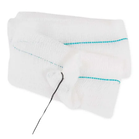
Because it is a sponge left in a body opening, it is handled like any counted sponge:
- It is counted with the other sponges, by you and the circulator. Some references do not count it with the sponges, so your facility's policy decides how it is recorded.
- Its placement is announced to the team. Facilities mark it so no one forgets it, for example with a hemostatHEE-moe-statA clamp with finger rings and a locking ratchet used in surgery to grasp and close off bleeding vessels.hem/o blood-stat stopping, standing stillFull entry hanging from the corner of the Mayo standMAY-oh standA small tray on an adjustable stand placed over or beside the patient during surgery to hold the instruments used most often.Full entry , a sticker, or its tail tied to the breathing tube.
- It comes out before the patient is extubated. If the jaws will stay wired together after surgery, it comes out before they are fixed together, or its end is brought out beside the back teeth so it can still be pulled at the end. The pharynxFAIR-inksThe throat: the passage behind the nose and mouth that carries air to the voice box and food to the esophagus.pharyng/o throatFull entry is suctioningSUK-shun-ingRemoving mucus, saliva or other secretions from the mouth, nose or airway with a tube connected to a suction source.Full entry with the Yankauer, and the pack is pulled with the same instrument that placed it.
- Its removal is confirmed aloud before the breathing tube comes out, and it is counted out with the sponges.
a throat pack left in place can be inhaled once the breathing tube is out and block the airway. It is counted like a sponge, it always comes out before extubation, and when the jaws will be wired shut, its removal is planned before they are fixed together.
6Position, prep and drape
Position
Most cases are supine, with the head on a foam or doughnut headrest (or a Mayfield headrest) that keeps it still, the upper body slightly raised, and the arms tucked at the sides with the ulnar nerves protected. For mouth cases the head is tilted back a little, sometimes over a rolled towel. The table is often turned so the surgeon can work from the head, with the anesthesia provideran-us-THEE-zhuh pruh-VY-derThe licensed professional who gives anesthesia and watches over the client's breathing and vital functions during a procedure.Full entry at the side; if the surgeon sits, the table may be reversed to give knee room. Long cases need padding at the heels, elbows, sacrumSAY-krumThe large, triangle-shaped bone at the base of the spine, between the two hip bones.sacr/o sacrumFull entry and shoulders, warming, and often a urinary catheter, and in children blood loss is watched closely.
The eyes are lubricated and protected, with ointment placed before the prep and corneaKOR-nee-uhThe clear, dome-shaped layer at the front of the eye, over the iris and pupil.Full entry shields when the work is near the eye. During a long mouth case the lips can dry and crack, so a cream or ointment may be put on them.
Prep
For facial surgery the prep covers the whole face, from the sternal notch up to the hairline, with the breathing tube included because it lies in the field. The usual antiseptic is povidone-iodinePOH-vih-done EYE-uh-dineA brown antiseptic solution of iodine used to kill germs on the skin before procedures and on wounds.Full entry without alcohol, after checking that the patient is not sensitive to iodineEYE-uh-dyneA mineral the thyroid gland uses to make thyroid hormone; it is also used to kill germs on the skin and in some contrast dyes.Full entry . Chlorhexidine (CHG) and alcohol-based preps damage the ear (they are ototoxic), and they are never used in the ear, the eye or the mouth, so neither CHG nor hexachlorophene is used on the face.
- Keep prep solution out of the eyes and the ears.
- With an injury, first remove gross debris, such as glass or dirt, gently, so you do not move a broken bone or damage the soft tissue further.
- Facial hair may be removed if needed, but the eyebrows and eyelashes are never shaved; the lashes may be trimmed if the surgeon asks.
- For a case only inside the mouth, a skin prep may not be needed. The teeth are brushed, or the inside of the mouth is swabbed, with an oral antiseptic.
AST's 2008 skin prep standard rules out both CHG and iodine (iodophor) preps for the eye and face, because either can injure the cornea, and suggests a nontoxic agent such as PCMX or triclosan instead, kept out of the eyes.
Drape
For a facial fracture, four towels held with clips frame the site, and a split sheet goes over the patient and around the face, leaving the mouth, the nose and the eyes in the field. Another way: towels outline the site, a head drape (turban) wraps the hair, a bar drape crosses the forehead and covers the head of the table, and a U-drape starts at the nose, its two arms running out past each side of the head and the rest of it covering the body. A tooth extraction needs only a head drape and a body sheet, and some mouth cases need no more than a half sheet across the body as a clean place to rest supplies.
The mouth is not sterile
The mouth holds many germs and cannot be made sterile, so mouth cases are done with the best technique possible rather than as sterile cases. When a case uses both the mouth and an incision through the skin onto the bone, the instruments used inside the mouth are kept apart from the sterile bone and plating setup, and the skin part is done with full surgical asepsisSUR-jih-kul ay-SEP-sisPractices that keep an area or object completely free of microorganisms; also called sterile technique.a- withoutsepsis decay, infectionFull entry . The arch bar instruments sit on their own Mayo standMAY-oh standA small tray on an adjustable stand placed over or beside the patient during surgery to hold the instruments used most often.Full entry .
7Incisions of the face
The surgeon reaches a fracture through the wound itself or through an incision chosen to give enough room and to hide the scar:
- Intraoral: inside the mouth, in the gum and cheek groove (the gingivobuccal, or vestibular, sulcus) above the upper teeth for the maxilla, or below the lower teeth for the front of the mandibleMAN-dih-bulThe lower jawbone, the only bone of the skull that moves.Full entry ; an incision in the cheek lining (buccal mucosamyoo-KOH-suhThe moist lining of body passages that open to the outside, such as the mouth, nose, gut and bladder.Full entry ) also reaches the mandible.
- Subciliary: through the skin of the lower eyelid, about 2 mm below the lashes.
- Transconjunctival: through the conjunctivakon-junk-TY-vuhThe thin, clear layer that lines the inside of the eyelids and covers the white of the eye.Full entry , inside the lower eyelid, to reach the orbital floor and the front of the maxilla. It is not sutureSOO-cherA stitch, or the thread used to stitch, that holds the edges of a wound or incision together.Full entry .
- Lateral eyebrow or upper eyelid: for the joint of the zygoma and the frontalFRUN-tulRelating to the front of the body, or to the forehead and the bone or brain area behind it.Full entry bone at the outer orbital rim.
- Coronal: across the top of the scalp from ear to ear, behind the hairline, with the scalp turned down over the face. It opens the forehead, the frontal sinusSY-nusA hollow space in the body, most often one of the air-filled spaces in the skull bones around the nose.Full entry , the upper orbits, the nasal bones and the zygomatic arches; the surgeon guards the frontal branch of the facial nerveFAY-shul NERVThe seventh cranial nerve, which moves the muscles of the face and carries taste from the front of the tongue.Full entry as the flap comes down over the temples.
- Submandibular: under the jaw, parallel to its lower border, for the angle, the ramus and the condyle, sparing the marginal mandibular branch. An incision in front of the ear (preauricular) also reaches the condyle and the joint, sparing the frontal branch.
- Temporal (Gillies): inside the hairline of the temple, so an instrument can pass under the zygomatic arch and lift it.
8Trays, instruments and supplies
Maxillofacial surgery works on bone and on soft tissue, so its setups borrow from several specialties: fine orthopedicor-thuh-PEE-dikHaving to do with the bones, joints, muscles, ligaments and tendons, and the branch of medicine that treats them.orth/o straightped/o child (the field began with straightening children's bones)-ic relating toFull entry instruments, plates and graftGRAFTTissue or a man-made material moved into place in surgery to replace or repair a damaged part.Full entry , plastic surgery instruments, and dental and nasal instruments. A frontalFRUN-tulRelating to the front of the body, or to the forehead and the bone or brain area behind it.Full entry bone injury may need neurosurgery instruments, and an eye injury eye instruments. Most mouth and jaw cases start from a dental set and a basic plastic set; fracture cases add a facial fracture set, an arch bar set and a plating system. The surgeon often wears a headlamp.
For the mouth and the teeth
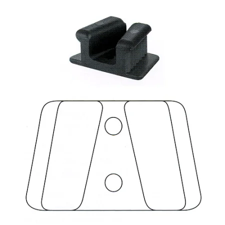
- Mouth props in several sizes hold the mouth open, and cheek retractorrih-TRAK-terA surgical instrument that holds back tissue or organs so the surgeon can see and reach the operative site.Full entry (such as the Minnesota or a plastic cheek retractor) and a Weider tongue depressor hold the cheek and tongue aside. A mouth mirror, precoated with antifog solution, can also hold back the cheek or tongue, and a probe with graduated marks, slid into the gumline, shows how much the gum around a tooth is damaged.
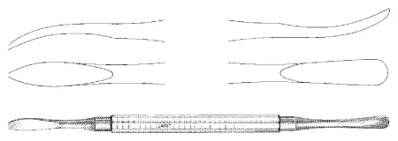
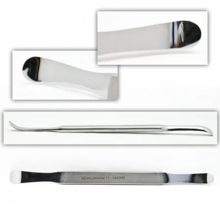
- Periosteal elevators, such as the #9 Molt or the double-ended Seldin, lift the gum and periosteum off the bone. Dental elevators, such as the right and left Cryer, loosen a tooth or a root in its socket. Extraction forcepsFOR-sepsA hand instrument with two blades or arms used to grasp, hold or pull tissue or objects.Full entry come in patterns and sizes chosen for the tooth and the patient's age.
- A periapical curettekyoo-RETA surgical instrument with a small spoon- or loop-shaped tip used to scrape tissue.Full entry , a bone file, a single-action bone rongeurron-ZHURA strong surgical instrument with sharp, cup-shaped jaws used to bite away small pieces of bone.Full entry , a dental drill and an osteotome with a mallet shape or remove bone around a tooth.
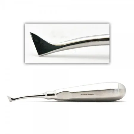
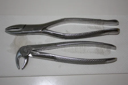
For the bones
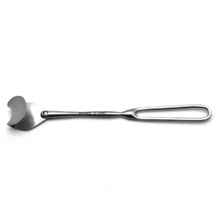
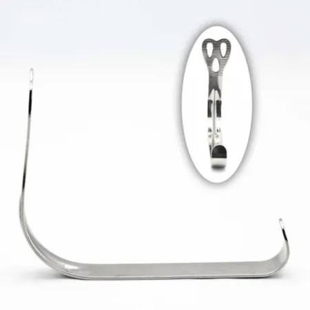
Fracture cases use periosteal elevators such as the Freer, bone hooks, Hohmann retractors, small bone clamps and bone-holding forceps (such as Dingman bone-holding forceps) to hold a reduced mandibleMAN-dih-bulThe lower jawbone, the only bone of the skull that moves.Full entry , and right and left Rowe disimpaction forceps to grip and free a maxilla driven back by the blow. Zygoma elevators and heavy hooks, such as the Dingman zygoma hook, lift a cheekbone back into place. Plates are held on the bone with a plate-holding forceps.
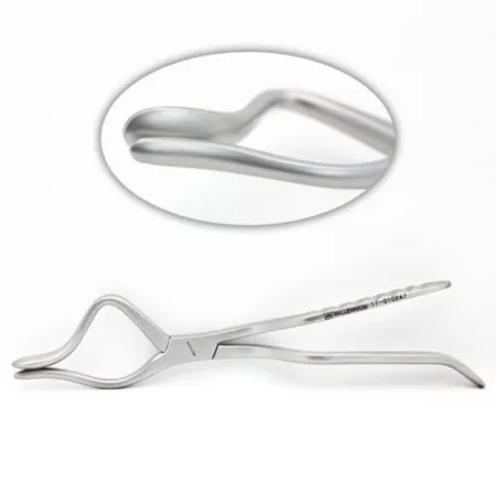
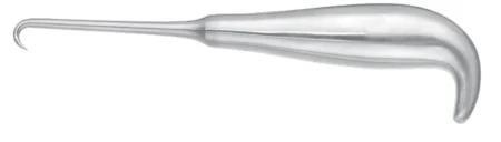
For arch bars and wiring
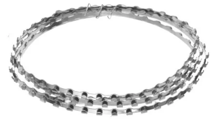
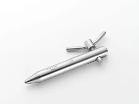
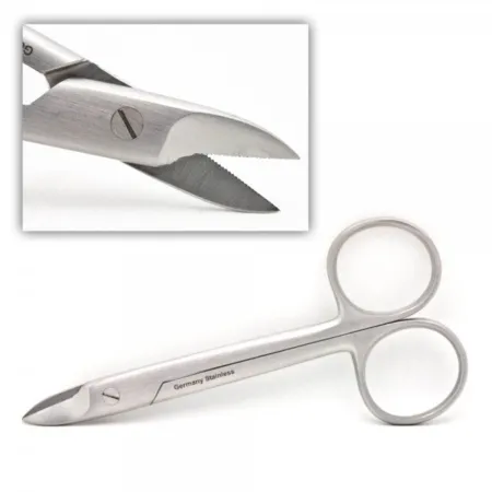
The arch bar setup sits on its own Mayo standMAY-oh standA small tray on an adjustable stand placed over or beside the patient during surgery to hold the instruments used most often.Full entry : arch bars, stainless steel wire (usually 24 to 26 gauge), wire twisters (wire tighteners), wire cutters, elastic (latexLAY-teksNatural rubber made from the sap of the rubber tree, used in some gloves, catheters and other medical supplies.Full entry ) bands, large and small Weider tongue depressors, a Freer septumSEP-tumA wall of tissue that divides two spaces or cavities, such as the two sides of the heart or nose.sept/o wall, partitionFull entry elevator and a small drain, with a Brown fasciaFASH-ee-uhA sheet or band of tough connective tissue that covers and separates muscles and organs.fasci/o bandFull entry needle if dentures or splints will be wired in. Arch bars, stainless steel wire and Silastic sheeting are the specialty supplies the Core Curriculum lists for this field. Stainless steel sutureSOO-cherA stitch, or the thread used to stitch, that holds the edges of a wound or incision together.Full entry is strong and inert but difficult to handle, and its cut ends puncture easily.
Suction, not sponges
The face and mouth have a rich blood supply, and suction, rather than sponges, keeps the field dry. Have a Yankauer and a smaller round-tipped tip, with Frazier tips for fine work, such as inside the gum or near the eye.
Power, plates and screws
Chapter 39 teaches drills, saws, plates and screws in general; here is what the face adds.
- Drills and saws: a small high-speed or air-powered drill places the screws, a dental drill cuts bone around teeth, and a saw cuts the jaws in orthognathic surgery. The drill heats bone, so irrigationeer-ih-GAY-shunWashing out a wound, body cavity, tube or eye with a steady flow of liquid.Full entry and suction run whenever it is used. Keep the controls in the safe position whenever you load, pass or rest the tool.
- Plating systems come from the makers as sets: plates and mesh, the matching screws, drill bits, plate benders and cutters, holding forceps and screwdrivers, often color coded by size. Plates come in straight, L, Y, H and T shapes. Titanium, which is strong, light and does not corrode, is used most; resorbable plates, which the body slowly absorptionub-ZORP-shunThe taking in of a substance, such as a drug or a nutrient, into the bloodstream.Full entry , are of most value in children. Resorbable plates and mesh are softened for shaping in a small sterile warm-water bath on the field, as the maker's instructions direct.
- Sizes: midface plates are usually 1.7 or 2 mm, orbital plates or mesh 1, 1.3 or 1.5 mm, and mandible plates 2.4 and 2.7 mm. Smaller screws suit the delicate facial bones, larger ones the mandible.
- Screws: cortexKOR-teksThe outer layer of an organ, especially the folded gray outer layer of the brain.cortic/o cortex, outer layerFull entry screws hold a plate to the bone. A bicortical screw passes through both hard outer layers of a bone and the spongy bone between them. A lag screw draws two pieces of bone tightly together as it goes in. Self-tapping screws cut their own thread; otherwise the hole is tapped first.
The plating rhythm is the same on every facial bone, so learn it once:
- The surgeon chooses a plate; you offer a selection. It is bent and cut to fit the bone with the plate benders and cutters.
- The plate is held against the bone with a plate-holding forceps.
- Load the drill with the bit that matches the screw. The surgeon drills, often through a drill guide, while you irrigate.
- Pass the depth gauge to measure the hole, and the tap if the hole needs a thread.
- Load the screw of that length onto the screwdriver and pass it.
- Reset the drill and the tools for the next hole. Repeat until the plate is secure: at least two screws on each side of the break.
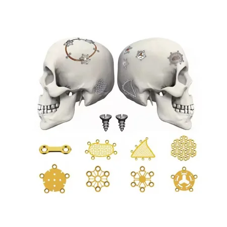
Every plate, screw, mesh and graft placed is an implant: confirm its type and size with the surgeon before it is opened, and record each one in the patient's record.
Grafts and sheeting
- Bone grafts fill gaps where bone was lost, fill cavities so new bone can grow, or support a weak repair. The patient's own bone (an autograft) comes from the iliacIL-ee-akHaving to do with the ilium, the large, wing-shaped upper part of the hip bone.Full entry crest, the ribs or the skull (calvarium); donor bone (an allograft) comes from a bone bank; coral (a xenograft) and synthetic materials can be used too. A graft site needs its own prep and drape and a separate setup. Keep harvested bone in a basin on the back table, covered with moist saline sponges or in antibiotic solution as the surgeon asks, never in water; chapters 25 and 29 teach grafts in general.
- Orbital floor materials include Silastic (silicone) sheeting, nylon sheeting, polypropylene mesh, Gelfilm, thin titanium plates or mesh, and porous polyethylene, with or without a titanium mesh inside. Sheeting is prepared and sterilizationstair-uh-luh-ZAY-shunA process that destroys all microorganisms, including spores, on an item; also a permanent birth control procedure.Full entry as its maker directs.
Dental sponges, drugs and dressings
- Sponges: small 2 by 2 gauze, dental (cotton) rolls and the throat pack. Gauze may be packed into a tooth socket to stop bleeding.
- Syringes: a 10 mL syringe with a 25-gauge needle for local anestheticLOH-kul an-es-THET-ikA drug that blocks feeling in one part of the body without making the person unconscious.Full entry , a dental cartridge syringe with anesthetican-us-THET-ikA drug that causes loss of feeling, with or without loss of consciousness.an- withoutesthesi/o feeling, sensation-ic relating toFull entry cartridges, and 20 mL syringes with blunt 19-gauge needles for irrigation. Keep two irrigation syringes so one can be refilled while the other is used.
- Local anesthetic with epinephrineep-ih-NEF-rinA hormone and drug, also called adrenaline, that raises heart rate and blood pressure and opens the airways.Full entry is often injected, with or without general anesthesiaJEN-er-ul an-es-THEE-zhuhDrug-induced loss of consciousness and feeling over the whole body, so a person feels nothing and is not aware during surgery.an- withoutesthesi/o feeling, sensation-ia conditionFull entry , to reduce bleeding and pain after surgery. Prepare it as the surgeon prefers, label the cup and every syringe with the drug, its strength and the epinephrine ratio, and tell the anesthesia provideran-us-THEE-zhuh pruh-VY-derThe licensed professional who gives anesthesia and watches over the client's breathing and vital functions during a procedure.Full entry that epinephrine is being used. Keep a running total of every mL used and report it to the circulator, because local anesthetics have maximum doses (chapters 12 and 13). Refill the syringe ahead when both sides will be injected.
- Eye care: ophthalmicoff-THAL-mikHaving to do with the eye, or describing a medicine made to be put in the eye.ophthalm/o eye-ic relating toFull entry ointment and balanced salt solution to keep the corneaKOR-nee-uhThe clear, dome-shaped layer at the front of the eye, over the iris and pupil.Full entry moist when the work is near the eye.
- Dressings: an incision in the mouth needs none. Skin incisions get antibiotic ointment, a nonadherent dressing such as Telfa, flat gauze or fluffs, and a rolled gauze wrap, which molds to the head and face. A submandibular incision with a drain gets a moderate pressure dressing, and an eye patch may follow cheekbone surgery. Ice reduces the bruising and swelling of the lips and cheeks.
9Arch bars and maxillomandibular fixation
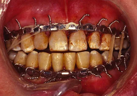
What and why: maxillomandibular fixation (MMF), also called intermaxillary fixation, holds the jaws closed in the correct bite. The usual hardware is the arch bar, a bendable stainless steel strip with small hooks along one edge. One bar is wired to the upper teeth and one to the lower teeth, the jaws are closed, and wire loops or elastic bands are hooked between the two bars. MMF may be used only during surgery, to set the bite while the fracture is plated, or kept on for weeks while a fracture heals; for some fractures it is the only treatment needed. Many surgeons consider arch bars the standard way to give MMF, though plates and screws have cut how often, and for how long, the jaws must be wired.
Before surgery: the surgeon checks the patient's bite, noting any malocclusion the patient had before the injury. general anesthesiaJEN-er-ul an-es-THEE-zhuhDrug-induced loss of consciousness and feeling over the whole body, so a person feels nothing and is not aware during surgery.an- withoutesthesi/o feeling, sensation-ia conditionFull entry goes through a nasal tube, so the teeth can close, unless the midface is broken (see the shared airway), and local anestheticLOH-kul an-es-THET-ikA drug that blocks feeling in one part of the body without making the person unconscious.Full entry with epinephrineep-ih-NEF-rinA hormone and drug, also called adrenaline, that raises heart rate and blood pressure and opens the airways.Full entry may be used with it, or alone, to reduce bleeding and pain after surgery. Prepare the wire ahead: cut segments a little longer than needed, about 10 cm, clamp each end in a wire twister, twist both a half turn and pull to stretch the wire so it will not stretch later, then trim the crimped ends. Have at least one wire for every tooth that will be wired, plus extras for wires that break or fall. For a patient without teeth, the bars are fixed to the patient's dentures or to splints made for them, and these are wired to the jaws.
Key steps:
- A mouth prop and cheek retractorrih-TRAK-terA surgical instrument that holds back tissue or organs so the surgeon can see and reach the operative site.Full entry open the field, and the throat pack goes in.
- The upper arch bar is measured, bent to the curve of the teeth and cut so it does not run past the last tooth or press into the gum. It sits between the widest part of the teeth and the gum, with its hooks facing away from the biting surfaces: up on the upper bar, down on the lower.
- A wire goes around the neck of each tooth, one end above the bar and one below, and is twisted tight, always clockwise, with the wire twister. Pass the wire mounted in its holder, then the twister and a probe to guide the wire, then the wire cutter. The twisted end is cut short and crimped in against the bar, away from the gum, so it does not cut the lip or cheek.
- The lower bar goes on the same way.
- The throat pack comes out, or its end is brought out beside the back teeth, before the jaws are fixed together; count it.
- The jaws are closed in the correct bite, and wire loops or elastic bands go over the hooks of both bars: at least three, one at the back on each side and one in front. Wires are twisted clockwise until snug and cut, and their ends are turned in.
- The mouth gets a final rinse and suction, the retractors come out, and every loose piece of wire is collected and discarded as a sharp.
Why clockwise: every wire on the jaws is tightened clockwise, so any other surgeon who has to remove it knows to untwist it counterclockwise.
Sharps: the cut ends of wire puncture gloves easily. Double glove; handle the wire with the twister rather than your fingers; keep the loose ends under control, grasping each one at its tip so no point is left free; pass it with care; and put cut pieces straight into the sharps container.
Elastics: the surgeon may place the elastic bands at the end of surgery, but often they go on later, once the patient is fully awake and alert.
hybrid arch bars are fixed with screws into the jaw bone instead of wires around every tooth, which makes MMF faster to place; other bone-supported devices, such as screws placed in the jaw for the loops, are used too.
Wound class: II, because the mouth is entered.
The wire cutters
A patient whose jaws are wired or banded shut cannot open the mouth. If they vomit, or if swelling blocks the airway, the vomit cannot get out and no one can reach the airway, so the patient can inhale it (aspirationas-puh-RAY-shunBreathing food, liquid, saliva or stomach contents into the airway and lungs; also, drawing fluid out with suction or a needle.Full entry ) or be unable to breathe. The mouth must be able to be opened quickly.
- Wire cutters (and scissors, if elastic bands are used) go with the patient from the OR, taped to the head of the stretcher or bed where everyone can see them.
- They stay with the patient the whole time the jaws are fixed: at the bedside in PACU and on the unit, and at home. The circulator shows the PACU nurse that they are taped to the head of the stretcher, and their placement is documented in the perioperativepair-ee-OP-er-uh-tivRelating to the whole time around a surgery: before, during and after it.peri- aroundoperative relating to surgeryFull entry record.
- In an emergency, the team cuts the tie wires or elastic bands that join the upper and lower arch bars, and the mouth opens. Only these links between the jaws are cut. The wires around each tooth hold each bar to its own jaw, so the bars stay on the teeth, just as they do before the ties go on.
- The patient and the nursing staff are taught how to use them, and a patient who goes home in MMF takes wire cutters and is taught to remove the wires in case of vomiting or trouble breathing.
let a patient whose jaws are wired or banded together leave the OR without wire cutters, or scissors for elastics, at the head of the stretcher. They let the team open the mouth quickly if the patient vomits or cannot breathe.
After surgery: the patient lives on liquids until the fixation comes off, often 4 to 6 weeks, and keeping weight on is hard, so nutritional supplements help. Food collects between the wires and the teeth, so the patient keeps the mouth very clean with a small, soft toothbrush and a home water irrigator. The bars usually come off in the office. Swelling can still threaten the airway in the first hours, and a tracheotomytray-kee-OT-uh-meeThe surgical cut made into the windpipe through the front of the neck.trache/o windpipe-tomy cutting, incisionFull entry may become necessary. Bleeding, infection and a bite that heals wrong are the main complications.
10Facial fracture repair
What every repair shares
In an ORIFOR-if, or said as lettersOpen reduction internal fixation: surgery that opens the skin to line up a broken bone and hold it with plates, screws or rods.Full entry (ORIF), the surgeon exposes the break, brings the pieces back into their normal position (reduction) and holds them there with plates and screws or wires (fixation). Plates and screws, or screws alone, are now the most common way to fix facial fractures.
The repair follows a few rules:
- The bite leads. Arch bars usually go on first, and MMF sets the teeth in their correct bite, so the broken bones can be lined up against it.
- An order. A broken mandibleMAN-dih-bulThe lower jawbone, the only bone of the skull that moves.Full entry is repaired before the midface, and the Le Fort fractures before the orbital walls.
- All the pieces before the plates. Every fracture is exposed and reduced before any plate is fixed.
- Check the bite again. After plating, the MMF is released and the surgeon checks the bite with the condyles seated in their sockets. If the teeth do not meet as they should, the reduction is revised.
X-rays or fluoroscopyfloo-ROS-kuh-peeAn x-ray method that shows moving pictures of the inside of the body in real time.fluor/o glowing (from the fluorescent screen)-scopy looking, viewingFull entry during the case may confirm the reduction, so let radiology know ahead and be ready to receive an X-ray cassette into the sterile fieldSTAIR-ul feeldAn area set up with sterile drapes and supplies, kept free of all germs, for a sterile procedure.Full entry .
ORIF of the mandible
What and why: the aim is to give back the patient's bite from before the injury. Some mandible fractures heal with closed reduction and MMF alone, but most are opened and fixed so the bone is held still enough to heal.
Before surgery: the patient lies supine, the head steadied on a doughnut headrest, arms tucked, under general anesthesiaJEN-er-ul an-es-THEE-zhuhDrug-induced loss of consciousness and feeling over the whole body, so a person feels nothing and is not aware during surgery.an- withoutesthesi/o feeling, sensation-ia conditionFull entry through a nasal tube. A badly swollen face can block the airway, so a tracheotomytray-kee-OT-uh-meeThe surgical cut made into the windpipe through the front of the neck.trache/o windpipe-tomy cutting, incisionFull entry may come before the repair. Prep and drape for a face case. The setup: a facial fracture set or minor orthopedicor-thuh-PEE-dikHaving to do with the bones, joints, muscles, ligaments and tendons, and the branch of medicine that treats them.orth/o straightped/o child (the field began with straightening children's bones)-ic relating toFull entry set, the arch bar set on its own Mayo, the plating system, a drill, two Dingman bone-holding forcepsFOR-sepsA hand instrument with two blades or arms used to grasp, hold or pull tissue or objects.Full entry , a nerve stimulator, marking pens, stainless steel wire and the electrosurgeryee-LEK-troh-SUR-jer-eeUsing high-frequency electric current through a handheld tool to cut tissue or seal bleeding vessels during surgery.Full entry .
Key steps:
- Arch bars and MMF set the bite, before or after the fracture is opened.
- The surgeon injects local anestheticLOH-kul an-es-THET-ikA drug that blocks feeling in one part of the body without making the person unconscious.Full entry with epinephrineep-ih-NEF-rinA hormone and drug, also called adrenaline, that raises heart rate and blood pressure and opens the airways.Full entry and makes the incision: inside the mouth through the gum and cheek lining with the ESU, or through the skin under the jaw with a #10 or #15 blade, parallel to the lower border. For a break at the angle or the back of the body, the nerve stimulator helps find the marginal mandibular branch of the facial nerveFAY-shul NERVThe seventh cranial nerve, which moves the muscles of the face and carries taste from the front of the tongue.Full entry . Senn retractorrih-TRAK-terA surgical instrument that holds back tissue or organs so the surgeon can see and reach the operative site.Full entry hold a skin incision open.
- The periosteum is lifted off the bone with a Freer or other periosteal elevator, exposing the fracture.
- The surgeon reduces the fracture and an assistant holds it with a small bone clamp or Dingman forceps. Steady the head and watch that the tube does not kink or slip.
- The plates go on in the plating rhythm. Most mandible fractures take two plates: a larger plate along the lower border (often 2.4 or 2.7 mm) and a miniplate or tension band along the upper border. Lag screws, without a plate, can also draw the pieces together.
- An X-ray may confirm the reduction. The wound is irrigationeer-ih-GAY-shunWashing out a wound, body cavity, tube or eye with a steady flow of liquid.Full entry , and a small drain may go in. An incision inside the mouth is closed with 3-0 absorbable sutureSOO-cherA stitch, or the thread used to stitch, that holds the edges of a wound or incision together.Full entry ; a skin incision is closed in layers with 3-0 absorbable suture and the skin with 4-0. The arch bars stay or come off, as the surgeon decides.
- A submandibular wound gets a moderate pressure dressing over it and its drain.
one reference still teaches holding a mandible fracture with stainless steel wire, passed through holes drilled on each side of the break and twisted tight, using plates only when rigid fixation is wanted.
Wound class: Class II when the repair enters the mouth. A repair done entirely through a skin incision, with no tract entered, is Class I by the CDC's definitions, as one reference lists it.
After surgery: the airway is watched closely, because swelling can block it. The lips and cheeks bruise and swell, so ice is applied. The elastics may go on now that the patient is awake, and if the jaws are fixed together the wire cutters go with the patient. Recovery takes about 4 to 6 weeks, and bleeding, infection and a bite that heals out of line are the main complications.
Splitting the jaw: mandibulotomy
To reach a large cancer at the back of the mouth, such as one of the tongue or the floor of the mouth, the surgeon may split the mandible in the midlineMID-lineAn imaginary line down the center of the body; also, a type of IV catheter placed in a vein of the upper arm.Full entry or just beside it (a mandibulotomy). Chapter 36 teaches the cancer operation itself; the jaw work follows the plating rhythm.
- The lip is split in the midline with a #15 blade and the cut carried down under the chin. The skin flaps are raised with the ESU and held with Senn rakes.
- Before the bone is cut, a titanium plate is fitted across the planned split, its screw holes are drilled with a small power drill, and it is screwed on, then taken off. Placing the plate first lets the jaw go back together in the patient's own bite.
- The surgeon splits the mandible in the midline with a small power saw, and the tumor is removed.
- The two halves are brought back together and fixed with the same plate. The lip, the mouth lining and the deep layers are closed over drains with 3-0 absorbable suture, and the skin with fine nonabsorbable suture.
Wound class: II, because the mouth is entered.
ORIF of a Le Fort I fracture
What and why: the repair reattaches the loose tooth-bearing maxilla to the face in the correct bite and rebuilds its buttresses. A Le Fort I fracture, and some Le Fort II fractures, may be treated by closed reduction and MMF alone; the more severe Le Fort II fractures and every Le Fort III need open reduction as well as MMF.
Before surgery: X-rays, computed tomographykum-PYOO-tid toh-MOG-ruh-feeAn imaging test that uses x-rays and a computer to make cross-section pictures of the inside of the body.tom/o cut, slice-graphy process of recordingFull entry and the exam make the diagnosis, and photographs from before the injury and the dental records show where the bones belong. A tracheotomy and the arch bars may come first, and dental impressions may be taken before the incision. The patient lies supine, the head on a doughnut or Mayfield headrest and the arms at the sides, under general anesthesia, with the breathing tube placed so the teeth can close (a nasal tube only when the skull base is not injured; see the shared airway), and the whole face is prepped, gently, without pressing on the loose bones. The setup: a facial fracture set, the arch bar set, a power drill, and plates and screws, with Rowe disimpaction forceps.
Key steps:
- Arch bars go on and the jaws are placed in MMF as needed.
- Local anesthetic with epinephrine is injected into the gum, and the surgeon makes an incision in the groove between the upper gum and the cheek with a #15 blade on a #7 handle, down through the mucosamyoo-KOH-suhThe moist lining of body passages that open to the outside, such as the mouth, nose, gut and bladder.Full entry to the maxilla. Have the ESU and suction ready.
- A Freer or a slightly larger periosteal elevator lifts the periosteum to expose the fracture lines.
- The surgeon frees and reduces the maxilla, by hand, with an elevator, or with the Rowe forceps placed in the nose and the mouth, careful not to disturb the breathing tube, and sets it in the bite with MMF, the condyles seated in their sockets.
- Plates fix the maxilla at four points: one at the buttress behind each cheekbone (the zygomaticomaxillary buttress), where chewing forces are greatest and a sturdy L, T, Y or straight plate is used, and one beside each side of the nasal opening (the piriform aperture). The plates are usually 1.7 or 2 mm, with at least two screws on each side of the break, placed so they miss the tooth roots.
- The MMF is released and the bite checked. An X-ray may confirm the reduction.
- The wound is irrigated and the mucosa closed with 3-0 absorbable suture. An incision in the mouth needs no dressing.
one reference still teaches fixing the maxilla with stainless steel wires passed through holes drilled on each side of the break, twisted clockwise, cut and buried in the hole, alone or with plates.
Wound class: Class II by the CDC's definitions, because the mouth and the maxillary sinusSY-nusA hollow space in the body, most often one of the air-filled spaces in the skull bones around the nose.Full entry are entered. One reference lists the Le Fort I repair as Class I.
After surgery: keeping the head raised before and after surgery reduces swelling and pain around the eyes. Watch for bleeding, swelling of the airway and clear fluid from the nose (a cerebrospinal fluidser-uh-broh-SPY-nul FLOO-idThe clear, watery fluid that bathes and cushions the brain and spinal cord.cerebr/o brainspin spine-al relating toFull entry leak). The patient eats a soft diet that needs no chewing for about 4 to 6 weeks, or liquids while the jaws are wired, and must keep the mouth clean: a mouth wound can split open when oral hygiene is poor, and a water jet used to clean the mouth is never aimed straight at an incision. The bone takes about 4 to 8 weeks to heal. Bleeding, infection and malocclusion are the main complications, and further surgery may be needed.
ORIF of Le Fort II and III fractures
What and why: these fractures reach the nose, the orbits and the skull base, so the repair rebuilds more buttresses and needs more approaches. Fluid from the nose after injury warns of a torn duraDOOR-uhThe tough outer layer of the membranes that cover the brain and spinal cord.Full entry , which the surgeon may seal with a stitch or a graftGRAFTTissue or a man-made material moved into place in surgery to replace or repair a damaged part.Full entry of fat, fasciaFASH-ee-uhA sheet or band of tough connective tissue that covers and separates muscles and organs.fasci/o bandFull entry or muscle.
Approaches: a Le Fort II is reached inside the mouth, through the lower eyelids (subciliary or transconjunctival), through any open wound and, for the nasal bones, through a small incision between the eyebrows or a coronal incision. A Le Fort III is usually reached through a coronal incision; incisions in both upper eyelids and between the eyebrows, with one in front of each ear for the arches, can avoid it.
Setup: as for a Le Fort I, with right and left Rowe forceps, periosteal elevators, the plating system and the sets for each approach. The setup card below is the Le Fort II repair's; a Le Fort III uses the same setup, with the sets its approaches need, such as for a coronal incision.
Key steps:
- Local anesthetic with epinephrine is injected and the approaches are made to expose the fractures at the orbital rims below the eyes, at the joints of the zygomas and the frontalFRUN-tulRelating to the front of the body, or to the forehead and the bone or brain area behind it.Full entry bone and at the root of the nose.
- Arch bars and MMF set the bite. A broken mandible is fixed first, then the MMF is released to let the condyles settle and the bite is checked again.
- The maxilla is freed and reduced, with the Rowe forceps in the nose and the mouth, or with Tessier mobilizers behind it. The Rowe forceps can apply great force, and forcing the midface can crack the orbit or the skull base and cause blindness, bleeding or a CSF leak; many surgeons instead make a Le Fort I cut so the lower maxilla can be moved freely.
- Plates fix the buttresses. For a Le Fort II: a larger plate at each zygomaticomaxillary buttress, a thin, low-profile plate along the rim below each eye (the forces there are small, and the patient may feel a larger plate under the skin), and plates or wires at the root of the nose. For a Le Fort III: plates at the joints of the zygoma and the frontal bone, at the root of the nose, and on the zygomatic arches when they need it. The bite is checked once the first plates are in and again before closing.
- The orbital walls are repaired after the Le Fort fractures, the incisions are closed, and the nasal fracture is set at the end.
two references still teach hanging the maxilla from the skull with suspension wires: holes are drilled in the frontal bone through incisions at the outer eyebrows, and a Brown fascia needle passes wires behind the zygomatic arches into the mouth to the upper arch bar, with pullout wires brought out at the hairline and tied over polyethylene buttons on foam pads.
Wound class: Class II, because the mouth, the nose and the sinuses are entered; a fresh open wound makes it Class III.
After surgery: as for a Le Fort I, with close watch for a CSF leak, for bleeding and for changes in vision.
ORIF of the zygoma
What and why: the repair lifts a flattened cheek, frees a pushed-in arch that blocks the jaw, and puts the outer wall and floor of the orbit back where the eye needs them. A break of the arch alone is most often lifted without opening the fracture (closed reduction); a trimalar fracture is most often opened and fixed. A zygoma is usually fixed at more than one of its joints, so it often needs more than one incision.
Setup: for a closed reduction, an orthopedic tray, a zygoma hook elevator and a jaw hook. For an open reduction, add an air-powered drill, stainless steel wire (26 to 30 gauge), a Kerrison rongeurron-ZHURA strong surgical instrument with sharp, cup-shaped jaws used to bite away small pieces of bone.Full entry , two Blair retractors, a bipolarby-POH-lerHaving two poles or two opposite ends; most often used as a short form of bipolar disorder.bi- twopolar having polesFull entry ESU, marking pens and a miniplating set. The patient is supine.
Closed reduction: for an arch fracture, the surgeon makes a short incision in the hairline of the temple (the Gillies approach), slides an elevator, such as a Gillies zygoma elevator, down beneath the temporalis fascia and under the arch, and lifts the arch back out. A depressed trimalar fracture may be lifted with a bone hook through the skin, then held with a Kirschner wire passed across through the maxillary sinuses, from the broken side to the normal side.
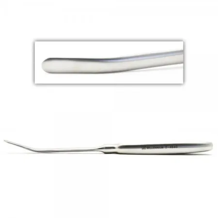
Open reduction:
- The surgeon marks incisions at the outer eyebrow, in the lower lid and over the rim below the eye, injects local anesthetic with epinephrine, and opens down to the bone.
- The zygoma is lifted with a periosteal elevator or a Kelly clamp placed behind it through the eyebrow incision, or with bone hooks or Hohmann retractors.
- At least two of its three fracture sites are fixed, usually with small plates and screws, or with wires through drill holes.
- The incisions are closed, and an eye patch may be applied.
Wound class: no reference gives one, so it follows the approach, by the CDC's definitions. Skin incisions that enter no tract are Class I; an incision inside the mouth, or entry into the maxillary sinus, makes it Class II.
After surgery: watch the eye. A zygoma left out of place lets the eye sink back.
ORIF of the orbital floor (blowout fracture)
What and why: the repair frees muscle and fat caught in the break, so the eye moves again and double vision clears, and it bridges the hole in the floor so the eye is held up and does not sink back. If the eye itself is injured, ask the surgeon which eye sets are needed, such as an eye muscle, cataractKAT-uh-raktClouding of the lens of the eye.Full entry or enucleation set; a cut through the tear duct near the inner corner of the eye needs a stentstentA small tube, often of metal mesh, placed inside a vessel or duct to hold it open.Full entry within about 72 hours. Chapter 35 teaches eye injuries.
Before surgery: the CT and X-rays stay in the room. The patient is positioned and prepped as for eye surgery, under general anesthesia, with ophthalmicoff-THAL-mikHaving to do with the eye, or describing a medicine made to be put in the eye.ophthalm/o eye-ic relating toFull entry ointment in the eyes before a careful prep that keeps solution out of the eyes and ears. Have a washed corneaKOR-nee-uhThe clear, dome-shaped layer at the front of the eye, over the iris and pupil.Full entry shield, the surgeon's implant material, prepared and sterilizationstair-uh-luh-ZAY-shunA process that destroys all microorganisms, including spores, on an item; also a permanent birth control procedure.Full entry as its maker directs, orbital retractors and the plating system; let radiology know an X-ray may be needed.
Key steps:
- To test whether a muscle is trapped, the surgeon grips the tendon of the inferior rectusREK-tusLatin for straight; the name of several straight muscles, such as the rectus abdominis of the abdomen and the rectus femoris of the thigh.Full entry with forceps and turns the eye as far as it will go (a forced duction test).
- A corneal protector goes on the eye, and balanced salt solution keeps the eye moist at this stage and as needed throughout; the scrub may do this.
- The incision is marked and injected with local anesthetic with epinephrine. The surgeon makes a subciliary incision about 2 mm below the lashes with a #15 blade, or a transconjunctival incision inside the lower lid. A tractionTRAK-shunA steady pulling force applied to a body part with weights, ropes and pulleys.Full entry stitch in the lid margin may help exposure. Keep cotton-tipped applicators or small eye sponges ready to blot blood and fluid.
- Fine tenotomy scissors and forceps dissectdy-SEKT, or dih-SEKTTo cut apart or separate tissues carefully during surgery.Full entry down to the orbital rim. The periosteum is opened with a #15 blade and lifted off the floor with a Freer.
- The orbital contents are lifted gently with a moistened orbital retractor or a small malleable or brain spatula retractor. The trapped fat and muscle are freed from the break, and loose bone splinters are removed.
- The floor is bridged: the surgeon cuts the implant to size with plate cutters or scissors and slides it between the floor and the orbital contents. An orbital plate is screwed to the rim below the eye. If the reduction is unstableun-STAY-bulDescribes a client whose condition is changing quickly or is hard to predict.Full entry , a rigid fixation plate goes in first, then the sheeting. The surgeon may instead use a bone graft from the iliacIL-ee-akHaving to do with the ilium, the large, wing-shaped upper part of the hip bone.Full entry crest.
- An X-ray may confirm the position. The subciliary incision is closed with 5-0 absorbable suture; a transconjunctival incision is not closed. The corneal protector comes out after closure, and antibiotic ophthalmic ointment may go in the eye and on the incision.
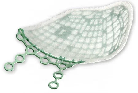
Wound class: Class II by the CDC's definitions when the repair enters the maxillary sinus, the airway space under the floor, as it does when trapped tissue is freed from the sinus or the sinus itself is opened (a Caldwell-Luc antrostomy). One reference lists the repair through the eyelid as Class I.
After surgery: vision may blur for a while from the swelling, and the eyes and nose bruise and swell; ice helps. Bleeding, infection and a change in vision from the injury to the eye are the complications to watch for. A small scar remains.
ORIF of the frontal bone and sinus
What and why: a frontal sinus repair has three jobs. The front wall is repaired so the forehead keeps its shape. The sinus must either drain into the nose or be closed off for good (obliterated), or it will become chronically infected. The back wall is managed to protect the brain in the front of the skull. A break that is not displaced may need only care of the skin wound, and the sinus may be watched.
Approach and setup: the surgeon reaches the forehead through a coronal incision or an open wound, and an endoscopeEN-duh-skohpA thin tube with a light and a camera that is passed into the body to look inside an organ or cavity.endo- within-scope instrument for viewingFull entry through a small incision can repair some front-wall fractures. The hair need not be shaved for a coronal incision, though a narrow hairless strip keeps hair out of the wound. Have the facial plating set, a drill, and neurosurgery instruments when the back wall or the dura is involved; chapter 42 teaches the craniotomykray-nee-OT-uh-meeSurgery in which a piece of the skull is removed to reach the brain, and usually put back at the end.crani/o skull-tomy cutting intoFull entry .
Key steps, by what is broken:
- Front wall only, the drainage passages intact: the pieces are lifted back into place and fixed with the smallest plates, or with small plates or wires for small fragments, because there is little force on the repair.
- The drainage passages injured: the surgeon may obliterate the sinus. Every bit of its lining is removed, so it cannot grow back and form a cyst (a mucocele), the drainage openings are plugged, for example with a plug of muscle glued in with fibrin sealant, and the sinus is filled, with fat, bone or another material, or left to fill in with bone.
- The back wall badly broken: neurosurgery repairs it from inside the skull, as chapter 42 teaches.
Wound class: no reference gives one, so it follows the CDC's definitions: Class I through the skin when no sinus or other tract is entered, Class II when the sinus, part of the airway, is entered, as in sinus surgery (chapter 36), and Class III with a fresh open wound over the break.
After surgery: watch for clear fluid from the nose (a CSF leak) and for infection.
11Orthognathic surgery
What and why: orthognathic surgery cuts and moves the jaws so the teeth meet properly and the face looks and works better. The surgeon may move the maxilla (a Le Fort I osteotomy), the mandibleMAN-dih-bulThe lower jawbone, the only bone of the skull that moves.Full entry (most often a bilateral sagittal split osteotomy), the chin (a genioplasty), or several at once. It is planned and elective, and it waits until enough permanent teeth are in place to hold the jaws still afterward.
Before surgery: braces line up the teeth first. The surgeon plans each movement and has splints made that the teeth fit into: an intermediate splint that sets the first jaw moved against the jaw not yet moved, and a final splint for the finished bite. Keep the splints and models with the patient's images in the room. Antibiotics are given, the breathing tube goes through the nose so the teeth can close into the splints, a shoulder roll extends the neck, and local anestheticLOH-kul an-es-THET-ikA drug that blocks feeling in one part of the body without making the person unconscious.Full entry with epinephrineep-ih-NEF-rinA hormone and drug, also called adrenaline, that raises heart rate and blood pressure and opens the airways.Full entry is injected in the groove above the upper teeth. The setup adds a reciprocating or piezoelectric saw, osteotomes and a mallet, a bone hook, Freer and Cottle elevators, arch bars and wire, and the plating system.
Key steps of a Le Fort I osteotomy:
- Arch bars go on. An incision in the groove above the upper teeth runs from one first molar to the other, and the periosteum and the lining of the nose are lifted off the maxilla with a periosteal elevator and a Freer or Cottle elevator.
- A saw cuts the maxilla straight across on each side, above the tooth roots, from the back of the maxilla through the buttress to the edge of the nasal opening, while a retractorrih-TRAK-terA surgical instrument that holds back tissue or organs so the surgeon can see and reach the operative site.Full entry guards the lining of the nose.
- Osteotomes tapped with a mallet free the maxilla from the pterygoid plates behind it, from the septumSEP-tumA wall of tissue that divides two spaces or cavities, such as the two sides of the heart or nose.sept/o wall, partitionFull entry and from the side walls of the nose. Keep the room quiet during these cuts: the surgeon listens for the change in sound that marks the back of the cut.
- The maxilla is fractured down (downfractured) with the thumbs and a bone hook, and freed until it moves into place without force.
- The maxilla is set into the intermediate splint, the jaws are put in MMF, and the maxilla is fixed with plates and screws. Bone graftGRAFTTissue or a man-made material moved into place in surgery to replace or repair a damaged part.Full entry fill any gaps.
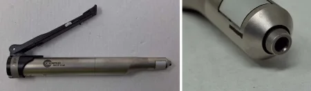
Sagittal split of the mandible: through incisions inside the mouth, the surgeon splits each ramus lengthwise (in the sagittal plane), so the tooth-bearing part can slide forward, slide back or rotate, and fixes it with plates or screws. It is the most widely used cut for the mandible. The inferior alveolusal-VEE-uh-lus (plural alveoli, al-VEE-uh-ly)One of the tiny air sacs at the ends of the smallest airways in the lungs.Full entry nerve runs inside the bone being split, so numbness or tingling (paresthesiapair-es-THEE-zhuhAn abnormal feeling in the skin, such as tingling, prickling or "pins and needles," with nothing touching it.para- abnormalesthesi/o feeling, sensation-ia conditionFull entry ) from that nerve is its most common complication.
Wound class: II, because the work is done inside the mouth.
After surgery: when the jaws are wired with arch bars, they stay 4 to 6 weeks, on a liquid diet with supplements, and the wire cutters go with the patient. Watch the airway: swelling after a sagittal split can block it, so no pressure dressing goes on, because it can push the swelling inward toward the airway.
12Teeth: odontectomy, implants and bone grafting
Instruments for removing teeth, for placing implants and for other surgical work on the teeth and jaws enter tissue that is normally sterile, so they are critical items and must be sterile, even though the mouth itself is not sterile.
Odontectomy and wisdom tooth removal
What and why: an odontectomy is the surgical removal of a tooth. In a simple extraction, the tooth is lifted out of its socket and drawn with forcepsFOR-sepsA hand instrument with two blades or arms used to grasp, hold or pull tissue or objects.Full entry . Removing an impactedim-PAK-tidFirmly packed or wedged in place so it cannot move or pass normally.Full entry tooth, most often a wisdom tooth, also means cutting the gum and removing the bone around it, and sometimes cutting the tooth into pieces. The operation is often done in the office; the OR is used for the reasons above, such as a bleeding disorder, extensive disease or a patient who needs general anesthesiaJEN-er-ul an-es-THEE-zhuhDrug-induced loss of consciousness and feeling over the whole body, so a person feels nothing and is not aware during surgery.an- withoutesthesi/o feeling, sensation-ia conditionFull entry .
Before surgery: the diagnosis comes from the exam and the panoramic X-ray, which stays in the room so the right teeth are removed. The patient lies supine with the arms tucked, under general anesthesia through a nasal tube (or monitored sedationsih-DAY-shunA calm, relaxed or sleepy state brought on by drugs, or the use of drugs to cause it.sed calm, settle-ation processFull entry ), draped with a head drape and a body sheet so the mouth is open to the surgeon. The setup: a dental set (an extraction tray), a dental drill with its bits, suction with irrigationeer-ih-GAY-shunWashing out a wound, body cavity, tube or eye with a steady flow of liquid.Full entry , a headlamp, the throat pack, 2 by 2 gauze and dental rolls, and antifog for the mirror.
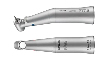
Key steps:
- The mouth prop goes in, then the throat pack. Cheek and tongue are held back with a Minnesota retractorrih-TRAK-terA surgical instrument that holds back tissue or organs so the surgeon can see and reach the operative site.Full entry , a cheek retractor or a mirror, and the site may be injected with local anestheticLOH-kul an-es-THET-ikA drug that blocks feeling in one part of the body without making the person unconscious.Full entry in a control syringe.
- For an impacted tooth, the surgeon cuts the gum down to the bone with a #15 blade, and a Molt or other periosteal elevator lifts the gum and periosteum away from the tooth.
- A dental drill removes the bone over the tooth, and the tooth may be split with the drill or an osteotome and mallet so it comes out in sections. Run suction and irrigation every time the drill runs, and pass the drill with its safety on.
- An elevator breaks the ligament that holds the tooth, so it loosens in its socket, and an extraction forceps of the right size removes it. 2 by 2 gauze may pack the socket for a moment.
- The socket is checked and irrigated so no debris stays, using a syringe of irrigation fluid with a blunt needle. Pressure and plain or chromic gut stop the bleeding as needed; after an impacted tooth the gum is closed with 4-0 chromic or silk on a cutting needle. Dental packs may be placed to prevent bleeding.
- The pharynxFAIR-inksThe throat: the passage behind the nose and mouth that carries air to the voice box and food to the esophagus.pharyng/o throatFull entry is suctioningSUK-shun-ingRemoving mucus, saliva or other secretions from the mouth, nose or airway with a tube connected to a suction source.Full entry with the Yankauer and the throat pack is removed with the instrument that placed it, then counted out.
Wound class: II, because the mouth is entered.
After surgery: be ready to help with extubation, and keep a laryngoscope and a breathing tube within reach in case the tube must go back in; a tracheotomytray-kee-OT-uh-meeThe surgical cut made into the windpipe through the front of the neck.trache/o windpipe-tomy cutting, incisionFull entry tray should be available in the OR. Ice reduces the swelling and bruising of the lips and cheeks. An outpatient must keep liquids down before going home, and most return to normal activity in 3 to 5 days. Bleeding, infection and malocclusion are the main complications.
Dental implants
What and why: a dental implant replaces a lost tooth. A small threaded post of titanium is placed into the maxilla or mandibleMAN-dih-bulThe lower jawbone, the only bone of the skull that moves.Full entry , and bone grows onto its surface and locks it in place (osseointegration). A crown, bridge or denture is later attached to it, sometimes through a connecting piece called an abutment. Implants are usually placed in the office; the oral surgeon may bring the patient to the OR for general anesthesia.
Before surgery: the patient lies supine, the head on a doughnut or other padded headrest; a patient having only a local anesthetic may first rinse the mouth with an oral antiseptic. Most implants go in under a local anesthetic block with epinephrineep-ih-NEF-rinA hormone and drug, also called adrenaline, that raises heart rate and blood pressure and opens the airways.Full entry . When sedation is needed, general anesthesia through a breathing tube is used with the local, or the local is given with moderate sedationMOD-er-it seh-DAY-shunA drug-induced state of relaxed, lowered awareness in which the client still responds to voice or light touch and breathes on their own.Full entry . A moistened throat pack goes in, and a self-retaining retractor or a bite block holds the mouth open.

Key steps: the surgeon cuts the gum with a #15 or #12B blade, lifts a flap of gum and periosteum, and prepares the implant site in the bone with the dental drill, with irrigation aimed at the drilling to keep heat from building up. The implant is placed. When the tooth will be attached later, a cover or healing screw closes the implant's hollow center so scar tissue cannot form in it. The gum is closed with absorbable sutureSOO-cherA stitch, or the thread used to stitch, that holds the edges of a wound or incision together.Full entry . Impressions taken before surgery are used to make the temporary teeth.
Healing: an implant was once left covered for about 3 to 6 months to fuse with the bone before it carried a tooth; now it may be loaded early, even the same day, with similar success. Smoking slows healing, and in one large study it raised the risk that an implant failed.
Wound class: II, because the mouth is entered.
Bone grafting
Bone graftGRAFTTissue or a man-made material moved into place in surgery to replace or repair a damaged part.Full entry in the mouth and jaws fill defects where bone was lost, fill cavities so new bone can grow, rebuild a jaw too thin to hold implants, and close the gap in the gum ridge of a child with a cleft. The graft may be the patient's own bone, from the chin, the ramus, the iliacIL-ee-akHaving to do with the ilium, the large, wing-shaped upper part of the hip bone.Full entry crest, a rib or the skull, or bone from a bone bank, coral or a synthetic material. A sinusSY-nusA hollow space in the body, most often one of the air-filled spaces in the skull bones around the nose.Full entry lift packs graft under the lifted lining of the maxillary sinus to give an upper implant enough bone.
The alveolusal-VEE-uh-lus (plural alveoli, al-VEE-uh-ly)One of the tiny air sacs at the ends of the smallest airways in the lungs.Full entry cleft graft is done in middle childhood, usually between about 7 and 11 years of age, before the permanent lateral incisor and canine next to the cleft come in; one reference gives 6 to 9 years. Spongy (cancellous) bone, most often from the iliac crest, is packed into the cleft and covered with a flap of gum. It gives the erupting canine bone to come through, closes any remaining hole between the mouth and the nose, joins the two sides of the gum ridge, and supports the base of the nose.
A second setup is opened for the donor site, which is prepped and draped as its own field. The harvested bone waits in a basin, covered with moist saline sponges or in antibiotic solution as the surgeon asks, never in water. Record every graft.
Wound class: II when the graft goes in through the mouth.
After surgery: most patients have only minor pain at an iliac crest donor site, eased by a long-acting local anesthetic injected at the end of the case, and a liquid diet follows an alveolar cleft graft for 2 to 3 weeks. Smoking slows wound healingwoond HEE-lingThe body's process of repairing damaged skin and tissue.Full entry and is linked with failed bone grafts.
13Cleft lip and palate repair
These repairs belong to plastic surgery as much as to oral and maxillofacial surgery, and chapter 38 meets them again. The patients are babies, so the room is warmed before the baby comes in, a warming blanket protects the baby's temperature, and the team keeps voices low and quiet while the baby is awake. The table is turned 90 degrees, with the head at the edge, so the surgeon can stand or sit at the head and the anesthesia provideran-us-THEE-zhuh pruh-VY-derThe licensed professional who gives anesthesia and watches over the client's breathing and vital functions during a procedure.Full entry works from the side.
Cheiloplasty (cleft lip repair)
What and why: a cheiloplasty rebuilds the upper lip by rearranging its own tissue to look as close to normal as possible: the vermilion (the red part of the lip), the Cupid's bow, the ridges of the philtrum (the groove from the nose to the lip), the orbicularis oris muscle under the skin, and often the floor of the nostril. Early repair helps the baby feed and helps the parents bond with their baby.
Timing: the first repair is usually done at about 10 to 12 weeks, around 3 months of age, by the "rule of 10": the baby is about 10 weeks old, weighs 10 pounds and has a hemoglobin of 10 g/dL. An older reference gives a range of about 3 to 18 months.
Before surgery: supine on a doughnut or foam headrest, arms tucked; general anesthesiaJEN-er-ul an-es-THEE-zhuhDrug-induced loss of consciousness and feeling over the whole body, so a person feels nothing and is not aware during surgery.an- withoutesthesi/o feeling, sensation-ia conditionFull entry , with local anestheticLOH-kul an-es-THET-ikA drug that blocks feeling in one part of the body without making the person unconscious.Full entry with epinephrineep-ih-NEF-rinA hormone and drug, also called adrenaline, that raises heart rate and blood pressure and opens the airways.Full entry for bleeding. The surgeon marks the landmarks with a marking pen, calipers and a ruler before the prep, so matching points line up on each side of the cleft. The face is prepped with povidone-iodinePOH-vih-done EYE-uh-dineA brown antiseptic solution of iodine used to kill germs on the skin before procedures and on wounds.Full entry , starting at the upper lip, and draped with a head drape. The setup: a plastic set with cleft lipkleft lipA split or opening in the upper lip that is present at birth.Full entry instruments, small blades (#11 and #15, and Beaver blades), calipers, lip clamps, a Logan bow, and a bipolarby-POH-lerHaving two poles or two opposite ends; most often used as a short form of bipolar disorder.bi- twopolar having polesFull entry electrosurgeryee-LEK-troh-SUR-jer-eeUsing high-frequency electric current through a handheld tool to cut tissue or seal bleeding vessels during surgery.Full entry .
Key steps (rotation-advancement repair):
- The incisions follow the markings, starting along the vermilion border toward the cleft and into the nose, made with a #11 or #15 blade. Skin hooks retract, and your main job now is retractionrih-TRAK-shunA drawing back or pulling in of tissue; in breathing, the skin sinking in between, below or above the ribs with each breath.Full entry .
- The mucosamyoo-KOH-suhThe moist lining of body passages that open to the outside, such as the mouth, nose, gut and bladder.Full entry is separated from the muscle, and the inner part of the lip is freed from the maxilla.
- Flaps are cut: one rotates down to make the Cupid's bow and the philtrum, and one advances across into the gap that leaves; a small flap helps form the floor of the nostril. Bleeding is controlled with the bipolar ESU.
- The lip is closed in three layers: the mucosa first, then the orbicularis oris muscle, then the skin, with every effort to rebuild the Cupid's bow. The sutureSOO-cherA stitch, or the thread used to stitch, that holds the edges of a wound or incision together.Full entry are tiny, so keep careful track of each needle.
- A small dressing of 2 by 2 gauze and tape, or adhesive strips, goes on, and nostril retainers may be placed.
Wound class: II, because the mouth is entered.
After surgery: arm (elbow) restraintrih-STRAYNTAny device, drug or method used to limit a person's movement or behavior that they cannot easily remove or refuse.Full entry keep the baby's hands away from the mouth and the dressing. Swelling can block the airway, so the baby is watched closely, and the back table and Mayo stay sterile until the baby has left the room. The scar fades but stays; speech therapy, orthodontics and nasal surgery may follow.
Palatoplasty (cleft palate repair)
What and why: a palatoplasty closes the cleft in the palate so the mouth is sealed off from the nose. The soft palateSOFT PAL-itThe soft, muscular back part of the roof of the mouth, with no bone in it.Full entry matters for speech, and an intact hard palateHARD PAL-itThe bony front part of the roof of the mouth.Full entry keeps air from escaping through the nose during speech and food and liquid from coming out of the nose. Every palate repair moves flaps of tissue from beside the cleft into the middle to close it.
Timing: usually at about 6 months of age, and before the child begins to talk; the child's weight and other health problems can change the timing. Repairs are typically done between about 4 and 18 months, and one reference waits until 11 to 12 months so the repair does not disturb the growth of the face.
Before surgery: supine, a shoulder roll to extend the head, the head on a doughnut headrest at the edge of the table, and the head section of the table may be lowered slightly. The surgeon may place the mouth gag and inject local anesthetic with epinephrine before the prep, so the epinephrine has time to work; then the mouth and face are prepped with povidone-iodine, the mouth gag included if it is already in, and draped with a head drape. The setup: a cleft palateKLEFT PAL-itA birth defect in which the roof of the mouth has an opening because it did not close during development.Full entry set with a Dingman mouth gag and assorted blades, a Blair palate hook, palate knives and Blair palate elevators, Freer and Cottle elevators, a bipolar ESU and a power drill with small bits.
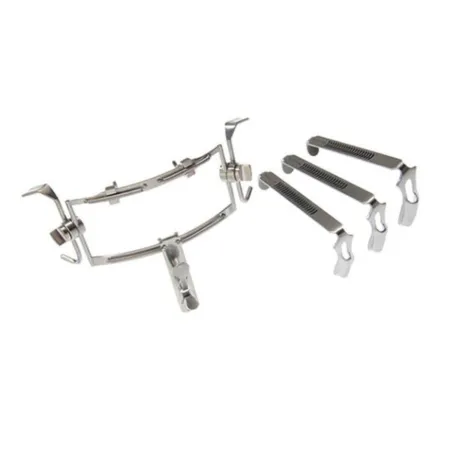
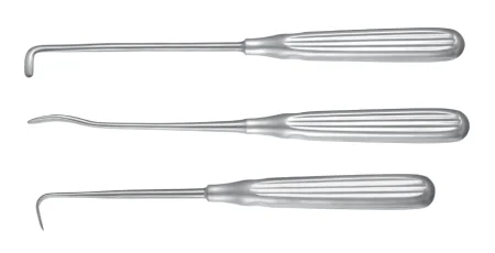
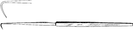
Key steps (V-Y palatoplasty):
- The Dingman mouth gag opens the mouth and its frame is fixed to the Mayo standMAY-oh standA small tray on an adjustable stand placed over or beside the patient during surgery to hold the instruments used most often.Full entry . From now on, never bump the Mayo, because a jolt goes straight to the child's mouth. The position of the breathing tube is checked as the gag goes in.
- A throat pack may be placed to absorptionub-ZORP-shunThe taking in of a substance, such as a drug or a nutrient, into the bloodstream.Full entry blood, and a second suction may be placed at the back of the throat. Help with irrigationeer-ih-GAY-shunWashing out a wound, body cavity, tube or eye with a steady flow of liquid.Full entry and suction throughout.
- The flaps are marked and injected. A V-shaped incision with a #15 blade, carried through with a palate blade, cuts through the oral mucosa, the muscle and the nasal mucosa.
- With Freer and Cottle elevators, the surgeon lifts the nasal mucosa off the muscle on both sides, then the oral mucosa, making three layers, and guards the greater palatine vessels, which feed the flaps. Small holes may be drilled in the hard palate for sutures.
- The palate is closed in three layers, nasal mucosa first, then muscle, then the oral mucosa, with 4-0 or 5-0 absorbable suture. The Y-shaped closure also lengthens the palate.
- The wound is irrigated and checked for bleeding, the throat pack is removed and counted, and the mouth gag comes out.
Wound class: II.
After surgery: the anesthesia provider extubates very carefully so the new palate is not hurt, and a soft bite block or a nasal airway may hold the airway open. Swelling can block the airway, so be ready to help, and keep the back table and Mayo sterile until the child has left the room. Soon after surgery the child may drink from a cup, but nothing that needs sucking, such as a bottle, a straw or a cup with a valve, which can pull the repair apart, and arm restraints may be used for some weeks.
If the child's speech later sounds nasal because air escapes through the nose (velopharyngeal insufficiencyin-suh-FISH-un-seeA state in which an organ or part does not work well enough to meet the body's needs.in- notsufficient enoughFull entry ), a second operation, a pharynxFAIR-inksThe throat: the passage behind the nose and mouth that carries air to the voice box and food to the esophagus.pharyng/o throatFull entry flap, can improve it.
14Wound classes and care after surgery
Wound classes follow the CDC's definitions (chapter 11). The mouth and the throat behind it (the oropharynxor-oh-FAIR-inksThe middle part of the throat, behind the mouth, from the soft palate down to the top of the voice box.or/o mouthpharyng/o throatFull entry ) are part of the digestive and breathing passages, so an operation that enters them is Class II, clean-contaminated; the CDC names the oropharynx in that class.
- Class I, clean: no tract is entered, as when a fracture is reached only through the skin and neither the mouth nor a sinusSY-nusA hollow space in the body, most often one of the air-filled spaces in the skull bones around the nose.Full entry is opened.
- Class II, clean-contaminated: the mouth, the nose or a sinus is entered: tooth extraction, arch bars, implants, intraoral fracture repairs, orthognathic surgery and cleft repairs.
- Class III, contaminated: a fresh, open traumatic wound, such as a facial lacerationlas-uh-RAY-shunA cut or tear in the skin or other tissue, often with jagged edges.Full entry over a fracture.
- Class IV, dirty: an infection already present, such as a dental abscessAB-sesA closed pocket of pus in tissue, walled off by the body.Full entry drained, or an old wound with dead tissue.
What to watch for after any of these cases:
- The airway first. Swelling and bleeding can block it in the first hours. The head is kept up, and a tracheotomytray-kee-OT-uh-meeThe surgical cut made into the windpipe through the front of the neck.trache/o windpipe-tomy cutting, incisionFull entry tray and reintubation equipment are kept close. Before the tube comes out, the anesthesia provideran-us-THEE-zhuh pruh-VY-derThe licensed professional who gives anesthesia and watches over the client's breathing and vital functions during a procedure.Full entry may empty the stomach through a tube to lower the risk of aspirationas-puh-RAY-shunBreathing food, liquid, saliva or stomach contents into the airway and lungs; also, drawing fluid out with suction or a needle.Full entry .
- The sterile fieldSTAIR-ul feeldAn area set up with sterile drapes and supplies, kept free of all germs, for a sterile procedure.Full entry stays up. Keep the back table and Mayo sterile until the patient has left the room, so the team is ready if the airway is lost on waking.
- Wire cutters go with every patient whose jaws are fixed together, and elastics are often added once the patient is awake.
- Bleeding, cerebrospinal fluidser-uh-broh-SPY-nul FLOO-idThe clear, watery fluid that bathes and cushions the brain and spinal cord.cerebr/o brainspin spine-al relating toFull entry leak and the eyes: watch for bleeding from the mouth or the wounds, clear fluid from the nose, and changes in vision.
- Mouth care and food: a mouth wound can split open when oral hygiene is poor, so the mouth is kept clean, with a water jet never aimed straight at an incision. A liquid or soft diet follows most jaw cases, and ice helps the swelling.
What to take from this chapter
- Oral and maxillofacial surgery repairs the bones and soft tissue of the face and mouth. Almost every jaw and midface repair aims to give the patient back their own bite from before the injury: arch bars and MMF set the bite first, and plates rebuild the buttresses that carry the force of chewing.
- Le Fort I runs straight across the maxilla above the teeth; Le Fort II is a pyramid from the nasal bones across the orbital floors and the rims below the eyes; Le Fort III cuts the whole midface off the skull through the root of the nose, the orbits and the zygomatic arches. A pure pattern is rare.
- The airway is shared. A tube through the nose (or under the chin, or a tracheostomy) leaves the teeth free to close, but when the midface is broken nasal tubes are avoided, because a tube can pass through a broken cribriform plate into the brain. The tube is part of the field, and oxygen near the ESU is a fire risk.
- The throat pack is counted with the sponges, as your facility's policy records it. Its placement is announced, its removal is confirmed before the breathing tube comes out, and it comes out (or its end is brought out) before the jaws are fixed together.
- A patient whose jaws are wired or banded shut leaves the OR with wire cutters (and scissors for elastics) taped at the head of the stretcher. They stay at the bedside to cut the links between the upper and lower bars if the patient vomits or cannot breathe, and the patient is taught to use them. Wires are always twisted clockwise.
- Prep the face with povidone-iodine without alcohol, after checking for an iodine allergy, and never chlorhexidine on the face, eyes, ears or mouth. The mouth is not sterile: keep mouth instruments apart from the plating setup, though instruments that enter tissue and bone are still sterile items.
- Learn the plating rhythm once: plate, hold, drill with irrigation, depth gauge, tap, screw. Confirm and record every implant and graft. An orbital floor repair frees trapped muscle and slides an implant under the eye; a frontal sinus repair restores the front wall, the drainage and, with neurosurgery, the back wall.
- Cleft lip is usually repaired at about 10 to 12 weeks (the rule of 10) and the palate at about 6 months, before the child talks. The Dingman gag hangs from the Mayo, so never bump it, and afterward the child wears arm restraints and does not suck. Any case that enters the mouth is at least Class II, and the back table stays sterile until the patient leaves the room.
Check yourself5 quick questions
Free for everyone. Pick an answer for each, then check them all at once.
In the ORWhere you'll use this
The chapter's whole question pool in sets of 10, with the answer explained after each one.
Part of the educators programThe chapter's words on spaced cards: the ones you miss come back sooner.
Part of the educators programGiven by your class's teacher. Each class draws its own 25 from the chapter's pool.
Part of the educators program
Suggest a fix
Something wrong, unclear or out of date in this chapter? Tell us and we will fix it for everyone. Your email is only used to reply.