Plastic and reconstructive surgery
Skin grafts and flaps, skin cancers, the face, the breast, the body and the hand: how plastic surgery restores form and function, and how the scrub keeps living tissue alive between one site and the next.
1What plastic surgery covers
Plastic surgery repairs, rebuilds or replaces parts of the body whose form or function was lost or never formed: the skin, the bones and soft tissue of the face and skull, the hand and limbs, the breast and trunk, and the external genitals. The word comes from the Greek plastikos, "to mold or give form".
It has two branches:
- Reconstructive surgery restores function and a normal appearance after a birth defect, an injury, a burnBURNAn injury to the skin or deeper tissues caused by heat, chemicals, electricity, radiation or friction.Full entry or a disease such as cancer. Breast reconstruction after a mastectomyma-STEK-tuh-meeSurgery to remove all or part of a breast.mast/o breast-ectomy surgical removalFull entry and cleft lipkleft lipA split or opening in the upper lip that is present at birth.Full entry repair are examples. It is considered medically necessary, so most health insurance covers it.
- Aesthetic (cosmetic) surgery reshapes normal structures to improve how they look, without necessarily changing how they work, as in a facelift, a breast augmentation or an abdominoplasty. It is generally not considered medically necessary.
Most of the operations' names are built from word parts (chapter 1). The suffix -plasty means surgical repair, as in blepharoplasty (blephar/oBLEF-uh-rohA word part that means eyelid.Full entry , the eyelid) and mammoplasty (mamm/oMAM-ohA word root that means breast; mast/o means the same.Full entry , the breast); -pexy means surgical fixation, as in a mastopexy (mast/o, the breast); -ptosis means drooping, as in a sagging brow or breast; and cutane/okyoo-TAY-nee-ohA word root that means skin; derm/o and dermat/o mean the same.Full entry means skin, as in a myocutaneous flap.
Plastic surgery crosses nearly every other specialty, its operations are often done in stages, and many are done in ambulateAM-byuh-laytTo walk, or to help a client walk.Full entry surgery centers.
When a wound or a defect needs closing, the surgeon weighs the options from the simplest to the most complex, a plan called the reconstructive ladder:
- Letting the wound heal by second intention
- Closing it directly, at once or after a delay (primary or delayed primary closure)
- A skin graftGRAFTTissue or a man-made material moved into place in surgery to replace or repair a damaged part.Full entry or a composite graft
- A local flap, which may follow tissue expansion
- A regional flap
- A free flap
- A transplant of composite tissue from a donor, such as a hand or a face
The surgeon does not have to climb one rung at a time. A large defect may go straight to a free flap; the lower rungs are the plan's contingencies.
Many operations here share instruments, positions and steps, which the Core Curriculum calls co-related procedures. Learn the skin graft once and it comes back in burn care, syndactyly release and nipple reconstruction; learn one pedicled flap and the latissimus and TRAM flaps follow. Each case below links to its setup card, which holds the instrument list and every step. Here you get the reasons, and the moments when the scrub matters most.
The plastic surgery patient
Plastic surgery touches body imageBOD-ee IM-ijHow a person sees, thinks and feels about their own body and its appearance and function.Full entry and self-esteem, so even a wanted change can bring anxiety and fear, and a patient having cosmetic surgery may lack the usual support of family and friends or feel judged for choosing elective surgery. What helps is an honest, calm, unhurried manner: explain what will happen and what the patient will feel, listen, keep the doors closed and expose only what the case needs.
Unrealistic expectations, surgery to please a partner or to cure grief, and body dysmorphia make a poor candidate, and diabetes, high blood pressure, obesity, heart disease and smoking raise the risks. Before a breast reduction, for example, patients stop smoking at least a month ahead and stop aspirin, nonsteroidal anti-inflammatory drugnon-STAIR-oy-dul an-tee-in-FLAM-uh-tor-ee DRUGA drug that relieves pain, fever and inflammation and is not a steroid, such as ibuprofen or naproxen.non- notanti- againstinflamm/o inflammationFull entry , vitamin EVY-tuh-min EEA fat-soluble vitamin that protects cells from damage, found in nuts, seeds, plant oils and green leafy vegetables.Full entry and herbal supplements at least 2 weeks ahead, because they slow healing or disturb clottingKLOT-ingThe process by which blood thickens into a gel to plug a damaged vessel and stop bleeding; also called coagulation.Full entry .
Each section names the chapter that already teaches the basics it builds on, such as the skin in chapter 3, burns and scars in chapter 11, local anestheticLOH-kul an-es-THET-ikA drug that blocks feeling in one part of the body without making the person unconscious.Full entry in chapters 12 and 13, dressings in chapter 30 and skin prep and the tourniquet cuff in chapter 31. The Core Curriculum lists cleft lip and palate repair and gender confirmation surgery under plastics too, but chapters 37 and 34 teach them.
2Anatomy that matters
Skin, tension lines and the depth of a graft
Chapter 3 teaches the skin's layers. Two facts matter most here: the dermisDER-misThe thick layer of skin under the epidermis that holds blood vessels, nerves, hair roots and glands.derm/o skinFull entry , not the epidermisep-ih-DER-misThe thin outer layer of the skin.epi- on, uponderm/o skinFull entry , carries the skin's vessels and nerves, so it is the layer that bleeds, and every skin graftGRAFTTissue or a man-made material moved into place in surgery to replace or repair a damaged part.Full entry takes some of it; and it holds the hair folliclehair FOL-ih-kulA small tube-shaped pocket in the skin from which a hair grows.Full entry and glands whose cells grow new skin, so a donor site that keeps part of its dermis heals on its own.
The surgeon places an incision along one of the skin's natural tension lines, such as the Langer lines, which run with the collagenKOL-uh-jenThe main structural protein of the body, which gives strength to skin, bones, tendons, ligaments and scar tissue.coll/a glue-gen producingFull entry and muscle fibers of the dermis. A scar along them is under less tension and shows less. On the face, incisions hide next to or within the hairline and around the ear.
The face
The soft tissue of the face is skin, fat, muscle, fasciaFASH-ee-uhA sheet or band of tough connective tissue that covers and separates muscles and organs.fasci/o bandFull entry and ligaments. A sheet of fibrousFY-brusMade of or containing tough threads (fibers), especially of connective tissue.fibr/o fiber-ous full of, relating toFull entry tissue called the superficial musculoaponeurotic system (SMAS) divides the fat under the skin into a superficial and a deep layer, and the facial ligaments tie the soft tissue to the bone. A facelift tightens the SMAS, close to the facial nerveFAY-shul NERVThe seventh cranial nerve, which moves the muscles of the face and carries taste from the front of the tongue.Full entry , which moves the muscles of expression.
The eyelids: the orbicularis oculi muscle closes them, and the levator muscle opens the upper lid through its tendon, the levator aponeurosis. Behind the skin and muscle, the orbital septumSEP-tumA wall of tissue that divides two spaces or cavities, such as the two sides of the heart or nose.sept/o wall, partitionFull entry holds back the orbital fat, and the tarsus, a plate of dense tissue, shapes the lid. With age the lid skin loosens (dermatochalasis) and the septum weakens, so the fat behind it bulges as "bags".
Chapter 8 teaches the breast and chapters 3 and 32 the abdominal wall; the hand is described with its operations below.
3Diseases and injuries of the skin
Skin cancers
Most skin cancers begin in the epidermisep-ih-DER-misThe thin outer layer of the skin.epi- on, uponderm/o skinFull entry , and most are caused by ultraviolet light from the sun or from tanning beds; damage from years of exposure can show up as cancer long afterward. The three most common are basal cell carcinomaBAY-sul SEL kar-sih-NOH-muhThe most common type of skin cancer, which starts in the basal cells at the bottom of the skin's outer layer.Full entry , squamous cell carcinomaSKWAY-mus SEL kar-sih-NOH-muhA cancer that starts in squamous cells, the thin, flat cells of the skin's outer layer and the linings of some organs.Full entry and melanomamel-uh-NOH-muhA cancer that starts in melanocytes, the cells that give skin its color.melan/o black, dark-oma tumorFull entry , and melanoma is the one of most concern:
- Basal cell carcinoma: grows in the basal layer of the epidermis, usually on the face, head and neck. It grows slowly and rarely spreads, but left alone it keeps growing. It makes up about 80 percent of skin cancers.
- Squamous cell carcinoma: grows from the flat cells of the upper epidermis, usually on the face, ears, lips, neck and backs of the hands, and sometimes in scars or chronic sores. It is more aggressive than basal cell carcinoma, can grow deeper and can spread, and makes up about 20 percent of skin cancers.
- Melanoma: grows from the melanocytemuh-LAN-uh-syteA cell in the bottom layer of the skin's outer layer that makes melanin, the pigment that gives skin and hair their color.melan/o black-cyte cellFull entry , the pigment cells in the bottom layer of the epidermis, and typically develops from a mole. It is only about 1 percent of skin cancers but causes most skin cancer deaths, so it is treated aggressively, and its excisionek-SIZH-unCutting out tissue, such as a growth or damaged area, during surgery.Full entry may include sentinel node mapping (chapter 32).
The ABCDEay-bee-see-dee-EE (said as letters)A memory aid with two common uses: the warning signs of melanoma in a mole, and the order of the first check of a seriously ill or injured client.Full entry warning signs of melanoma are Asymmetry, an uneven or notched Border, more than one Color, a Diameter of about 0.6 cm or more, and Evolving: any change in size, shape, color or height, or new bleeding, itching or crusting. A mole that looks unlike the patient's others, the "ugly duckling", is another warning.
Other skin lesions
- Nevus: a mole or birthmark, a well-defined pigmented spot, usually present from birth; some can become cancer.
- Keratosis: a horny growth of the skin. An actinic keratosis is a rough, sandpaper-like patch from sun damage that can become cancer; a seborrheic keratosis is a common, harmless growth of older adults.
- Wart: a raised growth of the skin caused by the human papillomavirusHYOO-mun pap-ih-LOH-muh-vy-rusA common group of viruses spread by close skin-to-skin contact, most often during sex.Full entry and spread by direct contact; common warts favor the fingers, the backs of the hands and the skin around the nails.
cellulitissel-yoo-LY-tisA bacterial infection of the deeper layers of the skin and the tissue just under it.cellul small cell-itis inflammationFull entry , the spreading infection of the skin and the tissue beneath it, is taught in chapter 11.
How a lesion is diagnosed
Many problems that bring patients to plastic surgery are diagnosed by looking at them, a visual examination. A suspicious lesionLEE-zhunAn area of tissue that has been damaged or has changed from normal, such as a sore, wound, lump or spot.Full entry is sampled. A punch biopsy takes a small round core of skin, the punch's size chosen for the lesion, which keeps the tissue's structure for the pathologist and usually shows how deep a melanoma has grown, the measure that guides its treatment. X-rays, computed tomographykum-PYOO-tid toh-MOG-ruh-feeAn imaging test that uses x-rays and a computer to make cross-section pictures of the inside of the body.tom/o cut, slice-graphy process of recordingFull entry and magnetic resonance imagingmag-NET-ik REZ-uh-nunts IM-uh-jingAn imaging test that uses a strong magnet and radio waves to make detailed pictures of the inside of the body.Full entry help plan hand surgery and surgery of the skull and face.
Injuries and congenital defects
Chapter 11 teaches the skin's injuries as injuries: burns, frostbite, hypothermiahy-poh-THER-mee-uhA body temperature that has fallen below the normal range.hypo- belowtherm/o heat-ia conditionFull entry and hyperthermiahy-per-THER-mee-uhA body temperature that is higher than normal because the body takes in or makes more heat than it can lose.hyper- abovetherm/o heat-ia conditionFull entry , chemical, electrical, mechanical and radiationray-dee-AY-shunEnergy that travels as waves or particles, such as X-rays, used to make images or treat cancer; also pain that spreads from its starting point.radi/o rays, radiation-ation processFull entry injuries, pressure injuryPRESH-er IN-juh-reeDamage to the skin and the tissue beneath it from pressure, or pressure with shear, usually over a bony area or under a device.Full entry and scars. This chapter teaches the surgery they need, and the birth defects plastic surgeons repair, such as syndactyly, polydactyly and radial dysplasia, with their operations.
4Before surgery: marking, anesthesia, position, prep and drape
Marks and photographs
The surgeon marks the patient before surgery, often in the holding area with the patient sitting or standing, because lying on the OR table moves the skin and shifts the landmarks: the lines of a facelift, a flap and its donor site, the new nipple position in a breast reduction, the bulges for liposuction. The marks are made with a surgical skin markerMAR-kerSomething that can be measured, such as a substance in the blood, that shows a disease or body process is present.Full entry , because other inks can wash off during the prep.
Plastic surgeons take photographs before, during and after surgery and may want them displayed during the case, because positioning changes the patient's features: make sure each is labeled correctly, and at the end take them down and secure them in the record, for the patient's privacy.
Anesthesia and fire
A plastic case may be done under local, regional, tumescent or general anesthesiaJEN-er-ul an-es-THEE-zhuhDrug-induced loss of consciousness and feeling over the whole body, so a person feels nothing and is not aware during surgery.an- withoutesthesi/o feeling, sensation-ia conditionFull entry , or with moderate sedationMOD-er-it seh-DAY-shunA drug-induced state of relaxed, lowered awareness in which the client still responds to voice or light touch and breathes on their own.Full entry , and the patient's blood pressure, breathing, heart rate and rhythm, saturationsach-uh-RAY-shunHow full something is; in nursing, usually the percentage of hemoglobin in the blood that is carrying oxygen.Full entry and end-tidal carbon dioxideKAR-bun dy-OK-sideA gas made by the body's cells as waste, carried in the blood to the lungs and breathed out.Full entry are monitored. local anestheticLOH-kul an-es-THET-ikA drug that blocks feeling in one part of the body without making the person unconscious.Full entry , with or without epinephrineep-ih-NEF-rinA hormone and drug, also called adrenaline, that raises heart rate and blood pressure and opens the airways.Full entry , is injected in many cases, even under general anesthesia, and the team agrees on the most that can be used, so the total stays below a toxicitytok-SIS-ih-teeThe harm a substance does to the body, or a state in which a drug has built up to a harmful level.tox/o poison-icity state or quality ofFull entry dose (chapters 12 and 13).
When oxygen flows through an open mask or cannulaKAN-yoo-luhA thin tube placed into the body or a body opening to deliver or remove fluid or air.Full entry during a procedure above the xiphoid, such as on the face, head or neck, the surgeon tells the anesthesia provideran-us-THEE-zhuh pruh-VY-derThe licensed professional who gives anesthesia and watches over the client's breathing and vital functions during a procedure.Full entry before using the electrosurgeryee-LEK-troh-SUR-jer-eeUsing high-frequency electric current through a handheld tool to cut tissue or seal bleeding vessels during surgery.Full entry or a laser, and the provider lowers the oxygen as far as is safe and says when it is clear to go ahead (chapter 20 teaches fire at the field).
Position
Most plastic cases are supine, but any position may be needed, and a case may change position partway: a back flap is raised with the patient on the side, then the patient is turned supine to set it into the chest. The OR bed is often flexed with the head raised at closure, as in an abdominoplasty, to take the tension off the wound.
Long cases, such as free flaps and replantations that run 12 to 16 hours, need every pressure point padded, a urinary catheter, compression devices on the legs and active warming. Warming starts in the holding area, and IV and irrigating fluids are warmed.
Prep
The surgeon chooses the prep solution and its strength; povidone-iodinePOH-vih-done EYE-uh-dineA brown antiseptic solution of iodine used to kill germs on the skin before procedures and on wounds.Full entry is common in plastic surgery, and it must not run into the eyes or ears. As chapter 31 teaches, CHG is never used on the face, in or around the eyes or ears, or in the mouth, and alcohol-based preps stay out of the ears, mouth and eyes; an iodineEYE-uh-dyneA mineral the thyroid gland uses to make thyroid hormone; it is also used to kill germs on the skin and in some contrast dyes.Full entry prep without alcohol, such as povidone-iodine, may be used on the outer ear once the patient is checked for iodine sensitivitysen-sih-TIV-ih-teeHow strongly something responds; in lab work, which drugs stop a germ, or how well a test finds people who have a disease.Full entry . A brow lift or facelift is prepped over the whole head, scalp, face, ears and neck, with the ear canals protected from pooling solution, and long hair is parted into sections and held with sterile rubber bands or a water-soluble lubricant.
AST's 2008 skin prep standard rules out both CHG and iodine (iodophor) preps for the eye and face, because either can injure the corneaKOR-nee-uhThe clear, dome-shaped layer at the front of the eye, over the iris and pupil.Full entry , and suggests a nontoxic agent such as PCMX with a warm sterile water rinse, kept out of the eyes.
A graftGRAFTTissue or a man-made material moved into place in surgery to replace or repair a damaged part.Full entry from the patient's own body gets two preps with separate setups, as chapter 31 teaches: the donor site first, with a colorless solution, most often CHG, so the surgeon can see the skin, and the recipient site, often an open wound such as a burnBURNAn injury to the skin or deeper tissues caused by heat, chemicals, electricity, radiation or friction.Full entry , last, as the contaminated one.
Drapes
For the face, a head drape (turban) keeps the hair out of the field and leaves the whole face in view, with a split sheet below; a limb gets a fenestrated or split sheet; and an impervious pocket drape catches the runoff when a lot of fluid is used, as in debridementdih-BREED-muntThe removal of dead, damaged or infected tissue from a wound so it can heal.Full entry . Many cases have more than one operative site, so have extra towels, plain sheets and adhesive drapes ready.
When the recipient site holds a cancer or an open wound and skin is taken from somewhere else, keep the instruments for each part of the case apart: once the recipient site is readied, the team changes gloves and the scrub switches to the clean instruments for the donor site, which counts as the clean one.
5Instruments, equipment and supplies
The plastic set
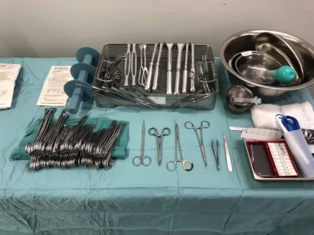
Plastic surgery on the skin and the tissue just beneath it uses short, fine instruments, about 5 to 7 inches long:
- Skin hooks and small retractorrih-TRAK-terA surgical instrument that holds back tissue or organs so the surgeon can see and reach the operative site.Full entry : single and double skin hooks, small rakes, Senn and Ragnell retractors and small spring-loaded self-retaining retractors hold back the skin edges.
- forcepsFOR-sepsA hand instrument with two blades or arms used to grasp, hold or pull tissue or objects.Full entry : fine toothed forceps such as the Adson and the Adson-Brown hold skin. hemostatHEE-moe-statA clamp with finger rings and a locking ratchet used in surgery to grasp and close off bleeding vessels.hem/o blood-stat stopping, standing stillFull entry never go on skin, but fine mosquito clamps catch small vessels and tag sutureSOO-cherA stitch, or the thread used to stitch, that holds the edges of a wound or incision together.Full entry .
- Scissors: fine, sharp scissors such as tenotomy, Westcott and iris scissors, with specialty patterns such as the Kaye; most plastic surgeons have one or two favorites, so have them ready.
- Needle holders: small, fine ones such as the Webster, for fine needles.
- Marking: sterile skin markerMAR-kerSomething that can be measured, such as a substance in the blood, that shows a disease or body process is present.Full entry or methylene blue, a ruler or tape measure, and calipers.
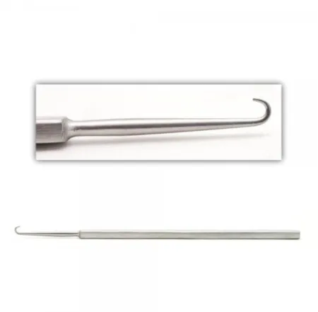
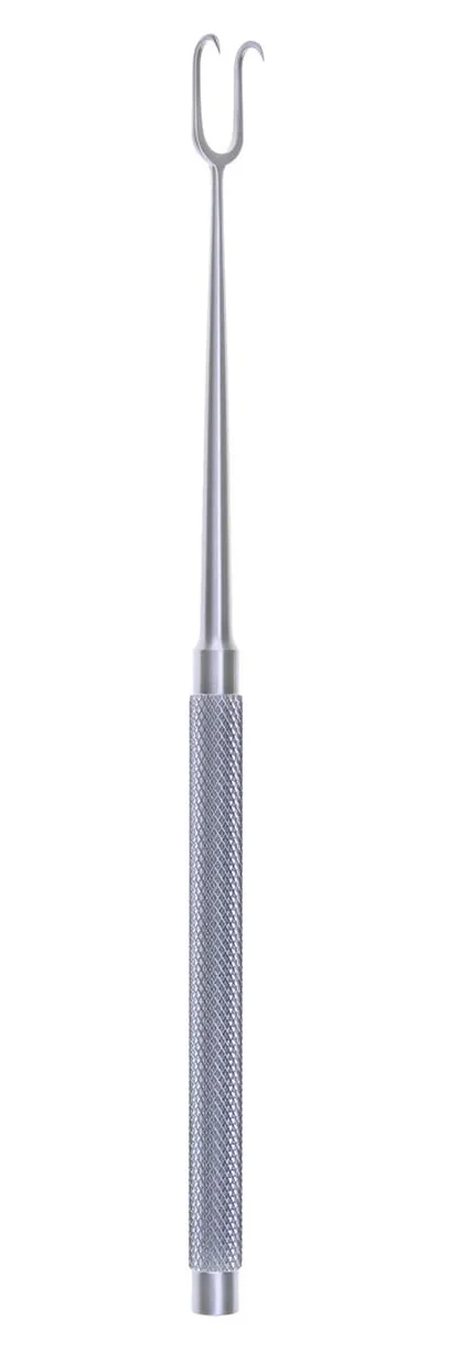
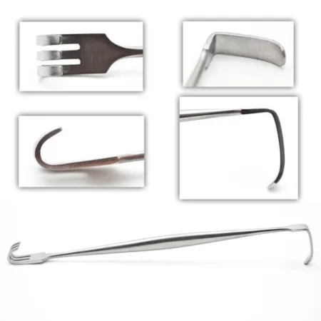
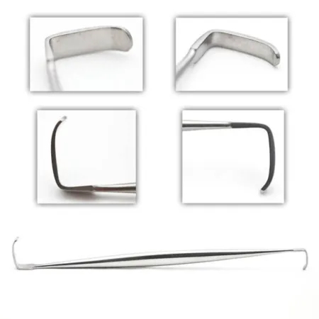
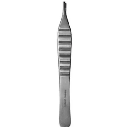
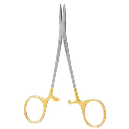
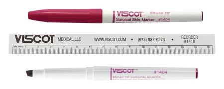
Deeper work, on the breast and the abdominal wall, adds general surgery retractors, and an abdominoplasty needs retractors that lift the body wall. Lighted (fiber-optic) retractors and headlights light deep pockets in breast, face and abdominal surgery. Work on bone adds rasps, small osteotomes, curettekyoo-RETA surgical instrument with a small spoon- or loop-shaped tip used to scrape tissue.Full entry , small bone clamps, small Kocher clamps and power drills and saws, which the scrub assembles and manages; curved and straight Mayo scissors trim cartilageKAR-tih-lijFirm, smooth, rubbery tissue that cushions joints and gives shape to parts such as the ear, nose and windpipe.chondr/o cartilageFull entry and soft implants. Many facilities build custom sets around their surgeons' preferences.
Equipment
- electrosurgeryee-LEK-troh-SUR-jer-eeUsing high-frequency electric current through a handheld tool to cut tissue or seal bleeding vessels during surgery.Full entry : monopolar and bipolarby-POH-lerHaving two poles or two opposite ends; most often used as a short form of bipolar disorder.bi- twopolar having polesFull entry units. A needle tip on a low setting does fine work, and bipolar keeps the heat from spreading in delicate tissue. A smoke evacuator is used whenever an energy device makes smoke, its tip within 2 inches (5 cm) of the site (chapter 20).
- Magnification: loupes, magnifying lenses worn like glasses, or the operating microscope, for vessels and nerves sewn with suture as fine as 9-0 to 11-0.
- Doppler ultrasoundDOP-ler UL-truh-soundAn ultrasound that uses sound waves to hear or show blood flowing through vessels or the fetal heartbeat.Full entry : a handheld ultrasound unit, used with a sterile probe or probe cover, that finds and checks the vessels feeding a flap.
- Pneumatic tourniquet: for surgery on the arm, hand or leg; chapter 31 teaches putting on the cuff, and chapter 23 how its pressure is worked out.
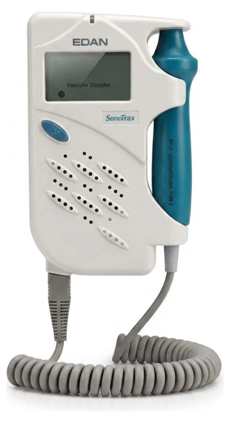
The Core Curriculum's specialty supplies for plastic surgery, the dermatome blade, the liposuction cannulaKAN-yoo-luhA thin tube placed into the body or a body opening to deliver or remove fluid or air.Full entry and tubing, the mesh graftGRAFTTissue or a man-made material moved into place in surgery to replace or repair a damaged part.Full entry carrier, prostheticpros-THET-ikDescribes an artificial part made to replace a missing or damaged body part, such as a limb, joint, eye or heart valve.prosthe an addition (from Greek)-tic relating toFull entry implants and tissue expanders, are taught with their operations below.
Implants
Implants range from human tissue to synthetic materials. The patient's own tissue is preferred when it is available. Donated human tissue includes acellular dermisDER-misThe thick layer of skin under the epidermis that holds blood vessels, nerves, hair roots and glands.derm/o skinFull entry matrix, such as AlloDerm, a sheet of donated skin from which the cells have been removed; it can cover and support a breast implant, and it grows into the patient's own tissue. Synthetic implants include silicone and polyethylene pieces for the chin and cheeks, and silicone breast implants and tissue expanders.
Every implant is documented, and the regulations require certain ones to be tracked (chapter 29): confirm its type and size with the surgeon before the circulator opens it, and its details go into the patient's record. Bacteria on an implant can make it fail, so handle every implant as little as possible, with strict surgical asepsisSUR-jih-kul ay-SEP-sisPractices that keep an area or object completely free of microorganisms; also called sterile technique.a- withoutsepsis decay, infectionFull entry .
Dressings
In plastic surgery the dressing is part of the result, and it often goes on while the patient is still asleep. A clear dressing lets the team watch a flap's color, and a compression garment is sized by its maker's instructions so it does not cut off blood flow; chapter 30 teaches pressure dressings, bolsters and the surgical bra.
Medications on the field
| Drug or solution | Use in plastic surgery |
|---|---|
| local anestheticLOH-kul an-es-THET-ikA drug that blocks feeling in one part of the body without making the person unconscious.Full entry with epinephrineep-ih-NEF-rinA hormone and drug, also called adrenaline, that raises heart rate and blood pressure and opens the airways.Full entry (lidocaineLY-doh-kaynA local anesthetic that numbs an area, also given by IV to treat some fast, abnormal heart rhythms.Full entry , bupivacainebyoo-PIV-uh-kaynA long-acting local anesthetic used to numb an area, block nerves, or give epidural and spinal anesthesia.Full entry ) | Injected to numb the area and narrow its vessels; label it with its exact ratio and keep a running total. sodium bicarbonateSOH-dee-um by-KAR-buh-nateAn alkaline drug, given by mouth or IV, that neutralizes acid in the stomach or the blood.Full entry may be added to ease the sting of the injection |
| Topical epinephrine 1 mg/mL (1:1,000) | Diluted in saline as the surgeon orders, on sponges that slow oozing from burnBURNAn injury to the skin or deeper tissues caused by heat, chemicals, electricity, radiation or friction.Full entry wounds and donor sites; kept in a shallow labeled dish, never in a syringe |
| phenylephrinefen-il-EF-rinA drug that tightens blood vessels, given IV to raise low blood pressure and used in nose sprays and eye drops.Full entry or thrombinTHROM-binAn enzyme in the blood that turns fibrinogen into fibrin, the threads that hold a blood clot together.thromb/o clot-in substanceFull entry solution | On sponges that stop oozing at donor sites |
| Tumescent solution | IV fluid with lidocaine and epinephrine, sometimes with hyaluronidase, infused into fat for liposuction and some other cases |
| Topical cocaine | On cottonoids in the nose for rhinoplasty; a controlled substancekun-TROHLD SUB-stunsA drug whose making, prescribing and use are strictly regulated by law because it can be misused or cause dependence.Full entry |
| Tetracaine eye drops; EMLA cream | Numb the eye before corneaKOR-nee-uhThe clear, dome-shaped layer at the front of the eye, over the iris and pupil.Full entry shields go in; numb intact skin before an injection |
| Papaverine, verapamilver-AP-uh-milA calcium channel blocker that slows the heart rate and relaxes blood vessels.Full entry or lidocaine | On a patty over vessels to reverse spasmSPAZ-umA sudden, involuntary tightening of a muscle or of the muscle in a hollow organ or tube.Full entry in microsurgery |
| heparinHEP-uh-rinAn anticoagulant drug, given by injection or IV, that keeps blood clots from forming or growing.Full entry | Given into a vein by the anesthesia provideran-us-THEE-zhuh pruh-VY-derThe licensed professional who gives anesthesia and watches over the client's breathing and vital functions during a procedure.Full entry before a microvascular anastomosisuh-nas-tuh-MOH-sisA connection between two tube-shaped structures, such as two ends of bowel or two blood vessels, made by surgery or found naturally.Full entry ; heparinized saline flushes the vessels |
| Topical antimicrobialan-tee-my-KROH-bee-ulA drug or substance that kills or slows the growth of microorganisms such as bacteria, viruses, fungi or parasites.anti- againstmicro- smallbi/o life-al relating toFull entry (silver sulfadiazineSIL-ver sul-fuh-DY-uh-zeenA white antibacterial cream, made of silver joined to a sulfa drug, put on burns.Full entry , mafenide, bacitracinbas-ih-TRAY-sinAn antibiotic used mostly as an ointment on the skin to prevent infection in minor cuts, scrapes and burns.Full entry ) | Dress burn wounds |
| Methylene blue | Marks skin and cartilage |
a skin graft or burn case can have two strengths of epinephrine on one field: a dilute one for injection (a local anesthetic with epinephrine, or epinephrine diluted with injectable saline) and epinephrine 1 mg/mL (1:1,000) for sponges. Label each the moment it arrives, keep the strong one in a labeled shallow dish away from every syringe, and say the strength aloud each time you pass it (chapter 12).
6Skin grafts
A skin graftGRAFTTissue or a man-made material moved into place in surgery to replace or repair a damaged part.Full entry is skin taken from one place, the donor site, and laid on a wound, the recipient site, where it lives only if new blood vessels grow into it from the wound bed. Skin grafting is a common operation in plastic surgery, and the type of graft planned decides the prep, the setup and the steps, so confirm it before the case.
Kinds of graft
Chapter 25 names the kinds of graft by where they come from: an autograft from the patient's own body, an allograft (homograft) from a donor, kept by a tissue bank, and a xenograft (heterograft) from another species, such as pig (porcine) or cow skin. Plastic surgery adds names for what a graft is made of:
- Biological graft: any graft from living tissue, human or animal. A composite graft holds more than one kind of tissue, such as skin with cartilageKAR-tih-lijFirm, smooth, rubbery tissue that cushions joints and gives shape to parts such as the ear, nose and windpipe.chondr/o cartilageFull entry or fat.
- Synthetic graft: a manufactured material the body tolerates, which may be soft, semisolid or liquid. A biosynthetic graft is made of a synthetic absorbable material, or helps healing without coming from living tissue.
Chapter 29 teaches how grafts from a tissue bank are tracked and stored, and chapter 30 the biosynthetic skin substitutes used as dressings.
Split-thickness and full-thickness grafts
A split-thickness skin graft (STSG) takes the epidermisep-ih-DER-misThe thin outer layer of the skin.epi- on, uponderm/o skinFull entry and part of the dermisDER-misThe thick layer of skin under the epidermis that holds blood vessels, nerves, hair roots and glands.derm/o skinFull entry , and how much of the dermis varies, from thin to thick. A full-thicknessful-THIK-nisGoing through every layer of the skin, or every layer of another tissue such as the wall of an organ.Full entry skin graft (FTSG) takes the epidermis and all of the dermis.
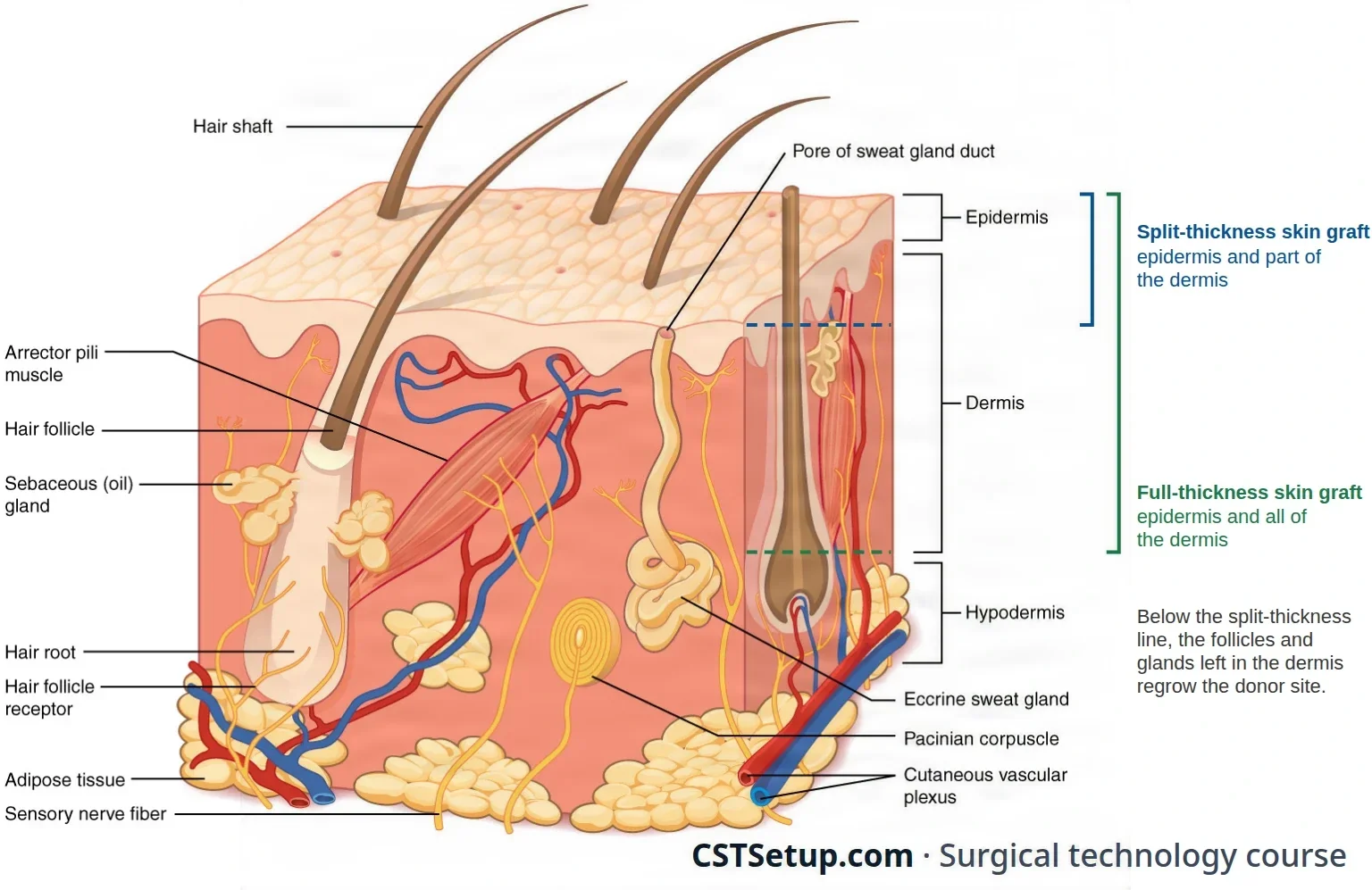
| Split-thickness (STSG) | Full-thickness (FTSG) | |
|---|---|---|
| Layers | Epidermis and part of the dermis | Epidermis and all of the dermis |
| Taken with | A dermatome or graft knife; a very small one with a #15 blade | A #15 blade and fine scissors |
| Donor sites | Broad, fleshy areas: the thighs, buttocks, back, chest and abdomen | Areas of looser skin: the groin, lower abdomen and the area below the collarbone |
| The donor site heals | On its own, as new epitheliumep-ih-THEE-lee-umThe tissue that covers the body's surfaces and lines its organs, vessels and cavities.Full entry grows from the dermis left behind | Only when it is sutureSOO-cherA stitch, or the thread used to stitch, that holds the edges of a wound or incision together.Full entry closed, or covered with a split-thickness graft |
| At the recipient site | Picks up a blood supply quickly, but shrinks more as it heals, wears less well and looks and feels different from normal skin | Shrinks very little, holds up to wear, works over joints that bend and looks better |
| Used for | Large areas, such as a burnBURNAn injury to the skin or deeper tissues caused by heat, chemicals, electricity, radiation or friction.Full entry | Small, deep defects, such as after a skin cancer is cut out |
Surgeons choose donor sites that clothing will cover where they can, because both kinds of graft leave a scar where the skin was taken.
For any graft to live, three things must hold: a recipient bed with a good blood supply, close contact between the graft and the bed, and no movement between them. Blood or serumSEER-umThe clear, yellow liquid left from blood after it clots and the cells are removed.Full entry collecting under the graft, or a dressing that lets it slide (shear), lifts it off the bed, and new vessels cannot grow in. That is why grafts are meshed, fixed at their edges and pressed down under a bolster.
The dermatome
A dermatome cuts thin, even slices of skin for grafts. There are three basic kinds: knife dermatomes, drum dermatomes and powered dermatomes run by electricity or compressed gas.
- Powered (oscillating) dermatome, the most common powered type: a flat blade moves side to side under an adjustable head as the surgeon pushes it across the skin.
- Knife dermatomes, such as the Watson, with a roller that sets the depth, and the Weck: for small grafts and burn debridementdih-BREED-muntThe removal of dead, damaged or infected tissue from a wound so it can heal.Full entry .
- Drum dermatomes, hand-operated half drums such as the Padgett and the Reese: they hold the skin with adhesive, and their bulk limits them to large, flat areas.
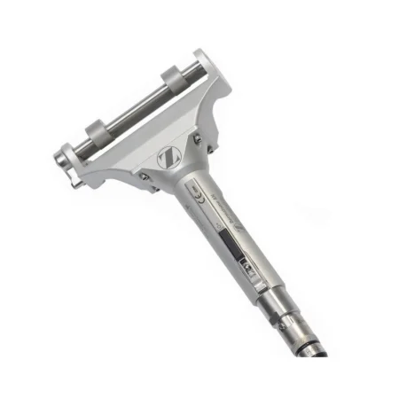
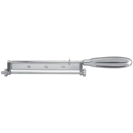
The scrub prepares the powered dermatome ahead of time, though some surgeons prefer to do it themselves. It is a critical step, because a blade or guard that is not seated right can work loose, injure the donor site or tear the graft:
- Open a new sterile blade for each patient, and keep more in the room, because several grafts may need several blades. Take the blade out of its package carefully, so it is not damaged and you are not cut.
- Check that the blade is the right one for that dermatome, seat it, and cover it with the guard plate the surgeon wants; the plate holds the blade and sets the graft's width. Tighten the screws with the screwdriver from the set.
- Connect the power only after the blade is loaded, with the safety switch on. Pass the cord off the field to the circulator and secure it so it does not get in the way of either operative site.
- Test the dermatome before it is needed.
- Hand it to the surgeon with the thickness setting at zero, so the surgeon chooses the depth on the lever at its side.
Have sterile mineral oilMIN-er-ul oylA lubricant laxative, taken by mouth or as an enema, that coats stool so it passes more easily.Full entry to lubricate the donor site so the blade glides, and sterile tongue blades to stretch the skin ahead of the dermatome as the surgeon draws the skin behind it back.
The graft mesher
A split-thickness graft is often meshed: laid on a plastic carrier and fed through a hand-cranked mesher, which cuts rows of small slits that open into diamond-shaped holes. Meshing lets the graft stretch over a larger or uneven area and lets blood and serum drain out through it instead of collecting underneath. The carrier's ridges set the size of the slits, and its package is marked with the expansion ratio: a 3:1 carrier makes a graft that covers three times its original area. Use the carrier the surgeon asks for. You may be asked to turn the mesher while the surgeon keeps the carrier in line.
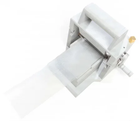
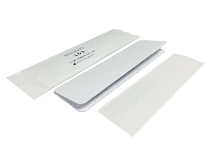
Split-thickness skin graft
What and why: an STSG covers a wound too large to close or to leave to heal by itself, so the tissues beneath it are protected from infection and injury: a burn, a wound after an injury, or the defect left by removing a tumor or releasing a scar.
Before surgery: the surgeon may mark both sites in the holding area. The patient is positioned so both sites are well exposed, supine for the thighs and trunk or prone for the buttocks and back, under general anesthesiaJEN-er-ul an-es-THEE-zhuhDrug-induced loss of consciousness and feeling over the whole body, so a person feels nothing and is not aware during surgery.an- withoutesthesi/o feeling, sensation-ia conditionFull entry or local anesthesiaLOH-kul an-es-THEE-zhuhNumbing medicine applied to or injected into one small area, so a procedure there is not felt while the person stays awake.Full entry with monitored anesthesiaan-us-THEE-zhuhLoss of feeling or sensation, often brought on by drugs so that surgery or a procedure can be done without pain.an- withoutesthesi/o feeling, sensation-ia conditionFull entry care.
Setup: a minor plastic set with fine scissors and fine Adson and Brown forcepsFOR-sepsA hand instrument with two blades or arms used to grasp, hold or pull tissue or objects.Full entry , the dermatome and its blades, the mesher and carriers, sterile mineral oil, sterile tongue blades, 10 mL Luer-Lok syringes with fine needles, a sterile marking pen, and the solutions and dressings the surgeon prefers.
Key steps:
- The recipient site is prepared first: a lesionLEE-zhunAn area of tissue that has been damaged or has changed from normal, such as a sore, wound, lump or spot.Full entry is cut out, or the wound is debrided down to healthy tissue that bleeds a little, which shows it is alive. It is covered with moist sponges or a towel while the graft is taken.
- The donor site may be injected with dilute epinephrineep-ih-NEF-rinA hormone and drug, also called adrenaline, that raises heart rate and blood pressure and opens the airways.Full entry to limit bleeding, then lubricated with mineral oil.
- The scrub or assistant holds the skin taut with tongue blades or 4x4 sponges while the surgeon advances the dermatome. As the graft comes out of the back of the head, lift it gently with fine Adson or Brown forceps, keeping slight tension so it does not curl up or tear; the scrub may be asked to do this.
- When there is enough, the surgeon tilts the dermatome up to cut the graft free, or frees it with fine scissors. Place it at once in a small basin, kept moist but not soaking, and covered, as your facility's policy directs (one reference keeps it in body-temperature saline until it is meshed).
- Sponges soaked in saline, a dilute epinephrine solution, thrombinTHROM-binAn enzyme in the blood that turns fibrinogen into fibrin, the threads that hold a blood clot together.thromb/o clot-in substanceFull entry or a phenylephrinefen-il-EF-rinA drug that tightens blood vessels, given IV to raise low blood pressure and used in nose sprays and eye drops.Full entry solution go on the donor site to stop the oozing. They are counted, and they come off and the count is reconciled before the donor site is dressed.
- If the graft is to be meshed, it is laid outer side up on the carrier and passed through the mesher. Keep a meshed graft on its carrier, under a moist sponge, until it goes onto the wound, so it does not roll up, dry out or get lost; a second graft goes on its own carrier.
- The graft is spread over the bed, trimmed with curved iris scissors and fixed with staples or 3-0 or 4-0 interrupted sutures: sutures on exposed areas such as the hands and face, staples where clothing covers it. The surgeon may leave the suture ends long, for tying over a bolster. The surgeon may rinse under the graft to wash out clots.
- Nonadherent gauze and a bolster (stentstentA small tube, often of metal mesh, placed inside a vessel or duct to hold it open.Full entry ) dressing tied over it hold the graft down, and the donor site gets its own dressing (chapter 30); a meshed graft always gets a moist dressing so it does not dry out and fail.
Wound class: the donor site is Class I, clean. The recipient site's class follows the wound it covers: Class I for a clean surgical wound, such as the bed left by cutting out a skin cancer; Class III for a fresh wound from an injury; Class IV for an old wound with dead tissue or infection. One reference lists every recipient site as Class II.
After surgery: the team watches the graft for changes in color or temperature, blanching, swelling, congestion, signs of infection and unusual pain, and reports any change to the surgeon. Infection, bleeding and scar contracturekun-TRAK-cherA lasting shortening and tightening of muscle, tendon, skin or joint tissue that keeps a joint from moving through its full range.Full entry at either site are the main complications, and a meshed graft heals with a waffle-like pattern.
Full-thickness skin graft
What and why: an FTSG covers a small, deep defect left by removing a lesion, an injury or disease, where a good look, little shrinkage or a durable cover over a joint matters.
Before and setup: a minor plastic set and a bipolarby-POH-lerHaving two poles or two opposite ends; most often used as a short form of bipolar disorder.bi- twopolar having polesFull entry electrosurgeryee-LEK-troh-SUR-jer-eeUsing high-frequency electric current through a handheld tool to cut tissue or seal bleeding vessels during surgery.Full entry ; the donor site is prepped and draped with the recipient site.
Key steps:
- The recipient site is prepared: it must be free of dead tissue, and the surgeon may dissectdy-SEKT, or dih-SEKTTo cut apart or separate tissues carefully during surgery.Full entry further to expose a bed with a good blood supply.
- The surgeon sizes the graft with a template, often cut from a sterile gloveSTAIR-ul gluvA glove that has been sterilized and packed so it can touch a sterile field or an open body area without bringing in germs.Full entry wrapper or a piece of unexposed X-ray film laid over the defect, then traces it on the donor site with a skin markerMAR-kerSomething that can be measured, such as a substance in the blood, that shows a disease or body process is present.Full entry .
- The graft is cut with a #15 blade, fine scissors such as iris or Stevens scissors, and fine forceps. The ESU is kept to a minimum, because its debris slows healing at the recipient site. The graft goes straight into a small basin on Telfa moistened with saline.
- All the fat is trimmed off the graft's underside with Stevens or curved iris scissors, and the graft is placed quickly to keep it alive.
- The graft is sewn in with interrupted 4-0 or 5-0 synthetic absorbable sutures, with a tacking stitch or two in the middle to hold it against the bed, and dressed with petrolatum or Xeroform gauze and gauze, often under a bolster.
- The donor site is closed with sutures.
After surgery and wound class: the graft is watched as a split-thickness graft is; Class I at the donor site, and the recipient site follows its wound.
Composite grafts
A composite graft, skin with cartilage or fat beneath it, moves with no blood supply of its own and lives only on vessels growing in from the edges of its bed, so it is kept small, no more than about 2 cm across. Keep it moist and apart until the surgeon sews it in.
7Flaps
A flap is tissue moved from one area to another while some or all of its own blood supply keeps working. Because it brings its own blood, a flap can cover what a graftGRAFTTissue or a man-made material moved into place in surgery to replace or repair a damaged part.Full entry cannot: a bed with a poor blood supply, a full-thicknessful-THIK-nisGoing through every layer of the skin, or every layer of another tissue such as the wall of an organ.Full entry loss, or exposed bone, tendon or nerve. A flap also keeps more of the qualities of normal skin. Its drawbacks are bulk, a color or texture that may not match, and the need for more than one operation and often a hospital stay.
Kinds of flap
- By blood supply: a random pattern flap is skin and fat fed by small unnamed vessels, so it must stay short for its width; an axial pattern flap runs along a named artery and vein, so it can be long and narrow.
- By movement: an advancement flap slides forward into a nearby defect; a transposition flap swings sideways across an angle; a rotation flap is a half circle that turns into place; and an island flap is tunneled under the skin to its new site. A Z-plasty, taught with scar revision below, is a transposition of two small flaps.
- pediclePED-ih-kulA narrow stem or stalk that attaches a structure, such as a tumor or a skin flap, to the body.Full entry or free: a pedicle flap stays attached to its blood supply at its base, the pedicle. A free flap is cut completely free and its artery and veins are joined to vessels at the new site under magnification.
- By tissue: myocutaneous (musculocutaneous) flaps carry skin, fat and muscle; fasciocutaneous flaps carry skin, fat and fasciaFASH-ee-uhA sheet or band of tough connective tissue that covers and separates muscles and organs.fasci/o bandFull entry ; osteocutaneous flaps add bone. A pedicle may also carry a nerve, giving feeling or movement to the new site.
Pedicle flaps can come from tissue close to the defect, such as the palm for a finger, or from far away, such as the trunk for a limb. Some pedicle flaps stay attached at the base until they have grown into the wound, and the base is divided later. Before it is divided, the surgeon checks the flap's circulation: rubber-shod clamps go across the base, fluorescein is injected into a vein, and after about 10 minutes the room lights go off and a Wood lamp, a handheld ultraviolet light, shows whether the flap glows.
Raising a local flap
Before surgery: the surgeon marks the flap and the defect, often in the holding area. The patient is positioned so both the defect and the donor tissue are reachable; a long case adds a urinary catheter, compression devices and active warming. If the donor site may need a skin graft, add the dermatome and mesher to the setup.
Key steps (an advancement flap):
- The defect and the planned flap are measured so the flap will cover it completely, and the lines are drawn on the skin.
- The edges of the defect are trimmed with a #15 blade and scissors back to healthy tissue.
- The flap is cut and lifted with the knife or tenotomy scissors while double skin hooks hold it, then advanced over the defect.
- The surgeon closes the deep layer with 3-0 or 4-0 absorbable sutureSOO-cherA stitch, or the thread used to stitch, that holds the edges of a wound or incision together.Full entry and the skin with fine 5-0 or 6-0 nonabsorbable suture. A drain may go under the flap.
- The donor site is closed directly or covered with a split-thickness graft, and the dressing holds the flap still, sometimes with a splint.
Free flaps and microsurgery
Microsurgery is any operation that needs magnification, through loupes or the operating microscope; microvascular work joins vessels about 1 mm across. Plastic surgeons use it for free flaps to the breast and the head and neck, muscle transfers, toe-to-hand transfers, replantation and nerve repair.
The scrub's work in a free flap:
- The room: the surgeon and assistant may stand or sit; for seated work have fully adjustable stools, and wrist and arm supports against fatigue in the long, fine work.
- Two teams: one team raises the flap and closes its donor site while the other prepares the vessels at the recipient site, often each with its own scrub and setup.
- Microinstruments: handle them as chapter 24 teaches, in their protective tray with tip guards on when not in use. A spring-handled needle holder is held like a pencil, so pass it that way, with light pressure on a nonlocking one so the needle does not drop.
- Microvascular clamps: small spring clamps, usually disposable, sized to the vessel and marked A for artery or V for vein. Hand the one the surgeon names.
- Joining the vessels: arteries are sewn by hand with very fine nylon or polypropylene, 8-0 to 12-0; veins may be sewn or joined with a coupler, two rings with pins that hold the vessel ends together and stay in as an implant. The stitches are tied with tying or jeweler's forcepsFOR-sepsA hand instrument with two blades or arms used to grasp, hold or pull tissue or objects.Full entry , and an approximator clamp may hold the two vessel ends together while they are sewn. A colored silicone background behind the vessels gives contrastKON-trastA dye or other substance given before an imaging test so body parts show up more clearly.Full entry .
- Small bleeders: bipolarby-POH-lerHaving two poles or two opposite ends; most often used as a short form of bipolar disorder.bi- twopolar having polesFull entry jeweler's forceps, whose very fine tips spread little heat, coagulationkoh-ag-yuh-LAY-shunThe process by which blood changes from a liquid into a gel, forming a clot.coagul clot-ation processFull entry them, and cellulose eye spears soak up small amounts of blood.
- Keeping the vessels open: the anesthesia provideran-us-THEE-zhuh pruh-VY-derThe licensed professional who gives anesthesia and watches over the client's breathing and vital functions during a procedure.Full entry gives heparinHEP-uh-rinAn anticoagulant drug, given by injection or IV, that keeps blood clots from forming or growing.Full entry into a vein before the anastomosisuh-nas-tuh-MOH-sisA connection between two tube-shaped structures, such as two ends of bowel or two blood vessels, made by surgery or found naturally.Full entry ; heparinized saline flushes the vessels, and a patty soaked in papaverine, verapamilver-AP-uh-milA calcium channel blocker that slows the heart rate and relaxes blood vessels.Full entry or lidocaineLY-doh-kaynA local anesthetic that numbs an area, also given by IV to treat some fast, abnormal heart rhythms.Full entry reverses spasmSPAZ-umA sudden, involuntary tightening of a muscle or of the muscle in a hollow organ or tube.Full entry .
- Keeping tissue alive: keep the vessels and the flap moist with saline or lactated Ringer's solutionLAK-tay-tid RING-erz suh-LOO-shunAn isotonic IV fluid containing water, sodium, chloride, potassium, calcium and lactate.Full entry (never water), and suction gently or through a moist sponge, because suction can injure a vessel. Move harvested tissue to and from the field in a Petri dish or small basin, and keep it well away from the edge of the back table.
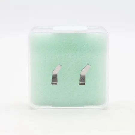
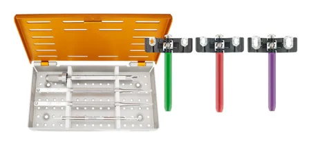
The surgeon checks the flap's color, warmth and Doppler ultrasoundDOP-ler UL-truh-soundAn ultrasound that uses sound waves to hear or show blood flowing through vessels or the fetal heartbeat.Full entry signal during the case. A marking stitch shows where to listen with the Doppler afterward, and for a buried flap that cannot be seen, a tiny implantable Doppler probe may be wrapped around the anastomosis; when monitoring ends its wire is pulled out and the probe stays in.
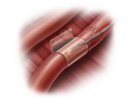
Flaps for the head and neck
After cancer surgery of the mouth, throat or jaw, the defect is often filled with a flap, which survives where the blood supply is poor. Know each flap by its source:
- Pectoralis major flap: the chest muscle, pedicled on a branch of the thoracoacromial artery.
- Deltopectoral flap: chest skin on branches of the internal mammary artery.
- Latissimus dorsi flap: the back muscle on the thoracodorsal artery, pedicled or free.
- Radial forearm free flap: forearm skin on the radial artery, the free flap used most often for the floor of the mouth and the tongue.
- Anterolateral thigh free flap: thigh tissue, chosen when bulk is wanted.
- fibulaFIB-yuh-luhThe thin outer bone of the lower leg, beside the larger tibia.Full entry free flap: the fibula, often with a paddle of skin, to rebuild the jaw.
Before a radial forearm flap, or any operation that may cut off the radial or ulnar artery, the patient has an Allen testAL-en testA bedside check of blood flow to the hand through the ulnar artery, done by pressing and releasing the wrist arteries.Full entry : both arteries at the wrist are pressed until the hand blanches, then the ulnar artery is released while the radial stays pressed, and the palm should turn red again at once, showing the ulnar artery alone can feed the hand. A Doppler marks the flap's vessels on the skin before it is raised and checks its blood flow during the case. A second team often raises the flap at the same time, a fibula flap under a thigh tourniquet for example, with its own scrub and setup. The team working in the mouth is not sterile, so no instruments, power equipment or supplies pass between the two fields.
Hand and face transplants
At the top of the ladder, one rung above the free flap, is a transplant of a hand or a face from a donor, a vascularized composite allograft (the older name is composite tissue allograft). The first partial face transplant restored light touch, the feeling of heat and cold and closing of the mouth, but such transplants need drugs that suppress the immune system, and that patient had episodes of rejection and infection.
Flap checks and flap failure
A flap is checked often in the first days after surgery, when its vessels are most likely to clot: most blockages happen within the first 48 hours, and the first 72 hours are watched most closely. Watch for these signs and report them at once:
- Venous congestion: the flap turns dusky and firm and swells. A blocked vein is the commonest cause of flap failure, behind 80 to 90 percent of failures in one reference.
- Arterial insufficiencyin-suh-FISH-un-seeA state in which an organ or part does not work well enough to meet the body's needs.in- notsufficient enoughFull entry : the flap turns pale and cool.
- Either one: a capillary refill that is very fast or absent, and a lost Doppler signal, which is often the last sign to appear.
A flap without enough blood dies (necrosisneh-KROH-sisThe death of cells or tissue in a part of the body.necr/o death-osis conditionFull entry ), often only at its far end at first. A hematomahee-muh-TOH-muhA collection of blood, often clotted, that has leaked out of the blood vessels into the tissue.hem/o blood (here as hemat-)-oma swelling or massFull entry under a flap can press its vessels shut, and too much tension or a twisted pedicle can do the same. A failing free flap means a fast return to the OR, where a prompt repair saves many flaps. Medicinal leeches may be used on a congested flap to draw off blood, with an antibiotic to prevent the infection that leeches can carry.
a flap lives on its pedicle. Nothing presses on it: no tight dressing, bra or strap over the flap or its vessels, and no pulling or twisting of its pedicle.
8Skin lesions, Mohs surgery and scar revision
Excision of a skin lesion
What and why: a lesionLEE-zhunAn area of tissue that has been damaged or has changed from normal, such as a sore, wound, lump or spot.Full entry is cut out to diagnose it or to cure it. An early basal cell carcinomaBAY-sul SEL kar-sih-NOH-muhThe most common type of skin cancer, which starts in the basal cells at the bottom of the skin's outer layer.Full entry can be cured by simple excisionek-SIZH-unCutting out tissue, such as a growth or damaged area, during surgery.Full entry and closure, with the pathologist checking that the margins are free of cancer.
Before and setup: local anestheticLOH-kul an-es-THET-ikA drug that blocks feeling in one part of the body without making the person unconscious.Full entry with epinephrineep-ih-NEF-rinA hormone and drug, also called adrenaline, that raises heart rate and blood pressure and opens the airways.Full entry , sometimes with moderate sedationMOD-er-it seh-DAY-shunA drug-induced state of relaxed, lowered awareness in which the client still responds to voice or light touch and breathes on their own.Full entry or general anesthesiaJEN-er-ul an-es-THEE-zhuhDrug-induced loss of consciousness and feeling over the whole body, so a person feels nothing and is not aware during surgery.an- withoutesthesi/o feeling, sensation-ia conditionFull entry ; a minor tray, a skin markerMAR-kerSomething that can be measured, such as a substance in the blood, that shows a disease or body process is present.Full entry and an electrosurgeryee-LEK-troh-SUR-jer-eeUsing high-frequency electric current through a handheld tool to cut tissue or seal bleeding vessels during surgery.Full entry ; a skin graftGRAFTTissue or a man-made material moved into place in surgery to replace or repair a damaged part.Full entry or flap setup if the defect may need one.
Key steps: after the local anesthetic, the surgeon cuts out the lesion with a margin of normal skin, or scrapes it out with a curettekyoo-RETA surgical instrument with a small spoon- or loop-shaped tip used to scrape tissue.Full entry , and sends it for a frozen section or permanent sections; then bleeding is stopped and the wound is closed directly or with a flap or a graft.
A specimen checked for margins is oriented, with a stitch or ink, so the pathologist can tell which edge is which (chapter 29).
Wound class: I, clean.
Mohs micrographic surgery
What and why: Mohs micrographic surgery (MMS) removes a basal or squamous cell carcinomaSKWAY-mus SEL kar-sih-NOH-muhA cancer that starts in squamous cells, the thin, flat cells of the skin's outer layer and the linings of some organs.Full entry layer by layer, examining each layer under the microscope at once, until no cancer is left. Its aim is to cure as many cancers as possible while keeping as much healthy skin as it can and leaving the smallest scar.
Before surgery: it is usually done as an outpatient case under local anesthesiaLOH-kul an-es-THEE-zhuhNumbing medicine applied to or injected into one small area, so a procedure there is not felt while the person stays awake.Full entry , in stages, with the lab work done while the patient waits, so it is long, and the patient's comfort matters. The head is steadied on a gel donut for a face lesion.
Setup: a minor tray, fine 5-0 or 6-0 sutureSOO-cherA stitch, or the thread used to stitch, that holds the edges of a wound or incision together.Full entry , skin markers and marking dyes, several specimen containers and Telfa strips.
Key steps:
- The surgeon draws the lesion's edge and marks the orientation with ink, small hash cuts like the hours of a clock, or sutures, then cuts a thin layer with a #15 blade while skin hooks hold the edges, not with the ESU, because heat distorts tissue meant for a frozen section.
- Receive each piece in a clear Petri dish or shallow container, on Telfa moistened with saline, apart from the others and with every mark intact; it leaves the field at once.
- The tissue is cut into sections, color-coded and mapped, then frozen, sliced, stained and read, and wherever cancer remains is marked on the map.
- Another layer is taken only where the map shows cancer, until the margins are clear; the wound is then left to heal, closed with 4-0 or 5-0 nylon or polypropylene, or covered with a flap or graft.
After surgery: because most skin cancers come from ultraviolet light, the patient is taught sun protection: avoid intense sun from 11 a.m. to 3 p.m., wear clothes that cover the skin, such as a wide-brimmed hat and long sleeves, and reapply sunscreen every 2 hours outdoors.
Wound class: I, clean.
Scar revision
What and why: a scar revision makes an existing scar less noticeable, or lengthens one that has contracted. Infection, tension or an injury can leave a ragged, raised or tight scar; chapter 11 teaches hypertrophyhy-PER-truh-feeAn increase in the size of an organ or tissue because its cells grow larger.hyper- above, excessive-trophy nourishment, growthFull entry scars and keloidKEE-loydA raised, firm overgrowth of scar tissue that spreads beyond the edges of the original wound.-oid like, resemblingFull entry . Lasers smooth many scars in the clinic.
Setup: a plastic instrument tray and a sterile marking pen, with the position, prep and drape set by where the scar is.
Key steps: the surgeon marks the plan and injects local anesthetic, cuts out the scar with a #15 blade and fine toothed forcepsFOR-sepsA hand instrument with two blades or arms used to grasp, hold or pull tissue or objects.Full entry while fine skin hooks hold it, undermines the skin around it (separates it from the deeper tissue with Metzenbaum or curved iris scissors) so the edges come together without tension, and closes it, often with 4-0 or 5-0 absorbable sutures under Telfa and flat gauze.
A wide scar can simply be cut out and closed again. More often the surgeon uses a Z-plasty, the most widely used revision: the scar becomes the central limb of a Z, two more limbs, each as long as the central limb, are cut at equal angles from its ends, and the two triangular flaps are swapped. The single long scar becomes three shorter ones, one of them along the skin's natural lines, where it shows least, and the swap lengthens a scar that had contracted. W-plasty, M-plasty and Y-V-plasty are other patterns. Z-plasties also release tight burnBURNAn injury to the skin or deeper tissues caused by heat, chemicals, electricity, radiation or friction.Full entry scars.
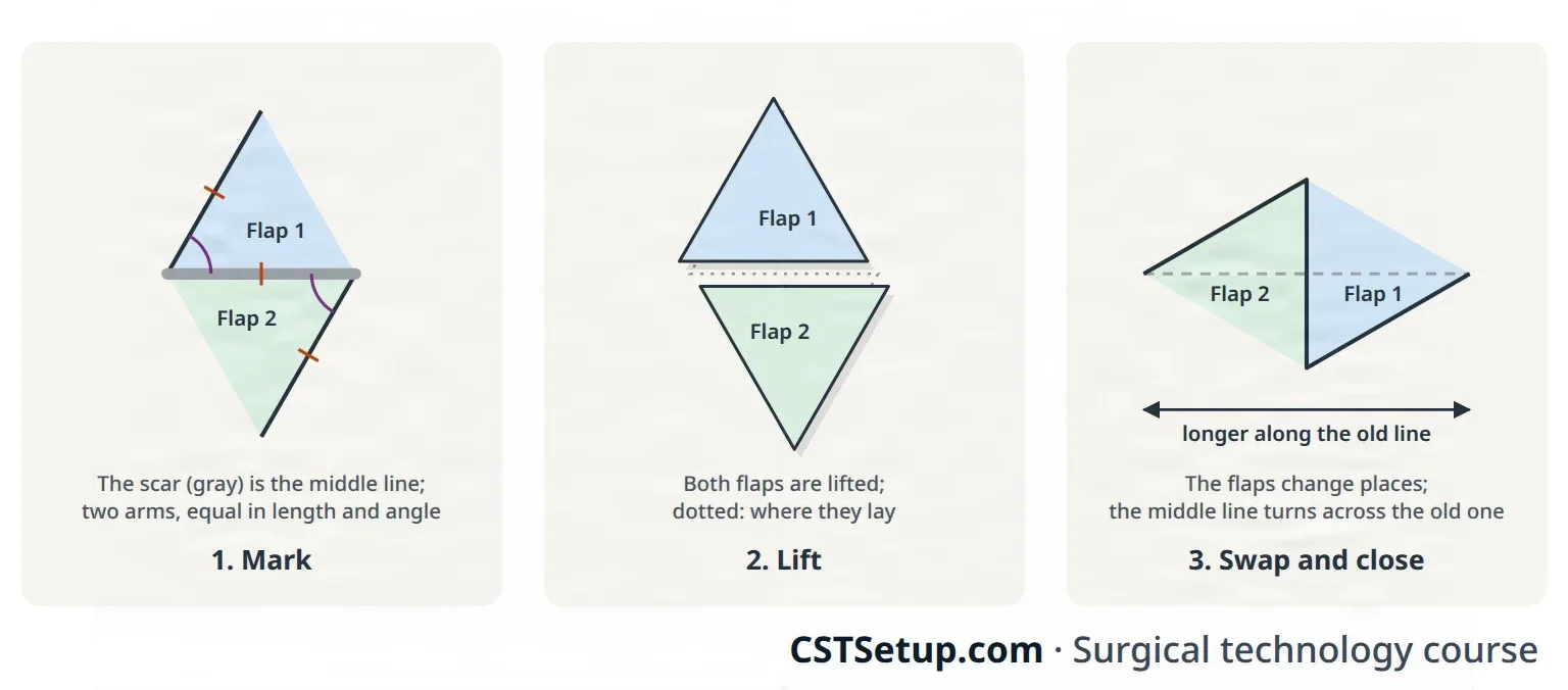
After surgery: the patient usually goes home the same day; infection, a scar that contracts again, or a look no better, or worse, are the risks.
Wound class: I, clean.
9Burns and pressure injuries
Burn surgery
Chapter 11 teaches how burns are graded by depth and how the rule of nines sizes them; a chart by age, the Lund and Browder chart, is used for children, whose bodies are proportioned differently. Patients with large burns are among the sickest who come to the OR, and burnBURNAn injury to the skin or deeper tissues caused by heat, chemicals, electricity, radiation or friction.Full entry centers have their own specially trained surgical teams.
Before surgery: a burned patient loses heat fast through the open wounds, so the room is warmed and only the area being worked on is uncovered; work with the anesthesia provideran-us-THEE-zhuh pruh-VY-derThe licensed professional who gives anesthesia and watches over the client's breathing and vital functions during a procedure.Full entry on fluid replacement. Because the pain is severe, the patient may be anesthetized before being moved onto the OR bed, and the burn dressings come off only after inductionin-DUK-shunThe starting of a process on purpose, such as bringing on labor or putting a client under anesthesia.Full entry , which spares pain and heat; protect the airway during the move.
Escharotomy and fasciotomyfash-ee-OT-uh-meeSurgery that cuts open the fascia, the tough tissue around a group of muscles, to relieve pressure inside it.fasci/o fascia, a band of connective tissue-tomy cutting intoFull entry : a full-thickness burnFULL THIK-nis BERNA burn that destroys both layers of the skin, the epidermis and all of the dermis, and may reach the tissue below.Full entry forms escharES-karThick, dry, dark dead tissue that covers part or all of a wound.Full entry , thick, stiff, dead tissue. Eschar all the way around a limb or other body part acts like a tourniquet as the tissue beneath swells, and can damage muscle, bone and vessels, so the surgeon cuts through it (escharotomy) or through the fasciaFASH-ee-uhA sheet or band of tough connective tissue that covers and separates muscles and organs.fasci/o bandFull entry (fasciotomy) to release the pressure, as in compartment syndromekum-PART-ment SIN-drohmA buildup of pressure inside a closed group of muscles that cuts off their blood supply.Full entry .
debridementdih-BREED-muntThe removal of dead, damaged or infected tissue from a wound so it can heal.Full entry : removing dead, damaged or infected tissue so the wound can heal. Burns need it again and again, because burned tissue keeps dying after the injury:
- Excisional debridement cuts away all the dead tissue down to the fascia over the muscle.
- Tangential excisionek-SIZH-unCutting out tissue, such as a growth or damaged area, during surgery.Full entry shaves thin layers parallel to the surface with a knife dermatome, down to fat that bleeds, which shows it is alive.
Setup: a minor plastic set with fine scissors and fine forcepsFOR-sepsA hand instrument with two blades or arms used to grasp, hold or pull tissue or objects.Full entry , a knife dermatome, #10 or #20 blades, an electrosurgeryee-LEK-troh-SUR-jer-eeUsing high-frequency electric current through a handheld tool to cut tissue or seal bleeding vessels during surgery.Full entry , topical thrombinTHROM-binAn enzyme in the blood that turns fibrinogen into fibrin, the threads that hold a blood clot together.thromb/o clot-in substanceFull entry , a pneumatic tourniquet for a burned limb, body-temperature saline irrigationeer-ih-GAY-shunWashing out a wound, body cavity, tube or eye with a steady flow of liquid.Full entry , bulb syringes, an impervious pocket drape and the surgeon's topical antimicrobialan-tee-my-KROH-bee-ulA drug or substance that kills or slows the growth of microorganisms such as bacteria, viruses, fungi or parasites.anti- againstmicro- smallbi/o life-al relating toFull entry and dressings. Keep a small basin ready for the tissue removed.
Key steps:
- With the burn exposed, prepped and draped, the surgeon removes the dead tissue with the knife or a knife dermatome until only living tissue is left.
- Bleeding is controlled with the ESU, topical thrombin and lap sponges soaked in saline with topical epinephrineep-ih-NEF-rinA hormone and drug, also called adrenaline, that raises heart rate and blood pressure and opens the airways.Full entry 1:1,000 added as the surgeon orders (one reference's mix is 4 mL in 1,000 mL of saline).
- The clean wound is covered for now with an allograft or a xenograft (pig skin), stapled on, or with nonadherent dressings such as Xeroform and fluffs, with compression wraps on the limbs.
The patient's own skin (an autograft) usually goes on several days later, once the bed has a good enough blood supply for a graftGRAFTTissue or a man-made material moved into place in surgery to replace or repair a damaged part.Full entry to take. A fourth-degree burn, into muscle, tendon or bone, cannot be grafted and needs special care.
Wound class: by the CDC's definitions, a fresh wound from an accident is Class III, and an old wound with dead tissue or infection is Class IV; your facility's policy decides how a burn is classed.
Pressure injury repair
Chapter 11 teaches how pressure injuryPRESH-er IN-juh-reeDamage to the skin and the tissue beneath it from pressure, or pressure with shear, usually over a bony area or under a device.Full entry form and are staged, and chapter 31 how positioning prevents them. When one needs surgery, the plastic surgeon generally debrides it and the patient gets IV antibiotics; many then heal on their own, but the surgeon may cut out the whole injury and close it, because healing by second intention can leave a weak scar that breaks down again. For a deep injury, the surgeon cuts out the injured tissue and the dead bone with rongeurron-ZHURA strong surgical instrument with sharp, cup-shaped jaws used to bite away small pieces of bone.Full entry , osteotomes and curettekyoo-RETA surgical instrument with a small spoon- or loop-shaped tip used to scrape tissue.Full entry and covers the defect with a graft or a flap, sometimes after tissue expansion, over large drains and under a tie-over bolster. Position, prep and drape so the injury and every donor site are exposed.
Wound class: IV, dirty, when dead tissue or infection is present.
10The face and skull
Facial cases are short-instrument cases on a small field. Most are supine on a donut or foam headrest with the arms tucked, under local anesthesiaLOH-kul an-es-THEE-zhuhNumbing medicine applied to or injected into one small area, so a procedure there is not felt while the person stays awake.Full entry with sedationsih-DAY-shunA calm, relaxed or sleepy state brought on by drugs, or the use of drugs to cause it.sed calm, settle-ation processFull entry or general anesthesiaJEN-er-ul an-es-THEE-zhuhDrug-induced loss of consciousness and feeling over the whole body, so a person feels nothing and is not aware during surgery.an- withoutesthesi/o feeling, sensation-ia conditionFull entry , with a head drape and a split sheet, and the face is prepped so no solution runs into the eyes or ears (above). cleft lipkleft lipA split or opening in the upper lip that is present at birth.Full entry and palate repair (cheiloplasty and palatoplasty), which plastic surgeons often do, is taught in chapter 37.
Blepharoplasty
What and why: with age the eyelid skin and the orbital septumSEP-tumA wall of tissue that divides two spaces or cavities, such as the two sides of the heart or nose.sept/o wall, partitionFull entry relax, the fat behind the septum bulges, and the lids look heavy and tired. A blepharoplasty removes extra lid skin, removes or moves the bulging fat and tightens the muscle, on the upper lids, the lower lids or both, often with a facelift; removing a hooded upper lid can also improve the side vision. Chapter 35 teaches the other eyelid operations.
Before surgery: the surgeon marks the lines with the patient sitting up, before the local anestheticLOH-kul an-es-THET-ikA drug that blocks feeling in one part of the body without making the person unconscious.Full entry , because the injection swells and distorts the lid. It is usually done under local anesthesia with moderate sedationMOD-er-it seh-DAY-shunA drug-induced state of relaxed, lowered awareness in which the client still responds to voice or light touch and breathes on their own.Full entry . No prep solution may pool in the eyes; corneaKOR-nee-uhThe clear, dome-shaped layer at the front of the eye, over the iris and pupil.Full entry shields, put in after numbing drops, protect the corneas, and ointment keeps the eyes from drying.
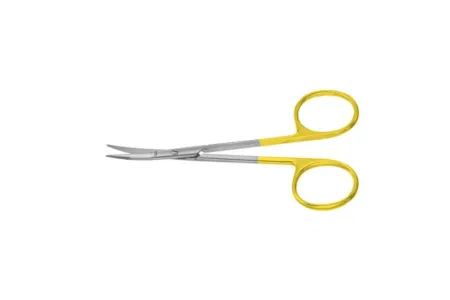
Setup: a plastic tray with short, delicate instruments: curved Kaye blepharoplasty scissors, fine toothed Adson or jeweler's forcepsFOR-sepsA hand instrument with two blades or arms used to grasp, hold or pull tissue or objects.Full entry , Westcott scissors, calipers, fixation forceps and a Webster or Castroviejo needle holder; bipolarby-POH-lerHaving two poles or two opposite ends; most often used as a short form of bipolar disorder.bi- twopolar having polesFull entry , or monopolar on a low setting with a needle tip; corneal shields, cotton-tipped applicators and cellulose (Weck-Cel) spears.
Key steps (upper lid):
- Local anesthetic goes into the lid through a fine (30-gauge) needle, and the surgeon cuts out the marked ellipse of skin with a #15 blade; calipers keep the two sides equal and stop too much being taken, which could leave the eye unable to close.
- A strip of orbicularis muscle is trimmed, the septum is opened, and the bulging fat is trimmed, guardingGAR-dingTightening of muscles, or protecting a body part with the hands or posture, to avoid pain when it is touched or moved.Full entry the levator muscle. Dab the small bleeders with the cellulose spears so the surgeon can see and seal them.
- The skin is closed with fine (6-0) sutureSOO-cherA stitch, or the thread used to stitch, that holds the edges of a wound or incision together.Full entry , often blue polypropylene, which stands out from the lashes when it is removed, and the other lid is done the same way.
Key steps (lower lid): the incision runs about 1 mm below the lashes (one reference gives 2 to 3 mm) and out into a natural line at the outer corner. A flap of skin and muscle is lifted, the extra fat is removed or moved, the skin is laid back upward and outward so the lid does not turn out (ectropion), the extra is trimmed, and the muscle at the outer corner is sewn to the periosteum.
After surgery: iced moist pads or cold compresses over the eyes, changed as they warm, and the head raised; usually no dressing, only antibiotic ointment; sutures out in 3 to 5 days. Complications include infection, unequal lids and, if too much skin was taken, a lid that cannot close.
Wound class: I, clean.
Brow lift
What and why: with age the forehead skin loosens, the brow sags (brow ptosis) and hoods the upper lids, and repeated muscle action carves forehead lines and frown creases, the frowns made by two small muscles between the brows, the corrugator and the procerus. A brow lift raises the brow through an incision across the top of the head (coronal), at the hairline, at the brow itself, or endoscopically through a few short scalp incisions.
Before surgery: check the endoscopicen-doh-SKOP-ikDone with an endoscope, a thin tube with a light and camera passed into the body.endo- within-scopic viewingFull entry equipment before the patient comes in. The patient lies supine with the head at the very top of the bed so the long instruments can work, the eyes protected with lubricant, the head of the bed perhaps raised, and the whole head prepped (above). General anesthesia is preferred, or local with MAC; a dilute local anesthetic with epinephrineep-ih-NEF-rinA hormone and drug, also called adrenaline, that raises heart rate and blood pressure and opens the airways.Full entry may be injected under the periosteum with a 60 mL syringe and an 18-gauge needle, and with several drugs on the back table, label every one.
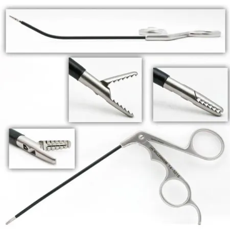
Setup: an endoscopic brow set with a 30-degree, 4 or 5 mm scope, periosteal elevators, endoscopic dissectors, scissors and graspers, the camera and light source, an electrosurgeryee-LEK-troh-SUR-jer-eeUsing high-frequency electric current through a handheld tool to cut tissue or seal bleeding vessels during surgery.Full entry , a power drill, and the fixation the surgeon uses: screws, absorbable tacking devices, bone anchors or sutures.
Key steps:
- Three to five short incisions are made behind the hairline, and the forehead is lifted off the skull under the periosteum with elevators as the scope goes in.
- Under the scope, the surgeon frees the tissue down to the brows, protects the supraorbital nerve, and divides, removes or weakens the corrugator and procerus muscles.
- The outer brow is lifted and fixed with sutures tied around screws in the outer table of the skull, or with tacks or anchors; the inner brow usually is not, because lifting it too far looks unnatural.
- The incisions are stapled or sutured, and a head dressing of fluffs and rolled gauze may go on.
After surgery: temporary screws come out in the office after about 10 to 14 days. Complications are uncommon: the brow sitting wrong or dropping again, hair loss or scarring at the incisions, numbness of the forehead and scalp, and weakness of the forehead muscle, usually temporary.
Wound class: I, clean.
Rhytidectomy (facelift)
What and why: aging, gravity, sun damage and smoking loosen the skin and the tissue beneath it, leaving jowls, deep folds beside the nose and mouth, and loose skin under the chin. A rhytidectomy (rhytid, wrinkle, and -ectomy, removal) lifts and repositions the deeper tissue, the SMAS and the platysma muscle of the neck, then redrapes the skin and removes the excess. There are many techniques, so ask the surgeon which one is planned.
Before surgery: the surgeon marks the patient first. The patient lies supine with the head and shoulders slightly raised, the eyes protected and the whole head prepped (above). Under local anesthesia with MAC or general anesthesia, the surgeon injects local anesthetic with epinephrine along the incisions with a fine spinal needle and a 10 mL syringe.
Setup: a plastic tray with facelift scissors of several lengths, such as the Kaye, smooth and toothed forceps and several needle holders, a fiber-optic lighted retractorrih-TRAK-terA surgical instrument that holds back tissue or organs so the surgeon can see and reach the operative site.Full entry , bipolar, skin hooks and small Deaver retractors, and drains if the surgeon uses them.
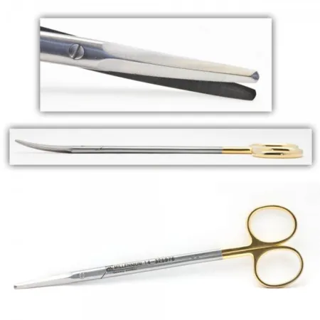
Key steps:
- After any liposuction of the neck, jowls or cheeks, the incision runs from the temple, inside the hairline, down in front of the ear, around the earlobe, up behind the ear and back into the scalp.
- The skin is lifted off the deeper tissue with facelift scissors while double hooks and then retractors hold the edges, placed with care because the facial nerveFAY-shul NERVThe seventh cranial nerve, which moves the muscles of the face and carries taste from the front of the tongue.Full entry lies beneath, and bipolar seals the small bleeders.
- The SMAS is folded and stitched (plicated) upward, and through a small incision under the chin the loose bands of the platysma are tightened.
- The skin is pulled up and back, held with tacking sutures if the surgeon wants, and the excess is trimmed.
- Small flat drains go under the flaps, fibrin sealant may be applied, and the incisions are closed with fine sutures and staples. Wash the blood out of the hair before the dressing, gauze held with an elastic bandage or a facial splint.
After surgery: the head stays raised and cold compresses ease swelling. Complications include infection, bleeding, a hematomahee-muh-TOH-muhA collection of blood, often clotted, that has leaked out of the blood vessels into the tissue.hem/o blood (here as hemat-)-oma swelling or massFull entry under the skin and scarring.
Wound class: I, clean.
Facial implants
What and why: implants give the chin or cheeks shape lost to a birth defect, an injury or cancer surgery, or that the patient wants for looks. Putting in a chin implant is a mentoplasty; raising the cheekbones is a malar augmentation. Implants may be silicone, polyethylene, Gore-Tex or mesh, or the patient's own fat, bone or cartilageKAR-tih-lijFirm, smooth, rubbery tissue that cushions joints and gives shape to parts such as the ear, nose and windpipe.chondr/o cartilageFull entry , and fillers can be injected instead.
Before surgery: the surgeon studies the face, its photographs and X-rays and marks the landmarks before the patient is numbed. The patient is supine or semi-sitting with the arms tucked, under local anesthesia with MAC or general anesthesia, and the draping leaves the whole face in view so the surgeon can judge the result.
Setup: a plastic set (a minor orthopedicor-thuh-PEE-dikHaving to do with the bones, joints, muscles, ligaments and tendons, and the branch of medicine that treats them.orth/o straightped/o child (the field began with straightening children's bones)-ic relating toFull entry set for the chin), periosteal elevators such as the Joseph, Senn retractors, cheek retractors for the mouth approach, bipolar, implant sizers and implants in several sizes. Never open the permanent implant until the surgeon names its size: confirm the size with the surgeon and tell the circulator, who opens it onto the field.
Key steps (chin):
- A short incision under the chin, or inside the mouth, is carried down to the periosteum, which is lifted off the bone to make a pocket below the mental nerves, which are protected.
- Sizers choose the implant, which is stitched to the periosteum so it does not slip, and the pocket is rinsed with antibiotic solution.
- The periosteum is closed with absorbable suture and the skin with fine suture.
Key steps (cheek): through an incision inside the mouth, where the gum meets the upper lip, a pocket is lifted over the cheekbone, guarding the infraorbital nerve; after sizers, the implant is held with a pull-out suture tied over a bolster on the skin, or small screws.
After surgery: an implant can shift and may need another operation; infection, a hematoma and nerve injury are other risks, and the cheek patient eats soft food and avoids clenching the teeth for a while.
Wound class: a chin implant placed through the skin is Class I, clean. A cheek implant placed through the mouth, or a chin implant by the mouth approach, enters the mouth, so by the CDC's definitions it is Class II, clean-contaminated; one reference lists both operations as Class I.
Otoplasty
What and why: otoplasty reshapes the outer ear, or builds one. It corrects ears that stick out (prominent ears, usually because the fold of the antihelix never formed), a missing or very small ear (microtia), an abnormal pointed fold in the upper ear (Stahl ear), and ears lost to injury or disease. In a child it is usually done before the child starts school, around age 5 (one reference gives 4 to 10). The ear's rim is the helix; inside it, the Y-shaped antihelix shapes most of the ear, and the bowl next to the ear canal is the concha.
Before surgery: supine with the head on a headrest, turned operative side up, and the head of the bed raised; both ears prepped and draped when both are done; general anesthesia for a child, local with MAC for an adult. Pad the ear underneath well, because microtia surgery can take 6 to 8 hours, and keep prep solution out of the ear canal with a cotton ball that comes out after the prep.
Setup: a minor plastic set with a cartilage rasp, fine (25, 27 or 28-gauge) needles, methylene blue, local anesthetic with epinephrine and a headrest; for microtia also a minor orthopedic set, a power drill and burs, a thoracotomythor-uh-KOT-uh-meeA surgical cut through the chest wall to reach the lungs, heart or other organs in the chest.thorac/o chest-tomy cutting intoFull entry set, chest tubechest toobA tube placed through the chest wall into the space around the lung to drain air, blood or fluid.Full entry and a chest drainage system ready.
Key steps (prominent ears):
- The surgeon folds the ear back with a finger to show where the new fold belongs, and marks it at several points with a fine needle passed through the cartilage, its tip dipped in methylene blue.
- An ellipse of skin is cut from the back of the ear and the cartilage is exposed.
- The cartilage is scored or rasped so it bends, and permanent sutures hold the new fold and set the ear back toward the head, some anchored to the fasciaFASH-ee-uhA sheet or band of tough connective tissue that covers and separates muscles and organs.fasci/o bandFull entry over the mastoid.
- The skin is closed, and a bulky dressing of petrolatum gauze, fluffs and rolled gauze, or cotton soaked in ointment and a mastoid dressing, holds the shape.
Microtia is rebuilt in stages. At the first, rib cartilage is taken through a chest incision and saline is poured into the wound: bubbles mean a torn pleuralPLOOR-ulHaving to do with the pleura, the thin two-layer membrane that covers the lungs and lines the chest wall.pleur/o pleura-al relating toFull entry , so hand the chest tube at once. The cartilage is carved with the drill, on a separate towel-padded table, against a sterilizationstair-uh-luh-ZAY-shunA process that destroys all microorganisms, including spores, on an item; also a permanent birth control procedure.Full entry template of the other ear, and set under a flap of the fascia at the side of the head (temporoparietal fascia); later stages build the earlobe and the folds.
After surgery: the dressing stays on for several days, then a loose headband is worn at night for weeks so the ear is not pulled forward in sleep. Complications include infection, a hematoma, an overcorrected or unnatural shape, and sutures working through the skin.
Wound class: I, clean.
Rhinoplasty
What and why: a rhinoplasty reshapes the outside of the nose, for looks, after an injury, for a birth defect, or to fix a breathing problem caused by its structure: it can make the nose bigger or smaller, remove a hump, reshape the tip or the bridge, narrow the nostrils or change the angle between the nose and the upper lip, using cartilage, bone graftGRAFTTissue or a man-made material moved into place in surgery to replace or repair a damaged part.Full entry or implants. In an open rhinoplasty the surgeon cuts across the base of the columella, the strip of skin between the nostrils, and lifts the skin off the nose; in a closed rhinoplasty every incision is inside the nostrils. A septoplasty, straightening the septum, is often done with it, and chapter 36 teaches it.
Before surgery: local anesthesia with sedation or general anesthesia, supine or semi-sitting; the surgeon usually wears a headlight. Ask whether the surgeon wants a skin prep; if so, it covers the face and neck. If a chin implant is done in the same case, the sterile procedure goes first, because the nose is not sterile. Before the prep and drapes, the surgeon packs the nose with cottonoids soaked in a vasoconstrictor, such as cocaine solution, from a separate small setup, and injects local anesthetic with epinephrine, even under general anesthesia, to shrink the lining, reduce bleeding and ease pain afterward (chapter 36 teaches these nasal drugs).
Setup: a rhinoplasty set built around the surgeon: nasal speculumSPEK-yoo-lumAn instrument used to open and hold open a body opening, such as the vagina, so the inside can be seen.Full entry , Freer and Cottle elevators, skin hooks, the Aufricht retractor-speculum and Fomon retractors, Cottle and Silver osteotomes and a mallet, Joseph saws and nasal rasps, bayonet forceps, a cartilage crusher, fine scissors, Frazier suction, and the nasal splint the surgeon prefers.
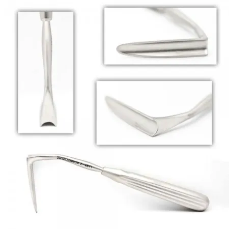
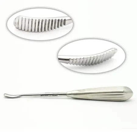
Key steps (open):
- The packing comes out, and the surgeon cuts across the base of the columella and up along the edge of one nostril.
- A double skin hook lifts the skin at the tip while the surgeon lifts the skin, periosteum and perichondrium off the framework with elevators and tenotomy scissors; keep the field clear with the Frazier suction.
- The upper lateral cartilages are trimmed, and a hump of bone is taken down with a chisel and mallet, which you may be asked to tap, and smoothed with a rasp.
- A septoplasty may follow, and cartilage or bone grafts may shape the tip, so keep any cartilage removed until told it is not needed. Lateral osteotomies, cuts through the side walls of the bony nose with the chisel, straighten it further.
- The skin is closed with fine absorbable suture, a metal or fiberglass splint goes on, and a small gauze pad is taped under the nostrils to catch drainage.
After surgery: the head stays raised, with a small ice pack on the nose; no blowing the nose or scrubbing the face for a week. Bruising, swelling and a stuffy nose are expected.
Wound class: II, clean-contaminated, because the nose, part of the respiratory tract, is entered.
Craniosynostosis correction
What and why: the bones of a baby's skull are joined by flexible seams, the sutures, which let the skull bend during birth and grow with the brain; the brain finishes most of its growth by about 2 years, and only then do the sutures begin to fuse. In craniosynostosis one or more sutures close too early. The skull cannot grow across the closed suture, so it grows misshapen; left untreated, the pressure inside the skull can rise to dangerous levels and the brain's development can stall. One suture closed is simple craniosynostosis, two or more compound; it may stand alone or be part of a syndrome. Early closure of the sagittal suture, along the top of the head, makes the skull long from front to back. Craniosynostosis is the commonest skull deformity in children that neurosurgeons treat; chapter 42 adds the neurosurgical part.
Before surgery: surgery is usually done between about 6 weeks and 6 months of age, and earlier repair gives a better result. The infant is placed supine for the front of the skull or prone for the back; a prone infant is anesthetized and endotracheal intubationen-doh-TRAY-kee-ul in-too-BAY-shunPutting a breathing tube through the mouth or nose into the windpipe (trachea).endo- withintrache/o windpipe-al relating toFull entry first, then turned. Warm the room, use a forced-air blanket and an overbed warmer during inductionin-DUK-shunThe starting of a process on purpose, such as bringing on labor or putting a client under anesthesia.Full entry , because an infant loses heat fast.
Setup: a craniosynostosis set with a #15 blade, Senn retractors, Penfield and periosteal elevators, Adson and bayonet forceps, hemostatHEE-moe-statA clamp with finger rings and a locking ratchet used in surgery to grasp and close off bleeding vessels.hem/o blood-stat stopping, standing stillFull entry , Mayo scissors, bone-cutting scissors, rongeurron-ZHURA strong surgical instrument with sharp, cup-shaped jaws used to bite away small pieces of bone.Full entry , bipolar and gelatin sponge; a 0-degree scope for the endoscopic operation; the craniotome and springs or distractors when they are used.
Key steps (endoscopic strip craniectomy):
- The incision lines are marked and injected with bupivacainebyoo-PIV-uh-kaynA long-acting local anesthetic used to numb an area, block nerves, or give epidural and spinal anesthesia.Full entry and epinephrine, and one or two short incisions are made over the fused suture.
- The scalp is lifted off the skull, and the duraDOOR-uhThe tough outer layer of the membranes that cover the brain and spinal cord.Full entry is gently separated from the inside of the bone. In a spring-assisted repair of the sagittal suture, the infant lies prone on a cerebellumser-uh-BEL-umThe part of the brain at the back, under the cerebrum, that coordinates movement, balance and posture.Full entry headrest, and the scalp is lifted widely under the galea through two incisions, with a tunnel under the skin between them.
- The bone on each side of the fused suture is cut with scissors or rongeurs under the scope, and the strip of fused bone comes out. Some surgeons place springs or distractors along the gap; a distractor is turned about 2 mm a day to widen it.
- Bleeding is stopped with bipolar and gelatin sponge soaked in thrombinTHROM-binAn enzyme in the blood that turns fibrinogen into fibrin, the threads that hold a blood clot together.thromb/o clot-in substanceFull entry , the wound is rinsed with antibiotic solution, and the galea and skin are closed with absorbable sutures.
After surgery: within a few days the baby is fitted with a soft helmet, worn for 6 to 8 months while the skull grows into a better shape. These operations are safe overall, but they can lose a great deal of blood, so a blood transfusionBLUD trans-FYOO-zhunGiving blood, or a part of blood, from a donor into a person's vein.trans- acrossfus/o pourFull entry plan is ready.
Wound class: I, clean.
Orbital decompression
What and why: in thyroid eye disease (Graves ophthalmopathy), an autoimmuneaw-toh-ih-MYOONDescribing a reaction in which the immune system attacks the body's own healthy cells and tissues.auto- selfimmune protected from diseaseFull entry process swells the eye muscles and the fat behind the eye, so the orbit's contents push the eye forward (proptosis) and can squeeze the optic nerveOP-tik nervThe nerve that carries visual signals from the back of the eye to the brain; the second cranial nerve.Full entry and threaten sight. When steroidSTAIR-oydA fat-based chemical built from cholesterol; the group includes hormones such as cortisol and estrogen, and many drugs.ster/o solid-oid likeFull entry and radiationray-dee-AY-shunEnergy that travels as waves or particles, such as X-rays, used to make images or treat cancer; also pain that spreads from its starting point.radi/o rays, radiation-ation processFull entry have not controlled it, the surgeon makes more room: removing part of one or more walls of the orbit, usually the inner (medial) wall and the floor, sometimes the outer wall, and opening the lining of the orbit (the periorbita) so fat can bulge into the sinusSY-nusA hollow space in the body, most often one of the air-filled spaces in the skull bones around the nose.Full entry next to it.
Approaches: through the eyelid or the conjunctivakon-junk-TY-vuhThe thin, clear layer that lines the inside of the eyelids and covers the white of the eye.Full entry , through the maxillary sinus, or endoscopically through the nose, or a combination; image guidance helps the surgeon find the way in the sinuses.
Before and setup (through the sinus): general anesthesia with an oral tube secured in place; ointment protects the eyes, which are left untaped so the pupils can be checked; IV antibiotics and a high-dose steroid are given first. Have the instruments for a Caldwell-Luc (chapter 36) and the ethmoid cells, a drill with cutting or diamond burs, small osteotomes, a #12 blade and, for some surgeons, the microscope.
Key steps: through an incision under the upper lip, the surgeon opens the front of the maxillary sinus and clears the ethmoid cells, guarding the infraorbital nerve. The orbital floor on the inner (medial) side of the nerve is removed with the drill or small osteotomes, and the thin inner wall is fractured and removed, leaving the periorbita intact. Last, the #12 blade cuts the periorbita from back to front, and the orbital fat bulges into the sinuses.
After surgery: the eye settles back, by about 3 to 5 mm on average in reported series, and vision usually improves when the nerve was squeezed. Double vision can appear afterward and may need eye muscle surgery; sinusitissy-nuh-SY-tisInflammation of the lining of the sinuses, the air spaces around the nose.sinus sinus-itis inflammationFull entry and a spinal fluid leak are other risks.
Wound class: II, clean-contaminated, when the sinuses are opened, as in the endoscopic and transantral approaches, by the CDC's definitions; an approach that stays outside the sinuses is Class I.
11The breast
Chapter 32 teaches the breast's anatomy, breast cancer, biopsy and mastectomyma-STEK-tuh-meeSurgery to remove all or part of a breast.mast/o breast-ectomy surgical removalFull entry ; here are the breast operations of plastic surgery: reconstruction, augmentation, reduction and lift. Most are done supine with the arms tucked or out on armboards at less than 90 degrees, and both breasts are prepped and draped so the surgeon can compare them: from the neck to the umbilicus and out to both sides, often squared off with four towels under a chest drape. The surgeon marks the patient sitting or standing beforehand, and during the case the head of the bed may be raised so the patient sits up and the surgeon can check the shape and match the sides.
Breast reconstruction
After a mastectomy, reconstruction can be done at the same operation, before or after other cancer treatment, or years later, as each patient decides. In the United States, the Women's Health and Cancer Rights Act of 1998 requires a health plan that pays for a mastectomy to also cover every stage of reconstruction, surgery on the other breast to make them match, breast prosthesispros-THEE-sisAn artificial body part that replaces one that is missing or does not work, such as an artificial leg, joint, eye or heart valve.Full entry and treatment of complications, lymphedemalimf-uh-DEE-muhSwelling of a body part from a buildup of lymph fluid in the tissues.lymph/o lymph, the clear fluid of the lymph systemedema swellingFull entry included. Reconstruction is rarely a single operation: revisions are the rule, not the exception.
The options are implants and tissue expanders, the patient's own tissue moved as a pedicled or free flap, or both. When reconstruction follows the mastectomy at once, the mastectomy wound is covered with a towel and the team may switch to a new sterile setup, so cancer cells are not carried into the reconstruction.
Tissue expander and implant reconstruction
What and why: a mastectomy may leave too little skin to cover a new breast mound. A tissue expander, an inflatable silicone shell shaped like a breast implant, goes under the chest muscle and is filled a little at a time with sterile saline through a fill port, built in or on tubing, until the skin has stretched enough to swap it for a permanent implant at a second, shorter operation; some expanders are made to stay in as the implant.
Before surgery: the surgeon marks the incision and the fold under each breast. Have expanders, implants and implant sizers in several sizes in the room or ordered ahead, as your facility does it.
Setup: a minor tray with fiber-optic breast retractorrih-TRAK-terA surgical instrument that holds back tissue or organs so the surgeon can see and reach the operative site.Full entry , the expander or implant, sizers, sterile saline and a closed system to fill with, drains and the surgical bra or band.
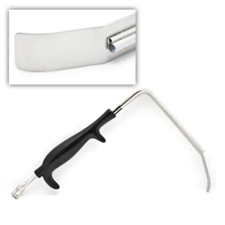
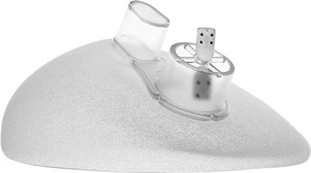
Key steps:
- After checking that the mastectomy skin flaps have a good blood supply, the surgeon opens the skin and fasciaFASH-ee-uhA sheet or band of tough connective tissue that covers and separates muscles and organs.fasci/o bandFull entry along the edge of the pectoralis major and makes a pocket under the muscle, from the breastbone over the lower ribs.
- The surgeon chooses the size, tests the expander for leaks or inspects the implant for flaws, and places it, handled as little as possible; a sheet of acellular dermisDER-misThe thick layer of skin under the epidermis that holds blood vessels, nerves, hair roots and glands.derm/o skinFull entry matrix may bridge the gap from the raised muscle to the fold under the breast.
- The wound is closed and the expander filled with sterile saline by the maker's instructions, until the skin over it blanches slightly (one reference fills it 60 mL at a time); record the volume and the implant details.
- Drains go in, because a hematomahee-muh-TOH-muhA collection of blood, often clotted, that has leaked out of the blood vessels into the tissue.hem/o blood (here as hemat-)-oma swelling or massFull entry or seroma could push the expander out of place.
The expander is then filled in the surgeon's office over about 6 to 8 weeks. At the second stage it is let down, removed and replaced with the permanent implant, the nipple may be built, and the other breast may be lifted, reduced or enlarged to match.
Wound class: I, clean.
Breast augmentation (augmentation mammoplasty)
What and why: an implant enlarges a small breast, evens out breasts of different sizes, or recreates a breast after mastectomy. Breast implants have a silicone shell, smooth or textured, filled with silicone gel or sterile saline. In the United States, the FDA approves silicone gel implants for augmentation from age 22 and saline implants from age 18, and both for reconstruction at any age. Implants can make mammograms harder to read.
Before surgery: supine with the arms out at less than 90 degrees or tucked; local anesthesiaLOH-kul an-es-THEE-zhuhNumbing medicine applied to or injected into one small area, so a procedure there is not felt while the person stays awake.Full entry with sedationsih-DAY-shunA calm, relaxed or sleepy state brought on by drugs, or the use of drugs to cause it.sed calm, settle-ation processFull entry or general anesthesiaJEN-er-ul an-es-THEE-zhuhDrug-induced loss of consciousness and feeling over the whole body, so a person feels nothing and is not aware during surgery.an- withoutesthesi/o feeling, sensation-ia conditionFull entry .
Setup: a plastic tray, fiber-optic breast retractors, implant sizers, the implants, saline-filled syringes or a closed fill system for saline implants, antibiotic for the irrigating saline, a bulb syringe, and the surgical bra; an endoscopeEN-duh-skohpA thin tube with a light and a camera that is passed into the body to look inside an organ or cavity.endo- within-scope instrument for viewingFull entry if the surgeon works endoscopically.
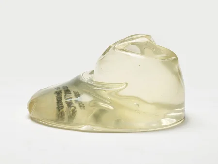
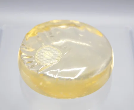
Key steps:
- local anestheticLOH-kul an-es-THET-ikA drug that blocks feeling in one part of the body without making the person unconscious.Full entry may be injected, and the incision goes around the lower edge of the areola, in the fold under the breast, in the armpit or at the umbilicus; the pocket goes under the breast tissue, under the pectoralis muscle or partly under it.
- Sizers go in, and the head of the bed is raised so the patient sits up and the surgeon can judge the size and shape; the bed comes back down and the pocket is adjusted.
- The circulator confirms the implant's type and size with the surgeon and passes it to the field; handle it as little as possible and as its maker directs. After the pocket is rinsed with saline or antibiotic solution, an unfilled saline implant goes in rolled up and is filled through the closed system (some saline implants come prefilled); a gel implant's shell already holds its silicone gel.
- The other side is done, the incisions are closed in two layers, and a surgical bra goes on.
A firm, painful breast after augmentation may be capsular contracturekun-TRAK-cherA lasting shortening and tightening of muscle, tendon, skin or joint tissue that keeps a joint from moving through its full range.Full entry , the scar capsule around the implant tightening; it is released (capsulotomy) or removed (capsulectomy) with the augmentation setup, and the patient, facing a second operation, needs your kindness.
Wound class: I, clean.
Reduction mammoplasty and mastopexy
What and why: a reduction mammoplasty removes breast tissue and skin from very large breasts (macromastia), whose weight causes back and neck pain, skin inflammation in the folds under the breasts and deep grooves from bra straps, and which can trouble a patient socially. It also evens out breasts of different sizes, or matches the breast that remains after a mastectomy. A mastopexy lifts and reshapes a drooping breast (breast ptosis) by moving the nipple up and removing extra skin, with little or no breast tissue removed; an implant may be added for fullness.
Before surgery: the surgeon marks the patient sitting, with an areola markerMAR-kerSomething that can be measured, such as a substance in the blood, that shows a disease or body process is present.Full entry , a keyhole-shaped pattern and a tape measure. Patients 35 and older, or with a family history of breast cancer, may need a mammogram first. Supine with the arms out on padded armboards at less than 90 degrees, with the hips at the bed's break so the patient can be sat up during the case.
Setup: a plastic tray with baby Deaver retractors, an electrosurgeryee-LEK-troh-SUR-jer-eeUsing high-frequency electric current through a handheld tool to cut tissue or seal bleeding vessels during surgery.Full entry , a skin stapler and sterile marking pens; many #10 blades if the surgeon removes the surface skin layer from areas of the breast (de-epithelializes) to keep the tissue beneath; extra sutureSOO-cherA stitch, or the thread used to stitch, that holds the edges of a wound or incision together.Full entry for tie-over bolsters if the nipple is moved as a free graftGRAFTTissue or a man-made material moved into place in surgery to replace or repair a damaged part.Full entry ; and a calibrated scale.
Key steps (reduction, inverted-T):
- The skin between the old and the new nipple sites is removed, leaving the nipple attached to the breast tissue beneath it. Sometimes the nipple is removed instead and put back as a free graft, tied under a bolster.
- The extra breast tissue below the nipple is cut away through an incision shaped like an upside-down T, or an anchor.
- The nipple and the tissue around it are moved up and sewn into their new place, the skin edges are brought together in a vertical line below it, and the fold incision is trimmed and closed across. Closed suction drains may go in.
- Keep the tissue from each breast apart. Weigh each, record the weights, and send both to pathologypuh-THOL-uh-jeeThe study of disease and its causes and effects, or the disease changes found in a person or tissue.path/o disease-logy study ofFull entry : the weights help the surgeon remove about the same from each side.
After surgery: elastic bandages or a surgical bra over gauze, with drains. The patient is told to expect less feeling in the nipples, possible trouble breastfeeding, mammograms that are harder to read and visible scars.
Mastopexy follows the same pattern with less removed, through an incision around the areola, around it and straight down (a vertical or "lollipop" lift), or the anchor, the most common for a full lift.
gynecomastiagy-nuh-koh-MAS-tee-uhEnlarged breast gland tissue in a male.gynec/o womanmast/o breast-ia conditionFull entry , enlarged breast tissue in a male, is removed by liposuction or through an incision at the edge of the areola, and the tissue goes to pathology, because males can get breast cancer.
Wound class: I, clean.
Flap reconstruction of the breast
Some patients choose to rebuild the breast with their own tissue, a myocutaneous flap of skin, fat and muscle from the back or the abdomen, or skin and fat from the buttock. Patients with a great deal of excess weight and those who have had abdominal surgery before may not be good candidates for an abdominal flap. For each of these flaps, have a Doppler ultrasoundDOP-ler UL-truh-soundAn ultrasound that uses sound waves to hear or show blood flowing through vessels or the fetal heartbeat.Full entry with a sterile probe in the room; two teams may work at once, one raising the flap and one preparing the chest.
Latissimus dorsi flap: used when the mastectomy left a large shortage of tissue or an abdominal flap is not an option. The latissimus dorsi, the wide, flat muscle of the mid back, stays attached to the humerusHYOO-mer-usThe long bone of the upper arm, from the shoulder to the elbow.Full entry and to its main blood supply, the thoracodorsal artery, and is usually paired with an implant.
Before: the surgeon marks the back's skin island and the chest with the patient sitting. The patient is placed on the side, donor side up, with the arm supported, then turned supine once the back is closed; have the padding and positioners ready.
Setup: a major tray, fiber-optic breast retractors, vascular instruments, long forcepsFOR-sepsA hand instrument with two blades or arms used to grasp, hold or pull tissue or objects.Full entry and scissors, sterile marking pens and a Doppler.
Key steps: an island of skin is cut across the back and the muscle is freed under the skin, then tunneled under the armpit to the chest with its vessels and its attachmentuh-TACH-mentThe deep emotional bond between an infant and a parent or caregiver.Full entry to the humerus untouched; the skin island and muscle are sewn into place over an implant, a drain goes in, and the back is closed.
Complications: watch for and report signs that the flap's blood supply is failing, a hematoma, a seroma, infection or the wound opening; partial or complete loss of the flap is the most serious.
Pedicled TRAM flap: a transversetrans-VERSLying across something, at a right angle to its length.Full entry rectusREK-tusLatin for straight; the name of several straight muscles, such as the rectus abdominis of the abdomen and the rectus femoris of the thigh.Full entry abdominis myocutaneous flap moves skin and fat from the lower abdomen, with one or both rectus muscles, up to the chest. The rectus muscle carries the flap's blood supply, the superior epigastric artery and vein, from above; its lower end is cut free and the flap is passed through a tunnel under the upper abdominal skin. The patient also gets a flatter abdomen, though a small implant may still be needed.
Before: the surgeon marks the abdomen and the chest with the patient standing. Supine with the arms tucked or out at less than 90 degrees; a urinary catheter and compression devices; prep from the lower neck to the mid-thighs. The bed is flexed with the head up for the abdominal closure, which eases the tension on the abdominal wound and supports the flap's circulation.
Setup: a major or laparotomylap-uh-ROT-uh-meeSurgery that opens the abdomen through a large cut to reach the organs inside.lapar/o abdomen-tomy cutting intoFull entry tray with fiber-optic breast retractors and a table-mounted retractor, vascular instruments, extra right-angle and long clamps, vessel loops, clips, long forceps and scissors, a Doppler with a sterile probe, and synthetic mesh for the abdominal wall.
Key steps:
- The mastectomy scar is cut out and the chest pocket opened and covered with a moist lap; then an ellipse of lower abdominal skin and fat, from hip to hip around the umbilicus, is cut down to the rectus sheathSHEETHA covering that wraps around a structure; in procedures, a short hollow tube left in a blood vessel to pass catheters through.Full entry .
- With the Doppler, the surgeon finds the epigastric vessels; the lower (inferior) ones are clamped, cut and tied, and the upper ones, which feed the flap, are kept.
- The rectus muscle is cut across at its lower end and freed up toward the ribs, and the flap is drawn up through a tunnel under the upper abdominal skin, by hand or with a grasping clamp, without twisting or pulling its pediclePED-ih-kulA narrow stem or stalk that attaches a structure, such as a tumor or a skin flap, to the body.Full entry .
- The flap is shaped to match the other breast, sewn in and drained; save the extra skin and fat trimmed from it, which may build the mound.
- The abdominal wall is closed in layers, sometimes reinforceree-in-FORSTo repeat and support teaching or care that has already been started, so the client remembers and follows it.Full entry with synthetic mesh, and the skin is closed over a drain. An abdominal dressing and loose fluffs on the chest go on, placed so nothing presses on the flap.
Complications: infection, bleeding, death of part or all of the flap when its blood supply fails, and the abdominal wound opening (dehiscencedee-HIS-ensThe splitting open of the edges of a wound that had been closed, such as a surgical incision.Full entry ), even far enough for the organs to push out (eviscerationih-vis-er-AY-shunThe pushing out of internal organs, usually loops of bowel, through a surgical wound that has come open.e- outviscer/o internal organs-ation processFull entry ).
Free flaps from the abdomen: the same lower abdominal tissue can be moved as a free flap and joined to vessels in the chest under the microscope:
- A free TRAM flap is lower abdominal skin, fat and rectus muscle that lives on the deep inferior epigastric artery. It is chosen when losing one or both rectus muscles is a concern, or when the blood supply of a pedicled flap is in doubt.
- A deep inferior epigastric perforator (DIEP) flap uses no muscle to build the breast: the surgeon follows the small perforating vessels through the rectus muscle and moves skin and fat on them, with at most a small piece of muscle around the vessels. Before surgery the perforators are found with a duplex Doppler and computed tomographykum-PYOO-tid toh-MOG-ruh-feeAn imaging test that uses x-rays and a computer to make cross-section pictures of the inside of the body.tom/o cut, slice-graphy process of recordingFull entry and marked on the skin.
DIEP flap, key steps:
- Two teams often work at once, one on the abdomen and one on the chest, after a prep from the neck to the pubisPYOO-bisThe front part of each hip bone, low in the pelvis; the two meet in the middle at the pubic symphysis.pub/o pubisFull entry , a urinary catheter and compression devices.
- The marked ellipse of lower abdomen is lifted with the ESU, and a sterile Doppler guides the dissectiondy-SEK-shunA tear in the inner layer of an artery wall that lets blood split the layers apart; also, the cutting apart of tissue in surgery.Full entry down through the muscle along the chosen perforators; keep irrigationeer-ih-GAY-shunWashing out a wound, body cavity, tube or eye with a steady flow of liquid.Full entry going so the vessels do not dry. If no perforator gives enough blood flow, the surgeon may change to a TRAM flap.
- At the chest, the internal mammary vessels are exposed, usually by removing a short piece of the third rib's cartilageKAR-tih-lijFirm, smooth, rubbery tissue that cushions joints and gives shape to parts such as the ear, nose and windpipe.chondr/o cartilageFull entry . After heparinHEP-uh-rinAn anticoagulant drug, given by injection or IV, that keeps blood clots from forming or growing.Full entry , the flap's vessels are clipped and divided and its artery is flushed with heparinized saline.
- Under the microscope, the artery is joined by hand-sewn sutures and the vein by sutures or a coupler; then the clamps come off, the flow is checked, and a marking stitch shows where to place the Doppler afterward.
- The flap is shaped and sewn in, and the abdomen is closed like an abdominoplasty, with the umbilicus set back in place and two drains.
After surgery: the patient often spends the first night in intensive care for flap checks and stays several days. Every free flap is checked as taught above.
Wound class: I, clean.
Nipple reconstruction
The nipple and areola are usually rebuilt last, often about 6 to 8 weeks after the breast mound (one reference waits 8 to 10 weeks), once the new breast has healed and settled, so the nipple goes in the right place. A common method is the skate or wing flap: three small flaps of the new breast's own skin and fat are raised from one point and wrapped around a core to make a nipple that sticks out, and the bases are closed. The areola may be a full-thicknessful-THIK-nisGoing through every layer of the skin, or every layer of another tissue such as the wall of an organ.Full entry graft from the groin, the inner thigh, the fold under the buttock, the ear or the labia, and color is added by tattooing, then or later. For the tattoo, the scrub sets up sterile tattoo equipment and ink as its maker directs. The dressing comes off at about 6 days; infection, a graft that does not take, and a result that disappoints are the risks.
Wound class: I, clean.
12The abdomen and body
Losing a lot of weight shrinks the fat but leaves the stretched skin behind, and it does not tighten stretched abdominal muscles. These operations remove the extra skin and fat and reshape the body. They are long, wide cases on the trunk: plan for warming, privacy during a prep that may cover most of the body, padding and repositioning. In one large review of plastic surgery done in an ambulateAM-byuh-laytTo walk, or to help a client walk.Full entry center, the patients at greatest risk of complications had combined procedures (above all abdominoplasty with liposuction), a BMI over 30, more than 3 L of fat suctioningSUK-shun-ingRemoving mucus, saliva or other secretions from the mouth, nose or airway with a tube connected to a suction source.Full entry , or surgery longer than 4 hours.
Abdominoplasty
What and why: an abdominoplasty ("tummy tuck") flattens the abdomen by removing extra skin and fat and tightening the abdominal muscles. It helps people who have lost weight or whose abdominal skin is lax, and is usually cosmetic, though loose skin can also interfere with movement. A mini abdominoplasty, through a short low incision with no move of the umbilicus, treats fullness of the lower abdomen; a full abdominoplasty, from hip to hip with the umbilicus moved, treats loose skin above and below the umbilicus with a separation of the rectusREK-tusLatin for straight; the name of several straight muscles, such as the rectus abdominis of the abdomen and the rectus femoris of the thigh.Full entry muscles (diastasis); an endoscopicen-doh-SKOP-ikDone with an endoscope, a thin tube with a light and camera passed into the body.endo- within-scopic viewingFull entry one repairs the muscles alone. A patient planning more pregnancies is advised to wait until they are done.
Before surgery: the surgeon plans and marks the incisions with the patient standing, in the surgeon's clinic. general anesthesiaJEN-er-ul an-es-THEE-zhuhDrug-induced loss of consciousness and feeling over the whole body, so a person feels nothing and is not aware during surgery.an- withoutesthesi/o feeling, sensation-ia conditionFull entry ; supine with the hips slightly flexed (semi-Fowler) to ease the tension on the abdomen; the pubic hair is removed; prep from the nipples to the mid-thighs and out to both sides; drapes that leave the whole abdomen, the lower rib edges and both iliacIL-ee-akHaving to do with the ilium, the large, wing-shaped upper part of the hip bone.Full entry spines in view. A full abdominoplasty takes about 2 to 5 hours.
Setup: a laparotomylap-uh-ROT-uh-meeSurgery that opens the abdomen through a large cut to reach the organs inside.lapar/o abdomen-tomy cutting intoFull entry or plastic set with long instruments, extra clamps such as Crile and Kocher, large Deaver retractorrih-TRAK-terA surgical instrument that holds back tissue or organs so the surgeon can see and reach the operative site.Full entry and a table-mounted self-retaining retractor, a lighted fiber-optic retractor (test it before the patient arrives), an electrosurgeryee-LEK-troh-SUR-jer-eeUsing high-frequency electric current through a handheld tool to cut tissue or seal bleeding vessels during surgery.Full entry with a long blade tip, a sterile umbilical template ("cookie cutter"), plenty of lap sponges, closed suction drains, and an abdominal binder or girdle. Much of the dissectiondy-SEK-shunA tear in the inner layer of an artery wall that lets blood split the layers apart; also, the cutting apart of tissue in surgery.Full entry is done with the ESU, so clean the charred buildup off its tip often.
Key steps:
- The surgeon cuts around the umbilicus and makes a low transversetrans-VERSLying across something, at a right angle to its length.Full entry incision from hip to hip, low enough to hide under underwear, then lifts the skin and fat off the fasciaFASH-ee-uhA sheet or band of tough connective tissue that covers and separates muscles and organs.fasci/o bandFull entry up to the rib edges and the xiphoid, leaving the umbilicus on its stalk, while table-mounted and lighted retractors hold the flap up.
- A separation of the rectus muscles is closed by folding and stitching the fascia (plication) from the xiphoid down to the pubisPYOO-bisThe front part of each hip bone, low in the pelvis; the two meet in the middle at the pubic symphysis.pub/o pubisFull entry .
- The flap is pulled down and the excess below the marked line is cut away; a small opening is cut for the umbilicus, often with the template, and it is sewn in.
- Drains go in, and the wound is closed in layers, often with staples on the skin; a dressing and a binder go on.
After surgery: keep the patient's hips bent, in a semi-sitting position in bed, so the sutureSOO-cherA stitch, or the thread used to stitch, that holds the edges of a wound or incision together.Full entry line is not pulled tight. Complications include infection, bleeding, severe swelling and a result that disappoints.
Wound class: I, clean, since no tract is entered.
Panniculectomy
What and why: a panniculectomy removes the panniculus, the loose apron of skin and fat that hangs from the lower abdomen of a patient who was very obese and has lost a great deal of weight. The apron can hang as low as the knees, making movement and daily activities very hard; it is a different operation from a cosmetic abdominoplasty, though the two may be combined. It may also be done with another operation, such as a herniaHER-nee-uhA bulge of an organ or tissue through a weak spot in the muscle or tissue wall that normally holds it in.Full entry repair, or pelvic surgery in a patient with severe obesity. Setup, position and closure follow the abdominoplasty.
Wound class: I, clean, by the CDC's definitions, unless an infection of the skin folds is present.
Body lifts after weight loss
After major weight loss, often from bariatric surgery, the patient may have loose skin and fat around the whole trunk, a sagging mons pubis, rolls on the back, flat low buttocks, and loose upper arms and thighs. The Core Curriculum lists the body lift as full or lower. The references describe the belt (circumferential) lipectomy, which removes the rolls of skin and fat that circle the whole trunk; it is combined as needed with an arm lift (brachioplasty), thigh lifts and a mastopexy to recontour the body. Because bariatric surgery can keep the body from absorbing nutrients well, wounds may heal slowly.
Before and setup: the surgeon measures and marks extensively in the holding area. The patient is turned during the case, so pad every pressure point and recheck the skin, the nerves, the safety strap and every device after each turn. A minor tray with extra Backhaus (piercing) towel clamps, a skin stapler, many drains and an ESU.
Key steps:
- Tumescent solution may be infused to limit bleeding and ease the undermining, and the incisions go down to the fascia, the flaps lifted to the marked lines, with liposuction for contouring and plication of the muscle as needed.
- The superficial fascia is closed with permanent sutures, the extra skin is cut off, and drains are placed.
- The patient is turned and the same is done in each new position until every area, the umbilicus included, is finished; compression garments go on at the end.
Wound class: I, clean.
Liposuction (suction lipectomy)
What and why: liposuction removes deposits of fat through a hollow cannulaKAN-yoo-luhA thin tube placed into the body or a body opening to deliver or remove fluid or air.Full entry under strong suction, to improve the body's contour. It is not a way to lose weight or a treatment for obesity: the ideal patient is near normal weight, with localized fat that diet and exercise have not shifted, and keeps the result only with good habits afterward. It can treat nearly any area from the face to the ankles, and it also treats gynecomastiagy-nuh-koh-MAS-tee-uhEnlarged breast gland tissue in a male.gynec/o womanmast/o breast-ia conditionFull entry and removes lipomas.
Before surgery: the surgeon marks the areas with the patient standing, often as rings like a contour map around each bulge, so more fat comes from the center than the edges. The patient may be prepped standing, all the way around the area, so protect privacy and keep the patient warm. It can be done under general or epidural anesthesiaep-ih-DOOR-ul an-us-THEE-zhuhPain relief or numbness from an anesthetic drug given through a thin catheter into the space just outside the covering of the spinal cord.epi- upon, overdur/o dura (the tough covering of the spinal cord)-al relating toFull entry or moderate sedationMOD-er-it seh-DAY-shunA drug-induced state of relaxed, lowered awareness in which the client still responds to voice or light touch and breathes on their own.Full entry , and with tumescent solution under local anesthesiaLOH-kul an-es-THEE-zhuhNumbing medicine applied to or injected into one small area, so a procedure there is not felt while the person stays awake.Full entry with light sedationsih-DAY-shunA calm, relaxed or sleepy state brought on by drugs, or the use of drugs to cause it.sed calm, settle-ation processFull entry . Agree on the order of the areas and the positions beforehand.
The wetting solution: before suctioning, the surgeon infuses IV fluid, saline or lactated Ringer's solutionLAK-tay-tid RING-erz suh-LOO-shunAn isotonic IV fluid containing water, sodium, chloride, potassium, calcium and lactate.Full entry , with lidocaineLY-doh-kaynA local anesthetic that numbs an area, also given by IV to treat some fast, abnormal heart rhythms.Full entry and epinephrineep-ih-NEF-rinA hormone and drug, also called adrenaline, that raises heart rate and blood pressure and opens the airways.Full entry , sometimes with hyaluronidase or sodium bicarbonateSOH-dee-um by-KAR-buh-nateAn alkaline drug, given by mouth or IV, that neutralizes acid in the stomach or the blood.Full entry , into the fat. The scrub may mix it to the surgeon's recipe, label it, keep the 30 mL infiltrationin-fil-TRAY-shunThe leaking of IV fluid or a drug that is not harmful to tissue out of the vein into the tissue around the IV site.Full entry syringes filled and warm the solution in a warmer designed for it. One published recipe adds 50 mL of 1% lidocaine (500 mg), 1 mL of epinephrine 1:1,000 (1 mg) and 10 mL of 8.4% sodium bicarbonate to 1,000 mL of normal saline. In the superwet technique the volume infused about equals the fat to be removed (1:1); in the tumescent technique the fat is filled until it is tense, about 2 to 3 mL of fluid for every 1 mL of fat. The lidocaine numbs, the epinephrine narrows the vessels so less blood is lost, and the swelling makes the cannula easier to pass.
the tumescent volume has limits. Too much fluid can overloadOH-ver-lohdMore of something than the body can handle; in nursing, most often too much fluid in the circulation.Full entry the patient (rising blood pressure, swollen neck veins, a bounding pulse, cough, shortness of breath, crackleKRAK-ulA short popping or crackling lung sound heard through a stethoscope, usually while breathing in.Full entry in the lungs), and the lidocaine in it counts toward the patient's total dose of local anestheticLOH-kul an-es-THET-ikA drug that blocks feeling in one part of the body without making the person unconscious.Full entry . Spread through the fat, the lidocaine is absorptionub-ZORP-shunThe taking in of a substance, such as a drug or a nutrient, into the bloodstream.Full entry slowly, and one researcher's study found lidocaine doses of 35 mg/kg safe for liposuction, where 7 mg/kg is the accepted maximum for lidocaine with epinephrine. That is a study's finding, never a dose to work to: the maximum doses in chapter 13 stand, and the surgeon and the anesthesia provideran-us-THEE-zhuh pruh-VY-derThe licensed professional who gives anesthesia and watches over the client's breathing and vital functions during a procedure.Full entry decide the limit. Keep an exact running total of every mL infused and report it.
Setup: a minor tray, the liposuction aspirator (a high-vacuum unit with graduated canisters), firm large-bore suction tubing, rigid cannulas of several lengths and diameters (from 2 or 3 mm for fat near the surface up to larger ones for deep fat), infiltration needles or cannulas and 30 mL syringes, a #15 blade, a marking pen, absorbent dressings and the compression garment. Some techniques add an ultrasonic or laser probe that breaks up the fat first.
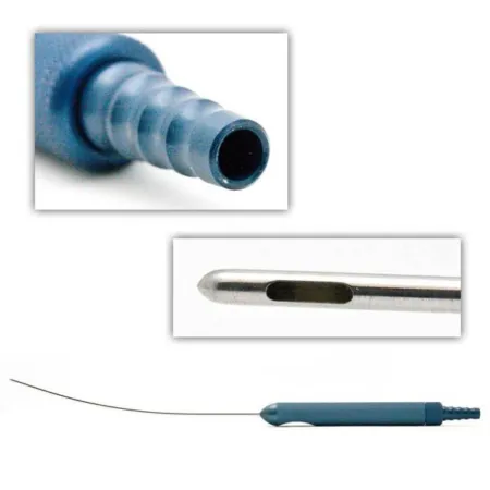
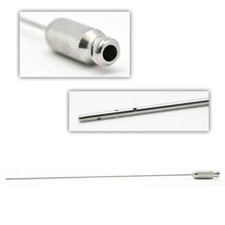
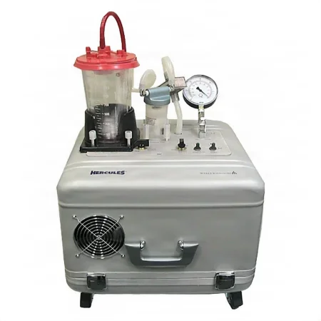
Key steps:
- Small stab incisions are made in hidden places, more than one for each area so the cannula reaches all of it without leaving dents, and the wetting or tumescent solution is infused; the surgeon waits for the epinephrine to work.
- The cannula, on its tubing to the aspirator, is pushed back and forth through the fat parallel to the skin, small cannulas first, and the high vacuum pulls the loosened fat into the canister.
- Watch the canister: track the volume removed and report it. One reference warns that removing more than 1,000 mL can lead to hypovolemiahy-poh-voh-LEE-mee-uhA lower than normal volume of fluid in the blood vessels; also called fluid volume deficit.hypo- belowvol volume-emia in the bloodFull entry and other fluid problems.
- The stab wounds are sutured or left open to drain, and absorbent dressings and the compression garment go on; open the garment on the stretcher first, so the patient can be moved onto it and it can be closed around them.
After surgery: the patient wears the compression garment for about 2 to 4 weeks, and the incisions may leak fluid for the first 24 hours. Bruising, swelling and moderate pain are expected; bleeding and severe swelling are complications.
Wound class: I, clean.
13The genitalia
The Core Curriculum's plastic surgery of the genitals is gender confirmation surgery (genitoplasty and phalloplasty), labiaplasty and vaginoplasty. Chapter 34 teaches gender confirmation surgery, phalloplasty and vaginoplasty included, with the respectful care every patient is owed, and chapter 33 teaches labiaplasty. Gender confirmation surgery is done in stages by a team of many specialists, a plastic surgeon among them, and it can also change the face and the chest: facial feminization or masculinization surgery changes the features of the face to a feminine or a masculine appearance, and top surgery removes breast tissue to make a masculine chest, or reshapes the chest to a feminine appearance with breasts.
14The hand
Plastic, orthopedicor-thuh-PEE-dikHaving to do with the bones, joints, muscles, ligaments and tendons, and the branch of medicine that treats them.orth/o straightped/o child (the field began with straightening children's bones)-ic relating toFull entry and hand surgeons all operate on the hand, and whatever the cause, the goal is to restore how it works first and how it looks second. Hand fractures are taught in chapter 39, and carpal tunnel release and nerve repair in chapter 42.
Hand anatomy in brief
- Directions: volar is the palm side and dorsalDOR-sulHaving to do with the back of the body, or the top of the hand or foot.Full entry the back of the hand; radial is the thumb side and ulnar the little finger side.
- Bones and nerves: chapter 3 counts the bones (8 carpals, 5 metacarpals numbered from the thumb, and 14 phalanges, two in the thumb) and chapter 4 the radial, median and ulnar nerves; the median nerve gives feeling to the thumb-side two-thirds of the hand and the ulnar nerve to the little finger side.
- Tendons: the flexor tendons run through the carpal tunnel on the palm side, with the median nerve; the extensor tendons run through six compartments on the back of the wrist. In the fingers, the tendons glide inside sheathSHEETHA covering that wraps around a structure; in procedures, a short hollow tube left in a blood vessel to pass catheters through.Full entry lined with synovium and are held to the bone by pulleys. The first dorsal compartment, at the thumb side of the wrist, holds the abductor pollicis longus and extensor pollicis brevis tendons.
Setting up for hand surgery
- The hand table: the arm rests on a hand table, and the surgeon and the assistant (often the scrub) sit facing each other across it. Only the top of the draped hand table is sterile; the field never extends below it.
- Tourniquet: a pneumatic tourniquet, applied as chapter 31 teaches, gives a bloodless field for nearly every hand case, after an Esmarch bandage empties the arm of blood.
- anesthesiaan-us-THEE-zhuhLoss of feeling or sensation, often brought on by drugs so that surgery or a procedure can be done without pain.an- withoutesthesi/o feeling, sensation-ia conditionFull entry : a block of the arm, an IV regional (Bier) block or local anestheticLOH-kul an-es-THET-ikA drug that blocks feeling in one part of the body without making the person unconscious.Full entry , often lidocaineLY-doh-kaynA local anesthetic that numbs an area, also given by IV to treat some fast, abnormal heart rhythms.Full entry mixed with a longer-acting drug such as bupivacainebyoo-PIV-uh-kaynA long-acting local anesthetic used to numb an area, block nerves, or give epidural and spinal anesthesia.Full entry ; general anesthesiaJEN-er-ul an-es-THEE-zhuhDrug-induced loss of consciousness and feeling over the whole body, so a person feels nothing and is not aware during surgery.an- withoutesthesi/o feeling, sensation-ia conditionFull entry for a long case or a child. epinephrineep-ih-NEF-rinA hormone and drug, also called adrenaline, that raises heart rate and blood pressure and opens the airways.Full entry is used with extreme caution, and as little as possible, where the blood supply is limited: the fingers, toes, tip of the nose and penis (chapter 13). Some references say it is not used in the fingers, or is contraindicationkon-truh-in-dih-KAY-shunA reason not to give a drug or do a treatment, because in that person it could cause harm.contra- againstindication a reason to useFull entry there.
- Instruments: short plastic instruments, with delicate orthopedic instruments for bone, a small drill and Kirschner wires (K-wires); small vascular clamps and short right angles for reconstruction; a fine-tipped bipolarby-POH-lerHaving two poles or two opposite ends; most often used as a short form of bipolar disorder.bi- twopolar having polesFull entry , which does not heat the tissue nearby; and Silastic vessel loops to hold tendons and ligaments aside. A lead hand, a malleable metal hand, can hold the fingers open; without one, you may hold them.
- Tissue and sutureSOO-cherA stitch, or the thread used to stitch, that holds the edges of a wound or incision together.Full entry : keep tendons, nerves and vessels moist with saline from a bulb or a syringe and moist patties, and use low suction with a fine tip; fine, inert sutures leave little scar, and double-armed sutures join tendons, vessels and nerves.
- Dressing: a bulky hand dressing, often with a splint, that leaves the fingertips showing so their color and circulation can be checked; the arm is kept raised afterward.
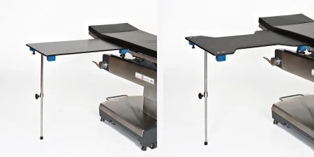
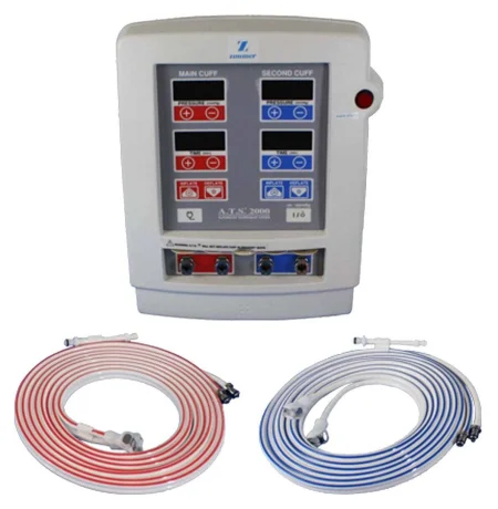
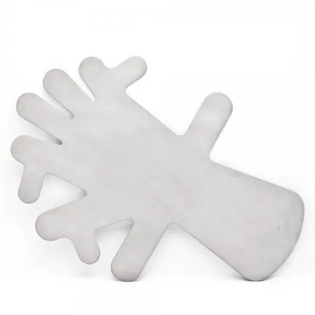
many hand surgeons now operate wide awake, under local anesthesiaLOH-kul an-es-THEE-zhuhNumbing medicine applied to or injected into one small area, so a procedure there is not felt while the person stays awake.Full entry with no tourniquet (WALANT): lidocaine with epinephrine 1:100,000 is injected so the patient can move the repaired tendon during surgery, and studies have not borne out the old fear that epinephrine in a finger causes gangreneGANG-greenThe death of body tissue because its blood supply has been lost or because of severe infection.Full entry ; phentolamine can reverse it if needed.
De Quervain release
What and why: in De Quervain tenosynovitis, the sheath of the first dorsal compartment thickens and narrows around its two tendons, the abductor pollicis longus and the extensor pollicis brevis, which rub where they change direction at the wrist. It causes pain over the tip of the radius (the radial styloid), often in adults 30 to 50 and in new parents from the wrist motions of caring for a newborn. In the Finkelstein test the examiner holds the thumb and quickly bends the hand toward the little finger, which causes sharp pain over the styloid. Rest, anti-inflammatory drugs and a steroidSTAIR-oydA fat-based chemical built from cholesterol; the group includes hormones such as cortisol and estrogen, and many drugs.ster/o solid-oid likeFull entry injection come first; when the pain persists, the sheath is released.
Before surgery: local anesthetic and a tourniquet, often an Esmarch bandage around the upper forearm.
Key steps:
- A short oblique, transversetrans-VERSLying across something, at a right angle to its length.Full entry or lengthwise incision over the first dorsal compartment goes through the skin only, sparing the branches of the superficial radial nerve just beneath.
- The sheath (retinaculum) is opened lengthwise and both tendons are freed. Many patients have a septumSEP-tumA wall of tissue that divides two spaces or cavities, such as the two sides of the heart or nose.sept/o wall, partitionFull entry splitting the compartment, or an extra tendon slip; a missed one leaves the pain, so the surgeon looks for it and may remove the septum, guardingGAR-dingTightening of muscles, or protecting a body part with the hands or posture, to avoid pain when it is touched or moved.Full entry the radial artery just beyond the end of the radius.
- A flap of the sheath is left over the tendons so they do not slip forward, the skin alone is closed, and a small pressure dressing goes on.
After surgery: the patient moves the thumb and hand at once, avoiding hard bending of the wrist for the first 2 weeks.
Wound class: I, clean.
Trigger finger release
Trigger finger is the same kind of problem in a flexor tendon, which catches at the first (A1) pulley of its sheath at the base of the finger or thumb, so the finger catches as it bends and straightens; splints and steroid injections come first. Under local anesthetic and a forearm Esmarch or an arm tourniquet, the surgeon cuts the pulley lengthwise through a short incision in the palm, guarding the digital nerves, and the awake patient then bends and straightens the finger to show it glides freely. A needle can cut the pulley through the skin instead.
Wound class: I, clean.
Dupuytren contracture release
What and why: in Dupuytren contracturekun-TRAK-cherA lasting shortening and tightening of muscle, tendon, skin or joint tissue that keeps a joint from moving through its full range.Full entry the fasciaFASH-ee-uhA sheet or band of tough connective tissue that covers and separates muscles and organs.fasci/o bandFull entry of the palm thickens and shortens. It shows as a painless lump in the palm near the ring or little finger, a dimple there, or a cord running toward the finger, and the shortening fascia pulls the finger down so it cannot straighten; it runs strongly in families. When it limits the hand's movement and use, the diseased fascia is removed (a palmarPAL-merHaving to do with the palm of the hand.Full entry fasciectomy).
Before and setup: a Bier block or general anesthesia, with a tourniquet. The patient may be sedationsih-DAY-shunA calm, relaxed or sleepy state brought on by drugs, or the use of drugs to cause it.sed calm, settle-ation processFull entry but awake, so keep talk and noise low. Have a hand set, or a plastic and minor orthopedic set, a lead hand, and a dermatome in case a skin graftGRAFTTissue or a man-made material moved into place in surgery to replace or repair a damaged part.Full entry is needed.
Key steps:
- Incisions are marked on the palm, a Z-plasty for an extensive contracture so the existing skin can cover the palm, and any free graft is taken first and kept moist.
- The surgeon cuts with a #15 or a small Beaver blade, changed often so it is never dull, and with fine forcepsFOR-sepsA hand instrument with two blades or arms used to grasp, hold or pull tissue or objects.Full entry and tenotomy scissors slowly dissectdy-SEKT, or dih-SEKTTo cut apart or separate tissues carefully during surgery.Full entry the tight fascia off each finger's nerves, vessels and tendons; it can be long and tedious, so stay alert.
- When the fingers move freely, the tourniquet is let down, bleeding is checked and the wound irrigationeer-ih-GAY-shunWashing out a wound, body cavity, tube or eye with a steady flow of liquid.Full entry . The skin is closed, a graft covers any gap or a small area is left to heal open, and a bulky dressing of petrolatum gauze, fluffs and an elastic wrap goes on, sometimes with a splint.
After surgery: the arm is kept raised; minor separation of the wound edges is common, and the contracture can come back.
Wound class: I, clean.
Syndactyly release
What and why: the paddle-shaped hands and feet of the embryo normally separate into digits between weeks 6 and 8; when they do not, the digits stay joined (syndactyly). It is the commonest malformationmal-for-MAY-shunA body part or organ that formed in an abnormal way before birth.mal- badformation shapingFull entry of the limbs, about 1 in 2,000 births, most often joining the middle and ring fingers, often on both hands, sometimes with other conditions, extra digits (polydactyly) among them. Simple syndactyly joins only skin and soft tissue and complex shares bone; complete joins the digits to their tips and incomplete only partway. Even a loose web is always short of skin once the fingers are separated, so plan for a skin graft, usually full-thicknessful-THIK-nisGoing through every layer of the skin, or every layer of another tissue such as the wall of an organ.Full entry .
Before and setup: usually between about 1 year of age and school age, under general anesthesia with a pediatric tourniquet and Esmarch; the graft donor site, often the groin, or the wrist or forearm, is prepped and draped separately. Have a hand or plastic set, a needle-tip electrosurgeryee-LEK-troh-SUR-jer-eeUsing high-frequency electric current through a handheld tool to cut tissue or seal bleeding vessels during surgery.Full entry or bipolar, a marking pen, a bulb syringe, and graft supplies with a dermatome on hand.
Key steps:
- The surgeon draws zigzag (Z-plasty) flaps on both sides of the web, and a full-thickness graft may be taken first and kept moist.
- The flaps are lifted, each finger's neurovascularnoor-oh-VAS-kyoo-lerHaving to do with both the nerves and the blood vessels of a body part.neur/o nervevascul blood vessel-ar relating toFull entry bundles are protected, and the band joining the fingers is divided.
- The flaps rebuild the web space and cover one finger; the bare skin on the other is measured with a template, and the graft is cut to it, its fat trimmed, and sewn on with fine interrupted sutures.
- Xeroform and a bolster go over the graft, padding goes around each finger with the fingers held widely apart, and a cast often protects the repair.
After surgery: the arm is kept raised, and the dressing or cast comes off in 4 to 6 days so the fingers can move; infection and contractures are the risks.
Wound class: I, clean.
Polydactyly
What and why: polydactyly is an extra digit, from a small extra piece to a complete duplicate, on the hand or foot, more often on one side. A duplicated (bifid) thumb is sorted by how much is doubled, from part of the tip to the whole thumb and its metacarpal. Usually the outer (radial) thumb is removed, which keeps the important ulnar collateral ligament of the remaining thumb, and the parents or guardians are told that thumb will always be a little smaller than the other.
Before and setup: usually at about 18 months, and before age 5; general anesthesia, a pediatric tourniquet, a warm room, and little talk or noise as the child comes in, so the child is not startled; a hand or orthopedic set, a wire driver and K-wires, and casting supplies.
Key steps (removing a radial thumb):
- Through an incision over the back of the outer thumb, keeping the tendon of the abductor pollicis brevis, the radial collateral ligament is detached and the extra thumb comes off with part of its metacarpal.
- The remaining thumb is centered on its joint, the collateral ligament and the small tendon are sewn back to its base, the tendons are centered if needed, and a K-wire holds the joint.
- The skin is closed with fine sutures and a short-arm thumb spica cast goes on.
After surgery: the wire comes out at about 4 weeks and a splint follows; infection, stiffness and a late angled or unstableun-STAY-bulDescribes a client whose condition is changing quickly or is hard to predict.Full entry joint are among the risks.
Wound class: I, clean.
Radial dysplasia repair (centralization)
What and why: in radial dysplasia (radial clubhand, now also called radial longitudinal deficiency) the radius and the tissue on its side of the forearm fail to develop, from a small radius to none at all. The forearm is short, the hand bends toward the thumb side, the thumb is often small or missing, other body systems may be affected, and the tight tissue on the thumb side worsens as the child grows. Centralization, done most often, sets the wrist on the end of the ulna to straighten the hand and improve its use, while keeping the wrist and fingers moving and the forearm growing.
Before and setup: at about 1 year of age, under general anesthesia, well padded for a long case, the arm prepped from the fingertips to the armpit, in a warm room; an ulna bowed more than 30 degrees also needs an osteotomy. Have a hand or major orthopedic set and a plastic set, a wire driver and K-wires, osteotomy instruments, a marking pen, several #15 blades and casting supplies.
Key steps:
- A Z-plasty incision on the thumb side of the wrist crease protects the median nerve and releases the tight fibrousFY-brusMade of or containing tough threads (fibers), especially of connective tissue.fibr/o fiber-ous full of, relating toFull entry bands.
- A second incision across the back of the wrist exposes it; the ulnar nerve and the flexor carpi ulnaris are guarded and the extra tissue over the ulna is removed.
- The end of the ulna is squared off and set into a notch cut in the wrist bones, sparing its growth plate; tendons are moved to hold the wrist centered, and a K-wire through the wrist bones and the third metacarpal into the ulna holds it.
- The wound is irrigated, the skin is closed with the Z-plasty, and a splint or cast goes on.
After surgery: the K-wire stays at least 8 to 10 weeks, then a splint is worn; the deformity coming back is the commonest failure.
Wound class: I, clean.
Replantation
What and why: replantation reattaches a body part that has been completely cut off, such as a finger, a thumb or a hand; revascularization repairs a part still partly attached whose blood supply has been cut. The aim is a part that works better than any prosthesispros-THEE-sisAn artificial body part that replaces one that is missing or does not work, such as an artificial leg, joint, eye or heart valve.Full entry . Success depends on the type of injury (a clean cut does better than a crush or a tear), where it is, how badly the structures inside are damaged, how the part was cared for and how much time has passed. Ideally the part is replanted within 4 to 6 hours, though success has been reported up to 24 hours when it was kept cold.
the amputationam-pyoo-TAY-shunThe removal of all or part of a limb or other body part, by surgery or by injury.Full entry part is wrapped in gauze moistened with saline, sealed in a plastic bag, and the bag set in a container of iced saline. The part itself never touches the ice or the iced saline.
Before surgery: a regional block or general anesthesia; pad every pressure point for a case that can last 12 to 16 hours, and warm the room before the patient arrives so the limb's vessels do not clamp down. Have labeled X-rays of the part and the stump in the room, and a padded tourniquet on the arm.
Setup: a hand instrument set, microvascular instruments, an operating microscope, a wire driver and K-wires, bipolar, microsuture, heparinized irrigation and papaverine.
Key steps:
- Two teams prepare the stump and the cut-off part at once, the part kept cold until its turn; damaged tissue is trimmed, and each tendon, vessel and nerve is tagged with suture so it is easy to find and cannot pull back out of sight.
- The bone ends are shortened, to take the tension off the vessels and nerves, and fixed with K-wires (or plates and screws for a larger bone), and the flexor and extensor tendons are repaired.
- Under the microscope, the surgeon repairs at least one artery and two veins, the minimum for the part to live, and the nerves; after a long ischemiais-KEE-mee-uhToo little blood flow to a part of the body, so the tissue does not get enough oxygen.isch- hold back-emia blood conditionFull entry time (the time the part has gone without blood flow), the vessels may be joined first.
- The skin is closed loosely, to leave room for swelling, under a bulky dressing and a splint, with the fingertips left out so their color and warmth can be checked.
After surgery: the arm is kept raised and the part is checked often, especially in the first hours. The main complications are failure of the part from poor blood flow, weaker nerve function and deformity.
Toe-to-hand transfer: when a thumb or finger is lost, a toe can take its place as a free transfer joined under the microscope, a 12 to 16 hour case with two teams, two tourniquets and two setups, one at the foot and one at the hand.
Wound class: replantation of a fresh traumatic amputation is Class III, contaminated, by the CDC's definitions; a planned toe-to-hand transfer is Class I.
15Wound classes and care after plastic surgery
Most plastic surgery is on skin, fat, muscle and bone with no body tract opened, so most cases are Class I, clean. The exceptions follow the CDC's definitions (chapter 11):
- Class II: the nose, a sinusSY-nusA hollow space in the body, most often one of the air-filled spaces in the skull bones around the nose.Full entry or the mouth is entered, as in a rhinoplasty, a cheek implant through the mouth or an orbital decompression through the sinuses, and a cleft repair (chapter 37).
- By the wound: a graftGRAFTTissue or a man-made material moved into place in surgery to replace or repair a damaged part.Full entry 's or a flap's recipient site follows the wound it covers: Class I for a clean surgical bed, Class III for a fresh wound from an injury, Class IV for an old one with dead tissue or infection. burnBURNAn injury to the skin or deeper tissues caused by heat, chemicals, electricity, radiation or friction.Full entry debridementdih-BREED-muntThe removal of dead, damaged or infected tissue from a wound so it can heal.Full entry is Class III when fresh and Class IV with escharES-karThick, dry, dark dead tissue that covers part or all of a wound.Full entry or infection, as your facility's policy decides; the replantation of a fresh amputationam-pyoo-TAY-shunThe removal of all or part of a limb or other body part, by surgery or by injury.Full entry is Class III; pressure injuryPRESH-er IN-juh-reeDamage to the skin and the tissue beneath it from pressure, or pressure with shear, usually over a bony area or under a device.Full entry repair is Class IV.
After surgery
In a large review of cosmetic and reconstructive cases done in an accredited ambulateAM-byuh-laytTo walk, or to help a client walk.Full entry center, fewer than 1 percent of patients had a complication in the first 48 hours, and most returns to the OR, about 80 percent of them, were to drain a hematomahee-muh-TOH-muhA collection of blood, often clotted, that has leaked out of the blood vessels into the tissue.hem/o blood (here as hemat-)-oma swelling or massFull entry . The complications to watch for and report at once:
- Hematoma: blood collecting under a flap, a facelift or a breast. It can choke a flap's blood supply and push an implant out of place, so drains, pressure dressings and compression garments are used to prevent it. In facial flap surgery, holding the systolic pressure under 120 mm Hg for the first hours after surgery has been shown to lower the risk.
- Seroma: serumSEER-umThe clear, yellow liquid left from blood after it clots and the cells are removed.Full entry collecting in the dead space under a large flap or around an implant, prevented the same way.
- Infection, which can make an implant fail.
- Blood clots: long cases and combined procedures raise the risk, so compression devices go on the legs and patients walk early.
Report what you see at the end of the case: swelling, a change in the color or warmth of a graft or flap, or a dressing, bra or garment that is too tight.
in plastic surgery the dressing, the garment and the position after surgery protect the result. Nothing tight over a flap, fingertips left in view after hand surgery, the head raised after facial surgery, and the hips bent after an abdominoplasty.
Patients usually go home with written instructions, because anxiety makes it hard to remember what they are told: how to manage pain, care for the incisions and drains and wear the garment, which signs of infection or other trouble to watch for, when to come back, and how to reach the surgeon. After a breast reduction, for example, the patient walks every 2 hours during the day for the first day or two, against clots, and avoids heavy lifting for a month.
What to take from this chapter
- Plastic surgery restores form and function (reconstructive) or reshapes normal structures for looks (aesthetic). The surgeon chooses from the reconstructive ladder, from healing on its own to a free flap or a transplant, and marks the patient awake, sitting or standing, before the case.
- A split-thickness graft takes the epidermis and part of the dermis, and its donor site heals on its own; a full-thickness graft takes all the dermis, has its fat trimmed, and its donor site is sutured. Two-site cases get two preps and two setups, the donor site first with a colorless solution, and any cancer or open wound is kept apart from the donor site.
- Assemble the dermatome exactly as its maker directs, hand it over set at zero, and lift the graft as it comes off. Use the mesh carrier the surgeon names, keep a meshed graft on its carrier and moist, and expect a bolster dressing over a graft.
- A flap carries its own blood supply. Pedicled flaps stay attached; free flaps are joined under the microscope with heparin, fine sutures or a coupler, and antispasm drugs. After surgery, a pale and cool flap means arterial trouble and a dusky, firm one venous congestion, the commonest cause of failure; report either at once.
- Mohs specimens and lesion margins keep their orientation, each in its own labeled container; breast reduction tissue is weighed right and left. Burns are debrided again and again, covered for a time with allograft or xenograft, and autografted days later.
- Facial cases are short-instrument cases; a brow lift or facelift is prepped over the whole head, and CHG never goes on the face, near the eyes or ears, or in the mouth. Rhinoplasty enters the nose, so it is Class II; most other plastic cases are Class I.
- In breast surgery, handle implants and expanders as little as possible, confirm the type and size before they are opened, and record the saline volume and implant details. TRAM, DIEP and latissimus flaps rebuild the breast with the patient's own tissue.
- Tumescent solution is lidocaine and epinephrine in IV fluid: keep an exact total, because it can overload the patient and cause toxicity. In the hand, only the top of the hand table is sterile, the tourniquet and fingertips are watched, and a replanted part travels wrapped, bagged and on iced saline, never touching the ice.
Check yourself5 quick questions
Free for everyone. Pick an answer for each, then check them all at once.
In the ORWhere you'll use this
The chapter's whole question pool in sets of 10, with the answer explained after each one.
Part of the educators programThe chapter's words on spaced cards: the ones you miss come back sooner.
Part of the educators programGiven by your class's teacher. Each class draws its own 25 from the chapter's pool.
Part of the educators program
Suggest a fix
Something wrong, unclear or out of date in this chapter? Tell us and we will fix it for everyone. Your email is only used to reply.