Obstetrics and gynecology
Cesarean birth, hysterectomy and the rest: the cases of the female pelvis and of childbirth, the anatomy under your clamps, and how to be ready when a patient in labor and the baby need the room in minutes.
1What obstetrics and gynecology covers
Obstetrics and gynecologygy-nuh-KOL-uh-jeeThe branch of medicine that cares for the female reproductive organs and breasts.gynec/o woman-logy study ofFull entry is one specialty with two halves. Gynecology treats and prevents disease of the female reproductive organs. Obstetrics cares for pregnancy and birth, which is called parturition. Fertility medicine joins gynecology and endocrinologyen-doh-krih-NOL-uh-jeeThe branch of medicine that deals with hormones and the glands that make them.endo- within-crine to secrete-logy study ofFull entry to help a patient become and stay pregnant.
You will scrub these cases in the main OR, in an OR inside the labor and delivery unit and in outpatient centers. Many smaller procedures, such as a minor hysteroscopy, have moved to the physician's office. surgical technologistSUR-jih-kul tek-NOL-uh-jistA member of the operating room team who prepares the sterile field and instruments and hands them to the surgeon.Full entry also work in the obstetric department and in freestanding birth centers. AST's position is that whoever practices surgical technology in a cesareansih-ZAIR-ee-unRelating to the birth of a baby through a surgical cut in the mother's abdomen and uterus.Full entry or tubal ligation, in the main OR or in labor and delivery, should be a graduate of an accredited program and a CSTsee-es-tee (said as letters)Certified Surgical Technologist, a credential for surgical technologists; in maternity care, contraction stress test.Full entry .
Most abdominal gynecologic surgery now uses a scope. laparoscopylap-uh-ROS-kuh-peeSurgery or an exam done through small cuts in the belly, using a thin, lighted tube with a camera.lapar/o abdomen-scopy looking with an instrumentFull entry and robotic surgery reach the pelvis through ports, and hysteroscopy reaches the inside of the uterus through the cervix. Some patients still need open surgery, depending on their condition.
The cases sort by approach, and the approach tells you the setup:
- Transcervical: through the cervix into the uterus, as in a D&C or a hysteroscopy.
- Transvaginal: through the vagina, to reach the vagina itself or the pelvic cavity, as in a vaginal hysterectomyhis-tuh-REK-tuh-meeSurgery to remove the uterus.hyster/o uterus-ectomy surgical removalFull entry or a colporrhaphy.
- Abdominal: open (laparotomylap-uh-ROT-uh-meeSurgery that opens the abdomen through a large cut to reach the organs inside.lapar/o abdomen-tomy cutting intoFull entry ) or minimally invasive (laparoscopiclap-uh-roh-SKOP-ikDone through a few small cuts in the abdomen with a thin camera and long instruments, instead of one large opening.lapar/o abdomen-scopic viewingFull entry or robotic).
- Vulvar and perinealper-ih-NEE-ulHaving to do with the perineum, the area between the genitals and the anus.Full entry : the outside structures, such as a Bartholin glandsBAR-tuh-lin glandzTwo small glands, one on each side of the vaginal opening, that make fluid to moisten the vagina.Full entry or a perineal tear.
Cases that share instruments, supplies, positions and steps are taught together as co-related procedures, and any approach other than open counts as minimally invasive surgery (MIS). A laparoscopic hysterectomy and a vaginal hysterectomy, for example, remove the same organ with different equipment. Each case here links to its setup card, which holds the instrument list and every step; this chapter gives the reasons, the key steps and the moments when the scrub matters most.
Chapter 14 explains how these cases count in your case log, vaginal deliveries included.
The patient
Surgery on the reproductive organs touches privacy, body imageBOD-ee IM-ijHow a person sees, thinks and feels about their own body and its appearance and function.Full entry and the hope of having children. A young patient may find an exam of the genitals frightening or deeply embarrassing. A patient of childbearing age may fear losing fertility, while another feels relief that a long problem will end. Cancer surgery brings fear and grief. Protect modesty at all times, even under anesthesiaan-us-THEE-zhuhLoss of feeling or sensation, often brought on by drugs so that surgery or a procedure can be done without pain.an- withoutesthesi/o feeling, sensation-ia conditionFull entry , and explain each step in words the patient understands.
A patient who has lost a pregnancy needs care that is honest and gentle. Acknowledge the baby and the loss, use plain words rather than vague phrases, and avoid lines such as "it's for the best" or "you can always have another baby." A perinatal death can affectAF-ekt (stress on the first syllable)In mental health, the outward expression of a person's emotions, as seen in the face, voice and body language.Full entry the team too. Chapter 18 teaches grief and loss.
Gynecologic and pregnant patients are screened for intimate partner violenceIN-tih-mut PART-ner VY-uh-lensPhysical, sexual or emotional harm, stalking or control by a current or former partner or spouse.Full entry , and pregnancy raises the risk; chapter 15 teaches the hospital's duty to screen for abuse, teach staff the signs and report.
Chapter 18 teaches care of the pregnant patient as a susceptible population. Hysterectomy is also one stage of some gender-affirming care, which chapter 34 teaches.
2Anatomy that matters
Chapter 8 teaches the female reproductive organs and the menstrual cycle in full. Here is what you need at the field.
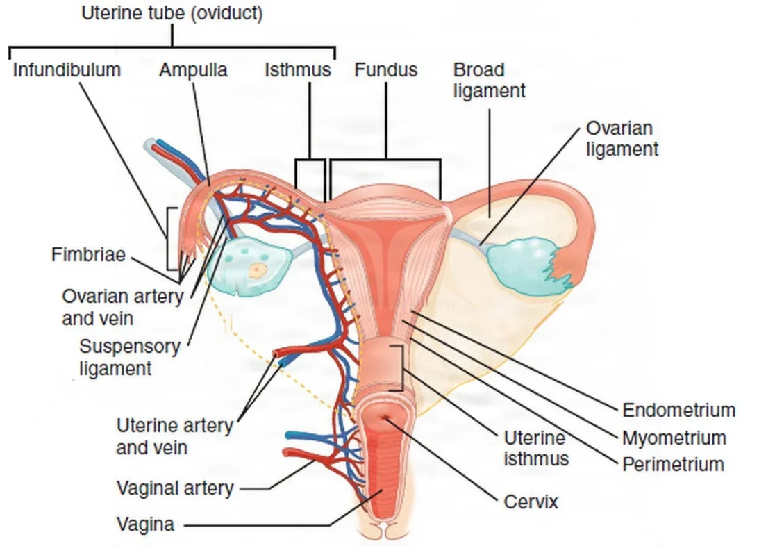
The uterus
The uterus is a pear-shaped muscular organ about 7.5 cm long that sits in the pelvis between the bladder in front and the rectum behind, usually tipped forward. Its parts are the fundusFUN-dusThe rounded top or base of a hollow organ, most often the top of the uterus.Full entry above the tubes, the body (corpus) and the cervix, the neck that pokes into the vagina. The cervix is 2 to 3 cm long; its canal runs from the internal os to the external os, which opens into the vagina and dilates in labor.
Its wall has three layers:
- Perimetrium: the outer serousSEER-usThin, watery and clear to pale yellow, like the fluid part of blood.Full entry layer, a fold of the pelvic peritoneumpair-ih-toh-NEE-umThe thin membrane that lines the belly cavity and covers most of the organs inside it.peri- aroundton stretched-eum a structureFull entry .
- Myometrium: thick smooth musclesmooth MUS-ulMuscle that works without conscious control and lines hollow organs such as the stomach, intestines, bladder, airways and blood vessels.Full entry , which contracts in labor and in menstruation. It is tough, which is why hysterectomyhis-tuh-REK-tuh-meeSurgery to remove the uterus.hyster/o uterus-ectomy surgical removalFull entry needs heavy scissors and toothed or grooved clamps.
- endometriumen-doh-MEE-tree-umThe inner lining of the uterus, which thickens each month and is shed as the menstrual period if no pregnancy begins.endo- withinmetr/o uterus-ium tissueFull entry : the lining, whose functional layer grows each cycle and is shed, over a basal layer that stays.
The peritoneum folds forward from the uterus onto the bladder. This fold is the bladder flap, and in a cesareansih-ZAIR-ee-unRelating to the birth of a baby through a surgical cut in the mother's abdomen and uterus.Full entry or an abdominal hysterectomyab-DOM-ih-nul his-ter-EK-tuh-meeSurgery to remove the uterus through a cut in the lower abdomen.hyster/o uterus-ectomy surgical removalFull entry the surgeon opens it and pushes the bladder down. Behind the uterus the peritoneum dips between the uterus and the rectum to form the posterior cul-de-sac, the pouch of Douglas, which the top of the vagina touches.
The ligaments
| Ligament | Where it runs | Why it matters at the field |
|---|---|---|
| Broad | A double fold of peritoneum from the side of the uterus to the pelvic wall, with the tube in its upper edge (the mesosalpinx) | Opened to free the uterus and tube |
| Round | From the uterus near the tube, forward through the inguinalING-gwih-nulRelating to the groin, the fold where the thigh meets the lower abdomen.Full entry canal | Often the first ligament clamped in an abdominal hysterectomy |
| Cardinal | At the base of the broad ligament, beside the cervix | The main support of the uterus; it carries the uterine artery and vein |
| Uterosacral | From the back of the uterus to the sacrumSAY-krumThe large, triangle-shaped bone at the base of the spine, between the two hip bones.sacr/o sacrumFull entry , on either side of the rectum | Clamped from below first in a vaginal hysterectomy |
| Infundibulopelvic (suspensory ligament of the ovary) | From the pelvic side wall to the ovary | Carries the ovarian artery and vein; clamped when the ovary comes out |
| Ovarian | From the ovary to the uterus | Clamped with the tube when the ovary stays |
One reference also calls the broad ligament a primary support of the uterus.
The tubes, ovaries and adnexa
Each uterine (fallopian) tube is about 10 to 13 cm long and only 3 to 5 mm wide. From the uterus out, it has an interstitialin-ter-STISH-ulRelating to the small spaces between cells or between the parts of a tissue or organ.Full entry part inside the uterine wall, a narrow isthmus, a wide ampulla where the egg is usually fertilizationfer-tuh-lih-ZAY-shunThe joining of a sperm and an egg to form a single new cell, the start of a pregnancy.Full entry , and a trumpet-shaped infundibulum whose fimbriae sweep the egg in. The tube is not attached to the ovary. The ovaries are almond-shaped, about 3.75 cm long, and hang from the back of the broad ligament by a fold of peritoneum (the mesovarium). The tubes, ovaries and their ligaments together are the adnexa.
The right tube and ovary lie near the cecum and appendixuh-PEN-diksA small, finger-shaped pouch attached to the first part of the large intestine, in the lower right abdomen.Full entry , and the left near the sigmoidSIG-moydShaped like the letter S; most often short for the sigmoid colon, the S-shaped last part of the colon before the rectum.Full entry colon.
Blood supply and the ureter
Most of the pelvis's blood comes from branches of the internal iliacIL-ee-akHaving to do with the ilium, the large, wing-shaped upper part of the hip bone.Full entry artery. The uterine and vaginal arteries are among them. The ovarian arteries come straight off the abdominal aorta and run in the infundibulopelvic ligaments. The uterus and tubes get blood from both the uterine and ovarian arteries, so both pediclePED-ih-kulA narrow stem or stalk that attaches a structure, such as a tumor or a skin flap, to the body.Full entry are tied in a hysterectomy.
The ureterYOOR-uh-terOne of the two tubes that carry urine from the kidneys down to the bladder.Full entry is the structure every pelvic surgeon guards. It comes down into the pelvis over the iliac vessels near the ovarian vessels, runs beside the cervix, and passes under the uterine artery, usually about 1 cm from the cervix, before it enters the bladder. A memory aid: water (the ureter) flows under the bridge (the uterine artery). A clamp or stitch meant for the uterine artery or the infundibulopelvic ligament can catch it, so the surgeon finds the ureter before clamping them.
The vagina, pelvic floor and vulva
The vagina is a muscular canal from the vestibule to the cervix. Its front wall, 6 to 8 cm long, lies against the bladder and urethrayoo-REE-thruhThe tube that carries urine from the bladder out of the body.Full entry , and its back wall, 7 to 10 cm, against the rectum. The recesses around the cervix are the fornices. The vagina allows sexual intercourse (coitus) and is the last part of the birth canal. The pelvic floor, the levator ani and coccyxKOK-siksThe small, triangle-shaped bone at the very bottom of the spine; the tailbone.Full entry muscles with their fasciaFASH-ee-uhA sheet or band of tough connective tissue that covers and separates muscles and organs.fasci/o bandFull entry , is a sling that holds up the pelvic organs; when it weakens, the bladder, rectum or uterus can bulge into the vagina.
The vulva is the external genitalia: the mons pubisPYOO-bisThe front part of each hip bone, low in the pelvis; the two meet in the middle at the pubic symphysis.pub/o pubisFull entry , labia majora and minora, clitorisKLIT-er-isA small, very sensitive organ at the top of the vulva, where the inner folds of skin meet.Full entry and the vestibule, which holds the urethral and vaginal openings. The Bartholin glandsBAR-tuh-lin glandzTwo small glands, one on each side of the vaginal opening, that make fluid to moisten the vagina.Full entry lie on each side of the vaginal opening at about the 5 and 7 o'clock positions. The perineumpair-ih-NEE-umThe area of skin and muscle between the genitals and the anus.perine/o perineumFull entry is the area between the vagina and the anus; under its skin lie the strong muscles of the pelvic floor.
lymphLIMFThe clear fluid that flows through the lymphatic vessels and carries white blood cells.Full entry from the pelvic organs drains to nodes along the iliac vessels, the obturatorOB-too-ray-terA smooth-tipped rod that fits inside a tube, such as a tracheostomy tube or cannula, to guide it in and then is taken out.Full entry space and the aorta, and from the vulva to the groin. That is where a cancer operation takes nodes.
3Diagnosis
Diagnosis begins well before surgery with a history and a physical exam that includes an internal (vaginal) exam. The history covers the menstrual cycle, from the first period (menarcheMEN-ar-keeA person's first menstrual period.men/o month, menstruationarche beginning (Greek)Full entry ) to menopauseMEN-uh-pawzThe time in a woman's life when menstrual periods stop for good, counted once there has been no period for 12 months in a row.men/o month, menstruation-pause stopFull entry , and any abnormal bleeding, each pregnancy and birth, contraceptionkon-truh-SEP-shunThe use of a method, device or drug to prevent pregnancy.contra- againstconception start of a pregnancyFull entry , infections, pain, discharge, urinary symptoms such as leaking urine with a cough, medicines and the family history. Chapter 8 explains gravidaGRAV-ih-duhA pregnant woman; in a pregnancy history, the number of times a person has been pregnant, including now.Full entry and paraPAIR-uhThe number of pregnancies a person has carried to 20 weeks or more, whatever the outcome.Full entry .
Many surgeons also examine the patient under anesthesiaan-us-THEE-zhuhLoss of feeling or sensation, often brought on by drugs so that surgery or a procedure can be done without pain.an- withoutesthesi/o feeling, sensation-ia conditionFull entry , after positioning and before the prep. Have nonsterile gloves and lubricant ready; cultures and a Pap testPAP testA screening test in which cells are gently scraped from the cervix and looked at under a microscope.Full entry may be taken then, so check that the lubricant will not spoil a cell sample.
Cell and tissue tests
- Pap test: cells brushed from the cervix and examined for changes that can lead to cervical cancerSER-vih-kul KAN-serCancer that starts in the cells of the cervix, the lower, narrow end of the uterus that opens into the vagina.Full entry . It is a screening testSKREE-ning testA test given to people who have no symptoms, to find a disease or risk early.Full entry , often done with testing for high-risk human papillomavirusHYOO-mun pap-ih-LOH-muh-vy-rusA common group of viruses spread by close skin-to-skin contact, most often during sex.Full entry (HPV).
- Colposcopy: the cervix is examined under magnification. Acetic acid turns abnormal cells white, and Lugol solution stains normal tissue brown and leaves abnormal tissue pale (the Schiller test); the surgeon biopsies the abnormal areas. Chapter 12 teaches these stains.
- cervicalSER-vih-kulRelating to the neck, or to the cervix, the lower end of the uterus.cervic/o neck, cervix-al relating toFull entry biopsy and endocervical curettage: small pieces taken with a biopsy punch or forcepsFOR-sepsA hand instrument with two blades or arms used to grasp, hold or pull tissue or objects.Full entry , or scraped from the canal, to rule out invasive cancer.
- Endometrial biopsy: a sample of the uterine lining, alone or as part of a D&C.
- Cultures: of the cervix and vagina for infections such as Chlamydiakluh-MID-ee-uhA sexually transmitted infection caused by the bacterium Chlamydia trachomatis; also the name of the bacterium.Full entry , herpes and trichomonas, and for group B Streptococcusstrep-tuh-KOK-usA group of round bacteria that grow in chains, some of which cause infections in people.strept/o twisted chain-coccus round bacteriumFull entry in pregnancy.
Imaging and blood tests
- Ultrasound, through the abdomen or the vagina, is the everyday test of the pelvis and of pregnancy. It shows fibroids, ovarian masses, fluid or blood in the pelvis, fetal age, growth and position, and the placentapluh-SEN-tuhThe organ that grows in the uterus during pregnancy and passes oxygen and food from the parent's blood to the fetus.Full entry 's location.
- Sonohysterography fills the uterus with fluid, such as saline, through a thin catheter before the ultrasound for a clearer picture, and is replacing hysterosalpingography because it is painless and uses no radiationray-dee-AY-shunEnergy that travels as waves or particles, such as X-rays, used to make images or treat cancer; also pain that spreads from its starting point.radi/o rays, radiation-ation processFull entry . Hysterosalpingography injects contrastKON-trastA dye or other substance given before an imaging test so body parts show up more clearly.Full entry into the uterus and tubes under fluoroscopyfloo-ROS-kuh-peeAn x-ray method that shows moving pictures of the inside of the body in real time.fluor/o glowing (from the fluorescent screen)-scopy looking, viewingFull entry , to look for a blocked tube or a cavity problem.
- magnetic resonance imagingmag-NET-ik REZ-uh-nunts IM-uh-jingAn imaging test that uses a strong magnet and radio waves to make detailed pictures of the inside of the body.Full entry shows the exact size and place of a tumor and how deep it has grown into the muscle; computed tomographykum-PYOO-tid toh-MOG-ruh-feeAn imaging test that uses x-rays and a computer to make cross-section pictures of the inside of the body.tom/o cut, slice-graphy process of recordingFull entry are also used before surgery.
- Pregnancy test: measures human chorionic gonadotropinHYOO-mun kor-ee-ON-ik goh-nad-oh-TROH-pinA hormone made by the placenta during pregnancy, measured in blood or urine as a pregnancy test.Full entry (hCG) in blood or urine; a blood test can track whether the level rises as expected early in pregnancy. With an ultrasound that shows no pregnancy inside the uterus, the hCG level helps confirm an ectopic pregnancyek-TOP-ik PREG-nun-seeA pregnancy in which the fertilized egg implants and grows outside the main cavity of the uterus, most often in a fallopian tube.ec- outtop/o place-ic relating toFull entry .
- CA-125see-ay wun-TWEN-tee-fyvA blood test for cancer antigen 125, a protein used as a tumor marker for ovarian cancer.Full entry : a blood tumor markerMAR-kerSomething that can be measured, such as a substance in the blood, that shows a disease or body process is present.Full entry that, with routine blood tests, helps estimate the risk of cancer before surgery; laparoscopylap-uh-ROS-kuh-peeSurgery or an exam done through small cuts in the belly, using a thin, lighted tube with a camera.lapar/o abdomen-scopy looking with an instrumentFull entry can assess further.
- Prenatal blood tests include the blood typeBLUD typeThe group a person's blood belongs to, based on markers on the red blood cells: A, B, AB or O, and Rh positive or negative.Full entry , Rh factorar-AYCH FAK-terA protein that may be present on the surface of red blood cells; people who have it are Rh-positive and those without it are Rh-negative.Full entry and antibodyAN-tih-bod-eeA Y-shaped protein made by the immune system that sticks to one specific germ or foreign substance and marks it for destruction.anti- againstbody Full entry screen, a CBC and screens for infections such as syphilisSIF-uh-lisA bacterial infection, spread mainly through sex, caused by Treponema pallidum.Full entry , hepatitis Bhep-uh-TY-tis BEEA liver infection caused by the hepatitis B virus.hepat/o liver-itis inflammationFull entry and HIVaych-eye-VEE (said as letters)Human immunodeficiency virus: a virus that infects and destroys immune cells called CD4 T cells.Full entry .
Laparoscopy and hysteroscopy are diagnostic procedures as well as operations; both are taught below.
4Diseases of the female reproductive system
These are the conditions the Core Curriculum lists for this system, and the ones that bring patients to the OR. Chapter 32 teaches breast disease, and chapter 38 breast reconstruction.
Benign gynecologic conditions
- uterine fibroidYOO-ter-in FY-broydA noncancerous growth of muscle and fibrous tissue in or on the wall of the uterus.fibr/o fiber-oid likeFull entry (leiomyomas): benign tumors of the smooth musclesmooth MUS-ulMuscle that works without conscious control and lines hollow organs such as the stomach, intestines, bladder, airways and blood vessels.Full entry of the uterus, the most common benign tumors. They are named for where they sit: submucosal under the lining, intramural inside the wall, subserosal on the outer surface, or pedunculated on a stalk. They can cause heavy bleeding and anemiauh-NEE-mee-uhA lower than normal number of red blood cells or amount of hemoglobin in the blood.a- without, lacking-emia blood conditionFull entry , pain and pressure on nearby organs, and fibroids under the lining can make it harder to become or stay pregnant.
- endometriosisen-doh-mee-tree-OH-sisA condition in which tissue like the lining of the uterus grows outside the uterus.endo- withinmetr/o uterus-osis conditionFull entry : tissue like the endometriumen-doh-MEE-tree-umThe inner lining of the uterus, which thickens each month and is shed as the menstrual period if no pregnancy begins.endo- withinmetr/o uterus-ium tissueFull entry growing outside the uterus, most often on the abdominal organs. It still responds to the cycle's hormones, so it bleeds and causes pain and scarring. Its cause is unknown; treatment starts with pain relief and hormones, and surgery removes or destroys the implants.
- Abnormal uterine bleeding (AUB): the newer, more exact term for what was called dysfunctional uterine bleeding (DUB). menorrhagiamen-or-AY-jee-uhMenstrual periods that are very heavy or last longer than usual.men/o menstruation-rrhagia heavy flow, bursting forthFull entry is excessive bleeding during menstruation. Heavy periods can cause anemia and pain. Endometrial hyperplasiahy-per-PLAY-zhuhAn increase in the number of cells in a tissue or organ, making it larger.hyper- more than normal-plasia formation, growthFull entry is an overgrowth of the lining; bleeding after menopauseMEN-uh-pawzThe time in a woman's life when menstrual periods stop for good, counted once there has been no period for 12 months in a row.men/o month, menstruation-pause stopFull entry is checked for it and for cancer.
- polypPOL-ipA growth that sticks out from the lining of a hollow organ, such as the colon, nose or uterus.Full entry : small growths from the endometrium or the cervicalSER-vih-kulRelating to the neck, or to the cervix, the lower end of the uterus.cervic/o neck, cervix-al relating toFull entry canal. Cervical polyps are soft, red and bleed at the slightest touch.
- Ovarian cysts: most enlargements of the ovary are functional cysts from the normal cycle, follicleFOL-ih-kulA small sac or cavity in the body, such as the pocket a hair grows from or the sac in the ovary that holds an egg.Full entry cysts being the most common, and they normally regress on their own. A dermoid cyst (benign cystic teratoma) is a common tumor that grows from the germ layers and can hold hair, teeth, skin and oil glands. polycystic ovary syndromepol-ee-SIS-tik OH-vuh-ree SIN-drohmA hormone disorder in women in which the ovaries make more male hormones than usual and often do not release an egg each month.poly- manycyst/o sac-ic relating toFull entry (PCOS) leaves many cysts in the ovaries and is linked with obesity and insulin resistanceIN-suh-lin rih-ZIS-tunsA state in which the body's cells respond poorly to insulin, so more insulin is needed to move glucose into them.Full entry .
- Ovarian torsion: the ovary, the tube or both twist on their blood supply. It is a gynecologygy-nuh-KOL-uh-jeeThe branch of medicine that cares for the female reproductive organs and breasts.gynec/o woman-logy study ofFull entry emergency: the tissue can die, and peritonitispair-ih-tuh-NY-tisInflammation of the peritoneum, the thin membrane that lines the inside of the belly and covers the organs there.peritone/o peritoneum-itis inflammationFull entry can follow. The signs are often vague, so diagnosis can be late. When the tissue is still alive, the surgeon untwists it, usually through the laparoscope, and keeps the adnexa.
- pelvic inflammatory diseasePEL-vik in-FLAM-uh-tor-ee dih-ZEEZInfection of the female upper reproductive organs: the uterus, fallopian tubes and ovaries.Full entry (PID): infection that climbs from the lower genital tract to the uterus, tubes and ovaries, most often from a sexually transmitted infectionSEK-shoo-uh-lee trans-MIT-id in-FEK-shunAn infection passed from one person to another mainly through sexual contact.Full entry such as Chlamydiakluh-MID-ee-uhA sexually transmitted infection caused by the bacterium Chlamydia trachomatis; also the name of the bacterium.Full entry or gonorrheagon-uh-REE-uhA bacterial infection, spread mainly through sex, caused by Neisseria gonorrhoeae.Full entry . It scars the tubes and causes adhesionad-HEE-zhunA band of scar tissue that joins two surfaces inside the body that are normally separate.ad- towardhes stick-ion conditionFull entry , infertilityin-fer-TIL-ih-teeNot being able to become pregnant after a year of regular sex without birth control, or after six months for women over 35.Full entry and ectopic pregnancyek-TOP-ik PREG-nun-seeA pregnancy in which the fertilized egg implants and grows outside the main cavity of the uterus, most often in a fallopian tube.ec- outtop/o place-ic relating toFull entry .
- toxic shock syndromeTOK-sik shok SIN-dromeA rare, life-threatening illness caused by toxins from certain bacteria, mainly Staphylococcus aureus or group A streptococcus.Full entry : a life-threatening illness caused by a toxinTOK-sinA poison made by a living thing, such as a bacterium, plant or animal.Full entry from certain strains of Staphylococcus aureusstaf-uh-loh-KOK-us AW-ree-usA common round bacterium that often lives on the skin and in the nose and can cause infection.Full entry or group A Streptococcusstrep-tuh-KOK-usA group of round bacteria that grow in chains, some of which cause infections in people.strept/o twisted chain-coccus round bacteriumFull entry . It was first described in young patients using tampons, and it has also followed surgical wound packing. It brings high fever, a sunburn-like rash that later peels, low blood pressure and failure of several organs.
- Pelvic organ prolapsePROH-lapsThe slipping or falling of an organ or part from its normal position.Full entry : the pelvic floor weakens and organs bulge into the vagina. A cystocele is the bladder pushing the front vaginal wall down; a rectocele is the rectum pushing into the back wall; an enterocele is peritoneumpair-ih-toh-NEE-umThe thin membrane that lines the belly cavity and covers most of the organs inside it.peri- aroundton stretched-eum a structureFull entry , often with small bowel, bulging through the top of the vagina; uterine prolapse is the uterus dropping into or out of the vagina. Vaginal childbirth is the biggest cause, with age, obesity, chronic straining and earlier pelvic surgery. Patients feel pressure or a bulge and may have trouble passing urine or stool. Pelvic floor exercises help milder cases, and a pessary worn in the vagina can support any stage; whether to operate depends on the symptoms, the patient's age, health and wishes.
- Genital fistulaFIS-choo-luhAn abnormal passage between two organs or vessels, or between an organ and the skin.Full entry : an abnormal tract between the bladder, urethrayoo-REE-thruhThe tube that carries urine from the bladder out of the body.Full entry , ureterYOOR-uh-terOne of the two tubes that carry urine from the kidneys down to the bladder.Full entry or bowel and the vagina, so urine or stool leaks into the vagina. They follow radiationray-dee-AY-shunEnergy that travels as waves or particles, such as X-rays, used to make images or treat cancer; also pain that spreads from its starting point.radi/o rays, radiation-ation processFull entry , radical pelvic surgery, birth injury, infection or trauma.
- Bartholin glandsBAR-tuh-lin glandzTwo small glands, one on each side of the vaginal opening, that make fluid to moisten the vagina.Full entry cyst or abscessAB-sesA closed pocket of pus in tissue, walled off by the body.Full entry : the gland's duct blocks, mucus collects, and it may become infected and very painful.
- Condylomata acuminata (genital wartJEN-ih-tul WORTA small, soft growth on or around the genitals or anus caused by certain types of human papillomavirus (HPV).Full entry ): growths caused by human papillomavirusHYOO-mun pap-ih-LOH-muh-vy-rusA common group of viruses spread by close skin-to-skin contact, most often during sex.Full entry (HPV), usually the low-risk types 6 and 11; they may spread into the vagina or around the anus.
- Cervical insufficiencyin-suh-FISH-un-seeA state in which an organ or part does not work well enough to meet the body's needs.in- notsufficient enoughFull entry (incompetent cervix): the cervix opens too early, usually in the second trimester, so the pregnancy is lost. It may follow injury to the cervix, such as a cone biopsy or a tear.
Gynecologic cancers
- cervical cancerSER-vih-kul KAN-serCancer that starts in the cells of the cervix, the lower, narrow end of the uterus that opens into the vagina.Full entry : HPV infection is one of its main causes. It starts as dysplasia, abnormal cells graded CIN1 (mild) to CIN3 (severe, up to carcinomakar-sih-NOH-muhA cancer that starts in the cells that line the skin or the inside and outside surfaces of organs.carcin/o cancer-oma tumorFull entry in situ), before it invades. Early cancer may cause no symptoms or only light spotting or bleeding after intercourse, which is why Pap screening saves lives.
- Endometrial (uterine) cancer: the most common gynecologic cancer in the United States. Bleeding after menopause is the main symptom, and the average age at diagnosis is about 60. It is linked with obesity and high levels of estrogenES-truh-jenA female sex hormone, made mainly by the ovaries, and the group of drugs that act like it.Full entry .
- ovarian canceroh-VAIR-ee-un KAN-serCancer that starts in the ovaries or in the nearby fallopian tubes.Full entry : the leading cause of death from gynecologic cancer. Its symptoms are vague, so most patients are diagnosed after it has spread across the peritoneum. Most ovarian cancers are epithelial, and many may begin in the far end of the fallopian tubefuh-LOH-pee-un TOOBOne of the two tubes that carry an egg from the ovary to the uterus, where fertilization usually happens.Full entry .
- Vulvar cancer: about 6 percent of gynecologic cancers, usually slow growing; invasive cancer is most common in older patients, and in younger patients it is often linked to HPV. Itching and sores that will not heal are typical signs.
Risk factors across these cancers include HPV infection, inherited syndromes such as Lynch syndrome and BRCA gene mutationmyoo-TAY-shunA change in the genetic code (DNA) of a cell.Full entry , obesity and age. A tumor markerMAR-kerSomething that can be measured, such as a substance in the blood, that shows a disease or body process is present.Full entry (CA-125see-ay wun-TWEN-tee-fyvA blood test for cancer antigen 125, a protein used as a tumor marker for ovarian cancer.Full entry ), imaging and often laparoscopylap-uh-ROS-kuh-peeSurgery or an exam done through small cuts in the belly, using a thin, lighted tube with a camera.lapar/o abdomen-scopy looking with an instrumentFull entry help judge an ovarian mass before surgery, and a mass that may be cancer is removed without spilling it, so cancer cells are not seeded in the abdomen.
Pregnancy loss and ectopic pregnancy
A spontaneous abortionspon-TAY-nee-us uh-BOR-shunThe loss of a pregnancy on its own, before the fetus can live outside the uterus, usually before 20 weeks; a miscarriage.Full entry (miscarriage) is the loss of a pregnancy before 20 weeks; a loss after 20 weeks is a stillbirthSTIL-burthThe death of a fetus in the uterus at 20 weeks of pregnancy or later.Full entry . The types guide treatment:
| Type | What has happened |
|---|---|
| Threatened | Bleeding before 20 weeks with the cervix still closed |
| Inevitable | The cervix has opened, with the membranes ruptured or vaginal bleeding |
| Incomplete | Part of the pregnancy has been passed and part, such as the placentapluh-SEN-tuhThe organ that grows in the uterus during pregnancy and passes oxygen and food from the parent's blood to the fetus.Full entry , remains |
| Complete | Everything has been passed |
| Missed | The embryo or fetus has died but stays in the uterus |
| sepsisSEP-sisA severe whole-body response to an infection that damages the body's own organs.Full entry | A severe infection of the uterus with the abortion |
A D&C or suction curettage empties the uterus after an incomplete or missed abortion. Fetal demise means the death of the fetus.
An ectopic pregnancy implants outside the uterine cavity. Most grow in the tube, and the ampulla is the most common site; a few grow on the ovary, the cervix or in the abdomen. Risk factors include PID, smoking, earlier tubal surgery or tubal ligation, an intrauterine devicein-truh-YOO-ter-in dih-VICEA small T-shaped device that a health care provider places inside the uterus to prevent pregnancy.Full entry , in vitro fertilizationin VEE-troh fer-tuh-lih-ZAY-shunA fertility treatment in which eggs are joined with sperm in a lab and an embryo is then placed in the uterus.Full entry and an earlier ectopic pregnancy. The patient usually has pain, abnormal bleeding and a mass beside the uterus. A ruptured tube bleeds into the abdomen and is a surgical emergency: severe pain, a racing pulse and low blood pressure, sometimes shoulder pain from blood under the diaphragmDY-uh-framThe dome-shaped sheet of muscle under the lungs that is the main muscle of breathing; also the flat side of a stethoscope.Full entry . An unruptured ectopic can be treated with methotrexatemeth-oh-TREK-sateA drug that blocks the use of folic acid by cells, used in high doses for cancer and in low weekly doses for autoimmune diseases.Full entry instead of surgery.
Pregnancy at a glance
Chapter 8 teaches the words for pregnancy; these are the ones you will hear in the OR. The egg is usually fertilizationfer-tuh-lih-ZAY-shunThe joining of a sperm and an egg to form a single new cell, the start of a pregnancy.Full entry in the tube and implants in the uterine lining about 10 days later. The embryo becomes a fetus after week 8, and a pregnancy lasts about 40 weeks; gestational age is the baby's age in weeks. The placenta, attached to the uterine wall, passes nutrients and oxygen from the patient's blood to the baby and carries wastes away, through the umbilical cord. Two amniotic membranes, the chorion and the amnion, enclose the baby in amniotic fluidam-nee-OT-ik FLOO-idThe fluid inside the amniotic sac that surrounds and protects the baby during pregnancy.Full entry , which cushions it and keeps it warm. Labor is the regular contractions that bring the birth, and the presentation is the part of the baby that comes first into the birth canal, normally the head.
Emergencies of pregnancy and labor
Most births need no help. When a problem does arise, it can threaten the patient, the baby or both within minutes, and the OR may be called with little warning.
- placenta previapluh-SEN-tuh PREE-vee-uhA placenta that lies low in the uterus and partly or completely covers the opening of the cervix.Full entry : the placenta implants partly or completely over the cervical os. As the cervix opens, the placenta separates and bleeds, more when it covers the os completely, and a sudden hemorrhageHEM-uh-rijA large loss of blood from a blood vessel, inside the body or out of it.hem/o blood-rrhage bursting forthFull entry can kill the patient and the baby. A cesareansih-ZAIR-ee-unRelating to the birth of a baby through a surgical cut in the mother's abdomen and uterus.Full entry before labor begins often prevents the bleeding. These patients are also at high risk of hemorrhage after the birth.
- abruptio placentaeab-RUP-shee-oh pluh-SEN-teeEarly separation of a normally placed placenta from the wall of the uterus, before the baby is born.Full entry (placental abruption): the placenta separates from the uterine wall after 20 weeks and before the baby is born. Trauma, high blood pressure, a short cord and an abnormal uterus can cause it. The bleeding may show from the vagina or stay hidden behind the placenta; the patient has a tender abdomen and back pain, and the baby shows distress. The baby can survive if about half the placenta stays attached; a complete abruption kills the fetus. If delivery is not close, a cesarean is needed.
- Toxemia (gestational hypertensionjes-TAY-shuh-nul hy-per-TEN-shunHigh blood pressure that starts after 20 weeks of pregnancy in a person whose blood pressure was normal before.Full entry , preeclampsiapree-ih-KLAMP-see-uhA disorder of pregnancy, starting after 20 weeks, that affects the blood vessels and organs such as the kidneys, liver and brain.pre- beforeeclampsia Full entry ): high blood pressure that begins after 20 weeks of pregnancy. Severe disease damages the kidneys (proteinuriaproh-tee-NOOR-ee-uhMore protein in the urine than normal.protein proteinur/o urine-ia conditionFull entry ) and liver and lowers the plateletPLAYT-litA tiny cell fragment in the blood that sticks together at a wound to help form a clot.Full entry , and it narrows blood flow to the placenta, so the baby may be small. eclampsiaih-KLAMP-see-uhSeizures in a pregnant or recently pregnant person with preeclampsia, when no other cause explains them.Full entry is seizureSEE-zherA sudden burst of abnormal electrical activity in the brain that changes movement, feeling, behavior or awareness.Full entry from it. magnesium sulfatemag-NEE-zee-um SUL-fateA magnesium salt given by IV or IM to raise a low magnesium level or prevent seizures in severe preeclampsia.Full entry by vein prevents and treats the seizures; during a seizure the airway comes first, and once the patient is stable a cesarean can be done.
- umbilical cord prolapseum-BIL-ih-kul KORD PROH-lapsA condition in which the umbilical cord slips through the cervix ahead of or beside the fetus's presenting part.Full entry (umbilical cord prolapse): when the membranes rupture before the baby's head is low in the pelvis, the cord can be swept down ahead of it. The head then squeezes the cord against the pelvis and cuts off the baby's oxygen. It is more likely with a breech presentationBREECH prez-en-TAY-shunA position of the fetus in which the buttocks or feet, instead of the head, are lowest in the uterus.Full entry or extra amniotic fluid. The person who finds it holds the baby's head up off the cord from below, and the patient goes straight to the OR for an emergency cesarean.
- Nuchal cord: the cord is wrapped around the baby's neck. It may slow the baby's heart rate during contractions; most babies tolerate it, and at birth the cord is slipped over the head or clamped and cut. If the heart rate keeps falling and birth is hours away, a cesarean may be needed.
- Breech presentation: the buttocks, feet or knees come first. The labor can be long and hard on both, with risks of cord compression or prolapse and a trapped head or arms. The baby may be turned beforehand (a version); otherwise a cesarean is usually planned, though some breech births are sudden or happen with twins.
- Cephalopelvic disproportion (CPD) and failure to progress: the birth canal is the birthing patient's pelvis and the soft tissues the baby passes through. In CPD the baby's head is too large for it, or the pelvis too small, or the head is badly placed, so labor stops moving forward. If changes of position or turning the head do not help, a cesarean is needed.
Other emergencies at a birth are a ruptured uterus, which is most often a scar from an earlier cesarean or other uterine surgery giving way in labor; amniotic fluid embolismEM-buh-liz-umThe blocking of a blood vessel by a mass carried there in the blood (an embolus), such as a clot, fat or an air bubble.embol/o plug-ism conditionFull entry , a rare, sudden collapse with low blood pressure, low oxygen and bleeding from failed clottingKLOT-ingThe process by which blood thickens into a gel to plug a damaged vessel and stop bleeding; also called coagulation.Full entry ; and postpartum hemorrhagepohst-PAR-tum HEM-uh-rijHeavy bleeding after birth: a total blood loss of 1,000 mL or more, or blood loss with signs of low blood volume, within 24 hours.post- afterpart/o birthhem/o blood-rrhage bursting forthFull entry , taught with the cesarean below. disseminated intravascular coagulationdih-SEM-ih-nay-tid in-truh-VAS-kyuh-ler koh-ag-yuh-LAY-shunA disorder in which clotting is switched on throughout the blood vessels until the clotting factors are used up.intra- withinvascul/o vesselcoagul/o clottingFull entry (DIC), which chapter 5 teaches, is a common complication of both an abruption and an amniotic fluid embolism: AST's DIC guideline reports it in more than half of these patients.
5Positions, prep and drape
Positions
- Supine for abdominal cases. Once the abdomen is open, the table usually goes into Trendelenburg positiontren-DEL-en-burg puh-ZISH-unA position in which the client lies on the back with the bed tilted so the head is lower than the feet.Full entry , head down, so the bowel falls away from the pelvis.
- lithotomylih-THOT-uh-meeA position lying on the back with the hips and knees bent and the legs raised and spread apart, often in stirrups.lith/o stone-tomy cutting intoFull entry for vaginal, cervicalSER-vih-kulRelating to the neck, or to the cervix, the lower end of the uterus.cervic/o neck, cervix-al relating toFull entry and vulvar cases, with the legs in candy cane or boot stirrups; chapter 31 teaches how to place a patient in it safely and its nerve and pressure injuryPRESH-er IN-juh-reeDamage to the skin and the tissue beneath it from pressure, or pressure with shear, usually over a bony area or under a device.Full entry .
- Low lithotomy for laparoscopiclap-uh-roh-SKOP-ikDone through a few small cuts in the abdomen with a thin camera and long instruments, instead of one large opening.lapar/o abdomen-scopic viewingFull entry and robotic pelvic cases, so the assistant can reach the vagina and work a uterine manipulator. Steep Trendelenburg is used for the robot, with an anti-skid pad or positioner so the patient cannot slide toward the head, and arms tucked and padded.
- A pregnant patient lies supine with a wedge under the right hip, or the table tilted about 15 degrees to the left, so the heavy uterus does not squeeze the vena cavaVEE-nuh KAY-vuhOne of the two large veins that return oxygen-poor blood from the body to the right atrium of the heart.Full entry and aorta and drop the blood pressure and the blood flow to the baby.
The scrub's place
In an abdominal case, a right-handed surgeon usually stands on the patient's left, and you stand on the patient's right unless told otherwise. In a vaginal case you stand behind and between the surgeons at the foot of the table, or beside a single surgeon, with the back table behind the surgeon or to the side. You must reach past them or pass between them, so guard the field. Unlike an abdominal field, there is a gap between the perineumpair-ih-NEE-umThe area of skin and muscle between the genitals and the anus.perine/o perineumFull entry and your table, and instruments easily slide off onto the floor: pockets, clips and magnetic pads on the drape help, and it pays to have spare forcepsFOR-sepsA hand instrument with two blades or arms used to grasp, hold or pull tissue or objects.Full entry , scissors and an electrosurgeryee-LEK-troh-SUR-jer-eeUsing high-frequency electric current through a handheld tool to cut tissue or seal bleeding vessels during surgery.Full entry pencil. Many vaginal cases use only a back table, with no Mayo standMAY-oh standA small tray on an adjustable stand placed over or beside the patient during surgery to hold the instruments used most often.Full entry .
Prep
- Vagina and perineum: chapter 31 teaches the steps. The solution has no alcohol, and the vagina is prepped as its own stage, with sponges on sponge forceps. Clots and blood are cleaned off first so the prep reaches the skin and mucosamyoo-KOH-suhThe moist lining of body passages that open to the outside, such as the mouth, nose, gut and bladder.Full entry . A narrow vagina, as after pelvic radiationray-dee-AY-shunEnergy that travels as waves or particles, such as X-rays, used to make images or treat cancer; also pain that spreads from its starting point.radi/o rays, radiation-ation processFull entry , may be prepped by irrigationeer-ih-GAY-shunWashing out a wound, body cavity, tube or eye with a steady flow of liquid.Full entry with a bulb syringe. A urinary catheter, if ordered, goes in after the prep.
- Combined abdominal and vaginal prep: two separate preps with separate kits and gloves, one after the other, never at the same time. The vagina and perineum come first, so prep solution splashing from them cannot contaminate an abdomen already prepped. A uterine manipulator goes in after the prep.
- Abdomen: as for a laparotomylap-uh-ROT-uh-meeSurgery that opens the abdomen through a large cut to reach the organs inside.lapar/o abdomen-tomy cutting intoFull entry . For a cesareansih-ZAIR-ee-unRelating to the birth of a baby through a surgical cut in the mother's abdomen and uterus.Full entry , the abdomen is prepped and the vagina is not.
Chapter 31 teaches which antiseptics may be used on the vagina, chlorhexidine (CHG) included.
Drapes
Abdominal cases use a laparotomy drape; a cesarean drape is a laparotomy drape with a fluid collection pouch built in. Vaginal cases use a lithotomy drape with leggings, often with a pouch under the buttocks that collects and measures fluid. Combined cases use drapes with openings for both the abdomen and the perineum. Chapter 28 teaches the draping steps.
6Instruments, equipment and supplies
Trays
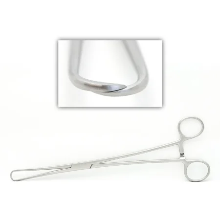
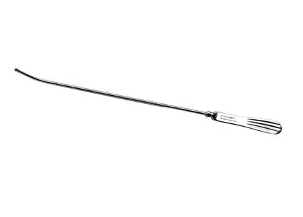
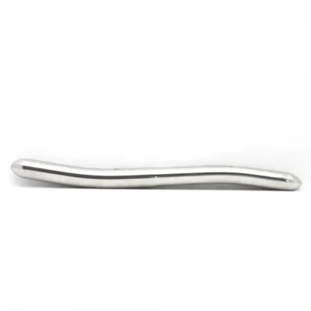
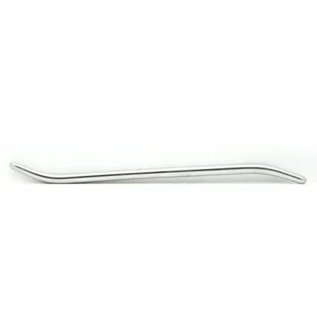
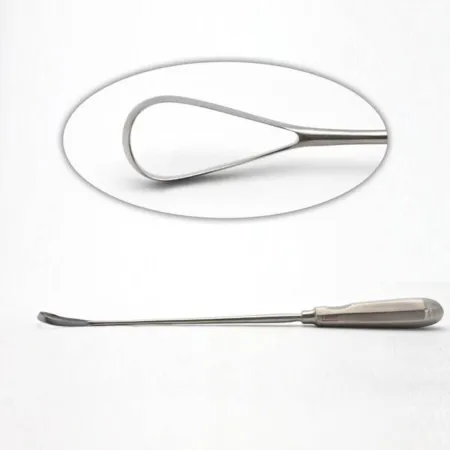
- D&C and vaginal set: vaginal speculumSPEK-yoo-lumAn instrument used to open and hold open a body opening, such as the vagina, so the inside can be seen.Full entry (Graves, and the weighted Auvard, placed in the back of the vagina), vaginal retractorrih-TRAK-terA surgical instrument that holds back tissue or organs so the surgeon can see and reach the operative site.Full entry (Heaney, Sims, Jackson and Deaver), a tenaculum to hold the cervix, a uterine sound, graduated cervicalSER-vih-kulRelating to the neck, or to the cervix, the lower end of the uterus.cervic/o neck, cervix-al relating toFull entry dilatorDY-lay-terAn instrument used to stretch or widen an opening, canal or passage in the body.Full entry (Hegar, Pratt or Hank, or a Goodell spreader), sharp and blunt curettekyoo-RETA surgical instrument with a small spoon- or loop-shaped tip used to scrape tissue.Full entry , polypPOL-ipA growth that sticks out from the lining of a hollow organ, such as the colon, nose or uterus.Full entry forcepsFOR-sepsA hand instrument with two blades or arms used to grasp, hold or pull tissue or objects.Full entry , sponge forceps and a supply of sponges. Vaginal surgery adds long uterine clamps, heavy dissecting scissors and Allis clamps.
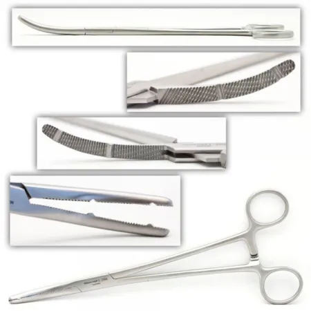
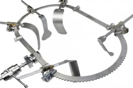
- Abdominal gynecologygy-nuh-KOL-uh-jeeThe branch of medicine that cares for the female reproductive organs and breasts.gynec/o woman-logy study ofFull entry (hysterectomyhis-tuh-REK-tuh-meeSurgery to remove the uterus.hyster/o uterus-ectomy surgical removalFull entry ) set: a laparotomylap-uh-ROT-uh-meeSurgery that opens the abdomen through a large cut to reach the organs inside.lapar/o abdomen-tomy cutting intoFull entry set plus heavy uterine clamps that will not slip on thick, vascular pediclePED-ih-kulA narrow stem or stalk that attaches a structure, such as a tumor or a skin flap, to the body.Full entry (Heaney, Heaney-Ballantine and Zeppelin clamps, Rochester-Ochsner and Pean clamps), Heaney needle holders, heavy curved scissors such as the Jorgenson, Russian and long tissue forceps, long instruments for a deep pelvis, and a self-retaining retractor such as the O'Sullivan-O'Connor, Balfour or table-mounted Bookwalter. Babcock clamps and vascular forceps handle the tubes, ovaries and bowel gently.
- cesareansih-ZAIR-ee-unRelating to the birth of a baby through a surgical cut in the mother's abdomen and uterus.Full entry set: the abdominal set with bandage scissors, a bladder retractor (DeLee, a Balfour bladder blade or a Doyen), Foerster ring forceps and Pennington clamps for the uterine edges, delivery forceps, cord clamps and a bulb syringe.
- laparoscopiclap-uh-roh-SKOP-ikDone through a few small cuts in the abdomen with a thin camera and long instruments, instead of one large opening.lapar/o abdomen-scopic viewingFull entry gynecologic set: graspers, a Maryland dissector, scissors, a hook electrode, bipolarby-POH-lerHaving two poles or two opposite ends; most often used as a short form of bipolar disorder.bi- twopolar having polesFull entry forceps and vessel sealers, a suction-irrigator, an aspirationas-puh-RAY-shunBreathing food, liquid, saliva or stomach contents into the airway and lungs; also, drawing fluid out with suction or a needle.Full entry needle for cysts, Babcock and Allis graspers, biopsy forceps and specimen bags, with a uterine manipulator from below.
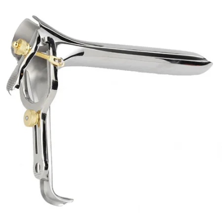
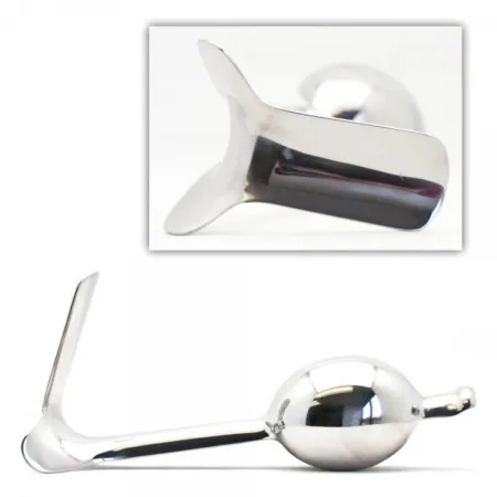
The Heaney is the classic hysterectomy clamp: heavy, with toothed, grooved jaws that hold a thick, vascular pedicle without slipping. The tubes are delicate, so they are held with a Babcock or another atraumatic clamp, never a toothed Kocher.
Equipment
- Uterine manipulator: a long instrument placed through the cervix into the uterus after the vaginal prep, so the assistant at the foot of the table can tilt the uterus and bring the tubes and ligaments into the scope's view. Many have a channel for injecting dye. For a hysterectomy, a colpotomy cup or ring (such as a Koh ring) fits around the cervix and shows the surgeon exactly where the cervix meets the vagina, and a balloon in the vagina keeps the gas in once the vagina is opened.
- Hysteroscope: a rigid or semirigid scope with a 0-degree or angled lens, passed through the cervix inside a sheathSHEETHA covering that wraps around a structure; in procedures, a short hollow tube left in a blood vessel to pass catheters through.Full entry . An obturatorOB-too-ray-terA smooth-tipped rod that fits inside a tube, such as a tracheostomy tube or cannula, to guide it in and then is taken out.Full entry , a blunt-tipped rod, may lead the sheath in to protect the tissue and is then taken out so the scope can go in. Channels with stopcocks carry the distentiondis-TEN-shunSwelling or stretching of a body part from pressure inside it.Full entry fluid in and out and take 3 mm or smaller instruments: scissors, graspers, biopsy forceps and electrodes. A light source and a video camera and monitor complete the setup, as for laparoscopylap-uh-ROS-kuh-peeSurgery or an exam done through small cuts in the belly, using a thin, lighted tube with a camera.lapar/o abdomen-scopy looking with an instrumentFull entry .
- Resectoscope: a sheathed scope with a spring-loaded handle that slides a wire electrode back and forth. A loop shaves tissue away bit by bit and seals as it cuts; a roller ball coagulationkoh-ag-yuh-LAY-shunThe process by which blood changes from a liquid into a gel, forming a clot.coagul clot-ation processFull entry a surface. It works in monopolar or bipolar mode, depending on the system.
- Fluid management: a pump fills the uterus at a set pressure and an under-buttocks drape or tubing collects what runs out, so fluid in and fluid out can be measured. Chapter 21 teaches the distention fluids and the fluid deficit.
- Endometrial ablationab-LAY-shunA procedure that destroys a small area of tissue, often with heat or cold, to stop it from causing a problem.Full entry devices, which destroy the lining without a resectoscope: a radiofrequency mesh device (such as NovaSure) that sizes the cavity and treats it in about 90 seconds, a heated fluid balloon, a heated saline system and a freezing probe.
- Hysteroscopic tissue removal device (such as MyoSure): passed through the hysteroscope, it draws a polyp or submucosal fibroid into a cutting window by suction and removes it.
- electrosurgeryee-LEK-troh-SUR-jer-eeUsing high-frequency electric current through a handheld tool to cut tissue or seal bleeding vessels during surgery.Full entry , monopolar and bipolar, with LEEP loops, ball electrodes and needle tips, and a smoke evacuator. Lasers (carbon dioxideKAR-bun dy-OK-sideA gas made by the body's cells as waste, carried in the blood to the lungs and breathed out.Full entry and others) treat endometriosisen-doh-mee-tree-OH-sisA condition in which tissue like the lining of the uterus grows outside the uterus.endo- withinmetr/o uterus-osis conditionFull entry , dysplasia and warts, usually with the colposcope, microscope or laparoscope. Chapter 20 teaches energy and lasers.
- Laparoscopic and robotic equipment: the video chain, insufflationin-suh-FLAY-shunBlowing a gas into a body cavity, such as carbon dioxide into the abdomen during laparoscopic surgery.Full entry , energy devices and, for large specimens, a power morcellator used inside a containment bag (below). Chapter 21 teaches them.
- For a birth: fetal heart rate monitoringFEE-tul hart rayt MON-ih-ter-ingChecking a fetus's heart rate during pregnancy or labor, with a handheld device or a continuous electronic monitor.Full entry (a Doppler ultrasoundDOP-ler UL-truh-soundAn ultrasound that uses sound waves to hear or show blood flowing through vessels or the fetal heartbeat.Full entry or ultrasound and the fetal heart rateFEE-tul hart raytThe number of times the unborn baby's heart beats per minute.Full entry monitor), an infant radiant warmer, infant resuscitationrih-sus-ih-TAY-shunEmergency treatment to restore breathing, heartbeat or blood circulation.Full entry equipment and drugs, and a suction line kept for the baby alone.

Supplies
The Core Curriculum's obstetric and gynecologic supply list:
- Cord blood tubes: filled from the cut umbilical cord at a birth; a cord pH kit or blood gas syringe may be added.
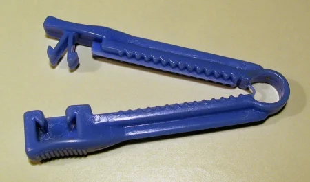
- Umbilical cord clamp: the plastic clamp that stays on the baby's cord; pull two for each baby.
- Falope ring system and Filshie clips: for laparoscopic tubal sterilizationstair-uh-luh-ZAY-shunA process that destroys all microorganisms, including spores, on an item; also a permanent birth control procedure.Full entry (below).
- intrauterinein-truh-YOO-ter-inInside the uterus.intra- withinuter uterus-ine relating toFull entry thermal balloon kit and NovaSure handpiece: endometrial ablation.
- MyoSure handpiece: hysteroscopic removal of fibroids and polyps.
- Transvaginal sling, tape and mesh: for stress urinary incontinenceYOOR-ih-nair-ee in-KON-tih-nunsLeaking of urine that a person cannot control.Full entry ; chapter 34 teaches bladder slings.
Other supplies you will meet: a DeLee suction catheterSUK-shun KATH-uh-terA thin, flexible tube attached to suction, used to remove mucus and other secretions from the airway.Full entry and bulb syringes for the baby, perinealper-ih-NEE-ulHaving to do with the perineum, the area between the genitals and the anus.Full entry pads, vaginal packing, a colpotomy ring and vaginal occluder, specimen retrieval and morcellation bags, and a drape with a fluid pouch. Packing is often moistened with saline or coated with an antibiotic, antifungalan-tee-FUNG-gulA drug that kills fungi or stops them from growing, used to treat fungal infections.anti- againstfung/i fungus-al relating toFull entry or estrogenES-truh-jenA female sex hormone, made mainly by the ovaries, and the group of drugs that act like it.Full entry cream. If more than one pack is used, or a pack is cut, it is recorded and reported at handoff reportHAND-off ree-PORTThe passing of information and responsibility for a client's care from one nurse or team to another.Full entry so all of it comes out later.
Sutures
- Uterine ligaments and vessels: synthetic absorbable, 0 or 2-0, on a heavy taper needle.
- Bladder flap: absorbable, 2-0 or 3-0, on a small taper needle.
- Ovary: absorbable, 3-0 or 4-0.
- Tube repair: very fine (5-0 to 7-0, finer still for microsurgery).
- Vaginal cuff and vaginal wall: absorbable, size 0.
- Vulvar plastic work: fine monofilament such as nylon or polypropylene on a cutting needle.
Keep two sutureSOO-cherA stitch, or the thread used to stitch, that holds the edges of a wound or incision together.Full entry ligatures of two sizes ready during a hysterectomy: each clamped pedicle is tied the moment it is cut.
Medications
Chapter 12 teaches how these drugs work; chapter 9 explains oxytocinok-sih-TOH-sinA hormone that makes the uterus contract and releases breast milk; also given by IV to start or strengthen labor and control bleeding after birth.Full entry as a hormone. At the field, label every drug, and say its name, strength and amount each time you pass it.
- Oxytocin (Pitocin): makes the uterus contract. After the baby is delivered, the anesthesia provideran-us-THEE-zhuh pruh-VY-derThe licensed professional who gives anesthesia and watches over the client's breathing and vital functions during a procedure.Full entry gives it into the vein, in IV fluid, to shrink the uterus and slow the bleeding, and some surgeons ask for a dose, such as 10 units, to be injected straight into the uterine muscle. Have it drawn up ahead of time, from a vial the circulator holds as chapter 12 teaches, and label the syringe. Given into a vein, oxytocin is a high-alert medicationHY-uh-lert med-ih-KAY-shunA drug that can cause serious harm to a client if it is used in error; also called a high-risk medication.Full entry .
- vasopressinvay-zoh-PRES-inA man-made form of antidiuretic hormone, given by IV mainly to raise blood pressure in shock.Full entry (Pitressin): injected, diluted in saline, into the cervix, a fibroid, the tube or the vulvar skin to clamp down the vessels and cut bleeding. Confirm the dilution with the surgeon and label it. Never confuse Pitressin with Pitocin: the names look and sound alike. Given into a vein, vasopressin is also a high-alert drug.
- methylergonovinemeth-il-er-GOE-noe-veenAn ergot drug that makes the uterus contract firmly, given after birth.Full entry (Methergine): makes the uterus contract to control bleeding after a birth or an abortion; it is given into a muscle or a vein.
- Other drugs for heavy bleeding after a birth: carboprostKAR-boh-prostA prostaglandin drug injected into muscle to make the uterus contract strongly.Full entry and misoprostolmy-soh-PROS-tolA prostaglandin drug that protects the stomach lining and also makes the cervix soften and the uterus contract.Full entry , prostaglandinpros-tuh-GLAN-dinA hormone-like fatty substance made in many tissues that acts on pain, inflammation, blood flow and uterine contractions; also drugs that copy it.Full entry that also make the uterus contract, and tranexamic acidtran-ek-SAM-ik AS-idA drug that reduces bleeding by stopping the body from breaking down blood clots.Full entry , which keeps clots from breaking down.
- magnesium sulfatemag-NEE-zee-um SUL-fateA magnesium salt given by IV or IM to raise a low magnesium level or prevent seizures in severe preeclampsia.Full entry : by vein, to prevent and treat the seizureSEE-zherA sudden burst of abnormal electrical activity in the brain that changes movement, feeling, behavior or awareness.Full entry of eclampsiaih-KLAMP-see-uhSeizures in a pregnant or recently pregnant person with preeclampsia, when no other cause explains them.Full entry . Tocolytics relax the uterus to stop early labor, as during surgery on a pregnant patient.
- methotrexatemeth-oh-TREK-sateA drug that blocks the use of folic acid by cells, used in high doses for cancer and in low weekly doses for autoimmune diseases.Full entry : can treat an unruptured ectopic pregnancyek-TOP-ik PREG-nun-seeA pregnancy in which the fertilized egg implants and grows outside the main cavity of the uterus, most often in a fallopian tube.ec- outtop/o place-ic relating toFull entry instead of surgery.
- Rho(D) immune globulinROH-dee ih-MYOON GLOB-yoo-linAn injection of antibodies given to an Rh-negative person to stop their body from making antibodies against Rh-positive blood.Full entry (RhoGAM): given to an Rh factorar-AYCH FAK-terA protein that may be present on the surface of red blood cells; people who have it are Rh-positive and those without it are Rh-negative.Full entry patient carrying an Rh-positive baby, at about 28 weeks and again within 72 hours of the birth, so the patient does not make antibodyAN-tih-bod-eeA Y-shaped protein made by the immune system that sticks to one specific germ or foreign substance and marks it for destruction.anti- againstbody Full entry that would harm a later baby. Chapter 12 teaches it.
- local anestheticLOH-kul an-es-THET-ikA drug that blocks feeling in one part of the body without making the person unconscious.Full entry with epinephrineep-ih-NEF-rinA hormone and drug, also called adrenaline, that raises heart rate and blood pressure and opens the airways.Full entry : injected into the cervix or vaginal walls before a cone or vaginal surgery, to numb and to reduce bleeding. Chapter 13 teaches the dose limits.
- Dyes and stains: acetic acid and Lugol solution on the cervix; methylene blue, often diluted, through a uterine cannulaKAN-yoo-luhA thin tube placed into the body or a body opening to deliver or remove fluid or air.Full entry to show whether the tubes are open (chromopertubation); indigo carmine into a vein to color the urine and show a bladder or ureterYOOR-uh-terOne of the two tubes that carry urine from the kidneys down to the bladder.Full entry injury. Do not confuse Lugol with Monsel solution, which looks the same and burns tissue to stop bleeding.
- Estrogen, antibiotic or antifungal cream on vaginal packing, which it also lubricates; estrogen cream treats vaginal atrophyAT-ruh-feeThe wasting away or shrinking of a body part, such as a muscle or organ.a- without-trophy nourishment, growthFull entry , the thin, dry vaginal lining that follows the fall in estrogen after menopauseMEN-uh-pawzThe time in a woman's life when menstrual periods stop for good, counted once there has been no period for 12 months in a row.men/o month, menstruation-pause stopFull entry .
- Antibiotics: to prevent a surgical site infection, given so the drug is already in the tissue when the incision is made; for a cesarean, before the skin incision.
- erythromycineh-rith-roh-MY-sinA macrolide antibiotic, also given to newborns as an eye ointment to prevent eye infection.Full entry 0.5% ointment for the newborn's eyes, on the Core Curriculum's cesarean list: it prevents conjunctivitiskun-junk-tih-VY-tisInflammation of the thin, clear layer that covers the white of the eye and lines the inside of the eyelids.conjunctiv/o conjunctiva-itis inflammationFull entry from gonorrheagon-uh-REE-uhA bacterial infection, spread mainly through sex, caused by Neisseria gonorrhoeae.Full entry or Chlamydiakluh-MID-ee-uhA sexually transmitted infection caused by the bacterium Chlamydia trachomatis; also the name of the bacterium.Full entry passed on from the birthing patient.
confuse Pitocin (oxytocin) with Pitressin (vasopressin). One contracts the uterus; the other clamps down blood vessels. Both are high-alert drugs when given into a vein, and the names are easy to mix up when a case moves fast.
7Procedures of the cervix
cervicalSER-vih-kulRelating to the neck, or to the cervix, the lower end of the uterus.cervic/o neck, cervix-al relating toFull entry procedures are done in lithotomylih-THOT-uh-meeA position lying on the back with the hips and knees bent and the legs raised and spread apart, often in stirrups.lith/o stone-tomy cutting intoFull entry , through the vagina, with a vaginal prep and a D&C set. Because the vagina and cervix are entered, they are Class II, clean-contaminated.
Cervical biopsy and conization
What and why: after an abnormal Pap testPAP testA screening test in which cells are gently scraped from the cervix and looked at under a microscope.Full entry , the surgeon looks at the cervix through the colposcope, stains it with acetic acid or Lugol solution, and takes biopsies of the abnormal areas. A cervical biopsy is usually an office procedure, done with a biopsy punch or biopsy forcepsFOR-sepsA hand instrument with two blades or arms used to grasp, hold or pull tissue or objects.Full entry , and it may be added to a D&C in the OR. A cone biopsy (conization) removes a cone of tissue around the cervical canal, to diagnose dysplasia or early cancer and to treat dysplasia by removing it.
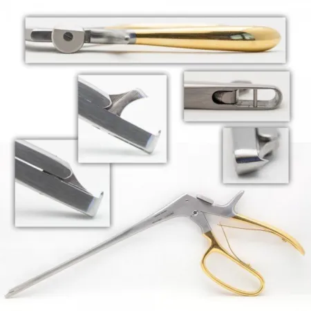
Approaches: the cone can be cut with a blade (a cold knife cone), with a laser, or with a thin electrosurgeryee-LEK-troh-SUR-jer-eeUsing high-frequency electric current through a handheld tool to cut tissue or seal bleeding vessels during surgery.Full entry wire loop, the loop electrosurgical excisionek-SIZH-unCutting out tissue, such as a growth or damaged area, during surgery.Full entry procedure (LEEP), also called LLETZ. LEEP is often done under local anesthesiaLOH-kul an-es-THEE-zhuhNumbing medicine applied to or injected into one small area, so a procedure there is not felt while the person stays awake.Full entry in a clinic, using the colposcope to see the lesionLEE-zhunAn area of tissue that has been damaged or has changed from normal, such as a sore, wound, lump or spot.Full entry ; it diagnoses and treats at once, because the loop gives a tissue specimen.
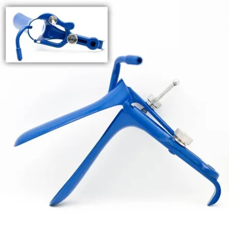
Setup: a D&C set, an ESU with loop electrodes and a ball electrode, a smoke evacuator, Lugol solution or acetic acid, local anestheticLOH-kul an-es-THET-ikA drug that blocks feeling in one part of the body without making the person unconscious.Full entry with epinephrineep-ih-NEF-rinA hormone and drug, also called adrenaline, that raises heart rate and blood pressure and opens the airways.Full entry , and a sutureSOO-cherA stitch, or the thread used to stitch, that holds the edges of a wound or incision together.Full entry for marking. Makers also offer speculumSPEK-yoo-lumAn instrument used to open and hold open a body opening, such as the vagina, so the inside can be seen.Full entry with an insulated coating and a vent for smoke evacuation.
Key steps:
- A speculum and retractorrih-TRAK-terA surgical instrument that holds back tissue or organs so the surgeon can see and reach the operative site.Full entry expose the cervix, and a tenaculum draws it toward the opening.
- The stain shows the abnormal tissue. Local anesthetic with epinephrine is injected into the cervix, often about 20 mL, to numb it and shrink its vessels.
- The surgeon cuts the cone with the loop or a blade. A stitch may mark 12 o'clock so the pathologist can tell the cone's position; label the specimen exactly as the surgeon names it.
- A 3 mm ball electrode coagulationkoh-ag-yuh-LAY-shunThe process by which blood changes from a liquid into a gel, forming a clot.coagul clot-ation processFull entry the bleeding base; the wide cone rarely needs suturing. Vaginal packing and a catheter may be placed, and a perinealper-ih-NEE-ulHaving to do with the perineum, the area between the genitals and the anus.Full entry pad goes on.
Smoke from the loop or the laser can carry human papillomavirusHYOO-mun pap-ih-LOH-muh-vy-rusA common group of viruses spread by close skin-to-skin contact, most often during sex.Full entry . Keep the smoke evacuator as close to the source as possible, within 2 inches (5 cm), without blocking the surgeon's view, and wear a fit-tested surgical N95en-NINE-tee-fiveA close-fitting respirator mask that filters at least 95% of tiny particles from the air the wearer breathes.Full entry .
some references still have the team wear high-filtrationfil-TRAY-shunThe passing of fluid through a filter or membrane, which holds back larger particles; in the kidneys, the first step in making urine.filtr strain through-ation processFull entry laser masks against the plume, which AST's laser guideline now keeps as the second line of defense after evacuation.
Wound class: II.
After surgery: heavy bleeding is uncommon but can happen, and infection is rare; later, though seldom, the cervix can narrow (stenosisstuh-NOH-sisNarrowing of a passage or opening in the body, such as a blood vessel, heart valve or the spinal canal.sten/o narrow-osis conditionFull entry ) or, in a later pregnancy, open too early (cervical insufficiencyin-suh-FISH-un-seeA state in which an organ or part does not work well enough to meet the body's needs.in- notsufficient enoughFull entry ).
Dilation and curettage (D&C)
What and why: a D&C opens the cervix and scrapes tissue from the cervical canal and the uterine lining. It diagnoses the cause of abnormal bleeding, confirms or rules out cancer of the endometriumen-doh-MEE-tree-umThe inner lining of the uterus, which thickens each month and is shed as the menstrual period if no pregnancy begins.endo- withinmetr/o uterus-ium tissueFull entry or cervix, helps study infertilityin-fer-TIL-ih-teeNot being able to become pregnant after a year of regular sex without birth control, or after six months for women over 35.Full entry and removes polypPOL-ipA growth that sticks out from the lining of a hollow organ, such as the colon, nose or uterus.Full entry ; it also empties the uterus after an incomplete or missed abortion. It may be combined with a hysteroscopy.
Before surgery: the patient is placed in lithotomy, and the bladder may be emptied with a straight catheterizationSTRAYT kath-uh-ter-ih-ZAY-shunPutting a catheter through the urethra into the bladder to drain urine, then taking it out right away.Full entry . A vaginal prep and a lithotomy drape follow. general anesthesiaJEN-er-ul an-es-THEE-zhuhDrug-induced loss of consciousness and feeling over the whole body, so a person feels nothing and is not aware during surgery.an- withoutesthesi/o feeling, sensation-ia conditionFull entry is preferred, but a regional block also works. If the surgeon examines the pelvis first, set that exam up as a separate tray, and the surgeon regloves afterward.
Key steps:
- A weighted speculum goes into the back of the vagina, a retractor holds the front wall, and a tenaculum grasps the front lip of the cervix.
- A uterine sound measures the depth and direction of the uterus, so the instruments that follow do not push through its wall.
- The cervix is opened with graduated dilatorDY-lay-terAn instrument used to stretch or widen an opening, canal or passage in the body.Full entry , smallest first (Hegar, Pratt or Hank, or a Goodell spreader). Line them up on the back table in size order, each with a little lubricant. In a tight, scarred cervix, as after menopauseMEN-uh-pawzThe time in a woman's life when menstrual periods stop for good, counted once there has been no period for 12 months in a row.men/o month, menstruation-pause stopFull entry , a LEEP or radiationray-dee-AY-shunEnergy that travels as waves or particles, such as X-rays, used to make images or treat cancer; also pain that spreads from its starting point.radi/o rays, radiation-ation processFull entry , very fine lacrimal probes may be needed first.
- The surgeon may explore for polyps with a polyp forceps.
- The surgeon scrapes the cervical canal first, then the endometrium, with sharp, blunt or serrationsair-AY-shunA row of small tooth-like ridges, such as the grooves on the jaws of a surgical clamp.Full entry curettekyoo-RETA surgical instrument with a small spoon- or loop-shaped tip used to scrape tissue.Full entry . Each sample is kept separate: often collected on a nonadherent pad laid in the back of the vagina, passed to you, and placed in its own labeled container.
- Bleeding at the tenaculum and biopsy sites is checked, a vaginal pack may go in, and a perineal pad is applied.
the endocervical and the endometrial specimens are two specimens. Keep them apart from the moment they come off the curette, because the pathologist needs to know which part of the uterus each came from.
Wound class: II.
After surgery: cramps and light bleeding for a few days are expected; heavy bleeding, fever or a bad-smelling discharge are reasons to call. The main risks are a hole through the uterine wall (perforationper-fuh-RAY-shunA hole that goes all the way through the wall of an organ or tissue.per- throughforat bored (a hole)-ion processFull entry ), injury to the cervix, bleeding and infection. Report any packing at handoff reportHAND-off ree-PORTThe passing of information and responsibility for a client's care from one nurse or team to another.Full entry .
Suction curettage and dilation and evacuation (D&E)
What and why: suction curettage empties the uterus with a vacuum cannulaKAN-yoo-luhA thin tube placed into the body or a body opening to deliver or remove fluid or air.Full entry . It is a safe way to treat a missed or incomplete spontaneous abortionspon-TAY-nee-us uh-BOR-shunThe loss of a pregnancy on its own, before the fetus can live outside the uterus, usually before 20 weeks; a miscarriage.Full entry , to end an early pregnancy and to remove a molar pregnancy (hydatidiform mole). It needs less dilationdy-LAY-shunThe widening or opening up of a body part, such as the cervix during labor, the pupils or a blood vessel.dilat widen-ion processFull entry than sharp curettage and causes less bleeding and less risk of perforation and infection. Dilation and evacuation (D&E), which the Core Curriculum lists as dilation, curettage and evacuation, is the term for emptying the uterus this way at about 13 weeks of pregnancy or later: it needs a wider opening, often made first with slowly swelling (osmoticoz-MOT-ikRelating to osmosis, the movement of water across a membrane toward the side with more dissolved particles.Full entry ) dilators, and forceps to remove the larger tissue.
Setup: a D&C set plus graduated dilators, sterile suction cannulas in several sizes, the vacuum aspirator and its tubing, placentapluh-SEN-tuhThe organ that grows in the uterus during pregnancy and passes oxygen and food from the parent's blood to the fetus.Full entry forceps and uterotonicyoo-ter-oh-TON-ikA drug that makes the uterus contract, used to start or strengthen labor or to control bleeding after birth.Full entry drugs.
Key steps: the cervix is exposed and held with a tenaculum, then dilated wide enough for a cannula, often 10 to 14 mm depending on the size of the uterus. The cannula goes in, the suction is turned on, and gentle movements empty the uterus. A curette may finish the job, but used gently, because scrapingSKRAY-pingA sample of cells collected by gently scraping a surface such as the skin, or the act of collecting it.Full entry away the deepest layer can scar the uterus shut inside (Asherman syndrome). The tissue is caught in a gauze trap inside the aspirator; it goes to pathologypuh-THOL-uh-jeeThe study of disease and its causes and effects, or the disease changes found in a person or tissue.path/o disease-logy study ofFull entry as the products of conception.
Wound class and after surgery: II. Watch for bleeding, and remember that the patient may be grieving a loss.
Cervical cerclage
What and why: in cervical insufficiency, the cervix opens without labor, usually in the second trimester, and the pregnancy is lost. A cerclage ties a band of nonabsorbable suture or polyester tape around the cervix to hold it closed. It is done before the cervix has dilated, usually late in the second trimester or early in the third, and an ultrasound showing a short, funneled cervix often leads to it. The stitch is usually removed in the office at about 36 to 37 weeks, when labor starts, or at a cesareansih-ZAIR-ee-unRelating to the birth of a baby through a surgical cut in the mother's abdomen and uterus.Full entry .
Before surgery: lithotomy, with spinal, epidural anesthesiaep-ih-DOOR-ul an-us-THEE-zhuhPain relief or numbness from an anesthetic drug given through a thin catheter into the space just outside the covering of the spinal cord.epi- upon, overdur/o dura (the tough covering of the spinal cord)-al relating toFull entry or general anesthesia. The fetal heart rateFEE-tul hart raytThe number of times the unborn baby's heart beats per minute.Full entry is watched. The vaginal prep is done gently, disturbing the cervix as little as possible.
Setup: a basic vaginal or D&C set, ligature carriers such as the Deschamps, the tape or heavy nonabsorbable suture, and absorbable suture for the mucosamyoo-KOH-suhThe moist lining of body passages that open to the outside, such as the mouth, nose, gut and bladder.Full entry . You may hold a retractor for the surgeon, and both of you may work from the back table.
Two methods:
- McDonald: the easier one. A purse-string stitch is run through the cervix at four to six points, such as 12, 3, 6 and 9 o'clock, and tied; unlike the Shirodkar, the stitch is not buried under the mucosa.
- Shirodkar: the mucosa at the front of the cervix is opened so the bladder can be pushed back, the back mucosa is opened too, and the tape is passed under the mucosa high around the cervix, tied, and covered by closing the incisions with absorbable suture, such as 2-0.
Wound class: II. After surgery: short bed rest, then activity with limits. Risks are a cervix that dilates anyway because the stitch is poorly placed, bleeding and infection.
8Procedures of the uterus
The uterus is reached three ways: through the cervix (hysteroscopy), through the vagina, or through the abdomen, open, laparoscopiclap-uh-roh-SKOP-ikDone through a few small cuts in the abdomen with a thin camera and long instruments, instead of one large opening.lapar/o abdomen-scopic viewingFull entry or robotic. The cesareansih-ZAIR-ee-unRelating to the birth of a baby through a surgical cut in the mother's abdomen and uterus.Full entry , the most common uterine operation of all, has its own section below.
Hysteroscopy
What and why: a hysteroscope passed through the cervix shows the inside of the uterus, the cervicalSER-vih-kulRelating to the neck, or to the cervix, the lower end of the uterus.cervic/o neck, cervix-al relating toFull entry canal and the openings of the tubes. It finds the cause of abnormal bleeding (often with an ablationab-LAY-shunA procedure that destroys a small area of tissue, often with heat or cold, to stop it from causing a problem.Full entry in the same visit), finds and removes a lost intrauterine devicein-truh-YOO-ter-in dih-VICEA small T-shaped device that a health care provider places inside the uterus to prevent pregnancy.Full entry , a polypPOL-ipA growth that sticks out from the lining of a hollow organ, such as the colon, nose or uterus.Full entry or a submucosal fibroid, frees scar tissue inside the uterus, cuts away a septumSEP-tumA wall of tissue that divides two spaces or cavities, such as the two sides of the heart or nose.sept/o wall, partitionFull entry that divides the cavity, and is part of the infertilityin-fer-TIL-ih-teeNot being able to become pregnant after a year of regular sex without birth control, or after six months for women over 35.Full entry workup. A diagnostic laparoscopylap-uh-ROS-kuh-peeSurgery or an exam done through small cuts in the belly, using a thin, lighted tube with a camera.lapar/o abdomen-scopy looking with an instrumentFull entry may be done at the same time.
Who should not have it: a patient who is pregnant or has a pelvic infection or cervical cancerSER-vih-kul KAN-serCancer that starts in the cells of the cervix, the lower, narrow end of the uterus that opens into the vagina.Full entry , and in some cases a patient who is bleeding heavily.
Setup: a D&C tray and a hysteroscopy tray, the scope and its sheathSHEETHA covering that wraps around a structure; in procedures, a short hollow tube left in a blood vessel to pass catheters through.Full entry , the camera, light source and monitor, pump tubing and the fluid pump, the electrosurgeryee-LEK-troh-SUR-jer-eeUsing high-frequency electric current through a handheld tool to cut tissue or seal bleeding vessels during surgery.Full entry , and, as the case needs, a resectoscope with its electrodes, an ablation device or a tissue removal device. An under-buttocks drape collects the outflowOWT-flohThe flow of a fluid out of an organ, space or vessel.Full entry .
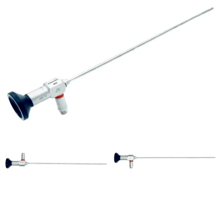
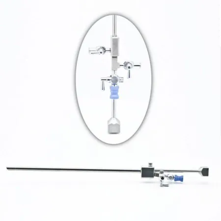
distentiondis-TEN-shunSwelling or stretching of a body part from pressure inside it.Full entry : fluid under pressure, or carbon dioxideKAR-bun dy-OK-sideA gas made by the body's cells as waste, carried in the blood to the lungs and breathed out.Full entry gas, opens the cavity; chapter 21 teaches the fluids, and the rule at the field is that saline, an electrolytic fluid (one that carries electric current), is used only with no electrosurgery or with a bipolarby-POH-lerHaving two poles or two opposite ends; most often used as a short form of bipolar disorder.bi- twopolar having polesFull entry system, never with a monopolar electrode, which uses a nonelectrolyte fluid such as 1.5% glycine.
Key steps:
- A weighted speculumSPEK-yoo-lumAn instrument used to open and hold open a body opening, such as the vagina, so the inside can be seen.Full entry and a front retractorrih-TRAK-terA surgical instrument that holds back tissue or organs so the surgeon can see and reach the operative site.Full entry expose the cervix; a tenaculum draws it forward.
- The uterus is sounded and the cervix dilated to fit the scope, often 6 to 8 mm.
- The tubing is primed with the chosen fluid, as the maker directs, and the scope goes in to the internal os.
- Fluid fills the cavity, and the surgeon looks around, on the monitor, and records images.
- Instruments go down the operating channel: biopsy forcepsFOR-sepsA hand instrument with two blades or arms used to grasp, hold or pull tissue or objects.Full entry , scissors, graspers, electrodes or a tissue removal device.
- At the end, the scope comes out, the fluid deficit is reported and recorded, and a perinealper-ih-NEE-ulHaving to do with the perineum, the area between the genitals and the anus.Full entry pad goes on.
The fluid deficit: chapter 21 teaches it; the fluid in and out is measured all through the case, and in a healthy patient the procedure is stopped at a deficit of about 1,000 mL of a nonelectrolyte fluid or about 2,500 mL of saline, lower for an older patient or one with heart or lung disease, with the deficit reported to the surgeon and the anesthesia provideran-us-THEE-zhuh pruh-VY-derThe licensed professional who gives anesthesia and watches over the client's breathing and vital functions during a procedure.Full entry and at handoff reportHAND-off ree-PORTThe passing of information and responsibility for a client's care from one nurse or team to another.Full entry .
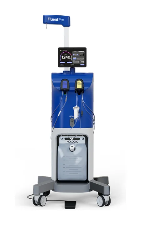
Risks: a hole through the uterine wall, which can injure bowel, bladder or the large vessels and may need a laparoscopy, and fluid overloadFLOO-id OH-ver-lohdToo much fluid in the body, in the blood vessels and the tissues.Full entry .
Wound class: II. After surgery: most patients go home the same day; cramping is common after a procedure inside the uterus.
Endometrial ablation
What and why: ablation destroys the lining of the uterus to stop or reduce heavy bleeding when medicines and other treatment have failed, as an alternative to hysterectomyhis-tuh-REK-tuh-meeSurgery to remove the uterus.hyster/o uterus-ectomy surgical removalFull entry . It is only for a patient who does not want a future pregnancy, because it prevents a normal implantationim-plan-TAY-shunThe attachment of a fertilized egg to the lining of the uterus, the start of a pregnancy.im- inplant to set-ation the process ofFull entry ; even so, pregnancy is still possible and unsafe afterward, so the patient still needs birth control. A pregnancy test comes first, and a D&C may be done first to check the cause of the bleeding. Suspected abnormal tissue or a large uterus rules it out.
Methods:
- Roller ball: through the resectoscope, a ball electrode about 3 mm across is rolled over the whole lining to coagulationkoh-ag-yuh-LAY-shunThe process by which blood changes from a liquid into a gel, forming a clot.coagul clot-ation processFull entry it, under the scope's view, in a nonelectrolyte fluid if monopolar. A loop can resect the lining instead.
- Global ablation, done "blind," without the scope during the treatment: a radiofrequency mesh device (such as NovaSure) that measures the cavity and treats it for about 90 seconds, stopping on its own if the tissue resistance gets too high; a balloon filled with hot fluid; heated saline circulated through a hysteroscope; or a freezing probe guided by ultrasound. The surgeon calls out the cavity measurements, and the circulator checks them and enters them into the controller.
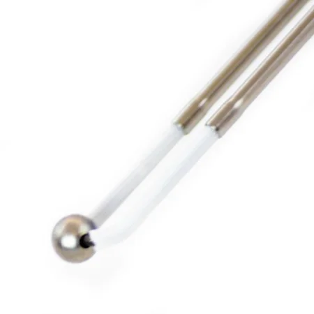
Wound class: II. After surgery: severe cramps and a watery discharge for several days are expected. Teach the signs of perforationper-fuh-RAY-shunA hole that goes all the way through the wall of an organ or tissue.per- throughforat bored (a hole)-ion processFull entry and infection. Few patients stop bleeding completely, and some need a repeat treatment.
Hysteroscopic myomectomy
What and why: a submucosal fibroid, under the lining, can be removed through the hysteroscope, with no incision.
Key steps: after the cervix is dilated, the resectoscope goes in with fluid flowing. The spring-loaded handle draws the cutting loop through the fibroid again and again, slicing off chips and sealing as it cuts, until the fibroid is level with the lining; the ball electrode stops bleeding. The chips float in the fluid and are flushed out through the sheath. A tissue removal device instead draws the fibroid in by suction and cuts it away.
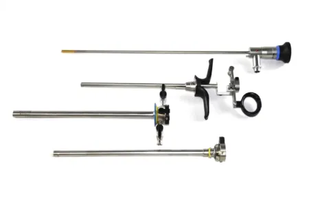
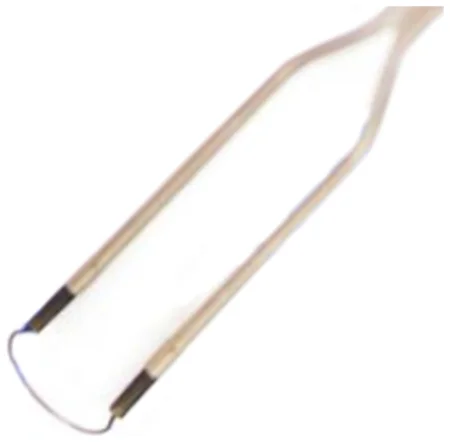

Specimen: every chip is collected for pathologypuh-THOL-uh-jeeThe study of disease and its causes and effects, or the disease changes found in a person or tissue.path/o disease-logy study ofFull entry . Wound class: II.
Hysterectomy: the words
- Total hysterectomy: the uterus and cervix.
- Supracervical (subtotal) hysterectomy: the body of the uterus only, leaving the cervix.
- Salpingo-oophorectomy: a tube and its ovary; bilateral means both sides (BSO). Salpingectomy alone takes the tubes and leaves the ovaries.
- Radical hysterectomy: the uterus and cervix with the tissue beside them (the parametria and supporting ligaments), the upper vagina and the pelvic lymph nodelimf nohdA small, bean-shaped organ of the immune system that filters lymph fluid and holds white blood cells.Full entry , for cervical cancer; the ovaries may stay.
Apart from operations related to pregnancy, hysterectomy is the most common operation performed on female patients in the United States. It treats fibroids, abnormal bleeding, endometriosisen-doh-mee-tree-OH-sisA condition in which tissue like the lining of the uterus grows outside the uterus.endo- withinmetr/o uterus-osis conditionFull entry , prolapsePROH-lapsThe slipping or falling of an organ or part from its normal position.Full entry , chronic pelvic pain, adenomyosis and early cancer.
Choosing the approach: when it is possible, a minimally invasive route (vaginal, laparoscopic or robotic) is preferred over an open one, for less pain, less bleeding and a faster recovery.
when the vagina is opened from above, the instruments that touched the cervix and vagina are dirty. Drop them, with the specimen, into a basin and pass them off the field, so vaginal germs cannot reach the abdominal wound.
Wound class (every hysterectomy): II, because the vagina is entered.
Total abdominal hysterectomy (TAH)
Before surgery: supine, then Trendelenburg positiontren-DEL-en-burg puh-ZISH-unA position in which the client lies on the back with the bed tilted so the head is lower than the feet.Full entry once the abdomen is open. The vagina and abdomen are prepped separately, the vagina first, and a urinary catheter goes in. A Pfannenstiel incision suits a normal-sized uterus; a midlineMID-lineAn imaginary line down the center of the body; also, a type of IV catheter placed in a vein of the upper arm.Full entry or paramedian incision is used for a large uterus, an obese patient or a look at the upper abdomen. sequential compression devicesih-KWEN-shul kum-PRESH-un dih-VYSInflatable leg sleeves that fill with air and squeeze the legs in turns to keep blood moving in the veins.Full entry and warming are routine.
Setup: a laparotomylap-uh-ROT-uh-meeSurgery that opens the abdomen through a large cut to reach the organs inside.lapar/o abdomen-tomy cutting intoFull entry set with the hysterectomy instruments (at least six heavy uterine clamps), long instruments, a self-retaining retractor, and often a vessel sealer or ultrasonic shears, which can replace many clamp, cut and tie steps.
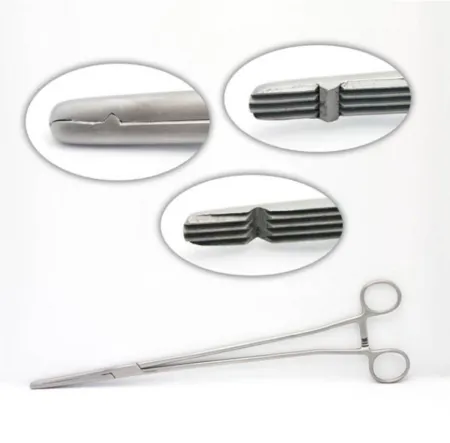
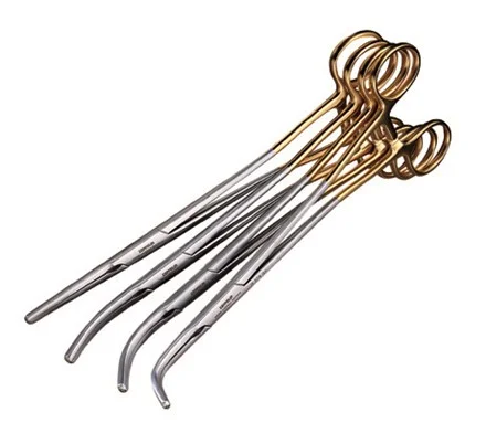
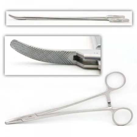
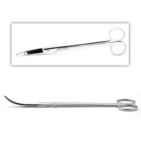
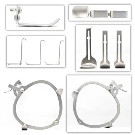
Key steps:
- The abdomen is opened, a self-retaining retractor placed and the bowel packed into the upper abdomen with moist laps.
- The round ligaments are double-clamped, cut and tied with sutureSOO-cherA stitch, or the thread used to stitch, that holds the edges of a wound or incision together.Full entry ligatures.
- The peritoneumpair-ih-toh-NEE-umThe thin membrane that lines the belly cavity and covers most of the organs inside it.peri- aroundton stretched-eum a structureFull entry over the bladder is opened and the bladder flap is pushed down off the uterus.
- If the ovaries come out, the surgeon finds the ureterYOOR-uh-terOne of the two tubes that carry urine from the kidneys down to the bladder.Full entry , then clamps (often three clamps), cuts and ties the infundibulopelvic ligament with the ovarian vessels. If they stay, the tube and the utero-ovarian ligament are clamped and cut beside the uterus instead.
- The uterine vessels are clamped, cut and tied on each side, as close to the uterus as possible, because the ureter lies only about 1 cm to the side.
- The cardinal and uterosacral ligaments are clamped, cut and tied down the sides of the cervix.
- The vagina is entered just below the cervix, and the cervix is cut free; the uterus is lifted out into a basin. The cervix and vagina instruments go with it.
- The vaginal cuff is closed, with angle stitches that anchor the ligaments to it for support. The peritoneum may be closed over it.
- The wound is irrigationeer-ih-GAY-shunWashing out a wound, body cavity, tube or eye with a steady flow of liquid.Full entry and closed in layers after the counts; an abdominal dressing and a perineal pad go on.
if you lose track of which pediclePED-ih-kulA narrow stem or stalk that attaches a structure, such as a tumor or a skin flap, to the body.Full entry is next, look at the field: hysterectomies proceed by anatomy, round ligament to cuff. Keep a suture ligature of each size ready for every clamp the surgeon places, so each pedicle is tied as soon as it is cut.
After surgery: nothing in the vagina for about 6 weeks. If the ovaries were removed, menopauseMEN-uh-pawzThe time in a woman's life when menstrual periods stop for good, counted once there has been no period for 12 months in a row.men/o month, menstruation-pause stopFull entry symptoms such as hot flashes can begin. Complications include bleeding, infection, wound separation, and injury to the bladder, bowel or ureter.
Vaginal hysterectomy
What and why: the uterus is removed through the vagina, with no abdominal incision. It suits a patient with good access, often one with prolapse, and allows a short stay; a large uterus or a cancer spreading beyond it rules it out. A laparotomy tray stays available.
Before surgery: lithotomylih-THOT-uh-meeA position lying on the back with the hips and knees bent and the legs raised and spread apart, often in stirrups.lith/o stone-tomy cutting intoFull entry ; a vaginal and perineal prep; a lithotomy drape. You will need long tissue forceps, long dissecting scissors and several sponge sticks, because the work goes deep into the pelvis.
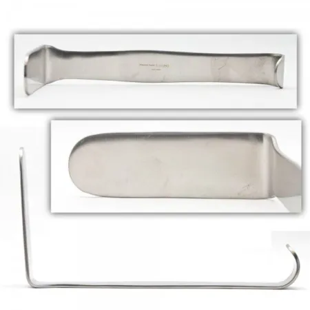
Key steps:
- The labia may be stitched aside, a weighted retractor holds the back wall and a tenaculum or a stitch grasps the cervix.
- Saline with vasopressinvay-zoh-PRES-inA man-made form of antidiuretic hormone, given by IV mainly to raise blood pressure in shock.Full entry , or local anestheticLOH-kul an-es-THET-ikA drug that blocks feeling in one part of the body without making the person unconscious.Full entry with epinephrineep-ih-NEF-rinA hormone and drug, also called adrenaline, that raises heart rate and blood pressure and opens the airways.Full entry , is injected around the cervix to cut bleeding and open the tissue planes.
- The surgeon cuts around the cervix with a #10 or #15 blade, pushes the bladder up off it and opens the peritoneum in front; then opens the posterior cul-de-sac. A Heaney or Sims retractor holds the bladder away.
- The uterosacral, then the cardinal ligaments and the uterine vessels are clamped with Heaney clamps, cut and tied with size 0 absorbable suture on a taper needle; the tie ends of the uterosacrals are often left long and tagged with a clamp. A handheld vessel sealer can replace clamp, cut and tie.
- The fundusFUN-dusThe rounded top or base of a hollow organ, most often the top of the uterus.Full entry is delivered, and the round and utero-ovarian ligaments and the tubes are clamped and cut. The uterus comes free.
- The ligaments are stitched to the top of the vagina to support it, a count is done, and the peritoneum and the vaginal cuff are closed. Any cystocele or rectocele is repaired now.
- A urinary catheter goes in; packing and a perineal pad may follow.
Wound class: II.
Laparoscopic and robotic hysterectomy
Laparoscopic-assisted vaginal hysterectomy (LAVH): the top of the operation is done through the scope (the round ligaments and, if the ovaries come out, the infundibulopelvic ligaments divided, and the bladder flap made), then the rest is done from below, as a vaginal hysterectomy. The patient avoids a large incision and has less pain and a shorter stay than with an abdominal hysterectomyab-DOM-ih-nul his-ter-EK-tuh-meeSurgery to remove the uterus through a cut in the lower abdomen.hyster/o uterus-ectomy surgical removalFull entry . Once the vaginal part begins, the team does not go back into the abdomen without fresh gowns and gloves: when the surgeon returns to the scope at the end to check the bleeding, the surgical team changes gowns and gloves first.
Total laparoscopic and robotic hysterectomy: everything is done from above. In low lithotomy, a uterine manipulator with a colpotomy cup is placed, and ports go in; for the robot, the patient goes into steep Trendelenburg and the robot is docked. The ligaments and vessels are sealed and divided down to the cup, and the surgeon cuts the vagina around the cup while the assistant or scrub pushes the uterus up with the manipulator, which lifts the cervix away from the ureters. The uterus is pulled into the vagina or out through it, an occluder keeps the gas from escaping, and the cuff is closed from above, often with a barbed absorbable suture. Chapter 21 teaches the ports and the robot.
A supracervical hysterectomy leaves the cervix, so the body of the uterus must come out in pieces (morcellated) or through a small incision. Because a power morcellator can spread an unsuspected cancer, morcellation is done inside a containment bag, and many facilities have stopped using power morcellation. A patient who keeps the cervix still needs cervical cancer screening.
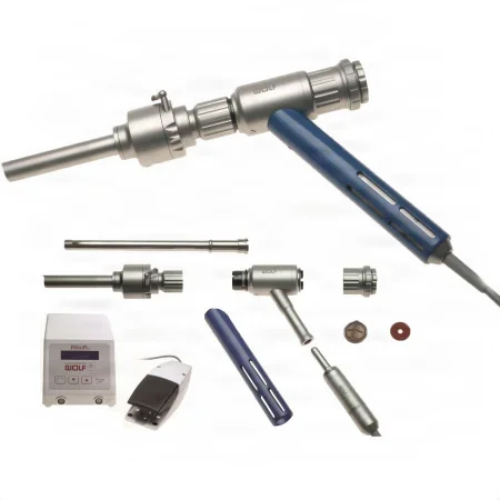
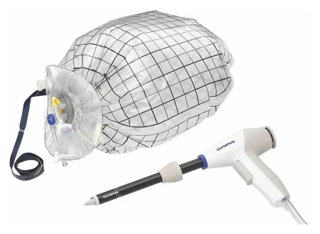
Wound class: II. After surgery: shoulder pain from the gas is common for a few days. Complications include bleeding, conversion to an open operation, injury to a large vessel, bowel, bladder or ureter, infection, a herniaHER-nee-uhA bulge of an organ or tissue through a weak spot in the muscle or tissue wall that normally holds it in.Full entry at a port site and, later, a fistulaFIS-choo-luhAn abnormal passage between two organs or vessels, or between an organ and the skin.Full entry into the vagina.
Abdominal myomectomy
What and why: a myomectomy removes fibroids and keeps the uterus, usually for a younger patient with symptoms who wants to keep the chance of pregnancy. It bleeds more and carries more risk than a hysterectomy. Months of a hormone drug (a GnRH agonistAG-uh-nistA drug or body chemical that attaches to a receptor on a cell and switches it on.Full entry such as leuprolide) may shrink the fibroids and their blood supply first. It can also be done laparoscopically or with the robot.
Key steps: the abdomen is opened as for a hysterectomy. Vasopressin diluted in saline is injected into the uterus or the fibroid to cut bleeding. The surgeon grasps each fibroid with a tenaculum or towel clip and shells it out of its bed with sharp and blunt dissectiondy-SEK-shunA tear in the inner layer of an artery wall that lets blood split the layers apart; also, the cutting apart of tissue in surgery.Full entry , the ESU or a laser. The uterine wall is then rebuilt with stitches.
Wound class: II. After surgery: risks include injury to the bowel, bladder, a tube or a ureter, wound infection and infertility, and one reference reports that about a fifth to a quarter of patients need more pelvic surgery later. A scar through the full-thicknessful-THIK-nisGoing through every layer of the skin, or every layer of another tissue such as the wall of an organ.Full entry of the uterine wall is one reason a later birth is done by cesarean.
Brachytherapy
What and why: brachytherapybrak-ee-THAIR-uh-peeRadiation treatment in which a radioactive source is placed inside the body, in or right next to a tumor.brachy- short (a short distance)-therapy treatmentFull entry places a radiationray-dee-AY-shunEnergy that travels as waves or particles, such as X-rays, used to make images or treat cancer; also pain that spreads from its starting point.radi/o rays, radiation-ation processFull entry source inside or right beside a cancer of the cervix or endometriumen-doh-MEE-tree-umThe inner lining of the uterus, which thickens each month and is shed as the menstrual period if no pregnancy begins.endo- withinmetr/o uterus-ium tissueFull entry , alone or with external radiation and antineoplastican-tee-nee-oh-PLAS-tikA drug used to treat cancer by killing cancer cells or stopping them from growing and dividing.anti- againstneo- newplas growth, formation-tic relating toFull entry . After a hysterectomy for endometrial cancer, a cylinder goes into the vagina; with the uterus still in place, a rod (tandem) goes through the cervix into the uterus with small holders (ovoids) against the cervix. Needles may also be placed into the tissue around the cervix.
In the OR: under general anesthesiaJEN-er-ul an-es-THEE-zhuhDrug-induced loss of consciousness and feeling over the whole body, so a person feels nothing and is not aware during surgery.an- withoutesthesi/o feeling, sensation-ia conditionFull entry in lithotomy, the surgeon places the empty applicators, a urinary catheter with its balloon filled with contrastKON-trastA dye or other substance given before an imaging test so body parts show up more clearly.Full entry and a rectalREK-tulRelating to the rectum, the last part of the large intestine; also, given or measured through the rectum.rect/o rectum, straight-al relating toFull entry markerMAR-kerSomething that can be measured, such as a substance in the blood, that shows a disease or body process is present.Full entry , so imaging can show the bladder and rectum. For these applicators, the radioactive source is loaded later, in the radiation department or the patient's shielded room. Radioactive seeds can also be placed at surgery, strung on suture, in tubes loaded afterward, or released through needles guided by ultrasound. Chapter 22 teaches radiation safety.
Wound class: II, worked out from the CDC definitions, since the applicators enter the genital tract through the vagina. After surgery: a low-dose-rate treatment keeps the patient in the hospital 1 to 3 days with the source in place, on bed rest with limited visitors, and the staff who care for them wear dosimeters; a high-dose-rate treatment is given as an outpatient.
9The pelvis through the scope: diagnostic laparoscopy
What and why: laparoscopylap-uh-ROS-kuh-peeSurgery or an exam done through small cuts in the belly, using a thin, lighted tube with a camera.lapar/o abdomen-scopy looking with an instrumentFull entry looks into the abdomen and pelvis to find the cause of pelvic pain or infertilityin-fer-TIL-ih-teeNot being able to become pregnant after a year of regular sex without birth control, or after six months for women over 35.Full entry and to judge a pelvic mass, and many procedures can follow at once: freeing adhesionad-HEE-zhunA band of scar tissue that joins two surfaces inside the body that are normally separate.ad- towardhes stick-ion conditionFull entry , burning away endometriosisen-doh-mee-tree-OH-sisA condition in which tissue like the lining of the uterus grows outside the uterus.endo- withinmetr/o uterus-osis conditionFull entry , draining a cyst, taking a biopsy or washings, removing a cyst, an ovary, a tube or an ectopic pregnancyek-TOP-ik PREG-nun-seeA pregnancy in which the fertilized egg implants and grows outside the main cavity of the uterus, most often in a fallopian tube.ec- outtop/o place-ic relating toFull entry , and tubal sterilizationstair-uh-luh-ZAY-shunA process that destroys all microorganisms, including spores, on an item; also a permanent birth control procedure.Full entry . Chapter 21 teaches the equipment, entry, the gas and its risks.
Before surgery: general anesthesiaJEN-er-ul an-es-THEE-zhuhDrug-induced loss of consciousness and feeling over the whole body, so a person feels nothing and is not aware during surgery.an- withoutesthesi/o feeling, sensation-ia conditionFull entry ; modified (low) lithotomylih-THOT-uh-meeA position lying on the back with the hips and knees bent and the legs raised and spread apart, often in stirrups.lith/o stone-tomy cutting intoFull entry ; the abdomen, perineumpair-ih-NEE-umThe area of skin and muscle between the genitals and the anus.perine/o perineumFull entry and vagina prepped and draped for a combined procedure; the bladder emptied to protect it from the trocarTROH-karA sharp or bladed instrument inside a tube (cannula), used to pierce a body wall and leave the tube in place as a port.Full entry . A D&C may come first. The surgeon sounds the uterus and places a uterine manipulator, often with a channel for dye. An open abdominal tray stays in the room.
Key steps: the port sites may be numbed first, and the abdomen is entered at the umbilicus with a Veress needle, an optical trocar or an open (Hasson) technique, and filled with carbon dioxideKAR-bun dy-OK-sideA gas made by the body's cells as waste, carried in the blood to the lungs and breathed out.Full entry , usually held at 12 to 15 mm Hg. The scope goes in, the patient is tilted head down, and other ports are added under view in the lower abdomen. For chromopertubation, diluted methylene blue or saline is pushed through the manipulator's cannulaKAN-yoo-luhA thin tube placed into the body or a body opening to deliver or remove fluid or air.Full entry with a 60 mL syringe and extension tubing; dye spilling from the ends of the tubes means they are open. At the end the gas is let out, the fasciaFASH-ee-uhA sheet or band of tough connective tissue that covers and separates muscles and organs.fasci/o bandFull entry at large ports is closed, and the manipulator comes out and the cervix is checked for bleeding.
peritonealper-ih-toh-NEE-ulHaving to do with the peritoneum, the thin lining of the belly cavity and the organs inside it.Full entry washings may be collected and sent for cytologysy-TOL-uh-jeeThe study of cells, or a lab test that looks at cells under a microscope.cyt/o cell-logy study ofFull entry , to look for cancer cells. Handle them as a specimen.
Wound class: II when a manipulator passes through the vagina into the uterus. After surgery: most go home the same day; shoulder pain from the gas, injury to an abdominal organ and gas embolismEM-buh-liz-umThe blocking of a blood vessel by a mass carried there in the blood (an embolus), such as a clot, fat or an air bubble.embol/o plug-ism conditionFull entry are the risks.
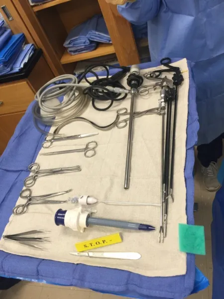
10Procedures of the uterine tubes
Ectopic pregnancy
Ruptured: a ruptured tube is an emergency, with blood filling the abdomen. Expect a fast laparoscopylap-uh-ROS-kuh-peeSurgery or an exam done through small cuts in the belly, using a thin, lighted tube with a camera.lapar/o abdomen-scopy looking with an instrumentFull entry if the patient is stable, or a laparotomylap-uh-ROT-uh-meeSurgery that opens the abdomen through a large cut to reach the organs inside.lapar/o abdomen-tomy cutting intoFull entry (often Pfannenstiel) if not. Have suction and irrigationeer-ih-GAY-shunWashing out a wound, body cavity, tube or eye with a steady flow of liquid.Full entry ready at once, a basin for clots, plenty of Mayo and Crile clamps and sutureSOO-cherA stitch, or the thread used to stitch, that holds the edges of a wound or incision together.Full entry ligatures; the surgeon scoops out the clots, finds the bleeding and clamps the tube and the mesosalpinx to stop it. Then the pregnancy, or the tube with it (salpingectomy), is removed. The patient may be in shockshokA life-threatening state in which blood flow is too low to bring enough oxygen to the body's tissues.Full entry from blood loss.
Unruptured: a patient who wants future pregnancies may keep the tube. In a salpingotomy (the Core Curriculum's term; some references call it salpingostomy), dilute vasopressinvay-zoh-PRES-inA man-made form of antidiuretic hormone, given by IV mainly to raise blood pressure in shock.Full entry is injected into the tube wall, the tube is opened along its free (antimesenteric) border over the bulge, and the pregnancy tissue is gently flushed or teased out with irrigation and forcepsFOR-sepsA hand instrument with two blades or arms used to grasp, hold or pull tissue or objects.Full entry . The tube is left to heal on its own or closed with very fine suture. The human chorionic gonadotropinHYOO-mun kor-ee-ON-ik goh-nad-oh-TROH-pinA hormone made by the placenta during pregnancy, measured in blood or urine as a pregnancy test.Full entry level is followed afterward, because a little tissue can stay behind and keep growing.
Specimen: the tube or the pregnancy tissue. Wound class: I, or II when a uterine manipulator is used.
Salpingectomy
A salpingectomy removes a tube, for an ectopic pregnancyek-TOP-ik PREG-nun-seeA pregnancy in which the fertilized egg implants and grows outside the main cavity of the uterus, most often in a fallopian tube.ec- outtop/o place-ic relating toFull entry , a blocked or infected tube, or a tumor. Laparoscopically, the mesosalpinx is coagulationkoh-ag-yuh-LAY-shunThe process by which blood changes from a liquid into a gel, forming a clot.coagul clot-ation processFull entry or sealed and cut with bipolarby-POH-lerHaving two poles or two opposite ends; most often used as a short form of bipolar disorder.bi- twopolar having polesFull entry or ultrasonic energy, staples or a pretied loop ligature, as close to the tube as possible, so the ovary's blood supply stays. Because many ovarian canceroh-VAIR-ee-un KAN-serCancer that starts in the ovaries or in the nearby fallopian tubes.Full entry may start in the far end of the tube, surgeons are encouraged to offer to remove both tubes during a benign hysterectomyhis-tuh-REK-tuh-meeSurgery to remove the uterus.hyster/o uterus-ectomy surgical removalFull entry or other pelvic surgery, or instead of a tubal ligation (an opportunistic salpingectomy).
Sterilization (tubal ligation)
What and why: blocking or removing a part of each tube prevents the egg and sperm from meeting, as permanent birth control. It is voluntary, needs careful counseling and its own informed consentin-FORMD kun-SENTA person's agreement to a treatment or procedure, given freely after the risks, benefits and other choices have been explained to them.Full entry , and cannot be counted on to be reversed. It can be done laparoscopically as an outpatient, through a small incision just after a vaginal birth, or at a cesareansih-ZAIR-ee-unRelating to the birth of a baby through a surgical cut in the mother's abdomen and uterus.Full entry . At a cesarean it is done only when the patient asked for it before labor began, after its own counseling and consent; where facility policy or law requires the two separate consents, the case has two time-outs.
laparoscopiclap-uh-roh-SKOP-ikDone through a few small cuts in the abdomen with a thin camera and long instruments, instead of one large opening.lapar/o abdomen-scopic viewingFull entry methods: low lithotomylih-THOT-uh-meeA position lying on the back with the hips and knees bent and the legs raised and spread apart, often in stirrups.lith/o stone-tomy cutting intoFull entry with a uterine manipulator to swing each tube into view; usually a camera port at the umbilicus and a second port just above the pubisPYOO-bisThe front part of each hip bone, low in the pelvis; the two meet in the middle at the pubic symphysis.pub/o pubisFull entry . The surgeon grasps the tube with a Babcock and blocks it with:
- Bipolar coagulation: at least 3 cm of the isthmus burned, starting 2 to 3 cm from the uterus; the tube may also be cut.
- A Falope (Silastic) ring: you load the small ring onto the applicator, which pulls a loop of tube in and slips the ring over its base; the loop dies and separates.
- A clip: a spring clip (Hulka) or a hinged titanium Filshie clip lined with silicone, closed across the isthmus about 1 to 2 cm from the uterus, so the whole width of the tube is inside the jaws.
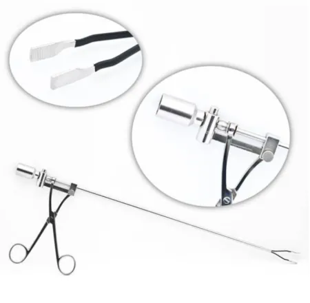
postpartumpohst-PAR-tumThe time after giving birth, usually the first six weeks, while the body returns to its non-pregnant state.post- afterpart/o birth, laborFull entry and open methods: through a small incision soon after a vaginal birth, or through the cesarean incision, a Babcock lifts the middle of each tube, a loop is tied at its base, and the loop is cut off (the Pomeroy method); other methods tie and divide the tube, as in the Irving method, which buries the cut end in the uterine wall. Each removed piece is a specimen, labeled "right fallopian tubefuh-LOH-pee-un TOOBOne of the two tubes that carry an egg from the ovary to the uterus, where fertilization usually happens.Full entry " or "left fallopian tube," so the pathologist can confirm the full width of each tube was taken.
Wound class: I, or II when a uterine manipulator is used. After surgery: most patients go home the same day; risks are rare: infection, bleeding inside the abdomen and short-lived pelvic pain. AST notes that a tubal ligation, even one done in labor and delivery, can still injure an organ or a large vessel and cause hemorrhageHEM-uh-rijA large loss of blood from a blood vessel, inside the body or out of it.hem/o blood-rrhage bursting forthFull entry , shock and death, which is why it calls for a CSTsee-es-tee (said as letters)Certified Surgical Technologist, a credential for surgical technologists; in maternity care, contraction stress test.Full entry .
Tuboplasty
What and why: tuboplasty reopens a blocked tube or rejoins the two ends after a tubal ligation, to restore fertility. Blockage follows infection, an earlier ectopic pregnancy, endometriosisen-doh-mee-tree-OH-sisA condition in which tissue like the lining of the uterus grows outside the uterus.endo- withinmetr/o uterus-osis conditionFull entry or a ligation. Diagnostic laparoscopy, hysterosalpingography or dye studies come first. The work is microsurgery, through a small laparotomy or the laparoscope, sometimes with the robot, under an operating microscope or loupes, and it may be done by fertility specialists.
Setup: a basic abdominal gynecologygy-nuh-KOL-uh-jeeThe branch of medicine that cares for the female reproductive organs and breasts.gynec/o woman-logy study ofFull entry tray with fine instruments, a set of microsurgical instruments used only for the microsurgery, micro-needle electrodes and fine bipolar forceps, a microscope or loupes, diluted dye with a uterine cannulaKAN-yoo-luhA thin tube placed into the body or a body opening to deliver or remove fluid or air.Full entry , and irrigation with lactated Ringer's solutionLAK-tay-tid RING-erz suh-LOO-shunAn isotonic IV fluid containing water, sodium, chloride, potassium, calcium and lactate.Full entry , sometimes with heparinHEP-uh-rinAn anticoagulant drug, given by injection or IV, that keeps blood clots from forming or growing.Full entry added, to keep the tissue moist and show bleeders.
Key steps: low lithotomy, so the cervix can be reached; a transversetrans-VERSLying across something, at a right angle to its length.Full entry incision; the bowel packed away; vasopressin injected to limit bleeding. The blocked part is cut out, each end is opened until dye flows through it, and the ends are joined in two layers with very fine (8-0 or 9-0) sutures, sometimes over a thin stentstentA small tube, often of metal mesh, placed inside a vessel or duct to hold it open.Full entry that is then removed. Dye injected through the cervix should spill from the end of the tube.
Wound class: I. After surgery: most go home the same day or the next. The risks include infection, bleeding, adhesionad-HEE-zhunA band of scar tissue that joins two surfaces inside the body that are normally separate.ad- towardhes stick-ion conditionFull entry , a later ectopic pregnancy and infertilityin-fer-TIL-ih-teeNot being able to become pregnant after a year of regular sex without birth control, or after six months for women over 35.Full entry that continues.
11Procedures of the ovary
Ovarian cystectomy
What and why: removing a cyst and keeping the ovary, for a cyst that persists or must be examined to rule out cancer. Most are done laparoscopically unless the mass is large. Before surgery, the surgeon judges the risk that the mass is cancer; be ready to convert to an open operation.
Key steps (laparoscopiclap-uh-roh-SKOP-ikDone through a few small cuts in the abdomen with a thin camera and long instruments, instead of one large opening.lapar/o abdomen-scopic viewingFull entry ): washings may be taken first. The ovary's surface (cortexKOR-teksThe outer layer of an organ, especially the folded gray outer layer of the brain.cortic/o cortex, outer layerFull entry ) is opened with scissors, a monopolar instrument or a laser, and the cyst is peeled out of the ovary with blunt dissectiondy-SEK-shunA tear in the inner layer of an artery wall that lets blood split the layers apart; also, the cutting apart of tissue in surgery.Full entry and fluid under pressure (hydrodissection). It goes into a retrieval bag and comes out whole through a port; a large cyst may be drained or cut up inside the bag. Bleeding is stopped with bipolarby-POH-lerHaving two poles or two opposite ends; most often used as a short form of bipolar disorder.bi- twopolar having polesFull entry energy; the ovary usually needs no stitches, or a few fine absorbable ones.
Spillage: a cyst that might be cancer must not be spilled, so it is bagged whole. If a cyst does rupture, the pelvis is washed out with large amounts of fluid. A dermoid cyst's thick, oily contents irritate the peritoneumpair-ih-toh-NEE-umThe thin membrane that lines the belly cavity and covers most of the organs inside it.peri- aroundton stretched-eum a structureFull entry , so laps are packed around it in an open case. The patient may be moved from head down to flat while the pelvis is washed, to keep the fluid in the pelvis.
Wound class: I, or II when a uterine manipulator is used.
Oophorectomy and salpingo-oophorectomy
What and why: an oophorectomy removes an ovary; a salpingo-oophorectomy removes the tube with it, one side or both (bilateral, BSO). Reasons include a tumor or cancer, a large cyst, abscessAB-sesA closed pocket of pus in tissue, walled off by the body.Full entry or torsion with dead tissue, endometriosisen-doh-mee-tree-OH-sisA condition in which tissue like the lining of the uterus grows outside the uterus.endo- withinmetr/o uterus-osis conditionFull entry , and lowering cancer risk in a patient with a BRCA mutationmyoo-TAY-shunA change in the genetic code (DNA) of a cell.Full entry or Lynch syndrome. Most are laparoscopic unless the mass is large. Removing both ovaries before menopauseMEN-uh-pawzThe time in a woman's life when menstrual periods stop for good, counted once there has been no period for 12 months in a row.men/o month, menstruation-pause stopFull entry brings on menopause at once and may raise the risk of heart disease and thin bones, which is one reason a premenopausal patient's ovaries may be kept.
Key steps: the surgeon grasps the tube with an Allis or Babcock, finds the ureterYOOR-uh-terOne of the two tubes that carry urine from the kidneys down to the bladder.Full entry , then clamps, cuts and ties (or seals) the infundibulopelvic ligament with the ovarian vessels. The mesosalpinx and the ovarian ligament are divided to free the tube and ovary from the uterus, and the raw edge is covered with peritoneum. Laps around the ovary catch any spill.
Specimen: if both ovaries come out, each goes in its own container, labeled right or left. Wound class: I, or II when a uterine manipulator is used.
After surgery (cystectomy or oophorectomy): a laparoscopic case often goes home the same day and an open one the next; the risks include infection, bleeding and, if a cyst spilled, infection inside the abdomen.
Surgery for ovarian cancer
ovarian canceroh-VAIR-ee-un KAN-serCancer that starts in the ovaries or in the nearby fallopian tubes.Full entry is staged and treated by surgery. The uterus, tubes and ovaries are removed, with the omentum when tumor shows on it (otherwise a biopsy of it), and sometimes a piece of bowel and enlarged lymph nodelimf nohdA small, bean-shaped organ of the immune system that filters lymph fluid and holds white blood cells.Full entry . The surgeon tries to remove every visible tumor (cytoreduction, or debulking), because the less that is left, the longer the patient lives. Have suction canisters set up in a chain for large amounts of fluid (ascitesuh-SY-teezA buildup of fluid in the abdominal cavity, the space around the organs of the belly.Full entry ), and several specimen containers for the lymph nodes and biopsies, each passed off and labeled as soon as it is filled.
12Procedures of the external genitalia
These cases are done in lithotomylih-THOT-uh-meeA position lying on the back with the hips and knees bent and the legs raised and spread apart, often in stirrups.lith/o stone-tomy cutting intoFull entry with a perinealper-ih-NEE-ulHaving to do with the perineum, the area between the genitals and the anus.Full entry and vaginal prep. They use small, fine instruments (7 to 9 inches) like a plastic surgery set, fine-tipped hemostatHEE-moe-statA clamp with finger rings and a locking ratchet used in surgery to grasp and close off bleeding vessels.hem/o blood-stat stopping, standing stillFull entry , forcepsFOR-sepsA hand instrument with two blades or arms used to grasp, hold or pull tissue or objects.Full entry , a small blade such as the #15, an electrosurgeryee-LEK-troh-SUR-jer-eeUsing high-frequency electric current through a handheld tool to cut tissue or seal bleeding vessels during surgery.Full entry and 4x4 sponges.
Ablation of condylomas
What and why: genital wartJEN-ih-tul WORTA small, soft growth on or around the genitals or anus caused by certain types of human papillomavirus (HPV).Full entry that are many or large are removed in the OR under anesthesiaan-us-THEE-zhuhLoss of feeling or sensation, often brought on by drugs so that surgery or a procedure can be done without pain.an- withoutesthesi/o feeling, sensation-ia conditionFull entry ; small ones are treated in the office. The surgeon may burnBURNAn injury to the skin or deeper tissues caused by heat, chemicals, electricity, radiation or friction.Full entry them with a monopolar ESU, cut them out, or vaporize them with a carbon dioxideKAR-bun dy-OK-sideA gas made by the body's cells as waste, carried in the blood to the lungs and breathed out.Full entry laser through a microscope, using a laser speculumSPEK-yoo-lumAn instrument used to open and hold open a body opening, such as the vagina, so the inside can be seen.Full entry for warts inside the vagina or anus.
Your role: human papillomavirusHYOO-mun pap-ih-LOH-muh-vy-rusA common group of viruses spread by close skin-to-skin contact, most often during sex.Full entry can travel in the plume. Run the smoke evacuator as close to the source as possible, within 2 inches (5 cm), without blocking the surgeon's view, and wear a fit-tested surgical N95en-NINE-tee-fiveA close-fitting respirator mask that filters at least 95% of tiny particles from the air the wearer breathes.Full entry ; follow laser safety, which chapter 20 teaches. A silver sulfadiazineSIL-ver sul-fuh-DY-uh-zeenA white antibacterial cream, made of silver joined to a sulfa drug, put on burns.Full entry cream may be applied to the treated skin at the end.
Wound class: II, the same as a vulvectomy; the warts can reach into the vagina or the anus.
Labiaplasty
What and why: a labiaplasty reshapes the labia minora, to correct enlarged or stretched labia (from birth, childbirth, injury or age) or to repair a tear. It may be cosmetic, and it is often done in a clinic.
Setup and steps: a vaginal or minor tray and, by preference, a laser; local anesthesiaLOH-kul an-es-THEE-zhuhNumbing medicine applied to or injected into one small area, so a procedure there is not felt while the person stays awake.Full entry with sedationsih-DAY-shunA calm, relaxed or sleepy state brought on by drugs, or the use of drugs to cause it.sed calm, settle-ation processFull entry , or general. The surgeon marks the tissue to remove and injects local anestheticLOH-kul an-es-THET-ikA drug that blocks feeling in one part of the body without making the person unconscious.Full entry , then trims the edge, cuts out a central wedge or removes only the surface lining, and closes with fine buried stitches, sometimes in a Z pattern. Bleeding is the main challenge, so keep the field clear with suction and sponges.
Wound class: II. After surgery: most go home the same day. Risks include bleeding, infection, an uneven result and painful nerve endings in the scar.
Marsupialization of a Bartholin gland cyst or abscess
What and why: when the duct of a Bartholin glandsBAR-tuh-lin glandzTwo small glands, one on each side of the vaginal opening, that make fluid to moisten the vagina.Full entry blocks, the cyst can fill and become a very painful abscessAB-sesA closed pocket of pus in tissue, walled off by the body.Full entry . Marsupialization opens the cyst and stitches its wall to the skin, turning a closed sac into an open pouch that keeps draining while it heals; or the cyst and gland are cut out.
Setup: a basic vaginal tray, a large needle and syringe, aerobic and anaerobic culture tubes for the fluid, and gauze packing or a small drain.
Key steps:
- The labia minora may be stitched to the skin of the thigh to hold them open.
- The surgeon cuts into the mucosamyoo-KOH-suhThe moist lining of body passages that open to the outside, such as the mouth, nose, gut and bladder.Full entry over the cyst, with a #15 blade, a curved or a star-shaped incision.
- The cyst is opened and drained; send the fluid for culture if it looks infected.
- The edges of the cyst wall are turned out and stitched to the vaginal mucosa with absorbable sutureSOO-cherA stitch, or the thread used to stitch, that holds the edges of a wound or incision together.Full entry , and the pouch is packed or drained.
- Count, then make sure the stitches holding the labia are taken out before the perineal pad goes on.
Wound class: IV when an abscess is drained, because infection is already present; one reference also lists Class III. Ice may be applied afterward.
Repair of a perineal laceration
What and why: during a vaginal birth the vagina, perineumpair-ih-NEE-umThe area of skin and muscle between the genitals and the anus.perine/o perineumFull entry or labia can tear. Tears are graded:
| Degree | What is torn |
|---|---|
| First | The vaginal mucosa or the skin of the perineum |
| Second | Deeper, into the muscles between the vagina and the rectum |
| Third | Into the anal sphincterSFINGK-terA ring of muscle that tightens to close an opening or passage in the body and relaxes to open it.Full entry , part or all of it |
| Fourth | Through the sphincter and the lining of the rectum |
First- and second-degree tears are common. Third- and fourth-degree tears are rarer and need a careful repair in layers, because a poor repair can leave the patient with bleeding, infection, a fistulaFIS-choo-luhAn abnormal passage between two organs or vessels, or between an organ and the skin.Full entry , pain with intercourse and loss of bowel control. They are linked with midlineMID-lineAn imaginary line down the center of the body; also, a type of IV catheter placed in a vein of the upper arm.Full entry episiotomyeh-pee-zee-OT-uh-meeA cut made in the perineum, the tissue between the vagina and the anus, during birth to widen the opening.episi/o vulva, perineum-otomy cutting intoFull entry , stirrups, oxytocinok-sih-TOH-sinA hormone that makes the uterus contract and releases breast milk; also given by IV to start or strengthen labor and control bleeding after birth.Full entry given too early, a long labor and a first birth.
An episiotomy is a cut in the perineum late in labor to widen the opening. There is no clear evidence that a routine episiotomy prevents later pelvic floor problems, so it is used selectively, where it helps prevent injury to the anal sphincter; the recommended cut runs at an angle (mediolateral).
an older reference describes episiotomy as a midline cut; a midline cut is the kind linked with third- and fourth-degree tears.
Setup: a minor set with Allis clamps, curved and straight Mayo scissors, bandage scissors, Crile or Halsted clamps and Mayo needle holders, a vaginal set with right-angle retractorrih-TRAK-terA surgical instrument that holds back tissue or organs so the surgeon can see and reach the operative site.Full entry (Heaney or Sims), and a self-retaining retractor such as a Gelpi for a deep tear. Local anesthetic is used if no regional or general anesthetican-us-THET-ikA drug that causes loss of feeling, with or without loss of consciousness.an- withoutesthesi/o feeling, sensation-ic relating toFull entry is in place; keep track of how much is used.
Key steps (fourth-degree): a third-degree repair is steps 1, 3 and 4 alone, since the lining of the rectum is intact.
- The surgeon checks how far the tear goes, and bleeders are clamped and tied; you may hold pressure.
- The torn lining of the rectum is held with Babcocks and closed with interrupted absorbable sutures.
- The torn ends of the external anal sphincter are grasped with Allis clamps and brought together with several stitches.
- The muscle and fasciaFASH-ee-uhA sheet or band of tough connective tissue that covers and separates muscles and organs.fasci/o bandFull entry , the fat and the skin are closed in layers.
After a rectalREK-tulRelating to the rectum, the last part of the large intestine; also, given or measured through the rectum.rect/o rectum, straight-al relating toFull entry exam or the sphincter repair, the surgeon changes into a fresh pair of sterile gloveSTAIR-ul gluvA glove that has been sterilized and packed so it can touch a sterile field or an open body area without bringing in germs.Full entry , to prevent cross-contamination.
Wound class: no reference gives a class for this repair. The CDC's definitions put open, fresh, accidental wounds in Class III, and a fourth-degree tear also opens the rectum; follow your facility's policy.
Vulvectomy
What and why: a vulvectomy removes vulvar tissue, mostly for vulvar cancer or widespread precancer. In a skinning (superficial) vulvectomy, only the skin is removed, often with a laser, for noninvasive disease. A simple vulvectomy removes the labia majora and minora, and sometimes the clitorisKLIT-er-isA small, very sensitive organ at the top of the vulva, where the inner folds of skin meet.Full entry and the skin around the anus. A radical vulvectomy removes the vulva widely and deeply, with the clitoris, the mons and any involved ends of the urethrayoo-REE-thruhThe tube that carries urine from the bladder out of the body.Full entry and vagina; for invasive cancer the groin lymph nodelimf nohdA small, bean-shaped organ of the immune system that filters lymph fluid and holds white blood cells.Full entry are also taken, now usually through separate groin incisions, or only the sentinel nodes for an early cancer.
Before surgery: lithotomy; a wide prep that may include the abdomen and thighs, and a skin graftGRAFTTissue or a man-made material moved into place in surgery to replace or repair a damaged part.Full entry donor site such as the outer thigh. A urinary catheter marks the urethra. For a radical case, expect many specimens, measure the blood loss, and have extra hands: a one-stage radical operation works best with a four-person team.
Setup: a vaginal tray with Richardson retractors, rakes and skin hooks, and closed suction drains; for the groin, an abdominal gynecologygy-nuh-KOL-uh-jeeThe branch of medicine that cares for the female reproductive organs and breasts.gynec/o woman-logy study ofFull entry tray with clips and appliers.
Key steps (simple):
- The surgeon marks the incisions with a pen; dilute vasopressinvay-zoh-PRES-inA man-made form of antidiuretic hormone, given by IV mainly to raise blood pressure in shock.Full entry may be injected along them.
- The skin is cut from above the clitoris around the labia to the perineum, and around the anus if needed, and the tissue is lifted with Allis clamps. Keep 4x4 sponges on the field.
- Bleeding vessels are clamped and tied, or coagulationkoh-ag-yuh-LAY-shunThe process by which blood changes from a liquid into a gel, forming a clot.coagul clot-ation processFull entry with the ESU; one reference says ESU coagulation is not used here, because the extra dead tissue it leaves can lead to necrotizing fasciitis.
- The specimen comes off in one piece. The wound is closed directly, often with skin flaps (Z-plasty), or covered with a split-thickness skin graft taken with a dermatome.
- Drains, a urinary catheter, and a dressing of nonadherent gauze, fluffs and an abdominal pad; the vagina may be packed.
Wound class: II. After surgery: rest, keep the area clean and dry, no sexual activity for 4 to 6 weeks, and watch for infection and swelling of the legs (lymphedemalimf-uh-DEE-muhSwelling of a body part from a buildup of lymph fluid in the tissues.lymph/o lymph, the clear fluid of the lymph systemedema swellingFull entry ) after groin node removal.
13Procedures of the vagina
Colpocleisis
What and why: colpocleisis closes the vagina surgically to treat severe prolapsePROH-lapsThe slipping or falling of an organ or part from its normal position.Full entry of the uterus or the vaginal vault. It suits an older or frail patient who is not sexually active and does not wish to be, or who could not undergo a longer, restorative repair. With the uterus in place, a partial (Le Fort) colpocleisis is done, after a normal cervicalSER-vih-kulRelating to the neck, or to the cervix, the lower end of the uterus.cervic/o neck, cervix-al relating toFull entry exam has been documented; after a hysterectomyhis-tuh-REK-tuh-meeSurgery to remove the uterus.hyster/o uterus-ectomy surgical removalFull entry , a total colpocleisis.
Key steps (partial): rectangles of lining are removed from the front and back walls of the vagina, and the raw surfaces are stitched together in layers, pushing the prolapse back up. Narrow, lined tunnels are left along each side so the cervix can still drain. The pelvic floor muscles may be tightened, and cystoscopy afterward checks that urine still flows from both ureterYOOR-uh-terOne of the two tubes that carry urine from the kidneys down to the bladder.Full entry .
Before surgery: general anesthesiaJEN-er-ul an-es-THEE-zhuhDrug-induced loss of consciousness and feeling over the whole body, so a person feels nothing and is not aware during surgery.an- withoutesthesi/o feeling, sensation-ia conditionFull entry , lithotomylih-THOT-uh-meeA position lying on the back with the hips and knees bent and the legs raised and spread apart, often in stirrups.lith/o stone-tomy cutting intoFull entry with every pressure point padded, and a urinary catheter. Wound class: II, since the vagina is the operative site. After surgery: limits on lifting and nothing in the vagina for 6 weeks; complications include injury to the bladder, ureter or bowel, infection and a prolapse that persists or comes back.
Fistula repair
What and why: a vesicovaginal fistulaFIS-choo-luhAn abnormal passage between two organs or vessels, or between an organ and the skin.Full entry joins the bladder and vagina, so urine leaks into the vagina, causing incontinencein-KON-tih-nensLoss of control over the bladder or bowel, so urine or stool leaks out without the person meaning it to.in- notcontin/o to hold back-ence state ofFull entry and irritation. A rectovaginal fistula joins the rectum and vagina, so stool or gas passes through it. Both follow childbirth injury, radiationray-dee-AY-shunEnergy that travels as waves or particles, such as X-rays, used to make images or treat cancer; also pain that spreads from its starting point.radi/o rays, radiation-ation processFull entry , pelvic surgery, infection or trauma. A long-standing tract is lined with tissue that keeps it from closing, so the repair cuts the tract out and closes each wall separately.
Vesicovaginal fistula, vaginal approach: lithotomy; films from earlier dye studies should be in the room. An Auvard or Sims retractorrih-TRAK-terA surgical instrument that holds back tissue or organs so the surgeon can see and reach the operative site.Full entry exposes the opening, and a malleable probe in the tract shows its course. The surgeon cuts around the tract, separates the bladder from the vagina, closes the bladder in layers with fine absorbable sutureSOO-cherA stitch, or the thread used to stitch, that holds the edges of a wound or incision together.Full entry , then closes the vaginal wall in a different direction. Saline may be run into the bladder to check for leaks, and a catheter drains it while it heals. A fistula high in the vaginal vault, or one that cannot be reached well from below, may need an abdominal approach instead.
Rectovaginal fistula, vaginal approach: the scar and tract are cut out, the rectum is closed with its lining turned in, and the vagina is closed in a different plane, often with a perinealper-ih-NEE-ulHaving to do with the perineum, the area between the genitals and the anus.Full entry repair. A large fistula after radiation may need a colostomykuh-LOS-tuh-meeA surgical opening that brings part of the colon through the belly wall, so stool leaves the body into a pouch.col/o colon-stomy surgical openingFull entry . If the surgeon does a rectalREK-tulRelating to the rectum, the last part of the large intestine; also, given or measured through the rectum.rect/o rectum, straight-al relating toFull entry exam, have another pair of sterile gloveSTAIR-ul gluvA glove that has been sterilized and packed so it can touch a sterile field or an open body area without bringing in germs.Full entry ready.
Wound class: II, because the bladder or rectum and the vagina are opened under control.
14Pelvic procedures
Anterior and posterior repair (colporrhaphy)
What and why: a colporrhaphy repairs the walls of the vagina. An anterior repair fixes a cystocele; a posterior repair fixes a rectocele, often with a repair of the perineumpair-ih-NEE-umThe area of skin and muscle between the genitals and the anus.perine/o perineumFull entry . Done together, it is an A and P repair. The repair tightens the tissue under the bladder or in front of the rectum and trims the extra vaginal lining. An enterocele is repaired by tying off and removing its peritonealper-ih-toh-NEE-ulHaving to do with the peritoneum, the thin lining of the belly cavity and the organs inside it.Full entry sac.
Before surgery: lithotomylih-THOT-uh-meeA position lying on the back with the hips and knees bent and the legs raised and spread apart, often in stirrups.lith/o stone-tomy cutting intoFull entry , a wide vaginal and perinealper-ih-NEE-ulHaving to do with the perineum, the area between the genitals and the anus.Full entry prep, a urinary catheter at the end of the prep, and lithotomy drapes with a fluid pouch. Setup: a vaginal set with extra Allis clamps, plenty of 4x4 sponges, and vaginal packing. A D&C tray should be in the room.
Key steps (anterior):
- A weighted speculumSPEK-yoo-lumAn instrument used to open and hold open a body opening, such as the vagina, so the inside can be seen.Full entry goes in, and a tenaculum holds the cervix. Saline with vasopressinvay-zoh-PRES-inA man-made form of antidiuretic hormone, given by IV mainly to raise blood pressure in shock.Full entry , or local anestheticLOH-kul an-es-THET-ikA drug that blocks feeling in one part of the body without making the person unconscious.Full entry with epinephrineep-ih-NEF-rinA hormone and drug, also called adrenaline, that raises heart rate and blood pressure and opens the airways.Full entry , may be injected to limit bleeding and open the planes.
- The surgeon cuts down the middle of the front vaginal wall. Several Allis clamps fan out the edges, and a Sims retractorrih-TRAK-terA surgical instrument that holds back tissue or organs so the surgeon can see and reach the operative site.Full entry holds the side.
- A 4x4 sponge pushes the bladder's fasciaFASH-ee-uhA sheet or band of tough connective tissue that covers and separates muscles and organs.fasci/o bandFull entry off the lining, alternating with curved Mayo scissors, up to the bladder neck. A wet sponge goes smooth and stops working, so keep handing fresh ones. You may hold a Heaney retractor to lift the top of the vagina: keep the pull gentle and even, to avoid bruising the lining.
- Stitches gather the fascia under the bladder and bladder neck, the extra lining is trimmed, and the lining is closed. Cystoscopy may check the urethrayoo-REE-thruhThe tube that carries urine from the bladder out of the body.Full entry and the ureterYOOR-uh-terOne of the two tubes that carry urine from the kidneys down to the bladder.Full entry openings.
Key steps (posterior): Allis clamps mark the corners at the vaginal opening, and a small cut is made between them. The lining is freed down to the rectum, the levator muscles and fascia are drawn together in front of the rectum, and the perineal body is rebuilt before the lining and skin are closed. The vagina may be packed with gauze coated with a cream, a catheter is secured and a perineal pad applied. The surgeon changes gloves after any rectalREK-tulRelating to the rectum, the last part of the large intestine; also, given or measured through the rectum.rect/o rectum, straight-al relating toFull entry exam.
In a vaginal case like this, clamps slide off the gap to the floor and fresh sponges run out fast, so keep extra Allis clamps, forcepsFOR-sepsA hand instrument with two blades or arms used to grasp, hold or pull tissue or objects.Full entry and scissors, a full stack of sponges and the packing ready before the surgeon asks.
Wound class: II. After surgery: watch the urine in the catheter bag. Complications include bleeding, a blood collection (hematomahee-muh-TOH-muhA collection of blood, often clotted, that has leaked out of the blood vessels into the tissue.hem/o blood (here as hemat-)-oma swelling or massFull entry ), urinary infection, trouble passing urine or new stress incontinencein-KON-tih-nensLoss of control over the bladder or bowel, so urine or stool leaks out without the person meaning it to.in- notcontin/o to hold back-ence state ofFull entry , a short or narrow vagina, a rectovaginal fistulaFIS-choo-luhAn abnormal passage between two organs or vessels, or between an organ and the skin.Full entry and a prolapsePROH-lapsThe slipping or falling of an organ or part from its normal position.Full entry that comes back. Chapter 34 teaches bladder slings and cystoscopy.
Radical hysterectomy (Wertheim-Meigs)
What and why: a radical hysterectomyhis-tuh-REK-tuh-meeSurgery to remove the uterus.hyster/o uterus-ectomy surgical removalFull entry treats cervical cancerSER-vih-kul KAN-serCancer that starts in the cells of the cervix, the lower, narrow end of the uterus that opens into the vagina.Full entry . It removes the uterus and cervix in one piece (en bloc) with the tissue on either side, the supporting ligaments far from the cervix and the upper vagina, and the pelvic lymph nodelimf nohdA small, bean-shaped organ of the immune system that filters lymph fluid and holds white blood cells.Full entry are removed along the iliacIL-ee-akHaving to do with the ilium, the large, wing-shaped upper part of the hip bone.Full entry vessels and from the obturatorOB-too-ray-terA smooth-tipped rod that fits inside a tube, such as a tracheostomy tube or cannula, to guide it in and then is taken out.Full entry space. The tubes and ovaries are often removed, and may stay in some cases. The Wertheim operation is one form of it.
Before surgery: supine, with sequential compression devicesih-KWEN-shul kum-PRESH-un dih-VYSInflatable leg sleeves that fill with air and squeeze the legs in turns to keep blood moving in the veins.Full entry , a warming mattress and a urinary catheter; a midlineMID-lineAn imaginary line down the center of the body; also, a type of IV catheter placed in a vein of the upper arm.Full entry incision. Blood loss and urine output are watched closely.
Setup: a laparotomylap-uh-ROT-uh-meeSurgery that opens the abdomen through a large cut to reach the organs inside.lapar/o abdomen-tomy cutting intoFull entry set, a long general set and the hysterectomy instruments; a self-retaining retractor such as a Bookwalter or Balfour; vessel loops, Penrose drains, a vessel sealer or ultrasonic shears, a stapler and a bowel bag. Keep many sponge sticks and sutureSOO-cherA stitch, or the thread used to stitch, that holds the edges of a wound or incision together.Full entry ligatures of two sizes loaded.
Key steps:
- The abdomen is opened, the retractor placed, and the bowel packed away or put in a bowel bag with about 20 mL of warm saline.
- The round and infundibulopelvic ligaments are clamped and tied, opening the space along the pelvic wall.
- Each ureter is found and freed along its whole length to the bladder, and held aside with a vessel loop or a Penrose drain dipped in saline, its ends held with a blunt clamp.
- The fat and lymph nodes are cleared from the iliac vessels and the obturator space. Have several specimen containers ready, and pass each set of nodes off and get it labeled at once.
- The uterine vessels are clamped, cut and tied twice; the bladder is pushed off the cervix and vagina; then the surgeon opens the cul-de-sac and divides the uterosacral and cardinal ligaments well away from the cervix.
- The upper vagina is clamped and cut, and the specimen comes out in one piece into a basin, kept apart from the node specimens.
- A count is done before the vagina and the space behind the peritoneumpair-ih-toh-NEE-umThe thin membrane that lines the belly cavity and covers most of the organs inside it.peri- aroundton stretched-eum a structureFull entry are closed. The vagina is closed with a running locked stitch, drains are placed, and the wound is closed in layers.
In a deep pelvic case, laps packed beside the bowel and in the pelvis are easy to lose. Keep track of every one placed, keep your instrument tips clean, and keep the water basin fresh through a long case.
Wound class: II. After surgery: PACU or the ICU; watch urine output, bleeding and infection. A suprapubic catheter may drain a bladder that is slow to recover.
Total pelvic exenteration
What and why: a total pelvic exenteration removes all the pelvic organs in one piece: the bladder and lower ureters, the uterus, cervix, vagina, tubes and ovaries, the rectum and lower sigmoidSIG-moydShaped like the letter S; most often short for the sigmoid colon, the S-shaped last part of the colon before the rectum.Full entry colon, and the lymph nodes, often with the pelvic floor, the levator muscles and the perineum. It is done for cancer of the cervix (or vulva or vagina) that returns in the center of the pelvis after radiationray-dee-AY-shunEnergy that travels as waves or particles, such as X-rays, used to make images or treat cancer; also pain that spreads from its starting point.radi/o rays, radiation-ation processFull entry , only when there is a reasonable chance of a cure. A partial exenteration, anterior or posterior, removes less, depending on where the cancer started and how far it has spread. The operation combines gynecologygy-nuh-KOL-uh-jeeThe branch of medicine that cares for the female reproductive organs and breasts.gynec/o woman-logy study ofFull entry , urologic and bowel surgery and lasts 6 to 9 hours.
Before surgery: modified lithotomy with careful padding for a long case, a prep from the nipple line to mid-thigh with the perineum and vagina, sequential compression devices and warming. irrigationeer-ih-GAY-shunWashing out a wound, body cavity, tube or eye with a steady flow of liquid.Full entry is measured and laps weighed, so the blood loss is known.
Setup: two back tables with separate purposes, one for the abdominal and one for the perineal part, never shared; laparotomy, gynecologic, intestinal, long and vascular instruments, staplers, clips and many suture ligatures. Bowel technique is used once the colon is divided.
Key steps: the abdomen is explored for spread first, and the operation goes on only if none is found. The ureters, iliac vessels and nodes are dissectdy-SEKT, or dih-SEKTTo cut apart or separate tissues carefully during surgery.Full entry , the colon is divided, and the organs are freed down to the pelvic floor. A second team, or the same team after, works from the perineum, around the vagina and anus, until the whole specimen comes out. Then the pelvis and abdomen are irrigated with warm saline, a new setup is used for the rest of the case, and the surgeons rebuild: usually a urinary conduit from a piece of ileumIL-ee-umThe last and longest part of the small intestine, which joins the large intestine.Full entry (chapter 34) and a permanent colostomykuh-LOS-tuh-meeA surgical opening that brings part of the colon through the belly wall, so stool leaves the body into a pouch.col/o colon-stomy surgical openingFull entry (chapter 32), and sometimes a new vagina by plastic surgery.
During a long case: wipe instruments often and keep the water basin fresh; you may be relieved for breaks, and chapters 16 and 28 teach the relief handoff reportHAND-off ree-PORTThe passing of information and responsibility for a client's care from one nurse or team to another.Full entry and its count.
Wound class: II, or III if bowel contents spill. After surgery: fluid and electrolyte problems, sepsisSEP-sisA severe whole-body response to an infection that damages the body's own organs.Full entry , bleeding and blood clots are the main risks.
15Wound classes and care after gynecologic surgery
Chapter 11 teaches wound healingwoond HEE-lingThe body's process of repairing damaged skin and tissue.Full entry and the wound classes. In this specialty the class follows the approach:
| Procedure | Class | Why |
|---|---|---|
| D&C, hysteroscopy, ablationab-LAY-shunA procedure that destroys a small area of tissue, often with heat or cold, to stop it from causing a problem.Full entry , LEEP and cone, cerclage, colporrhaphy, other vaginal surgery | II | The vagina or cervix is entered |
| Every hysterectomyhis-tuh-REK-tuh-meeSurgery to remove the uterus.hyster/o uterus-ectomy surgical removalFull entry , open, vaginal or laparoscopiclap-uh-roh-SKOP-ikDone through a few small cuts in the abdomen with a thin camera and long instruments, instead of one large opening.lapar/o abdomen-scopic viewingFull entry | II | The vagina is opened |
| laparoscopylap-uh-ROS-kuh-peeSurgery or an exam done through small cuts in the belly, using a thin, lighted tube with a camera.lapar/o abdomen-scopy looking with an instrumentFull entry with a uterine manipulator | II | An instrument passes through the vagina into the uterus |
| Surgery on an ovary or tube with nothing passed through the vagina, such as an oophorectomy, a cystectomy, a tuboplasty, a tubal ligation or an ectopic pregnancyek-TOP-ik PREG-nun-seeA pregnancy in which the fertilized egg implants and grows outside the main cavity of the uterus, most often in a fallopian tube.ec- outtop/o place-ic relating toFull entry | I | The vagina is not entered; the references class these as clean |
| Abdominal myomectomy | II | The references class it as clean-contaminated |
| Labiaplasty, vulvectomy, condyloma ablation | II | The references class these as clean-contaminated |
| Colpocleisis, fistulaFIS-choo-luhAn abnormal passage between two organs or vessels, or between an organ and the skin.Full entry repair, brachytherapybrak-ee-THAIR-uh-peeRadiation treatment in which a radioactive source is placed inside the body, in or right next to a tumor.brachy- short (a short distance)-therapy treatmentFull entry | II | The genital tract is entered under control, and in a fistula repair the bladder or rectum too |
| cesareansih-ZAIR-ee-unRelating to the birth of a baby through a surgical cut in the mother's abdomen and uterus.Full entry | II | The uterus, part of the genital tract, is entered |
| A Bartholin abscessAB-sesA closed pocket of pus in tissue, walled off by the body.Full entry drained | IV | Infection is already present; one reference also lists III |
| Repair of a perinealper-ih-NEE-ulHaving to do with the perineum, the area between the genitals and the anus.Full entry lacerationlas-uh-RAY-shunA cut or tear in the skin or other tissue, often with jagged edges.Full entry | Facility policy | No reference gives a class; a fresh accidental open wound is Class III |
| Pelvic exenteration | II, or III with spillage | The bowel and bladder are opened |
After surgery: perineal pads, often held with mesh pants, cover vaginal and perineal wounds. A urinary catheter is common, and all drains and catheters are secured; many contain latexLAY-teksNatural rubber made from the sap of the rubber tree, used in some gloves, catheters and other medical supplies.Full entry , so check for a latex allergyLAY-teks AL-er-jeeAn allergic reaction to proteins in natural rubber latex, found in some gloves, catheters and other products.Full entry first. Vaginal packing is removed later, so its count goes into the handoff reportHAND-off ree-PORTThe passing of information and responsibility for a client's care from one nurse or team to another.Full entry . Patients go home early, often the same day, so teaching starts before surgery: what bleeding to expect and when to call, the signs of infection, nothing in the vagina (no tampons, douching or intercourse) until the surgeon allows, and the symptoms of menopauseMEN-uh-pawzThe time in a woman's life when menstrual periods stop for good, counted once there has been no period for 12 months in a row.men/o month, menstruation-pause stopFull entry if both ovaries were removed. The problems the team works to prevent are a wound infection, trouble passing urine, a drop in body temperature and positioning injuries to nerves and skin, a special risk in a long case in lithotomylih-THOT-uh-meeA position lying on the back with the hips and knees bent and the legs raised and spread apart, often in stirrups.lith/o stone-tomy cutting intoFull entry or steep Trendelenburg positiontren-DEL-en-burg puh-ZISH-unA position in which the client lies on the back with the bed tilted so the head is lower than the feet.Full entry .
16Cesarean birth
A cesarean birthsih-ZAIR-ee-un berthBirth of a baby through surgical cuts in the belly and the uterus.Full entry (cesarean section, C-section) delivers one or more babies through an incision in the abdomen (a laparotomylap-uh-ROT-uh-meeSurgery that opens the abdomen through a large cut to reach the organs inside.lapar/o abdomen-tomy cutting intoFull entry ) and one in the uterus (a hysterotomy). It is ranked as the most commonly performed major operation in the United States, and one research report gives the US cesareansih-ZAIR-ee-unRelating to the birth of a baby through a surgical cut in the mother's abdomen and uterus.Full entry rate as 32 percent of births a year. It is done when waiting longer for a vaginal birth would put the baby, the patient or both in danger, or when a vaginal birth cannot be done safely. It may take place in an OR in the labor and delivery unit or in the main OR.
Why a cesarean is done
The Core Curriculum sorts the reasons into fetal ones (the baby's) and maternal ones (the birthing patient's).
For the baby (fetal):
- Cephalopelvic disproportion: the baby cannot fit through the pelvis.
- Fetal distress, a heart rate that stays low; fetal death (demise); growth restriction (intrauterine growth restrictionin-truh-YOO-ter-in grohth rih-STRIK-shunSlower than expected growth of a fetus in the uterus, with an estimated weight below the 10th percentile for its age.intra- withinuter/o uterus-ine relating toFull entry ), a baby much smaller than expected for its age, below the 10th percentileper-SEN-tileA number from 1 to 99 that shows how a measurement compares with others, as the share of the group that falls below it.Full entry .
- Malpresentation (breech presentationBREECH prez-en-TAY-shunA position of the fetus in which the buttocks or feet, instead of the head, are lowest in the uterus.Full entry , a sideways or transversetrans-VERSLying across something, at a right angle to its length.Full entry lie, or another position), twins or more, conjoined twins, and some birth defects (congenital anomalies).
- A umbilical cord prolapseum-BIL-ih-kul KORD PROH-lapsA condition in which the umbilical cord slips through the cervix ahead of or beside the fetus's presenting part.Full entry .
For the birthing patient (maternal):
- Failure to progress or a long labor, and failed inductionin-DUK-shunThe starting of a process on purpose, such as bringing on labor or putting a client under anesthesia.Full entry of labor.
- hemorrhageHEM-uh-rijA large loss of blood from a blood vessel, inside the body or out of it.hem/o blood-rrhage bursting forthFull entry from abruptio placentaeab-RUP-shee-oh pluh-SEN-teeEarly separation of a normally placed placenta from the wall of the uterus, before the baby is born.Full entry or placenta previapluh-SEN-tuh PREE-vee-uhA placenta that lies low in the uterus and partly or completely covers the opening of the cervix.Full entry .
- Toxemia (preeclampsiapree-ih-KLAMP-see-uhA disorder of pregnancy, starting after 20 weeks, that affects the blood vessels and organs such as the kidneys, liver and brain.pre- beforeeclampsia Full entry ) or eclampsiaih-KLAMP-see-uhSeizures in a pregnant or recently pregnant person with preeclampsia, when no other cause explains them.Full entry , diabetes, heart disease, immune thrombocytopeniathrom-boh-sy-toh-PEE-nee-uhA blood platelet count below normal, under 150,000/mm3.thromb/o clotcyt/o cell-penia lack, too fewFull entry (ITP), cervical cancerSER-vih-kul KAN-serCancer that starts in the cells of the cervix, the lower, narrow end of the uterus that opens into the vagina.Full entry or a pelvic tumor blocking the birth canal, and active genital herpes or another sexually transmitted infectionSEK-shoo-uh-lee trans-MIT-id in-FEK-shunAn infection passed from one person to another mainly through sexual contact.Full entry .
- Earlier surgery: a previous cesarean, especially a classical one, a previous rupture of the uterus, a full-thicknessful-THIK-nisGoing through every layer of the skin, or every layer of another tissue such as the wall of an organ.Full entry myomectomy, and earlier pelvic, vaginal or vulvar procedures such as a fistulaFIS-choo-luhAn abnormal passage between two organs or vessels, or between an organ and the skin.Full entry repair, a urinary sling or a colporrhaphy; extensive vulvar warts.
- Trauma, and the death of the birthing patient (maternal demise).
A cesarean is primary (the patient's first), repeat (after an earlier cesarean), elective (scheduled, for either reason or at the patient's request after counseling) or emergency (needed now).
Diagnostic studies
- fetal heart rate monitoringFEE-tul hart rayt MON-ih-ter-ingChecking a fetus's heart rate during pregnancy or labor, with a handheld device or a continuous electronic monitor.Full entry : the fetal heart rateFEE-tul hart raytThe number of times the unborn baby's heart beats per minute.Full entry , by Doppler ultrasoundDOP-ler UL-truh-soundAn ultrasound that uses sound waves to hear or show blood flowing through vessels or the fetal heartbeat.Full entry or electronic monitor; a fetal scalp blood sample for pH is listed too, though it is now seldom used in the United States.
- Ultrasound: the baby's age, size, position and the placentapluh-SEN-tuhThe organ that grows in the uterus during pregnancy and passes oxygen and food from the parent's blood to the fetus.Full entry 's location.
- Labs: a CBC with differential, a urinalysis and vaginal cultures, with the prenatal blood typeBLUD typeThe group a person's blood belongs to, based on markers on the red blood cells: A, B, AB or O, and Rh positive or negative.Full entry , Rh and antibodyAN-tih-bod-eeA Y-shaped protein made by the immune system that sticks to one specific germ or foreign substance and marks it for destruction.anti- againstbody Full entry screen.
Before surgery
The patient: a planned cesarean is usually done under a spinal or epidural anesthesiaep-ih-DOOR-ul an-us-THEE-zhuhPain relief or numbness from an anesthetic drug given through a thin catheter into the space just outside the covering of the spinal cord.epi- upon, overdur/o dura (the tough covering of the spinal cord)-al relating toFull entry anesthetican-us-THET-ikA drug that causes loss of feeling, with or without loss of consciousness.an- withoutesthesi/o feeling, sensation-ic relating toFull entry , so the patient is awake; the circulator helps the anesthesia provideran-us-THEE-zhuh pruh-VY-derThe licensed professional who gives anesthesia and watches over the client's breathing and vital functions during a procedure.Full entry place it, and the patient's skin is tested for numbness before the incision. General anesthesiaan-us-THEE-zhuhLoss of feeling or sensation, often brought on by drugs so that surgery or a procedure can be done without pain.an- withoutesthesi/o feeling, sensation-ia conditionFull entry , with a rapid induction to protect against aspirationas-puh-RAY-shunBreathing food, liquid, saliva or stomach contents into the airway and lungs; also, drawing fluid out with suction or a needle.Full entry , is used for many emergencies. The Core Curriculum lists local anesthesiaLOH-kul an-es-THEE-zhuhNumbing medicine applied to or injected into one small area, so a procedure there is not felt while the person stays awake.Full entry as well. Chapter 13 teaches spinal and epidural anesthesia.
under general anesthesiaJEN-er-ul an-es-THEE-zhuhDrug-induced loss of consciousness and feeling over the whole body, so a person feels nothing and is not aware during surgery.an- withoutesthesi/o feeling, sensation-ia conditionFull entry , everything is ready before the patient goes to sleep: the prep, the catheter, the drapes, the suction connected, the counts done and the team gowned and gloved. The anesthetic reaches the baby, so the faster the baby is delivered after induction, the less of it the baby gets.
Position: supine with a wedge under the right hip, or the table tilted about 15 degrees to the left, so the uterus does not squeeze the vena cavaVEE-nuh KAY-vuhOne of the two large veins that return oxygen-poor blood from the body to the right atrium of the heart.Full entry and aorta, which drops the blood pressure and the blood flow to the baby. Arms on armboards, bony points padded and the safety strap above the knees.
Monitoring: blood pressure, heart rate, saturationsach-uh-RAY-shunHow full something is; in nursing, usually the percentage of hemoglobin in the blood that is carrying oxygen.Full entry , breathing rate, temperature and urine output for the patient, with BIS and capnography under general anesthesia, and the fetal heart tones.
Prep and drape: an indwelling urinary catheterin-DWEL-ing YUR-ih-nair-ee KATH-uh-terA tube passed through the urethra into the bladder and left in place to drain urine into a bag.Full entry , usually already in place; an abdominal prep (the vagina is not prepped); folded towels to square off the incision, then a fenestrated drape with a fluid collection pouch, and an extra drape sheet if a second back table is set for the baby. A time-outTYM-owtA pause just before a procedure when the whole team confirms the right client, procedure and site; also, a short break to calm a child.Full entry verifies the patient and procedure; a tubal ligation planned at the same operation has its own consent and, where policy or law requires it, its own time-out.
one teaching hospital's infection prevention bundle for cesarean birth added a povidone-iodinePOH-vih-done EYE-uh-dineA brown antiseptic solution of iodine used to kill germs on the skin before procedures and on wounds.Full entry prep of the perineumpair-ih-NEE-umThe area of skin and muscle between the genitals and the anus.perine/o perineumFull entry and vagina before the abdominal prep.
The room: the electrosurgeryee-LEK-troh-SUR-jer-eeUsing high-frequency electric current through a handheld tool to cut tissue or seal bleeding vessels during surgery.Full entry , fetal monitoring devices, positioning aids, two suction systems, one of them kept for the baby, and an infant radiant warmer with resuscitationrih-sus-ih-TAY-shunEmergency treatment to restore breathing, heartbeat or blood circulation.Full entry equipment and drugs. In many hospitals, staff trained in newborn care and resuscitation attend the birth, because these newborns are treated as at risk until they show they are stable. The patient's support person may be allowed in, in scrubs or a cover gown, by hospital policy.
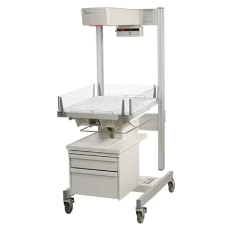
Instruments and supplies: a cesarean tray (a basic abdominal gynecologygy-nuh-KOL-uh-jeeThe branch of medicine that cares for the female reproductive organs and breasts.gynec/o woman-logy study ofFull entry set with bandage scissors, a bladder retractorrih-TRAK-terA surgical instrument that holds back tissue or organs so the surgeon can see and reach the operative site.Full entry , Pennington and Foerster forcepsFOR-sepsA hand instrument with two blades or arms used to grasp, hold or pull tissue or objects.Full entry ), delivery forceps and a head extractor on hand; a #10 blade; laps and sponges; two suctions with Yankauer tips; bulb syringes, one for each baby; a DeLee suction catheterSUK-shun KATH-uh-terA thin, flexible tube attached to suction, used to remove mucus and other secretions from the airway.Full entry ; cord clamps, two for each baby; cord blood tubes and a cord pH kit or blood gas syringes; and sutureSOO-cherA stitch, or the thread used to stitch, that holds the edges of a wound or incision together.Full entry for hemostasishee-moh-STAY-sisThe stopping of bleeding, either by the body's own clotting process or by medical or surgical methods.hem/o blood-stasis stopping, standing stillFull entry , the uterus, the bladder flap, the peritoneumpair-ih-toh-NEE-umThe thin membrane that lines the belly cavity and covers most of the organs inside it.peri- aroundton stretched-eum a structureFull entry , the fat and the skin.
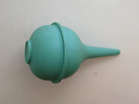
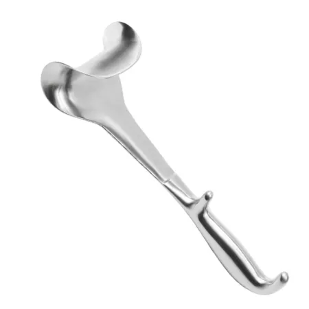
The name DeLee is on three things you may hand at a cesarean: a bladder retractor, an infant suction catheter and an obstetric forceps. Say which one you mean.
The emergency cesarean
An emergency cesarean, such as for a prolapsed cord or an abruption, can move from decision to incision in minutes. The patient may arrive with fetal monitoring in place and labor and delivery staff alongside. There may be only a few minutes to get ready, and often no time for a normal setup. As soon as you are gowned and gloved, set up what opens the abdomen and delivers the baby:
- a laparotomy drape
- a scalpel
- laps
- four to six Mayo clamps, and hemostatHEE-moe-statA clamp with finger rings and a locking ratchet used in surgery to grasp and close off bleeding vessels.hem/o blood-stat stopping, standing stillFull entry such as Kelly or Crile clamps
- Metzenbaum, curved Mayo and bandage scissors
- a bladder retractor (a DeLee or a Balfour bladder blade)
- two abdominal suctions and a bulb syringe
Keep this setup neat: in an emergency, the surgeon needs to find and get every item without a pause. There may be no time for the initial count, or for a full skin prep. If the initial count is waived, the team follows the facility's protocol for a waived count: the omission is documented on the operative record and an incident reportIN-sih-dent rih-PORTA written record of an unexpected event in a health care setting that caused or could have caused harm.Full entry explains why, later counts are done as the situation allows, including at closure, and an x-ray may be taken at the end to show that nothing was left behind. A checklist for emergency cesareans helps the team. The patient's support person waits in the surgical waiting area and may go with the baby to the nursery.
Incisions
Skin: a low transverse Pfannenstiel incision, the most common, or a vertical midlineMID-lineAn imaginary line down the center of the body; also, a type of IV catheter placed in a vein of the upper arm.Full entry incision below the umbilicus. It must be long enough to deliver the baby, so its length depends on the baby's size. Chapter 29 teaches the incisions.
Uterus:
- Kerr (low transverse): a crosswise cut in the thin lower segment of the uterus, about 2 cm above the bladder, the usual uterine incision. Its scar heals better and is far less likely to rupture in a later pregnancy.
- Classical: a vertical cut in the midline of the thick upper uterus, over the fundusFUN-dusThe rounded top or base of a hollow organ, most often the top of the uterus.Full entry , now seldom chosen. The assistant presses on its cut edges to control bleeding, it is closed in at least three layers, and its scar is much more likely to rupture in a later pregnancy, so a previous classical incision is a reason for a repeat cesarean.
- T incision: a low transverse cut extended upward in the midline; uncommon.
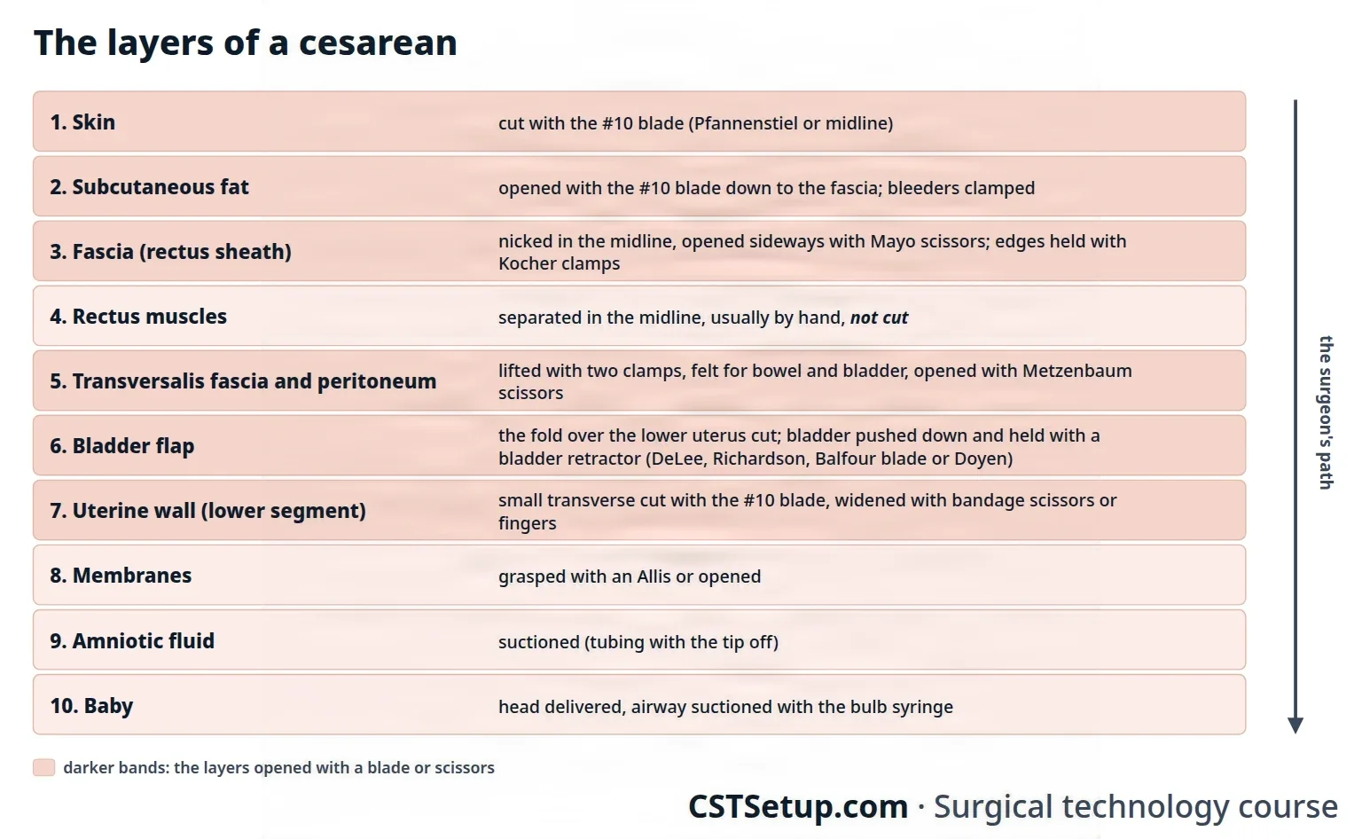
Key steps
- Skin and fat: the surgeon cuts the skin with a #10 blade to the fasciaFASH-ee-uhA sheet or band of tough connective tissue that covers and separates muscles and organs.fasci/o bandFull entry ; bleeders may be clamped and left untied until later, to save time.
- Fascia: the fascia is cut in the midline and opened to the sides with Mayo scissors; Kocher clamps lift its edges while it is freed from the rectusREK-tusLatin for straight; the name of several straight muscles, such as the rectus abdominis of the abdomen and the rectus femoris of the thigh.Full entry muscles above and below.
- Muscle: the rectus muscles are separated in the midline, usually by hand, to reach the peritoneum. The Core Curriculum's outline says they are divided.
- Peritoneum: two clamps lift the peritoneum, the surgeon feels it to make sure no bowel, omentum or bladder is caught, and opens it with Metzenbaum scissors.
- Bladder flap: the surgeon feels the uterus for the baby's position, cuts the fold of peritoneum over the lower uterus, and pushes the bladder down. A bladder retractor (a DeLee, a Richardson, a Balfour bladder blade or a Doyen) holds it out of the way.
- Uterus: a small cut with the #10 blade, widened with bandage scissors or the surgeon's fingers. Bring the suction (with its tip off, as many surgeons prefer), the bulb syringe and the bandage scissors close.
- Membranes: if still whole, they are grasped with an Allis or opened, and the amniotic fluidam-nee-OT-ik FLOO-idThe fluid inside the amniotic sac that surrounds and protects the baby during pregnancy.Full entry is suctioningSUK-shun-ingRemoving mucus, saliva or other secretions from the mouth, nose or airway with a tube connected to a suction source.Full entry off. Note its color: meconiummih-KOH-nee-umA newborn's first stools: thick, sticky and dark green to black.Full entry , the baby's first stool, normally passed only after birth, stains the fluid when the baby passes it early, often under stress, and the baby's airway then needs suctioning.
- The head: every retractor comes out of the wound, and sharp and metal objects come off the field, before the head is lifted out. The surgeon delivers the head by hand, or with forceps or a vacuum cup.
- The airway: as soon as the head is out, the mouth and nose are suctioned with the bulb syringe or a DeLee catheter.
- Fundal pressure: the assistant presses down on the top of the uterus through the abdomen to deliver the shoulders and body.
- The cord: the cord is clamped twice and cut between the clamps with curved Mayo or bandage scissors. With twins or more, each cord is marked differently so each cord blood sample matches its baby.
- The baby: hand the baby to the neonatal team member, who takes it in a sterile towel or sheet to the warmer, where its Apgar scoreAP-gar skorA 0-to-10 rating of a newborn's condition at 1 and 5 minutes after birth, based on five signs.Full entry is taken. Watch the nonsterile team's movements around your field.
- Cord blood: hold the cord blood tubes ready; the surgeon loosens one clamp to fill them, and you cap them and pass them to the circulator. A second sample may be taken for blood gases and pH.
- oxytocinok-sih-TOH-sinA hormone that makes the uterus contract and releases breast milk; also given by IV to start or strengthen labor and control bleeding after birth.Full entry : the anesthesia provider starts it in the IV fluid as the baby is delivered, so the uterus contracts and bleeds less; the surgeon may ask for an injection into the uterine muscle.
- The uterine edges: Pennington, Foerster ring, Duval or Allis clamps grasp the cut edges to control bleeding.
- The placenta: the surgeon delivers it by hand, or with gentle tractionTRAK-shunA steady pulling force applied to a body part with weights, ropes and pulleys.Full entry and fundal massage, into a large basin, and checks that it is whole. The inside of the uterus is wiped clean with a lap sponge.
- First closing count: before the uterus is closed, count sponges, sharps and other small items, because the uterus is a cavity within a cavity.
- The uterus: closed in one or two layers of absorbable suture, often running and locked or folded over (imbricated).
- The bladder flap is closed with a running absorbable suture, or may be left open. The pelvis is cleared of blood, clots, vernix caseosaVER-niks kay-see-OH-suhThe white, creamy, cheese-like coating on a newborn's skin.Full entry and fluid and may be irrigationeer-ih-GAY-shunWashing out a wound, body cavity, tube or eye with a steady flow of liquid.Full entry , and the tubes and ovaries are checked. A planned tubal ligation is done now.
- Second count, then the peritoneum, the muscle, the fascia and the fat are closed; a final count at skin closure; the skin is closed with an absorbable buried stitch or staples.
- The end: a dressing (4x4s and an abdominal pad, or an adhesive nonadherent dressing) and a perinealper-ih-NEE-ulHaving to do with the perineum, the area between the genitals and the anus.Full entry pad. The fundus is massaged and clots are pressed out of the vagina, which is cleaned.
Fundal pressure and the CST
Fundal pressure is a duty special to the cesarean. AST's position is that a CSTsee-es-tee (said as letters)Certified Surgical Technologist, a credential for surgical technologists; in maternity care, contraction stress test.Full entry from an accredited program is educated and competent to apply it under the direct superviseSOO-per-vyzeTo guide, watch over and check the work of another staff member who is doing a task.Full entry of the surgeon, who directs it, and that the facility's policy should require the CST's competenceKOM-puh-tunsHaving the knowledge, skill and judgment to do something well; in law, a person's ability to make their own decisions.Full entry to be documented, at hire and on an ongoing basis. Students learn it, with the first scrub role in cesareans, under a CST preceptor and the surgeon.
Medications
Oxytocin after the baby (into the vein by the anesthesia provider, or into the uterine muscle by the surgeon), other uterotonicyoo-ter-oh-TON-ikA drug that makes the uterus contract, used to start or strengthen labor or to control bleeding after birth.Full entry if the uterus will not contract, the antibiotic before the incision, and erythromycineh-rith-roh-MY-sinA macrolide antibiotic, also given to newborns as an eye ointment to prevent eye infection.Full entry 0.5% ointment for the baby's eyes. The medications section above has the details.
Counts
Counts are done before the incision; before the uterus is closed (a cavity within a cavity: sponges, sharps and small items, not instruments); when closure of the peritoneum and abdominal wall begins; and at skin closure. Ask for the uterine count as soon as you hand the first uterine suture: laps go inside the uterus to wipe it clean, so that count comes before the uterus is closed, not after. Chapters 28 and 29 teach counts.
Specimens, catheters and dressings
- Specimens: the cord blood samples, and the placenta with or without the cord. Facilities differ on whether every placenta goes to pathologypuh-THOL-uh-jeeThe study of disease and its causes and effects, or the disease changes found in a person or tissue.path/o disease-logy study ofFull entry ; follow your facility's policy.
- Catheters and drains: the urinary catheter, and a drain under the skin if needed.
- Dressing: 4x4s and an abdominal pad, or an adhesive, nonadherent dressing.
If a cell saver is used, the amniotic fluid is first suctioned away with a separate, clearly marked suction, and a leukocyte filter is used to reduce the fetal skin cells in the blood returned.
some references still teach that cell salvage is generally ruled out in a cesarean because of the amniotic fluid.
Wound class
Class II, clean-contaminated, because the uterus, part of the genital tract, is entered under control. The class rises, by the usual rules, with gross contamination or an infection already present. The Core Curriculum's outline lists a cesarean as Class I, Class II once the amniotic sac is broken and Class III with meconium, and one surgical technology textbook lists Class I with intact membranes.
After surgery
The patient goes to PACU. The return to normal activity takes about 6 to 8 weeks, and a cesarean makes a cesarean in a later pregnancy more likely.
Complications from the Core Curriculum's list:
- Hemorrhage from a uterus that will not contract, a placenta that will not separate, or a tear; it may end in an emergency hysterectomyhis-tuh-REK-tuh-meeSurgery to remove the uterus.hyster/o uterus-ectomy surgical removalFull entry (below).
- uterine ruptureYOO-tuh-rin RUP-churA tear through the wall of the uterus during pregnancy or labor.Full entry , most often a scar from an earlier cesarean giving way; like a hemorrhage, it can end in an emergency hysterectomy.
- Amniotic fluid embolismEM-buh-liz-umThe blocking of a blood vessel by a mass carried there in the blood (an embolus), such as a clot, fat or an air bubble.embol/o plug-ism conditionFull entry and disseminated intravascular coagulationdih-SEM-ih-nay-tid in-truh-VAS-kyuh-ler koh-ag-yuh-LAY-shunA disorder in which clotting is switched on throughout the blood vessels until the clotting factors are used up.intra- withinvascul/o vesselcoagul/o clottingFull entry , a clottingKLOT-ingThe process by which blood thickens into a gel to plug a damaged vessel and stop bleeding; also called coagulation.Full entry failure that AST's DIC guideline reports in more than half of these and of placentalpluh-SEN-tulRelating to the placenta, the organ that grows in the uterus in pregnancy and links the mother's blood supply to the baby's.Full entry abruptions.
- Injury to the patient (the bladder, bowel or ureterYOOR-uh-terOne of the two tubes that carry urine from the kidneys down to the bladder.Full entry ) or the baby, the death of the patient or the baby, and surgical site infection.
Postpartum hemorrhage
Bleeding of more than 500 mL in the first 24 hours after a birth is a postpartum hemorrhagepohst-PAR-tum HEM-uh-rijHeavy bleeding after birth: a total blood loss of 1,000 mL or more, or blood loss with signs of low blood volume, within 24 hours.post- afterpart/o birthhem/o blood-rrhage bursting forthFull entry , and 1,000 mL or more is a severe one. Blood loss is easy to underestimate, often by half, because it mixes with amniotic fluid and soaks into sponges, drapes and the floor, so measure what you can, such as by weighing the laps and reading the suction canisters, and report the amount. Its causes include uterine atonyYOO-ter-in AT-uh-neeWeak or absent tightening of the uterus muscle after childbirth.a- withoutton tone, tension-y conditionFull entry , a uterus that does not contract after the birth, a retained piece of placenta, a tear and a clotting problem. Chapter 12 teaches massive blood transfusionBLUD trans-FYOO-zhunGiving blood, or a part of blood, from a donor into a person's vein.trans- acrossfus/o pourFull entry .
The response steps up from the least invasive, and the team must be ready for each setup:
- Uterine massage and uterotonic drugs, oxytocin first (the medications section above).
- An intrauterinein-truh-YOO-ter-inInside the uterus.intra- withinuter uterus-ine relating toFull entry balloon tamponade: a balloon placed through the cervix and filled with warm saline to press on the bleeding wall.
- Compression sutures that wrap the uterus: have a long (36-inch) size 0 or 1 absorbable suture on a round-bodied needle ready.
- Ligation of the uterine or utero-ovarian arteries.
- If the bleeding still threatens life, an emergency hysterectomy, subtotal or total: have the heavy uterine clamps ready. A placenta that grows into the uterine wall and will not separate (placenta accreta), common at the site of an earlier cesarean scar, also ends in a hysterectomy.
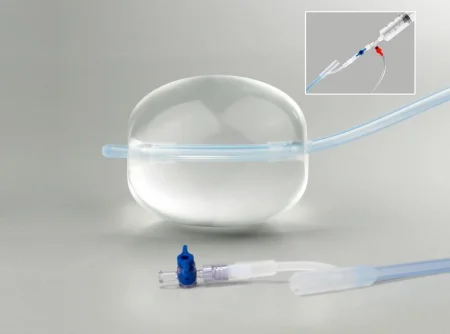
17Vaginal birth in the OR
A healthy vaginal birth, in a labor room, delivery room or birth center, seldom needs any intervention. A surgical technologistSUR-jih-kul tek-NOL-uh-jistA member of the operating room team who prepares the sterile field and instruments and hands them to the surgeon.Full entry in labor and delivery helps clamp and cut the cord, collects cord blood, supports the birthing patient, and gets ready for trouble with either patient. Vaginal deliveries count in your second scrub role.
Normal vaginal delivery
Labor has four stages: the first ends when the cervix is fully open (10 cm), the second with the birth of the baby, the third with the delivery of the placentapluh-SEN-tuhThe organ that grows in the uterus during pregnancy and passes oxygen and food from the parent's blood to the fetus.Full entry , and the fourth when the birthing patient is stable. As the head is born, the tissues of the vagina and perineumpair-ih-NEE-umThe area of skin and muscle between the genitals and the anus.perine/o perineumFull entry stretch and may tear. Once the baby is born, dried and the cord cut, the perineum is checked for tears; the placenta usually comes within 20 minutes, helped by gentle cord tractionTRAK-shunA steady pulling force applied to a body part with weights, ropes and pulleys.Full entry , an uterotonicyoo-ter-oh-TON-ikA drug that makes the uterus contract, used to start or strengthen labor or to control bleeding after birth.Full entry drug or breastfeeding, and then any tear or episiotomyeh-pee-zee-OT-uh-meeA cut made in the perineum, the tissue between the vagina and the anus, during birth to widen the opening.episi/o vulva, perineum-otomy cutting intoFull entry is repaired. For the first 2 hours, the patient's vital signs are checked often and the uterus is massaged until it is firm, because a firm uterus squeezes shut the vessels that fed the placenta.
Setup: a basic vaginal set with general instruments, right-angle retractorrih-TRAK-terA surgical instrument that holds back tissue or organs so the surgeon can see and reach the operative site.Full entry , Allis clamps, Kelly or Crile clamps, Metzenbaum and curved Heaney scissors, and needle holders; a placenta basin, cord clamps, cord blood tubes and a bulb syringe.
A breech presentationBREECH prez-en-TAY-shunA position of the fetus in which the buttocks or feet, instead of the head, are lowest in the uterus.Full entry baby born vaginally needs extra hands: to press above the pubisPYOO-bisThe front part of each hip bone, low in the pelvis; the two meet in the middle at the pubic symphysis.pub/o pubisFull entry to keep the head bent, to move the bed or the patient, and to resuscitationrih-sus-ih-TAY-shunEmergency treatment to restore breathing, heartbeat or blood circulation.Full entry the baby, which is often stressed and covered in thick meconiummih-KOH-nee-umA newborn's first stools: thick, sticky and dark green to black.Full entry .
Operative vaginal delivery: vacuum and forceps
When the second stage needs help, the obstetrician can guide the head out with a vacuum cup or obstetric forcepsFOR-sepsA hand instrument with two blades or arms used to grasp, hold or pull tissue or objects.Full entry . Both are used only when the baby is head first (or, for forceps, a few other positions), the cervix is fully open and the head is low in the pelvis; a vacuum also needs a full-term baby. Traction is applied only during contractions. If the head does not come down after three pulls, or the cup comes off twice, the attempt has failed: a failed vacuum is usually followed by forceps or a cesareansih-ZAIR-ee-unRelating to the birth of a baby through a surgical cut in the mother's abdomen and uterus.Full entry , and failed forceps by a cesarean.
- Vacuum: check every connection and test the vacuum on a gloved hand before you pass it. Scalp swelling under the cup is harmless and settles.
- Forceps: put them together before use to check that the blades fit and lock, and lubricate the blades. Types include the Simpson-Luikart, DeLee, Luikart and Kielland. Risks include injury to the baby's face, scalp or facial nerveFAY-shul NERVThe seventh cranial nerve, which moves the muscles of the face and carries taste from the front of the tongue.Full entry and tears of the birth canal.
Forceps or a vacuum may also be used through the uterine incision to lift the head at a cesarean.
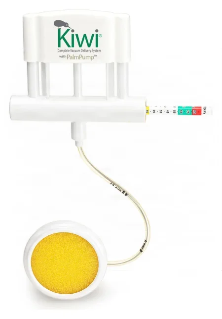
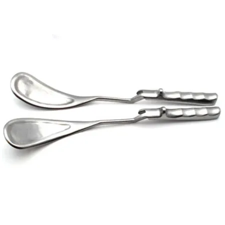
The newborn
The newborn is dried and stimulated at once and kept warm, at about 37°C (98.6°F), under the radiant warmer or skin to skin with a parent, covered; if all is normal and the patient agrees, the baby can stay on the patient's abdomen. The Apgar scoreAP-gar skorA 0-to-10 rating of a newborn's condition at 1 and 5 minutes after birth, based on five signs.Full entry , named for the anesthesiologist Virginia Apgar, rates five signs at 1 and 5 minutes after birth: heart rate, breathing, muscle tone, reflexREE-fleksAn automatic body response to something that stimulates it, which happens without thought, such as the knee jerk.Full entry response and color, each scored 0, 1 or 2. A total of 7 to 10 is normal, 4 to 6 means mild to moderate depression, and 0 to 3 severe depression.
Surgery during pregnancy
A pregnant patient may also need an operation unrelated to the pregnancy, most often for appendicitisuh-pen-dih-SY-tisInflammation of the appendix, the small pouch that hangs from the start of the large intestine.append/o appendix-itis inflammationFull entry or gallbladder disease. Necessary surgery is not put off because of the pregnancy. The team cares for two patients: the fetal heart rateFEE-tul hart raytThe number of times the unborn baby's heart beats per minute.Full entry is watched, the anesthesia provideran-us-THEE-zhuh pruh-VY-derThe licensed professional who gives anesthesia and watches over the client's breathing and vital functions during a procedure.Full entry uses a rapid inductionin-DUK-shunThe starting of a process on purpose, such as bringing on labor or putting a client under anesthesia.Full entry , drugs to stop labor are ready, and neonatal resuscitation equipment is in the room from the second trimester on. When the approach allows, a wedge under the right hip, or the table tilted about 15 degrees to the left, keeps the uterus from squeezing the vena cavaVEE-nuh KAY-vuhOne of the two large veins that return oxygen-poor blood from the body to the right atrium of the heart.Full entry and aorta. Ultrasound is the safest imaging. Chapter 18 teaches the pregnant patient as a person, and chapter 32 the gallbladder and appendixuh-PEN-diksA small, finger-shaped pouch attached to the first part of the large intestine, in the lower right abdomen.Full entry operations.
What to take from this chapter
- Obstetrics and gynecology reaches the female pelvis through the cervix, the vagina or the abdomen (open, laparoscopic or robotic). The approach sets the position (lithotomy, low lithotomy or supine), the prep (the vagina first, with separate kits, when both are prepped) and the tray.
- The ureter runs under the uterine artery, about 1 cm from the cervix, and near the infundibulopelvic ligament: the surgeon finds it before those are clamped. The cardinal ligaments are the uterus's main support and carry the uterine vessels.
- A D&C opens the cervix with graduated dilators after the uterus is sounded; the endocervical and endometrial specimens stay separate. In hysteroscopy, saline never goes with a monopolar electrode, and the fluid deficit is measured, reported and stopped at its limit.
- Every hysterectomy is Class II. When the vagina is opened from above, the instruments that touched the cervix and vagina go off the field with the specimen. Heaney clamps and a loaded suture ligature for every pedicle are the rhythm of the case.
- Placenta previa, abruptio placentae, a prolapsed cord, eclampsia and failure to progress can each turn a labor into an emergency cesarean in minutes: set up what opens the abdomen and delivers the baby first, and keep the setup neat. A ruptured ectopic pregnancy, with blood filling the abdomen, is an emergency too.
- In a cesarean, the patient lies with a wedge under the right hip; under general anesthesia everything is ready before induction; retractors come out before the head is lifted; the cord is clamped twice; and the first closing count comes before the uterus is closed. A cesarean is Class II.
- Oxytocin contracts the uterus after the birth, and Pitocin must never be confused with Pitressin (vasopressin). Postpartum hemorrhage is more than 500 mL; the response steps up from massage and uterotonics to a balloon, compression sutures, artery ligation and hysterectomy.
- Vacuum and forceps need a head-first baby, a fully open cervix and a low head, and stop after three pulls without progress. The Apgar score rates five signs at 1 and 5 minutes; 7 to 10 is normal.
Check yourself5 quick questions
Free for everyone. Pick an answer for each, then check them all at once.
In the ORWhere you'll use this
The chapter's whole question pool in sets of 10, with the answer explained after each one.
Part of the educators programThe chapter's words on spaced cards: the ones you miss come back sooner.
Part of the educators programGiven by your class's teacher. Each class draws its own 25 from the chapter's pool.
Part of the educators program
Suggest a fix
Something wrong, unclear or out of date in this chapter? Tell us and we will fix it for everyone. Your email is only used to reply.