General surgery
Gallbladders, appendixes, hernias, bowel, breast and thyroid: the core cases of general surgery, the anatomy under your instruments, and how to stay one step ahead of the surgeon.
1What general surgery covers
General surgery treats the abdomen and its organs, the abdominal wall, the breast, the thyroid and parathyroid glandpair-uh-THY-royd glandOne of four small glands behind the thyroid that control the amount of calcium in the blood.para- besidethyr/o thyroid gland-oid likeFull entry , and the anus and rectum, plus lumps in skin and soft tissue almost anywhere. It carries the largest case requirement of any specialty: accredited programs require at least 30 general surgery cases, 20 of them in the first scrub role.
The good news is that general surgery repeats itself. Almost every abdominal case opens and closes the same way, a short list of instruments does most of the work, and the same rules for counts, specimens and contamination apply every time. Learn the pattern once, and each new procedure is just a new middle.
Each case links to its full setup, with the instrument list and every step. Here you get the reasons and the moments when the scrub matters most.
2Anatomy that matters
The abdominal wall
Every abdominal case passes through the same layers, from outside in:
- Skin
- subcutaneoussub-kyoo-TAY-nee-usUnder the skin; in the fatty layer between the skin and the muscle.sub- undercutane/o skin-ous relating toFull entry fat
- fasciaFASH-ee-uhA sheet or band of tough connective tissue that covers and separates muscles and organs.fasci/o bandFull entry , called the rectusREK-tusLatin for straight; the name of several straight muscles, such as the rectus abdominis of the abdomen and the rectus femoris of the thigh.Full entry sheathSHEETHA covering that wraps around a structure; in procedures, a short hollow tube left in a blood vessel to pass catheters through.Full entry near the midlineMID-lineAn imaginary line down the center of the body; also, a type of IV catheter placed in a vein of the upper arm.Full entry
- Muscle: the two rectus abdominis muscles in front, and the external oblique, internal oblique and transversus abdominis at the sides
- Transversalis fascia and preperitoneal fat
- peritoneumpair-ih-toh-NEE-umThe thin membrane that lines the belly cavity and covers most of the organs inside it.peri- aroundton stretched-eum a structureFull entry , the thin membrane lining the cavity
The fascia holds a closure together, so its stitches are the strongest in the case. In the midline, the fascia from both sides fuses into the linea alba, a tough white band with few blood vessels. A midline incision goes through it without cutting muscle, which makes it fast and nearly bloodless.
The peritoneum lines the wall and wraps the organs. The mesentery is a fan-shaped fold of it that anchors the small intestine to the back wall and carries its vessels and lymph nodelimf nohdA small, bean-shaped organ of the immune system that filters lymph fluid and holds white blood cells.Full entry . The greater omentum is an apron of fat hanging from the stomach; it sticks to inflamed areas and often walls off infection. The kidneys, much of the duodenumdoo-uh-DEE-numThe first, short part of the small intestine, just after the stomach.Full entry and most of the pancreasPAN-kree-usA long, flat gland behind the stomach that makes digestive enzymes and the hormones insulin and glucagon.pan- allcreas fleshFull entry sit behind the peritoneum, in the retroperitoneum.
Of the four quadrants, the liver and gallbladder sit in the right upper, the spleenspleenAn organ in the upper left abdomen that filters the blood and helps fight infection.splen/o spleenFull entry in the left upper, and the appendixuh-PEN-diksA small, finger-shaped pouch attached to the first part of the large intestine, in the lower right abdomen.Full entry in the right lower.
The GI tract
The gastrointestinal (GI) tract is one long tube: esophagusih-SOF-uh-gusThe muscular tube that carries food and liquid from the throat to the stomach.esophag/o esophagusFull entry , stomach, small intestine (duodenum, jejunumjeh-JOO-numThe second of the three sections of the small intestine; it follows the duodenum and leads into the ileum.jejun/o jejunum (from the Latin for empty)Full entry , ileumIL-ee-umThe last and longest part of the small intestine, which joins the large intestine.Full entry ), then the large intestine. The ileum empties into the cecum, where the appendix hangs as a narrow blind tube. The colon then runs ascending, transversetrans-VERSLying across something, at a right angle to its length.Full entry , descending and sigmoidSIG-moydShaped like the letter S; most often short for the sigmoid colon, the S-shaped last part of the colon before the rectum.Full entry to the rectum and anal canal.
The wall has four layers: mucosamyoo-KOH-suhThe moist lining of body passages that open to the outside, such as the mouth, nose, gut and bladder.Full entry (the lining), submucosa (the strong layer that holds stitches), muscle, and serosa (the outer covering). The space inside is the lumenLOO-menThe open space inside a tube, such as a blood vessel, the intestine or a catheter.Full entry . The colon is full of bacteria, which is why opening the bowel changes how you handle instruments.
Blood reaches the bowel through the mesentery. The celiac trunk feeds the stomach, liver, gallbladder and spleen. The superior mesenteric artery feeds the small bowel, the right colon and most of the transverse colon. The inferior mesenteric artery feeds the left colon, sigmoid and upper rectum. When a piece of bowel comes out, its mesentery is divided with it; for cancer, a wedge of mesentery with its lymph nodes comes out too.
In the anal canal, the dentate line marks a change in the lining. Above it there are almost no pain nerves; below it the skin is very sensitive. That is why internal hemorrhoidHEM-uh-roydA swollen vein in or around the anus or lower rectum.Full entry bleed without pain and external ones hurt.
Liver, gallbladder and biliary tree
The liver fills most of the right upper quadrant. It has right and left lobes, eight segments by blood supply, and two blood sources: the hepatichih-PAT-ikRelating to the liver.hepat/o liver-ic relating toFull entry artery and the portal vein, which carries blood from the gut. It drains through the hepatic veins into the inferior vena cavain-FEER-ee-er VEE-nuh KAY-vuhThe large vein that carries blood from the lower body back to the right atrium of the heart.inferior lowervena veincava hollowFull entry . It is soft and bleeds easily.
The liver makes bilebileA yellow-green fluid made by the liver and stored in the gallbladder that helps digest fats.chol/e bileFull entry , which leaves through the right and left hepatic ducts. They join as the common hepatic duct. The gallbladder stores bile and connects by the cystic duct. Below that junction the tube is the common bile ductKOM-un BILE DUKTThe tube that carries bile from the liver and gallbladder into the small intestine.Full entry , which passes behind the duodenum, meets the pancreatic duct, and empties into the duodenum at the ampulla of Vater, guarded by the sphincterSFINGK-terA ring of muscle that tightens to close an opening or passage in the body and relaxes to open it.Full entry of Oddi.
The gallbladder has a fundusFUN-dusThe rounded top or base of a hollow organ, most often the top of the uterus.Full entry (the rounded end), a body, an infundibulum and a neck that narrows into the cystic duct. Its cystic artery usually branches from the right hepatic artery. The cystic duct and artery lie in the Calot triangle, bounded by the cystic duct, the common hepatic duct and the underside of the liver (many surgeons call it the hepatocystic triangle). It is the most important ground in a gallbladder operation, because the common bile duct runs right beside it.
Gallstones (cholelithiasiskoh-lee-lih-THY-uh-sisStones in the gallbladder.chol/e bilelith/o stone-iasis conditionFull entry ) form mostly from cholesterol or bile pigment. A stone stuck in the gallbladder neck causes inflammation (cholecystitiskoh-luh-sis-TY-tisInflammation of the gallbladder.chol/e bilecyst/o sac, bladder-itis inflammationFull entry ). A stone in the common bile duct blocks bile and causes jaundiceJAWN-disYellow color of the skin, the whites of the eyes and other tissues from too much bilirubin in the blood.Full entry , and if it blocks the pancreatic duct, pancreatitispan-kree-uh-TY-tisInflammation of the pancreas, which may come on suddenly (acute) or last for years (chronic).pancreat/o pancreas-itis inflammationFull entry .
Pancreas and spleen
The pancreas lies behind the stomach, its head in the C-shaped curve of the duodenum and its tail toward the spleen. Its ducts carry digestive enzymeEN-zimeA protein that speeds up a chemical reaction in the body without being used up.Full entry to the duodenum, and its islets release insulinIN-suh-linA hormone made by the pancreas that lets the body's cells take in glucose, also made as a medicine.Full entry (beta cellBAY-tuh SELA cell in the pancreas that makes and releases insulin.Full entry ) and glucagonGLOO-kuh-gonA hormone made by the pancreas that raises blood sugar; also given as a drug for severe low blood sugar.Full entry (alpha cells) into the blood.
The spleen sits under the left lower ribs, behind the stomach. It filters old red blood cells and germs from the blood. It is soft, full of blood and easily torn in trauma. Without a spleen, the body clears certain bacteria poorly, so patients get vaccines after a splenectomy.
Breast
The breast lies over the pectoralis major muscle, from about the second to the sixth rib, with a tail reaching into the axilla. It is made of 15 to 20 lobes of glands that drain through ducts to the nipple. Most of its lymphLIMFThe clear fluid that flows through the lymphatic vessels and carries white blood cells.Full entry drains to the axillary lymph nodes, grouped as level I (lateral to the pectoralis minor), level II (behind it) and level III (medial to it). Breast cancer usually spreads to these nodes first.
Two axillary nerves must be spared: the long thoracicthuh-RAS-ikRelating to the chest, or to the middle part of the spine.thorac/o chest-ic relating toFull entry nerve to the serratus anterior (injury leaves a "winged" shoulder blade) and the thoracodorsal nerve to the latissimus dorsi.
Thyroid and parathyroids
The thyroid is a butterfly-shaped gland in the front of the neck. Its two lobes sit beside the tracheaTRAY-kee-uhThe windpipe: the tube that carries air from the throat to the lungs.Full entry , joined by the isthmus across the second to fourth tracheal rings. It makes thyroid hormoneTHY-royd HOR-mohnA hormone made by the thyroid gland that controls how fast the body uses energy.Full entry (T3tee-THREETriiodothyronine: the more active of the two main thyroid hormones, or the blood test that measures it.Full entry and T4) and calcitoninkal-sih-TOH-ninA hormone made by the thyroid gland that lowers blood calcium; a lab-made form is used as a drug.Full entry . Strap muscles cover it, and the platysma lies under the skin.
Two structures make thyroid surgery delicate:
- The recurrent larynxLAIR-inksThe voice box: the part of the airway in the neck, above the windpipe, that holds the vocal cords.laryng/o larynxFull entry nerve runs up the groove between the trachea and esophagus, behind the thyroid, and moves the vocal cords. Injury on one side causes hoarseness; on both sides it can close the airway.
- Each parathyroid glandpair-uh-THY-royd glandOne of four small glands behind the thyroid that control the amount of calcium in the blood.para- besidethyr/o thyroid gland-oid likeFull entry is a tiny tan gland on the back of a thyroid lobe, usually four in all. They make parathyroid hormonepar-uh-THY-royd HOR-mohnA hormone from the parathyroid glands in the neck that raises the calcium level in the blood.para- besidethyr/o thyroid-oid resemblingFull entry , which controls blood calcium. If they are removed or lose their blood supply, calcium drops: tingling around the mouth and fingers, then cramps.
3Incisions, position, prep and drape
Incisions
- midlineMID-lineAn imaginary line down the center of the body; also, a type of IV catheter placed in a vein of the upper arm.Full entry : through the linea alba. Fast, bloodless and easy to extend; the standard for exploration and trauma.
- Paramedian: vertical, just beside the midline.
- subcostalsub-KOS-tulBelow the ribs.sub- belowcost/o rib-al relating toFull entry (Kocher): along the lower ribs; right for open gallbladder and liver work, left for the spleenspleenAn organ in the upper left abdomen that filters the blood and helps fight infection.splen/o spleenFull entry .
- McBurney: a short oblique cut through the McBurney pointmik-BUR-nee pointA spot on the lower right abdomen about one-third of the way from the front of the hip bone to the navel.Full entry , one-third of the way from the anterior superior iliacIL-ee-akHaving to do with the ilium, the large, wing-shaped upper part of the hip bone.Full entry spine to the umbilicus, splitting the muscles along their fibers. The transversetrans-VERSLying across something, at a right angle to its length.Full entry version is the Rocky-Davis. Both are for open appendectomyap-en-DEK-tuh-meeSurgery to remove the appendix.append appendix-ectomy surgical removalFull entry .
- inguinalING-gwih-nulRelating to the groin, the fold where the thigh meets the lower abdomen.Full entry : oblique, just above the inguinal ligament, for groin herniaHER-nee-uhA bulge of an organ or tissue through a weak spot in the muscle or tissue wall that normally holds it in.Full entry .
- Pfannenstiel: low transverse, just above the pubisPYOO-bisThe front part of each hip bone, low in the pelvis; the two meet in the middle at the pubic symphysis.pub/o pubisFull entry .
- Port sites: 5 to 12 mm stab incisions for laparoscopylap-uh-ROS-kuh-peeSurgery or an exam done through small cuts in the belly, using a thin, lighted tube with a camera.lapar/o abdomen-scopy looking with an instrumentFull entry , usually one at the umbilicus.
Five cases on one abdomen. Tap where the surgeon cuts, and see the incision.
This lab runs in the browser with JavaScript on.
This browser could not start the 3D room. Everything the lab practices is in the chapter.
Position
Most cases are supine, with each arm on a padded armboard at less than 90 degrees or tucked. The table then tilts so gravity moves the bowel away from the target: head up (reverse Trendelenburg positionree-VERS tren-DEL-un-burg puh-ZISH-unA position lying on the back on a bed or table that is tilted so the head is higher than the feet, with the body kept straight.Full entry ) for the gallbladder, stomach, spleen and thyroid; head down (Trendelenburg positiontren-DEL-en-burg puh-ZISH-unA position in which the client lies on the back with the bed tilted so the head is lower than the feet.Full entry ) for the appendixuh-PEN-diksA small, finger-shaped pouch attached to the first part of the large intestine, in the lower right abdomen.Full entry and pelvis; and a side tilt with the target side up. Anorectal cases use prone jackknife, lithotomylih-THOT-uh-meeA position lying on the back with the hips and knees bent and the legs raised and spread apart, often in stirrups.lith/o stone-tomy cutting intoFull entry or left lateral. Tilted patients need a safety strap and, for steep tilts, an anti-slide pad.
Four laparoscopic cases. Tilt the table so the bowel falls away from the target, then look inside.
This lab runs in the browser with JavaScript on.
This browser could not start the 3D room. Everything the lab practices is in the chapter.
Prep and drape
- Abdomen: nipple line to upper thighs, table to table. Clean the umbilicus first.
- Groin: above the umbilicus to the upper thigh; genitals, if included, last.
- Breast: base of the neck to below the breast (to the umbilicus for a mastectomyma-STEK-tuh-meeSurgery to remove all or part of a breast.mast/o breast-ectomy surgical removalFull entry ), past the midline to the table on the operative side, with the axilla and upper arm. For a mastectomy the arm is often prepped all around and draped free in stockinette.
- Neck: lower lip to nipples, including the shoulders.
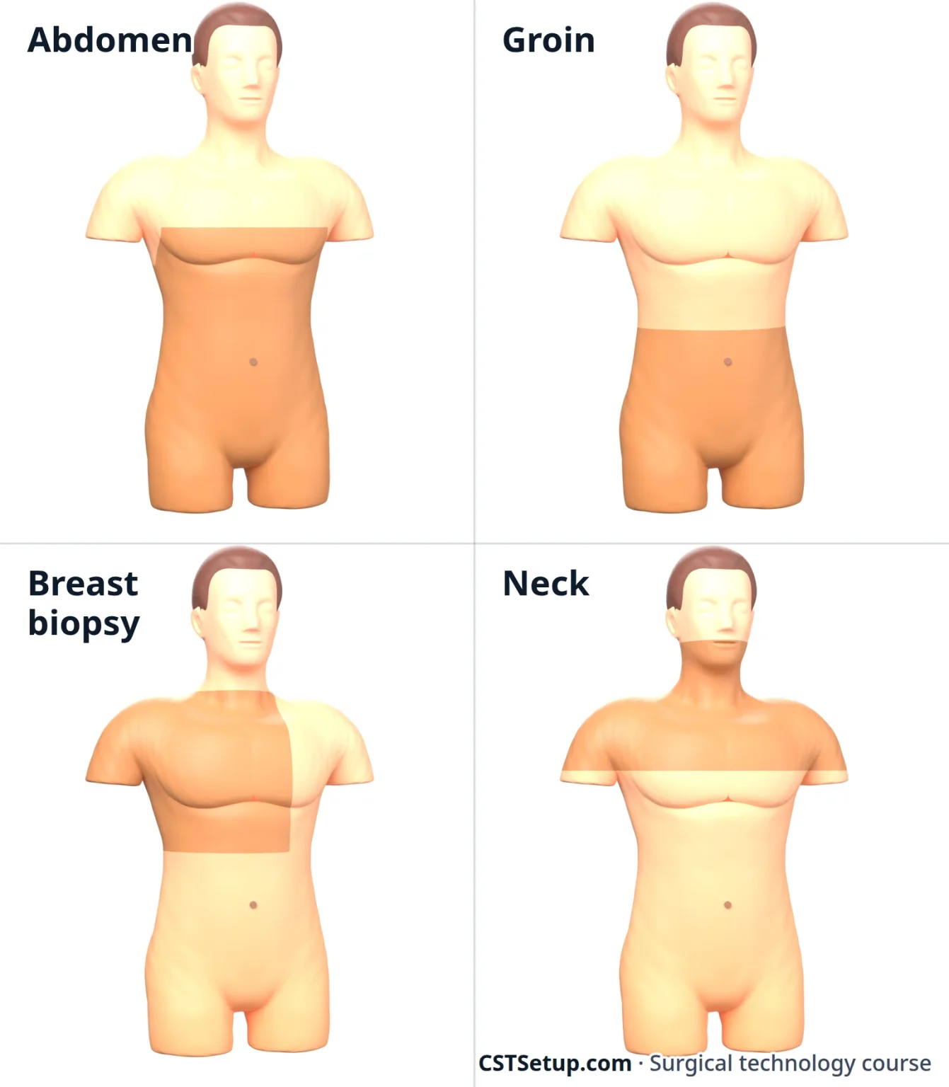
Draping is usually four towels to square off the site and a fenestrated sheet: a laparotomylap-uh-ROT-uh-meeSurgery that opens the abdomen through a large cut to reach the organs inside.lapar/o abdomen-tomy cutting intoFull entry sheet for the abdomen and groin, a breast or laparotomy sheet with an arm drape for the breast, and a neck sheet for the thyroid.
4Trays and instruments
- Basic or minor tray: short instruments for herniaHER-nee-uhA bulge of an organ or tissue through a weak spot in the muscle or tissue wall that normally holds it in.Full entry , breast biopsies and lumps: Army-Navy, Senn and Weitlaner retractorrih-TRAK-terA surgical instrument that holds back tissue or organs so the surgeon can see and reach the operative site.Full entry , hemostatHEE-moe-statA clamp with finger rings and a locking ratchet used in surgery to grasp and close off bleeding vessels.hem/o blood-stat stopping, standing stillFull entry , Allis and Babcock clamps, scissors, forcepsFOR-sepsA hand instrument with two blades or arms used to grasp, hold or pull tissue or objects.Full entry and needle holders.
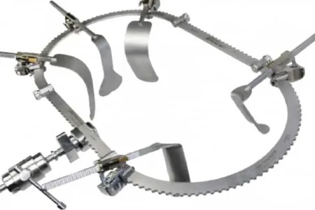
- Major laparotomylap-uh-ROT-uh-meeSurgery that opens the abdomen through a large cut to reach the organs inside.lapar/o abdomen-tomy cutting intoFull entry tray: the base for every open abdomen. It adds longer clamps (Pean, Kocher, right angle), deep retractors (Richardson, Deaver, Harrington, ribbon) and a self-retaining retractor such as the Balfour.
- Add-ons: long instruments; a GI tray with intestinal clamps; a bilebileA yellow-green fluid made by the liver and stored in the gallbladder that helps digest fats.chol/e bileFull entry tray with stone forceps and duct dilatorDY-lay-terAn instrument used to stretch or widen an opening, canal or passage in the body.Full entry ; a thyroid tray; an anorectal tray with rectalREK-tulRelating to the rectum, the last part of the large intestine; also, given or measured through the rectum.rect/o rectum, straight-al relating toFull entry speculumSPEK-yoo-lumAn instrument used to open and hold open a body opening, such as the vagina, so the inside can be seen.Full entry and retractors.
- laparoscopylap-uh-ROS-kuh-peeSurgery or an exam done through small cuts in the belly, using a thin, lighted tube with a camera.lapar/o abdomen-scopy looking with an instrumentFull entry tray: Veress needle, trocarTROH-karA sharp or bladed instrument inside a tube (cannula), used to pierce a body wall and leave the tube in place as a port.Full entry , scopes, light cord, graspers, dissectors, scissors, hook electrode, suction-irrigator, clip appliers and a needle holder.
What the common instruments do:
- Kocher: toothed tip for fasciaFASH-ee-uhA sheet or band of tough connective tissue that covers and separates muscles and organs.fasci/o bandFull entry and tough tissue. Never on bowel that stays.
- Allis: short teeth; holds tissue being removed and tough edges.
- Babcock: rounded jaws with no teeth; lifts bowel and the appendixuh-PEN-diksA small, finger-shaped pouch attached to the first part of the large intestine, in the lower right abdomen.Full entry without crushing them.
- Right angle: reaches behind a vessel or duct so a tie can pass around it.
- Kelly and Pean: workhorse clamps for tissue that will be cut and tied.
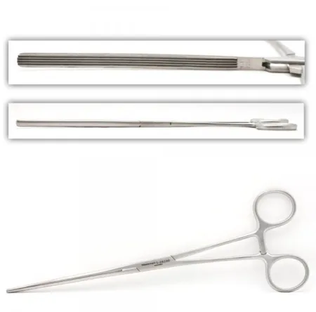
- Doyen: a noncrushing intestinal clamp that blocks the lumenLOO-menThe open space inside a tube, such as a blood vessel, the intestine or a catheter.Full entry without crushing the wall.
- Balfour and Bookwalter: self-retaining abdominal retractors. The Bookwalter is a ring on a table post that holds many blades.
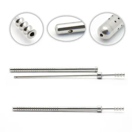
- Poole suction: a tip inside a sleeve full of holes, so a belly full of fluid empties without sucking up bowel. The sleeve comes off for a plain tip.
Once the peritoneumpair-ih-toh-NEE-umThe thin membrane that lines the belly cavity and covers most of the organs inside it.peri- aroundton stretched-eum a structureFull entry is open, use moist lap sponges for packing and keep any 4x4 sponge on a sponge stick; a loose small sponge is easily lost in an abdomen. Keep peanuts and Kittners on a clamp, and warm saline ready for laps and irrigationeer-ih-GAY-shunWashing out a wound, body cavity, tube or eye with a steady flow of liquid.Full entry .
5Energy and staplers
Energy devices
- Monopolar electrosurgeryee-LEK-troh-SUR-jer-eeUsing high-frequency electric current through a handheld tool to cut tissue or seal bleeding vessels during surgery.Full entry : cuts and coagulationkoh-ag-yuh-LAY-shunThe process by which blood changes from a liquid into a gel, forming a clot.coagul clot-ation processFull entry . Current flows through the patient to a return electrode pad on a well-padded muscle mass near the site, away from bone, scars and metal implants. Holster the pencil and keep the tip clean. laparoscopiclap-uh-roh-SKOP-ikDone through a few small cuts in the abdomen with a thin camera and long instruments, instead of one large opening.lapar/o abdomen-scopic viewingFull entry hooks are monopolar too.
- bipolarby-POH-lerHaving two poles or two opposite ends; most often used as a short form of bipolar disorder.bi- twopolar having polesFull entry : current flows only between the two tips. Good for small vessels near nerves.
- Ultrasonic shears (such as Harmonic): a blade vibrates about 55,000 times a second, cutting and sealing with frictionFRIK-shunThe rubbing of one surface against another, such as skin against bed sheets.Full entry heat and no current through the patient. The blade stays hot after use, so keep it off tissue and drapes.
- Vessel sealers (such as LigaSure): squeeze tissue and pass controlled bipolar current to seal vessels up to about 7 mm, then a blade cuts the seal. They replace many clamp, cut and tie steps.
All of them make surgical smoke, so use a smoke evacuator. In laparoscopylap-uh-ROS-kuh-peeSurgery or an exam done through small cuts in the belly, using a thin, lighted tube with a camera.lapar/o abdomen-scopy looking with an instrumentFull entry , cracked insulation or a live hook touching another metal instrument can burnBURNAn injury to the skin or deeper tissues caused by heat, chemicals, electricity, radiation or friction.Full entry bowel out of view. Check insulation before you pass an instrument.
Staplers
- Linear cutter (GIA): lays staggered rows of staples on both sides and cuts between them. It divides bowel with both ends sealed and makes side-to-side joins.
- Linear (TA): staples with no cut, to close an end or an opening; the surgeon trims with a knife.
- Circular (EEA): joins two bowel ends, often low in the colon and rectum. The anvil goes in one end, the body in the other. Firing staples a ring and cuts a "donut" from each end.
- endoscopicen-doh-SKOP-ikDone with an endoscope, a thin tube with a light and camera passed into the body.endo- within-scopic viewingFull entry staplers: linear cutters that fit a 12 mm port, many with a bending tip.
Reload colors mark staple height for thin or thick tissue, and they differ by maker, so read the package. Load each reload on the field and keep track of every one used. After a circular stapler fires, both donuts should be complete rings. Show them to the surgeon; they usually go to pathologypuh-THOL-uh-jeeThe study of disease and its causes and effects, or the disease changes found in a person or tissue.path/o disease-logy study ofFull entry .
6Open and laparoscopic approach
Open surgery (laparotomylap-uh-ROT-uh-meeSurgery that opens the abdomen through a large cut to reach the organs inside.lapar/o abdomen-tomy cutting intoFull entry ) gives a full view and touch, at the price of a large incision, more pain and a longer stay. Minimally invasive surgery (laparoscopylap-uh-ROS-kuh-peeSurgery or an exam done through small cuts in the belly, using a thin, lighted tube with a camera.lapar/o abdomen-scopy looking with an instrumentFull entry or robotics) works through small ports while the team watches a monitor. Most gallbladders and appendixes, and many herniaHER-nee-uhA bulge of an organ or tissue through a weak spot in the muscle or tissue wall that normally holds it in.Full entry and colon cases, are done this way. Open surgery stays the choice for many emergencies, trauma and dense scar tissue.
Getting in
The abdomen is inflated with carbon dioxideKAR-bun dy-OK-sideA gas made by the body's cells as waste, carried in the blood to the lungs and breathed out.Full entry (insufflationin-suh-FLAY-shunBlowing a gas into a body cavity, such as carbon dioxide into the abdomen during laparoscopic surgery.Full entry ), creating a pneumoperitoneum, usually at 12 to 15 mm Hg in adults. Entry is one of three ways:
- Veress needle: a spring-loaded blunt stylet pops forward once the needle enters the cavity. Test the spring before you pass it. The surgeon may confirm placement with a saline drop test, then swaps the needle for a trocarTROH-karA sharp or bladed instrument inside a tube (cannula), used to pierce a body wall and leave the tube in place as a port.Full entry .
- Open (Hasson): a small incision to the fasciaFASH-ee-uhA sheet or band of tough connective tissue that covers and separates muscles and organs.fasci/o bandFull entry , two fascia stay sutureSOO-cherA stitch, or the thread used to stitch, that holds the edges of a wound or incision together.Full entry , the peritoneumpair-ih-toh-NEE-umThe thin membrane that lines the belly cavity and covers most of the organs inside it.peri- aroundton stretched-eum a structureFull entry opened under view, and a blunt Hasson cannulaKAN-yoo-luhA thin tube placed into the body or a body opening to deliver or remove fluid or air.Full entry tied to the stay sutures.
- Optical trocar: the scope rides inside a clear trocar, so the surgeon watches each layer.
The trocar makes the path; the cannula (port) stays, with a valve that holds the gas in.
The scrub's laparoscopy jobs
Pass off the light cord, the camera in its sterile sleeve, the insufflation tubing and the suction-irrigator. White balance the camera, and warm the scope or use anti-fog. If you drive the camera, keep the horizon level and the working tip centered.
Count everything that goes through a port, such as needles and pre-tied loop sutures; a dropped needle must be found before closing. Fascia at ports of 10 mm and larger is usually closed to prevent a port-site hernia.
The gas can raise the patient's CO2, reduce blood return to the heart and chill the patient. Leftover gas often causes shoulder pain afterward.
Robotic surgery and conversion
In robotic surgery the surgeon sits at a console. The scrub drapes and docks the arms, changes instruments, and knows the emergency undocking steps.
Any minimally invasive case can turn open because of bleeding, injury or unclear anatomy. Keep the open tray in the room, stay calm when the surgeon says "open", and count in everything you add.
7Exploratory laparotomy and bowel technique
An exploratory laparotomylap-uh-ROT-uh-meeSurgery that opens the abdomen through a large cut to reach the organs inside.lapar/o abdomen-tomy cutting intoFull entry opens the abdomen to find and fix a problem: trauma, a perforationper-fuh-RAY-shunA hole that goes all the way through the wall of an organ or tissue.per- throughforat bored (a hole)-ion processFull entry , a blockage, bleeding or unexplained pain. It is also the template for every open abdominal case.
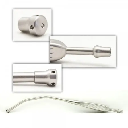
Position: supine, arms out. Prep and drape: full abdomen; towels and a laparotomy sheet. Setup: major laparotomy tray, long instruments and GI tray, self-retaining retractorrih-TRAK-terA surgical instrument that holds back tissue or organs so the surgeon can see and reach the operative site.Full entry , plenty of laps, Yankauer and Poole suction, warm irrigationeer-ih-GAY-shunWashing out a wound, body cavity, tube or eye with a steady flow of liquid.Full entry , cultures. Staplers, vascular clamps and hemostatichee-moh-STAT-ikStopping bleeding; also, a product or agent used to stop bleeding.hem/o blood-stasis stopping, standing still-ic relating toFull entry on hold.
Opening:
- Skin knife (often a #10 blade), then the electrosurgeryee-LEK-troh-SUR-jer-eeUsing high-frequency electric current through a handheld tool to cut tissue or seal bleeding vessels during surgery.Full entry through fat.
- fasciaFASH-ee-uhA sheet or band of tough connective tissue that covers and separates muscles and organs.fasci/o bandFull entry opened with ESU or knife; Kochers lift the edges.
- The peritoneumpair-ih-toh-NEE-umThe thin membrane that lines the belly cavity and covers most of the organs inside it.peri- aroundton stretched-eum a structureFull entry is lifted with two forcepsFOR-sepsA hand instrument with two blades or arms used to grasp, hold or pull tissue or objects.Full entry , nicked with Metzenbaum scissors and opened over the surgeon's fingers. From here on, laps only.
- The surgeon explores all four quadrants. Pass moist laps for packing, then the self-retaining retractor with laps under its blades.
For bleeding, think pressure first, then suction, clamps, ties and hemostatic agents. In damage-control trauma, the abdomen may be packed and closed temporarily; packs left in are documented.
Closing: irrigate with warm saline and suction with the Poole; remove retractor and packs; start the closing count before the peritoneum and fascia are closed; close fascia (many surgeons use a heavy looped running sutureSOO-cherA stitch, or the thread used to stitch, that holds the edges of a wound or incision together.Full entry , with retention sutures for high-risk patients); then skin with staples or sutures. A dirty wound's skin may be left open and packed.
Specimens: whatever is removed, plus cultures sent quickly and never in formalinFOR-muh-linA solution of formaldehyde in water used to preserve tissue specimens for the pathology lab.Full entry . Counts: initial, before closing the peritoneum, before closing the fascia, at skin closure, when items are added, and at any change of scrub or circulator. Dressing: sterile dressing and ABD pad, or negative pressureNEG-uh-tiv PRESH-erPressure that is lower than the pressure around it, so air or fluid is drawn in.Full entry therapy on an open wound.
Bowel technique
Once the GI lumenLOO-menThe open space inside a tube, such as a blood vessel, the intestine or a catheter.Full entry is open, its bacteria contaminate whatever touches it. Bowel technique keeps those germs out of the clean wound:
- It begins when the GI tract is opened and ends when it is closed. The team says aloud when the bowel is opened.
- Set up a separate dirty area, a second Mayo or a basin, for everything that touches open bowel, including staplers, suction tip, ESU tip and the specimen basin.
- Dirty items never return to the clean Mayo or back table. Fetch clean items without touching the clean setup, for example with a sponge stick kept for the purpose.
- Towels or a wound protector keep spillage off the wound edges.
- When the bowel is closed: isolate the dirty items where you can still count them, hand off the specimen, and change gowns and gloves, scrub first. Put new towels or a drape around the wound, a new suction tip and ESU tip, and close with clean instruments.
Wound class: I (clean) when there is no inflammation and the respiratory, GI, genital or urinary tract is not entered; II (clean-contaminated) when one of those tracts, the bilebileA yellow-green fluid made by the liver and stored in the gallbladder that helps digest fats.chol/e bileFull entry tract included, is entered under control; III (contaminated) with gross spillage from the GI tract, a major break in surgical asepsisSUR-jih-kul ay-SEP-sisPractices that keep an area or object completely free of microorganisms; also called sterile technique.a- withoutsepsis decay, infectionFull entry , acute inflammation without pus, or a fresh traumatic wound; IV (dirty) when pus, a perforation, or an old traumatic wound with dead tissue is already present.
The bowel is open. Set up the dirty side, sort what touched it, and close out clean.
This lab runs in the browser with JavaScript on.
This browser could not start the 3D room. Everything the lab practices is in the chapter.
8Gallbladder and appendix
Laparoscopic cholecystectomy
What and why: cholecystectomykoh-luh-sis-TEK-tuh-meeSurgery to remove the gallbladder.chol/e bilecyst/o sac, bladder-ectomy surgical removalFull entry removes the gallbladder, most often for painful gallstones, cholecystitiskoh-luh-sis-TY-tisInflammation of the gallbladder.chol/e bilecyst/o sac, bladder-itis inflammationFull entry or gallstone pancreatitispan-kree-uh-TY-tisInflammation of the pancreas, which may come on suddenly (acute) or last for years (chronic).pancreat/o pancreas-itis inflammationFull entry . Stones stuck in the common bile ductKOM-un BILE DUKTThe tube that carries bile from the liver and gallbladder into the small intestine.Full entry are often cleared before surgery by endoscopic retrograde cholangiopancreatographyen-doh-SKOP-ik RET-roh-grade koh-lan-jee-oh-pan-kree-uh-TOG-ruh-feeA procedure that uses a scope passed through the mouth, with dye and X-rays, to see and treat the bile and pancreatic ducts.endo- within-scopic viewingretrograde moving backwardcholangi/o bile ductpancreat/o pancreas-graphy making a pictureFull entry , done with a scope through the duodenumdoo-uh-DEE-numThe first, short part of the small intestine, just after the stomach.Full entry .
Position: supine, arms out or tucked. After entry, the reverse Trendelenburg positionree-VERS tren-DEL-un-burg puh-ZISH-unA position lying on the back on a bed or table that is tilted so the head is higher than the feet, with the body kept straight.Full entry with the right side up drops the bowel away. anesthesiaan-us-THEE-zhuhLoss of feeling or sensation, often brought on by drugs so that surgery or a procedure can be done without pain.an- withoutesthesi/o feeling, sensation-ia conditionFull entry often places an orogastric tube to empty the stomach.
Prep and drape: abdomen; laparotomylap-uh-ROT-uh-meeSurgery that opens the abdomen through a large cut to reach the organs inside.lapar/o abdomen-tomy cutting intoFull entry sheet.
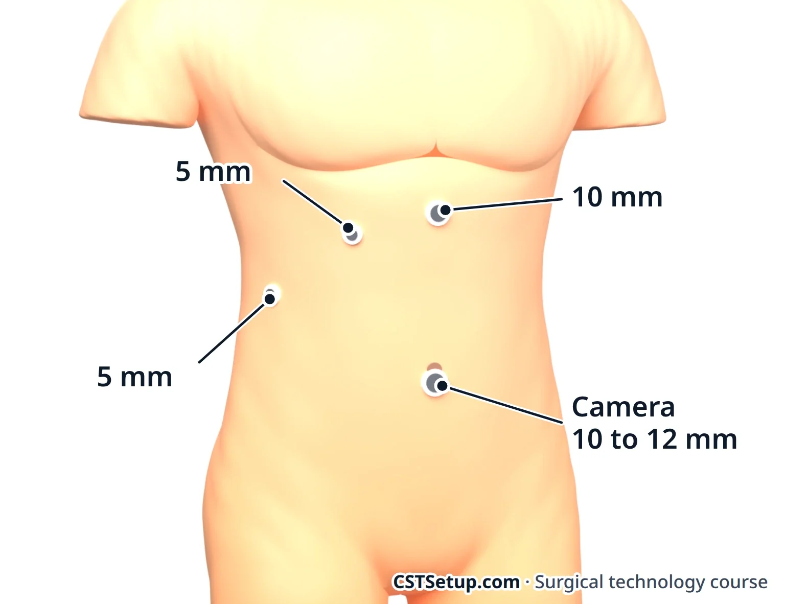
Setup: laparoscopylap-uh-ROS-kuh-peeSurgery or an exam done through small cuts in the belly, using a thin, lighted tube with a camera.lapar/o abdomen-scopy looking with an instrumentFull entry tray with a 30-degree or 0-degree scope, commonly two 10 to 12 mm and two 5 mm ports, graspers, Maryland dissector, hook, scissors, clip applier, suction-irrigator and retrieval bag. On hold: cholangiogram supplies, C-arm, lead aprons and an open tray.
Key steps:
- Umbilical entry and pneumoperitoneum; other ports under view, one high in the midlineMID-lineAn imaginary line down the center of the body; also, a type of IV catheter placed in a vein of the upper arm.Full entry and two below the right ribs.
- A locking grasper on the fundusFUN-dusThe rounded top or base of a hollow organ, most often the top of the uterus.Full entry pushes the gallbladder up toward the right shoulder; a second grasper pulls the infundibulum out to the side, opening the Calot triangle.
- The surgeon clears the triangle with the hook and Maryland until the critical view of safety: the triangle cleared of fat, the lower gallbladder lifted off the liver, and exactly two structures, the cystic duct and cystic artery, entering the gallbladder. Nothing is clipped before this.
- Clips go on the duct and artery, commonly two on the side that stays and one on the gallbladder side, and each is cut. Keep the clip applier loaded.
- Optional cholangiogram.
- The gallbladder is peeled off the liver bed, bagged and removed through the umbilical or upper port.
- irrigationeer-ih-GAY-shunWashing out a wound, body cavity, tube or eye with a steady flow of liquid.Full entry and check the bed and clips; release the gas; close fasciaFASH-ee-uhA sheet or band of tough connective tissue that covers and separates muscles and organs.fasci/o bandFull entry at 10 mm sites and the skin.
Cholangiogram: dye through a catheter in the cystic duct shows stones and the duct anatomy. Prepare labeled saline and contrastKON-trastA dye or other substance given before an imaging test so body parts show up more clearly.Full entry syringes (contrast diluted as ordered), a stopcock and the catheter, all free of air, because a bubble looks like a stone on the image. The team wears lead for the X-ray.
Specimen: the gallbladder in formalinFOR-muh-linA solution of formaldehyde in water used to preserve tissue specimens for the pathology lab.Full entry ; loose stones sent dry. Counts: initial and closing, including needles. Dressing: glue or strips. Wound class II; III with acute cholecystitis.
The worst complication is injury to the common bile duct, usually from mistaking it for the cystic duct. That is why the surgeon slows down at step 3, and why a quiet, ready scrub matters most there.
Appendectomy, open and laparoscopic
What and why: appendicitisuh-pen-dih-SY-tisInflammation of the appendix, the small pouch that hangs from the start of the large intestine.append/o appendix-itis inflammationFull entry starts when the appendixuh-PEN-diksA small, finger-shaped pouch attached to the first part of the large intestine, in the lower right abdomen.Full entry is blocked and infected. It can burst and cause peritonitispair-ih-tuh-NY-tisInflammation of the peritoneum, the thin membrane that lines the inside of the belly and covers the organs there.peritone/o peritoneum-itis inflammationFull entry , so removal is usually urgent. Most appendectomies are laparoscopiclap-uh-roh-SKOP-ikDone through a few small cuts in the abdomen with a thin camera and long instruments, instead of one large opening.lapar/o abdomen-scopic viewingFull entry .
Laparoscopic: supine, arms often tucked, Trendelenburg positiontren-DEL-en-burg puh-ZISH-unA position in which the client lies on the back with the bed tilted so the head is lower than the feet.Full entry with the right side up. Camera at the umbilicus; working ports often low in the midline and in the left lower quadrant. The surgeon follows the cecum to the appendix base, makes a window in the mesoappendix (which carries the appendix's artery), divides it with an energy device or a stapler with a vascular reload, then staples the base or ties it with pre-tied loops. The appendix leaves in a bag.
Open: a McBurney or Rocky-Davis incision; the external oblique opened and the muscles below split. The surgeon lifts the appendix with a Babcock, clamps and ties the mesoappendix, crushes and ties the base, and cuts the appendix off above the tie. Some surgeons bury the stump with a purse-string stitch. The knife, clamp and anything that touched the cut end go to the dirty basin: bowel technique on a small scale.
Prep and drape: abdomen; laparotomy sheet. Specimen: appendix in formalin; culture of any pus. Counts: initial and closing. Dressing: glue or strips on ports; dry dressing on an open wound, which may be left open after a rupture. Wound class: II if the appendix is not inflamed, III if inflamed without pus, IV if there is pus or it has ruptured (some textbooks class a ruptured appendix as III).
9Hernia repair
A herniaHER-nee-uhA bulge of an organ or tissue through a weak spot in the muscle or tissue wall that normally holds it in.Full entry is tissue, such as fat or bowel, bulging through a weak spot in the wall that should hold it in. It is reducible if it pushes back, incarcerated if it is stuck, and strangulated if its blood supply is cut off, which is an emergency.
Repairs either sew the edges (herniorrhaphy) or add mesh (hernioplasty). Mesh means less tension and fewer recurrencerih-KER-ensThe return of a disease or symptom after a period when it had gone away.re- againcur run-ence state ofFull entry , so most adult repairs use it. It is a permanent implant: open it when asked, handle it as little as possible, keep it off skin, and give the circulator the label for the implant record.
Inguinal hernia
The inguinalING-gwih-nulRelating to the groin, the fold where the thigh meets the lower abdomen.Full entry canal is a short tunnel just above the inguinal ligament. It runs from the deep ring to the superficial ring and carries the spermatic cord in males and the round ligament in females.
- Indirect: enters the deep ring and follows the cord, lateral to the inferior epigastric vessels. Often present from birth.
- Direct: pushes through the floor of the canal in the Hesselbach triangle, medial to the inferior epigastric vessels. The triangle is bordered by those vessels, the edge of the rectusREK-tusLatin for straight; the name of several straight muscles, such as the rectus abdominis of the abdomen and the rectus femoris of the thigh.Full entry muscle and the inguinal ligament. Comes with age and strain.
- femoralFEM-er-ulRelating to the thigh or the femur, the thigh bone.Full entry : passes below the inguinal ligament beside the femoral vein. Seen more often in women than in men, and the groin hernia most likely to strangulate.
Open mesh repair: supine; groin prep; laparotomylap-uh-ROT-uh-meeSurgery that opens the abdomen through a large cut to reach the organs inside.lapar/o abdomen-tomy cutting intoFull entry sheet. Often done under local anesthesiaLOH-kul an-es-THEE-zhuhNumbing medicine applied to or injected into one small area, so a procedure there is not felt while the person stays awake.Full entry with sedationsih-DAY-shunA calm, relaxed or sleepy state brought on by drugs, or the use of drugs to cause it.sed calm, settle-ation processFull entry , so keep voices low. Setup: minor tray, Penrose drain, mesh, local anestheticLOH-kul an-es-THET-ikA drug that blocks feeling in one part of the body without making the person unconscious.Full entry . Steps: inguinal incision; the external oblique aponeurosis opened while the ilioinguinal nerve is protected; a moist Penrose looped around the cord and clamped; the sac freed, then reduced or tied off and removed; mesh sewn over the canal floor with a slit for the cord; layers closed.
laparoscopiclap-uh-roh-SKOP-ikDone through a few small cuts in the abdomen with a thin camera and long instruments, instead of one large opening.lapar/o abdomen-scopic viewingFull entry or robotic repair: mesh goes behind the abdominal wall, covering the direct, indirect and femoral openings at once. In TAPP the surgeon enters the abdomen and opens a peritonealper-ih-toh-NEE-ulHaving to do with the peritoneum, the thin lining of the belly cavity and the organs inside it.Full entry flap; in TEP the surgeon stays outside the peritoneumpair-ih-toh-NEE-umThe thin membrane that lines the belly cavity and covers most of the organs inside it.peri- aroundton stretched-eum a structureFull entry , often opening the space with a balloon dissector. The mesh is tacked, glued or left unfixed. Trendelenburg positiontren-DEL-en-burg puh-ZISH-unA position in which the client lies on the back with the bed tilted so the head is lower than the feet.Full entry , arms tucked, bladder empty.
Specimen: sac, if removed. Counts: initial and closing, including the Penrose. Dressing: glue or strips. Wound class I.
Umbilical and ventral hernia
An umbilical hernia comes through the umbilical ring; many in infants close on their own. A ventralVEN-trulOn or toward the front, belly side of the body.ventr/o belly, front-al relating toFull entry hernia is any hernia of the front wall, and an incisional hernia forms through an old scar.
Umbilical repair: supine; minor tray; a curved incision beside the umbilicus. The sac is freed and reduced and the fasciaFASH-ee-uhA sheet or band of tough connective tissue that covers and separates muscles and organs.fasci/o bandFull entry closed with heavy permanent or slowly absorbable stitches; many surgeons add mesh for a larger hole. The navel skin is tacked back to the fascia. A small pressure dressing often goes in the navel.
Open ventral or incisional repair: wide prep; major tray. The old scar is often removed, adhesionad-HEE-zhunA band of scar tissue that joins two surfaces inside the body that are normally separate.ad- towardhes stick-ion conditionFull entry freed and the fascia edges found. Mesh goes on top of the fascia (onlay), behind the rectus muscle or in the preperitoneal space (sublay), or inside the abdomen (intraperitoneal, sometimes called underlay). Large repairs may need drains and a binder. In laparoscopic or robotic repairs, mesh inside the abdomen needs a coated side that is safe against bowel, fixed with tacks and sutureSOO-cherA stitch, or the thread used to stitch, that holds the edges of a wound or incision together.Full entry brought through the wall with a suture passer.
Specimen: sac or scar. Counts: initial, before closing the peritoneum if opened, and at skin. Wound class I.
10Bowel resection and anastomosis
A bowel resectionBOW-ul ree-SEK-shunSurgery to remove a section of the small or large intestine.Full entry removes diseased intestine: cancer, diverticulitisdy-ver-tik-yuh-LY-tisInflammation or infection of one or more small pouches that bulge out from the wall of the colon.diverticul/o a small pouch-itis inflammationFull entry , Crohn diseasekrohn dih-ZEEZA long-term inflammatory bowel disease that can affect any part of the digestive tract, through the full thickness of its wall.Full entry or ulcerative colitisUL-ser-uh-tiv koh-LY-tisA long-term inflammatory bowel disease that affects the inner lining of the colon and rectum.ulcer sorecol/o colon-itis inflammationFull entry , a blockage, a twist or dead bowel. The ends are joined (an anastomosisuh-nas-tuh-MOH-sisA connection between two tube-shaped structures, such as two ends of bowel or two blood vessels, made by surgery or found naturally.Full entry ) or brought to the skin as a stomaSTOH-muhAn opening on the surface of the body made by surgery, such as the end of bowel brought out through the belly wall.stoma mouth, openingFull entry when joining is not safe. The model case is the open right hemicolectomy, for cancer of the cecum or ascending colon; the same steps apply to other resectionrih-SEK-shunSurgery to remove part of an organ or tissue.re- back, againsect to cut-ion processFull entry , open or minimally invasive.
Position: supine; lithotomylih-THOT-uh-meeA position lying on the back with the hips and knees bent and the legs raised and spread apart, often in stirrups.lith/o stone-tomy cutting intoFull entry when a circular stapler goes through the anus. Prep and drape: abdomen; any stoma site marked before surgery. Setup: major and GI trays, self-retaining retractorrih-TRAK-terA surgical instrument that holds back tissue or organs so the surgeon can see and reach the operative site.Full entry , energy device, GIA and TA staplers with reloads, intestinal clamps, a dirty setup, and a separate clean closing setup.
Key steps:
- laparotomylap-uh-ROT-uh-meeSurgery that opens the abdomen through a large cut to reach the organs inside.lapar/o abdomen-tomy cutting intoFull entry and exploration, including the liver in cancer cases.
- The colon is freed from the side wall; the surgeon protects the ureterYOOR-uh-terOne of the two tubes that carry urine from the kidneys down to the bladder.Full entry and duodenumdoo-uh-DEE-numThe first, short part of the small intestine, just after the stomach.Full entry .
- The mesentery and its vessels are divided by clamp, cut and tie or a vessel sealer. Pass clamps in pairs with ties right behind.
- The bowel is divided, usually with a GIA. Bowel technique starts when a lumenLOO-menThe open space inside a tube, such as a blood vessel, the intestine or a catheter.Full entry is opened.
- Anastomosis. Stapled: the ends lie side by side, a small opening is cut in each, a GIA fork goes into each limb and fires, and the leftover opening is closed with a TA or GIA. Hand-sewn: often two layers, an inner running absorbable stitch and outer interrupted silk.
- The gap in the mesentery is closed and the abdomen irrigationeer-ih-GAY-shunWashing out a wound, body cavity, tube or eye with a steady flow of liquid.Full entry .
- Bowel technique ends: dirty items out, new gowns, gloves and drapes, clean closure.
Stomas: a colostomykuh-LOS-tuh-meeA surgical opening that brings part of the colon through the belly wall, so stool leaves the body into a pouch.col/o colon-stomy surgical openingFull entry brings colon to the skin; an ileostomyil-ee-OS-tuh-meeA surgical opening that brings the end of the ileum through the belly wall, so stool leaves the body there.ile/o ileum-stomy surgical openingFull entry brings ileumIL-ee-umThe last and longest part of the small intestine, which joins the large intestine.Full entry . An end stoma uses one cut end; a loop stoma opens a loop. The appliance goes on in the room.
Specimen: the segment with its mesentery and nodes, often stitch-marked for orientation; donuts separately. Counts: initial, before closing the peritoneumpair-ih-toh-NEE-umThe thin membrane that lines the belly cavity and covers most of the organs inside it.peri- aroundton stretched-eum a structureFull entry , at skin, and the clean setup counted in. Dressing: dry dressing, ABD pad, stoma appliance. Wound class II, or III with spillage.
11Breast and thyroid procedures
Breast biopsy
What and why: an excisional biopsy removes a lump or suspicious area whole, with a rim of normal tissue; a lumpectomy does the same for a known cancer. lesionLEE-zhunAn area of tissue that has been damaged or has changed from normal, such as a sore, wound, lump or spot.Full entry that cannot be felt are marked first in radiology with a hooked wire or a tiny seed.
Position: supine, arm out. Prep and drape: chest and axilla, gently, so the wire stays put. Setup: minor tray, skin hooks, small retractorrih-TRAK-terA surgical instrument that holds back tissue or organs so the surgeon can see and reach the operative site.Full entry , electrosurgeryee-LEK-troh-SUR-jer-eeUsing high-frequency electric current through a handheld tool to cut tissue or seal bleeding vessels during surgery.Full entry , marking stitches or ink.
Key steps: the surgeon removes the tissue around the wire or lump in one piece and marks its sides with stitches of different lengths or ink. A wire or seed specimen goes for an X-ray to prove the lesion and its markerMAR-kerSomething that can be measured, such as a substance in the blood, that shows a disease or body process is present.Full entry clip are inside, and the field stays sterile until the answer comes back. Extra "shave" margins may be cut, each labeled by side.
Specimen: oriented and sent fresh right away, or into formalinFOR-muh-linA solution of formaldehyde in water used to preserve tissue specimens for the pathology lab.Full entry if facility policy says so, so it is fixed within an hour; delay can spoil the tests that guide cancer treatment. Counts: sponges and sharps; the wire counts as a sharp. Dressing: glue or strips, gauze, surgical bra.
Sentinel node and mastectomy
A sentinel lymph nodelimf nohdA small, bean-shaped organ of the immune system that filters lymph fluid and holds white blood cells.Full entry biopsy removes the first node or nodes that drain the tumor. If they are free of cancer, the rest of the axilla can usually be left alone. A radioactive tracer is injected before surgery, and the surgeon may inject blue dye in the room. "Hot" nodes are found with a gamma probe in a sterile cover, "blue" nodes by sight. Blue dye can cause allergic reactions, even anaphylaxisan-uh-fuh-LAK-sisA sudden, severe allergic reaction that affects the whole body.ana- away, against-phylaxis protectionFull entry , and can make the pulse oximeter read falsely low.
Each sentinel node goes in its own container, labeled exactly as the surgeon names it. Never put two nodes in one container.
A simple mastectomyma-STEK-tuh-meeSurgery to remove all or part of a breast.mast/o breast-ectomy surgical removalFull entry removes the breast tissue, nipple and areola. A modified radical mastectomy removes the whole breast with the level I and II axillary nodes and leaves the chest muscles. A radical mastectomy, which also removes the pectoral muscles, is rare today.
Modified radical mastectomy: supine, the arm out and often draped free. Setup: tray with skin hooks and rakes, ESU with spare tips, clip appliers and one or two closed suction drains. Steps: an elliptical incision around the nipple and tumor; skin flaps raised with the ESU while hooks or rakes lift the edges; the breast peeled off the pectoralis fasciaFASH-ee-uhA sheet or band of tough connective tissue that covers and separates muscles and organs.fasci/o bandFull entry ; the axillary nodes removed below the axillary vein, sparing the long thoracicthuh-RAS-ikRelating to the chest, or to the middle part of the spine.thorac/o chest-ic relating toFull entry and thoracodorsal nerves; drains placed and stitched to the skin. Reconstruction may follow on a separate setup.
Specimen: oriented with a stitch, sent fresh. Counts: initial and closing. Dressing: fluffs and a wrap or surgical bra, drains to bulbs. Many hospitals then avoid blood pressure cuffs and needles in that arm.
Thyroidectomy
What and why: a thyroidectomythy-roy-DEK-tuh-meeSurgery to remove all or part of the thyroid gland in the neck.thyr/o thyroid-ectomy surgical removalFull entry removes the whole gland (total) or one lobe (hemithyroidectomy) for cancer, suspicious noduleNOD-yoolA small, firm, rounded lump of tissue that can be felt or seen, in or under the skin or in an organ.Full entry , a goiterGOY-terAn enlarged thyroid gland, seen or felt as a swelling at the front of the neck.Full entry that presses on the tracheaTRAY-kee-uhThe windpipe: the tube that carries air from the throat to the lungs.Full entry or esophagusih-SOF-uh-gusThe muscular tube that carries food and liquid from the throat to the stomach.esophag/o esophagusFull entry , or an overactivityoh-ver-ak-TIV-ih-teeMore activity than normal, in a person, an organ or a body system.Full entry gland.
Position: supine, a roll under the shoulders to extend the neck, head on a donut, arms tucked, slight head-up tilt. Many teams use a breathing tube with sensors that warn when the recurrent larynxLAIR-inksThe voice box: the part of the airway in the neck, above the windpipe, that holds the vocal cords.laryng/o larynxFull entry nerve is touched, and a sterile stimulator probe on the field.
Prep and drape: lower lip to nipples; towels and a neck sheet. Setup: thyroid tray with fine clamps, right angles, neck retractors such as the Green, and a thyroid tenaculum such as the Lahey; bipolarby-POH-lerHaving two poles or two opposite ends; most often used as a short form of bipolar disorder.bi- twopolar having polesFull entry , ultrasonic shears or small vessel sealer; clips; peanuts.
Key steps: a low collar incision in a skin crease; flaps raised under the platysma; strap muscles separated in the midlineMID-lineAn imaginary line down the center of the body; also, a type of IV catheter placed in a vein of the upper arm.Full entry ; the lobe rolled inward. Vessels are tied, clipped or sealed close to the gland while the surgeon protects the recurrent laryngeal nerve and each parathyroid glandpair-uh-THY-royd glandOne of four small glands behind the thyroid that control the amount of calcium in the blood.para- besidethyr/o thyroid gland-oid likeFull entry . Closure: strap muscles and platysma, then a buried skin stitch or glue.
If a parathyroid gland comes out by accident, the surgeon may mince it and implant it in a muscle. Keep it on the field in a labeled cup of saline until the surgeon decides.
Specimen: labeled right or left, often oriented with a stitch; sometimes frozen section. Counts: initial and closing. Dressing: glue or strips. A neck hematomahee-muh-TOH-muhA collection of blood, often clotted, that has leaked out of the blood vessels into the tissue.hem/o blood (here as hemat-)-oma swelling or massFull entry can squeeze the airway, so many hospitals keep a sutureSOO-cherA stitch, or the thread used to stitch, that holds the edges of a wound or incision together.Full entry removal kit at the bedside.
12Hemorrhoidectomy and other cases
Hemorrhoidectomy
A hemorrhoidHEM-uh-roydA swollen vein in or around the anus or lower rectum.Full entry is a normal cushion of blood vessels in the anal canal that becomes a problem when it swells, bleeds or prolapsePROH-lapsThe slipping or falling of an organ or part from its normal position.Full entry . Surgery is for large or prolapsing hemorrhoids that office treatments such as banding did not fix.
Position: prone jackknife with the buttocks taped apart, lithotomylih-THOT-uh-meeA position lying on the back with the hips and knees bent and the legs raised and spread apart, often in stirrups.lith/o stone-tomy cutting intoFull entry or left lateral; in jackknife, pad the chest, knees and toes. Prep and drape: perianal; fenestrated or lithotomy sheet. Setup: anorectal tray with a Hill-Ferguson retractorrih-TRAK-terA surgical instrument that holds back tissue or organs so the surgeon can see and reach the operative site.Full entry or Pratt speculumSPEK-yoo-lumAn instrument used to open and hold open a body opening, such as the vagina, so the inside can be seen.Full entry , Allis clamps, Metzenbaum scissors, electrosurgeryee-LEK-troh-SUR-jer-eeUsing high-frequency electric current through a handheld tool to cut tissue or seal bleeding vessels during surgery.Full entry or energy device, lubricant, local anestheticLOH-kul an-es-THET-ikA drug that blocks feeling in one part of the body without making the person unconscious.Full entry with epinephrineep-ih-NEF-rinA hormone and drug, also called adrenaline, that raises heart rate and blood pressure and opens the airways.Full entry , absorbable sutureSOO-cherA stitch, or the thread used to stitch, that holds the edges of a wound or incision together.Full entry .
Key steps: each hemorrhoid is grasped, cut out in a narrow ellipse up into the canal, and tied off at its top. The wound is sewn closed or left open, by surgeon preference. Strips of normal lining stay between the cuts so the anus does not scar narrow.
Specimen: each hemorrhoid, labeled by position. Counts: sponges, sharps and any packing. Dressing: gauze or hemostatichee-moh-STAT-ikStopping bleeding; also, a product or agent used to stop bleeding.hem/o blood-stasis stopping, standing still-ic relating toFull entry sponge, fluffs, ABD pad, mesh underwear. Trouble passing urine is common afterward.
Other general surgery cases
These follow the same patterns:
- endoscopyen-DOS-kuh-peeA procedure that uses a thin, flexible tube with a light and camera to look inside the body.endo- within-scopy looking with an instrumentFull entry : EGDee-jee-DEE (said as letters)Esophagogastroduodenoscopy: a test using a flexible scope through the mouth to look at the esophagus, stomach and start of the small intestine.esophag/o esophagusgastr/o stomachduoden/o duodenum-scopy looking, viewingFull entry , colonoscopykoh-luh-NOS-kuh-peeAn exam of the inside of the large intestine with a long, flexible tube that has a camera on its end.col/o colon-scopy looking with an instrumentFull entry and endoscopic retrograde cholangiopancreatographyen-doh-SKOP-ik RET-roh-grade koh-lan-jee-oh-pan-kree-uh-TOG-ruh-feeA procedure that uses a scope passed through the mouth, with dye and X-rays, to see and treat the bile and pancreatic ducts.endo- within-scopic viewingretrograde moving backwardcholangi/o bile ductpancreat/o pancreas-graphy making a pictureFull entry look inside with flexible scopes.
- Gastrectomy removes part or all of the stomach; a gastrostomy places a enteral feeding tubeEN-ter-ul FEE-ding toobA tube placed into the stomach or small intestine to give liquid food, water and drugs to a person who cannot eat enough by mouth.Full entry .
- Nissen fundoplication wraps the top of the stomach around the lower esophagusih-SOF-uh-gusThe muscular tube that carries food and liquid from the throat to the stomach.esophag/o esophagusFull entry to stop reflux.
- Weight-loss surgery: a sleeve gastrectomy staples away most of the stomach; a Roux-en-Y gastric bypass joins a small pouch to the jejunumjeh-JOO-numThe second of the three sections of the small intestine; it follows the duodenum and leads into the ileum.jejun/o jejunum (from the Latin for empty)Full entry . Expect a bariatric table and long instruments.
- Liver resectionrih-SEK-shunSurgery to remove part of an organ or tissue.re- back, againsect to cut-ion processFull entry removes segments; expect blood loss.
- Whipple procedure (pancreaticoduodenectomy) removes the head of the pancreasPAN-kree-usA long, flat gland behind the stomach that makes digestive enzymes and the hormones insulin and glucagon.pan- allcreas fleshFull entry , the duodenumdoo-uh-DEE-numThe first, short part of the small intestine, just after the stomach.Full entry , the gallbladder and the end of the common bile ductKOM-un BILE DUKTThe tube that carries bile from the liver and gallbladder into the small intestine.Full entry , and in the classic operation the lower stomach, then rebuilds with three joins.
- Splenectomy, open or laparoscopiclap-uh-roh-SKOP-ikDone through a few small cuts in the abdomen with a thin camera and long instruments, instead of one large opening.lapar/o abdomen-scopic viewingFull entry , for trauma or blood disorders.
- Anorectal: fistulotomy lays open a tract between the anal canal and the skin, found with a probe; pilonidal cyst excisionek-SIZH-unCutting out tissue, such as a growth or damaged area, during surgery.Full entry is done prone; sphincteroplasty repairs a torn anal sphincterSFINGK-terA ring of muscle that tightens to close an opening or passage in the body and relaxes to open it.Full entry .
What to take from this chapter
- General surgery has the largest case requirement of any specialty. Learn the open abdomen once, and every case becomes a new middle.
- The fascia holds the closure, and the linea alba makes the midline incision fast and nearly bloodless.
- In a laparoscopic cholecystectomy, nothing is clipped until the critical view of safety shows only the cystic duct and cystic artery entering the gallbladder.
- Babcocks hold bowel and the appendix gently; Kochers and Allis clamps hold tougher tissue; noncrushing clamps protect bowel that stays.
- Bowel technique starts when the GI tract is opened and ends when it is closed: dirty stays dirty, then new gowns, gloves and tips for a clean closure.
- A GIA staples and cuts, a TA staples only, and a circular stapler joins two ends and leaves two donuts. Reload colors differ by maker, so read the package.
- Direct inguinal hernias come through the Hesselbach triangle, medial to the inferior epigastric vessels; indirect hernias come through the deep ring, lateral to them.
- Breast tissue goes oriented and fast (fresh unless facility policy says formalin); each sentinel node gets its own container; a parathyroid gland that comes out stays on the field until the surgeon decides.
Check yourself5 quick questions
Free for everyone. Pick an answer for each, then check them all at once.
In the ORWhere you'll use this
Setups to see it in
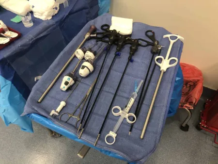 Laparoscopic CholecystectomyGeneral · 23 instruments · 6 photos
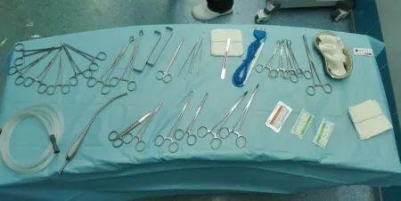 AppendectomyGeneral · 26 instruments · 1 photo
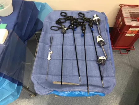 Laparoscopic AppendectomyGeneral · 29 instruments · 3 photos
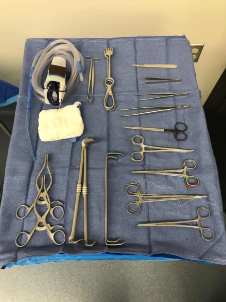 Inguinal Hernia RepairGeneral · 16 instruments · 2 photos
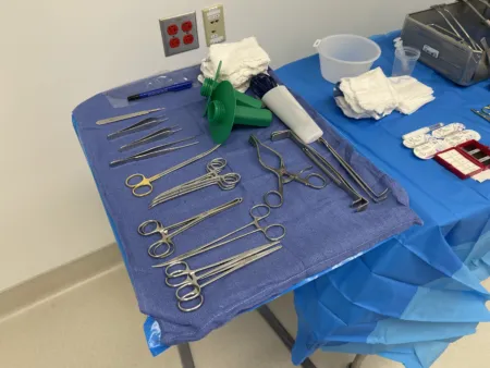 Umbilical Hernia RepairGeneral · 20 instruments · 3 photos Ventral (Incisional) Hernia Repair - OpenGeneral · 34 instruments
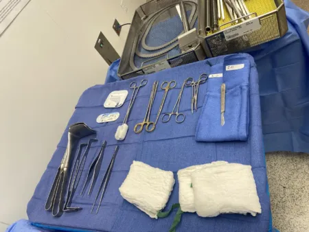 Exploratory LaparotomyGeneral · 32 instruments · 1 photo Open Right HemicolectomyGeneral · 40 instruments
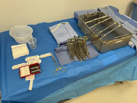 Breast BiopsyGeneral · 25 instruments · 3 photos Modified Radical MastectomyGeneral · 19 instruments
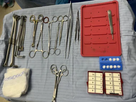 ThyroidectomyGeneral · 32 instruments · 1 photo
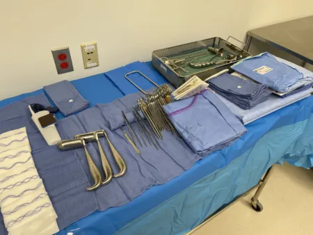 HemorrhoidectomyGeneral · 27 instruments · 3 photos
The chapter's instruments
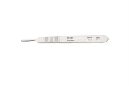 #3 Knife HandleCutting & Dissecting · On 319 setups
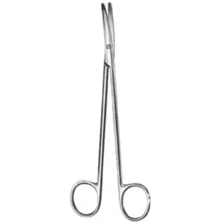 Metzenbaum ScissorCutting & Dissecting · On 271 setups
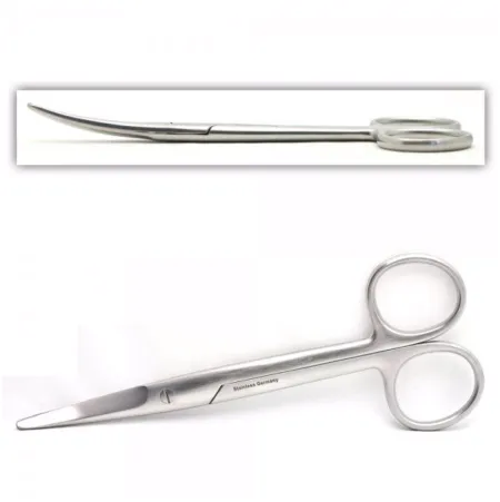 Curved Mayo ScissorCutting & Dissecting · On 121 setups
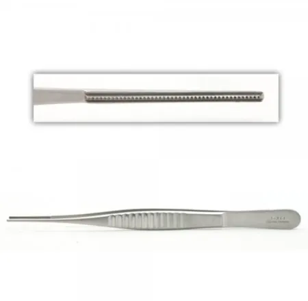 DeBakey ForcepsGrasping & Holding · On 159 setups
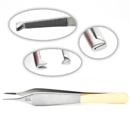 Adson ForcepsGrasping & Holding · On 308 setups
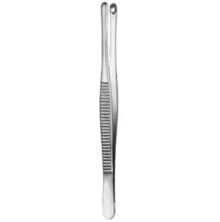 Russian Tissue ForcepsGrasping & Holding · On 77 setups
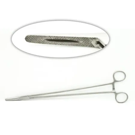 Mayo-Hegar Needle HolderSuturing & Stapling · On 257 setups
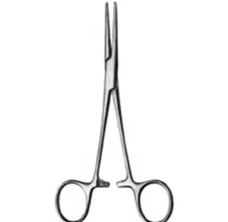 Kelly ForcepsClamping & Occluding · On 73 setups
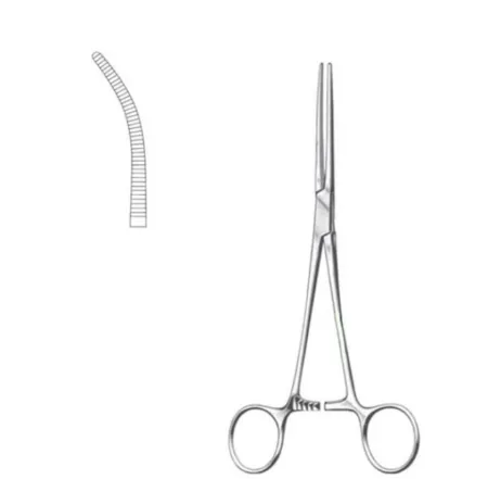 Rochester Pean ForcepsClamping & Occluding · On 92 setups
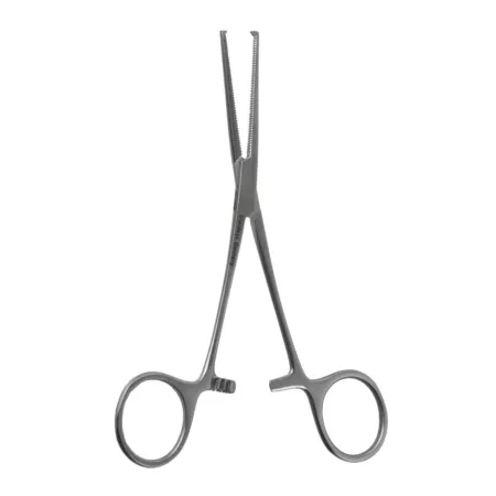 Kocher ForcepsClamping & Occluding · On 137 setups
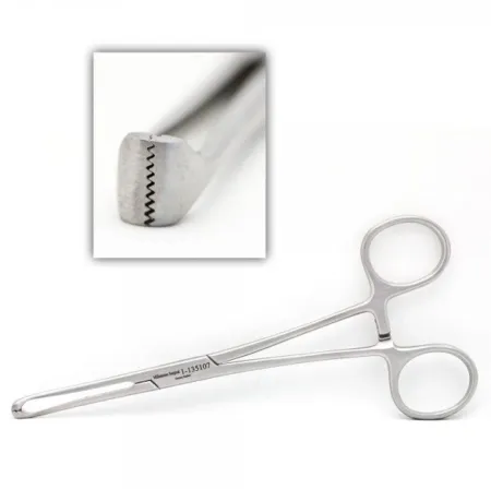 Allis ForcepsGrasping & Holding · On 141 setups
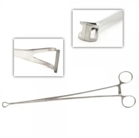 BabcockGrasping & Holding · On 70 setups
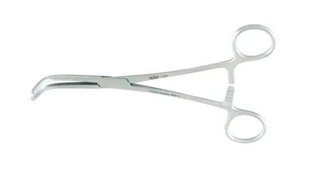 Right Angle ClampClamping & Occluding · On 102 setups
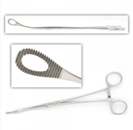 Foerster Sponge ForcepsGrasping & Holding · On 142 setups
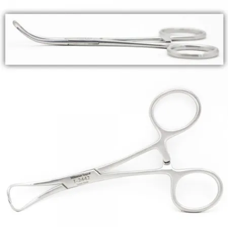 Backhaus Roeder Towel ForcepsGrasping & Holding · On 126 setups
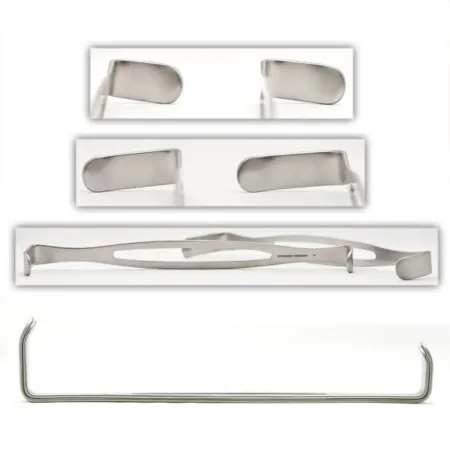 Army-Navy RetractorRetracting & Exposing · On 177 setups
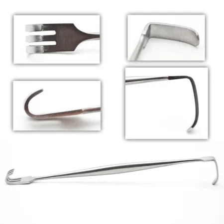 Senn RetractorRetracting & Exposing · On 169 setups
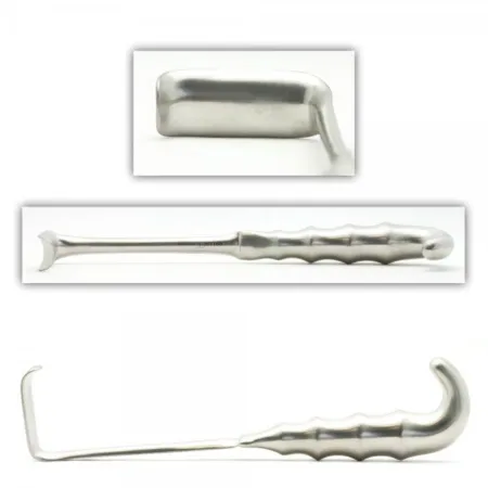 Richardson RetractorRetracting & Exposing · On 110 setups
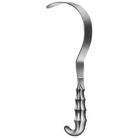 Deaver RetractorRetracting & Exposing · On 55 setups
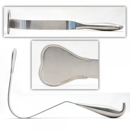 Harrington RetractorRetracting & Exposing · On 23 setups
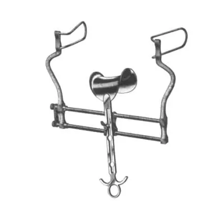 Balfour RetractorRetracting & Exposing · On 27 setups
 Bookwalter RetractorRetracting & Exposing · On 23 setups
 Poole Suction ProbeSuctioning · On 33 setups
 Yankauer SuctionSuctioning · On 228 setups
 Doyen Intestinal ForcepsGrasping & Holding · On 4 setups
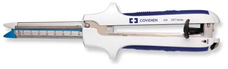 GIA StaplerSuturing & Stapling · On 12 setups
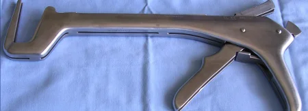 Linear StaplerSuturing & Stapling · On 15 setups
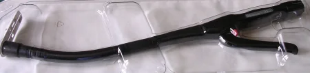 Intraluminal StaplerSuturing & Stapling · On 7 setups
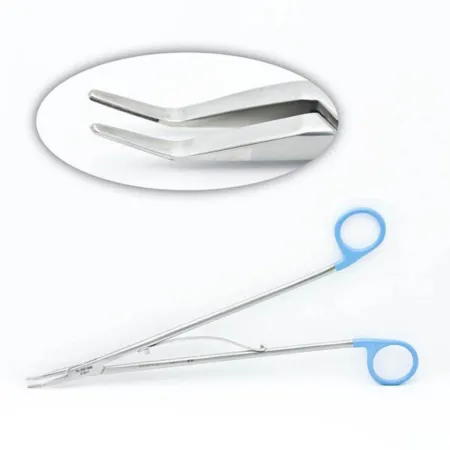 Hemostatic Clip ApplierClamping & Occluding · On 90 setups
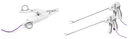 LigaSure Vessel SealerClamping & Occluding · On 22 setups
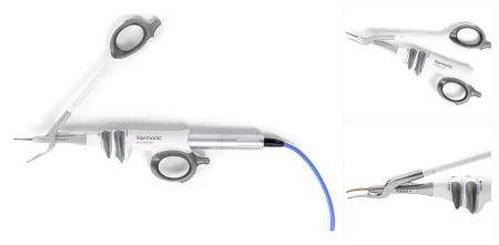 Harmonic Focus ShearsCutting & Dissecting · On 11 setups
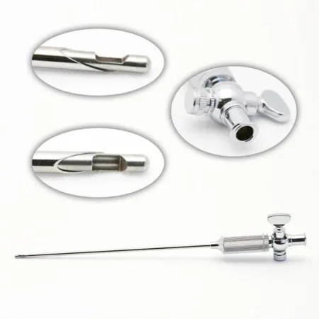 Veress NeedleCutting & Dissecting · On 29 setups
 Laparoscopic TrocarSpecialty · On 38 setups
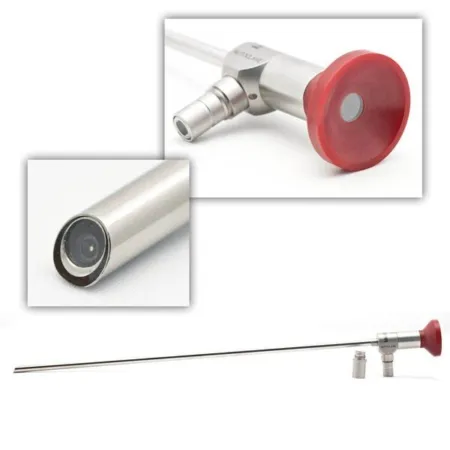 LaparoscopeSpecialty · On 28 setups
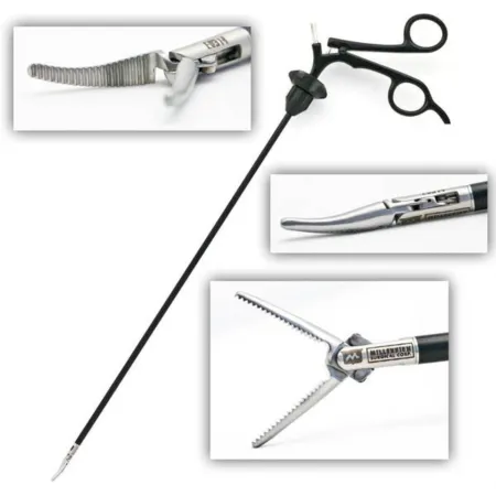 Laparoscopic Maryland Dissector GrasperGrasping & Holding · On 14 setups
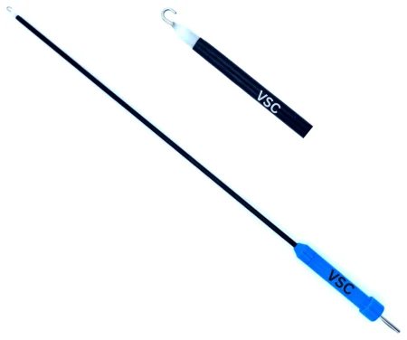 Laparoscopic J-Hook ElectrodeSpecialty · On 1 setup
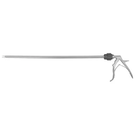 Laparoscopic Clip AppliersClamping & Occluding · On 30 setups
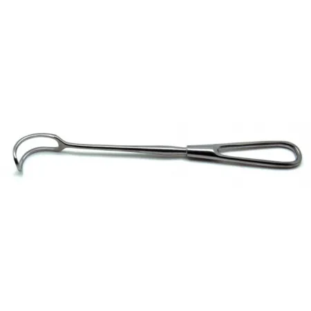 Green RetractorRetracting & Exposing · On 5 setups
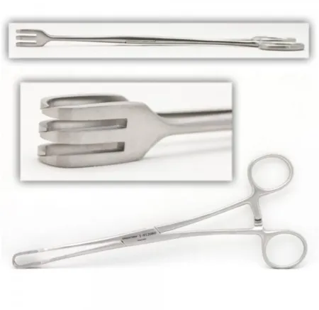 Lahey Traction ForcepsGrasping & Holding · On 9 setups
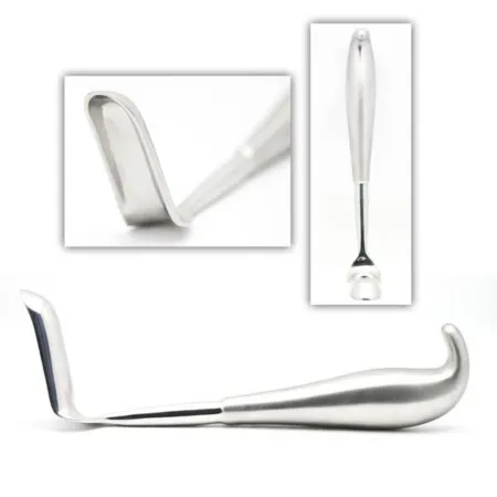 Hill Ferguson Rectal RetractorRetracting & Exposing · On 5 setups
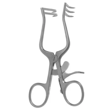 Weitlaner RetractorRetracting & Exposing · On 138 setups
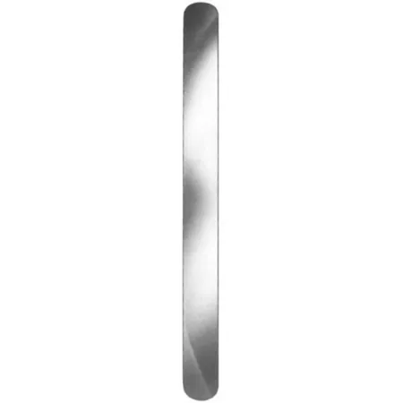 Ribbon RetractorRetracting & Exposing · On 65 setups
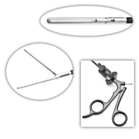 Laparoscopic GrasperGrasping & Holding · On 12 setups
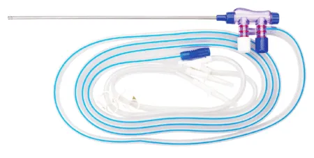 Suction IrrigatorSuctioning · On 35 setups
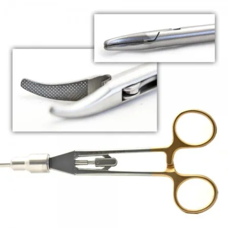 Laparoscopic Needle HolderSuturing & Stapling · On 12 setups
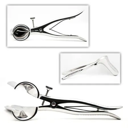 Pratt Rectal SpeculumRetracting & Exposing · On 7 setups
The chapter's whole question pool in sets of 10, with the answer explained after each one.
Coming soonThe chapter's words and instruments on spaced cards: the ones you miss come back sooner.
Coming soonGiven by your class's teacher. Each class draws its own 25 from the chapter's pool.
Coming soon
Suggest a fix
Something wrong, unclear or out of date in this chapter? Tell us and we will fix it for everyone. Your email is only used to reply.